Abstract
(1) Background: tooth agenesis is a very common dental anomaly of the human dentition most often affecting the maxillary anterior region, mandibular and maxillary premolar regions. (2) Purpose: the present study was aimed to evaluate the prevalence and patterns between bilateral and unilateral tooth agenesis among orthodontic individuals and to illustrate the treatment options for missing teeth and the outcome. (3) Materials and methods: Pre-treatment records, photographs and radiographs, of 3000 consecutively treated orthodontic individuals from the post-graduate clinic of Tel Aviv University were surveyed to detect permanent tooth agenesis in either dental arch. The data was recorded according to gender, and location and quantified between unilateral and bilateral agenesis. Descriptive and comparative statistical analysis were performed with t-test and Chi-square test (p < 0.05). (4) Results: permanent teeth agenesis, excluding third molars, was found in 326 individuals (11%), 139 males (43%) and 187 females (57%). Of them, 59% were missing in the maxilla and (41%) were missing in the mandible. A higher prevalence rate of bilateral missing lateral incisors in the maxilla (62 cases), followed by bilateral missing second premolars in the mandible (44 cases) compared with unilateral missing teeth. (5) Conclusions: this study found an overall prevalence of missing permanent teeth in orthodontic patients to be 11%. The female: male prevalence ratio was roughly 3:2, with a greater tendency in the maxilla than in the mandible. A higher prevalence of bilateral missing maxillary lateral incisors and mandibular second premolar than unilateral missing teeth.
1. Introduction
Tooth agenesis is a very common developmental dental anomaly of human dentition, and the most common dental anomaly among different ethnic groups, with third molars being the most common missing tooth. Congenital missing teeth is usually determined clinically, by the absence of tooth eruption in the oral cavity, and by radiographic examination where the dental bud cannot be detected in the radiograph. It is often detected in orthodontic patients and is found most frequently in the maxillary anterior region [1,2]. Tooth agenesis, based on the number of missing teeth, commonly termed hypodontia, is characterized by the congenital missing of one to five teeth, excluding the third molars. Oligodontia refers to congenital missing of six or more teeth, excluding third molars, and anodontia represents total missing of teeth in the dental arches which is extremely rare [3]. Anodontia or Oligodontia are usually associated with other systemic abnormalities such as ectodermal dysplasia or Down syndrome. However, Oligodontia may also appear as an isolated condition, referred to as non-syndromic oligodontia.
In addition, a strong correlation has been reported between the congenital absence of deciduous and that of their successor teeth [4,5]. When the deciduous tooth is missing, its permanent counterpart will also be absent. Additionally, agenesis can be linked to Sella Turcica Bridging (STB), or calcification of the interclinoid of Sella Turcica. This has been reported to be associated with some dental anomalies like canine impaction and transposition [6]. In addition, restriction of maxillary growth (maxillary hypoplasia) found in cleft lip and palate patients has been reported to be associated with dental agenesis [7].
The most frequently missing permanent teeth according to rank are the maxillary lateral incisors, second premolars and third molars, while missing maxillary central incisors, canines and first molars are very rare [5]. Hypodontia can be classified as non-syndromic or sporadic when the absence of a tooth is an isolated anomaly, or syndromic when it is a part of a complex of multiple congenital anomalies [8].
Etiological factors related to tooth agenesis can be environmental or genetic resulting in failure of tooth development. Heredity is the main factor for missing teeth due to an autosomal dominant gene with incomplete penetrance [9,10]. Several genes have been discovered to affect tooth development and cause tooth agenesis by mutations in the MSX1, PAX9, AX1N2, TGFA, IRF6, WNTA10, and FGFR1 genes [11,12]. It has been proposed that dental agenesis may also be an expression of an evolutionary process (phylogenetic tendency), in which the jaws of humans together with the quantity of dental units are being altered in size and number, although no correlation was found between the size of the jaws and the absence of teeth [13]. It can also be a result of systemic factors such as endocrine disturbances, a part of a syndrome or due to environmental factors such as radiation or facial trauma [14].
According to Butler’s Field Theory of tooth development in mammals, teeth were divided into “stable” and “unstable” groups where the missing tooth will always be the most distal tooth of any given group [15]. Therefore, the third molar is the most common missing in the molar group, the second premolar in the premolar group and the lateral incisor in the incisors group. While teeth such as central incisors, canines and first molars are rarely missing.
In patients with congenitally missing teeth, deformities in tooth size and shape (peg-shaped or small laterals) are more frequent. Peg-shaped lateral incisors can be considered as an incomplete expression of the gene responsible for agenesis of this tooth [13]. The high prevalence of tooth agenesis and peg-shaped lateral incisors suggests a common genetic etiology [2,16]. Hypodontia has additional localized influences such as its reported association with other dental anomalies such as palatially displaced maxillary canines [2,17].
The expression of anomalies has been reported according to ethnic and regional lines and the patterns of hypodontia and its frequency found to be variable for different populations [18,19,20,21,22,23,24,25]. For example, the prevalence of congenitally missing teeth is lower in American blacks compared with American whites. The hypodontia reported prevalence of permanent teeth in Caucasian populations ranges between 2.3% and 11.3% [18], and less than 1% in the deciduous dentition [19,20]. A prevalence of 2.6% has been reported for Arab orthodontic patients in Israel [21], 4.6% in Turkish orthodontic patients [22], 4.5% in the Norwegian population [23], 8.5% in Japanese orthodontic patients [24], and up to 14.7% in Hungarian orthodontic patients [25].
There are different opinions regarding the most frequently missing permanent tooth, excluding third molars. Some have concluded that this is the maxillary lateral incisor [22,26], while others claim that agenesis of the mandibular second premolar has a higher prevalence [20,27]. Currently, no extensive investigation on permanent tooth agenesis in the Israeli orthodontic population has been reported.
The present study was aimed to conduct a retrospective wide survey in order:
- (1)
- To evaluate the prevalence and patterns of permanent tooth agenesis and compare bilateral with unilateral agenesis in orthodontic individuals in a university clinic.
- (2)
- To demonstrate the clinical management of agenesis of maxillary lateral incisors specifically, because of esthetic concerns, and that of missing second premolars.
2. Materials and Methods
The present study was carried out based on clinical examination, pre-treatment photographs, panoramic and periapical radiographs and study models of 3000 consecutively treated orthodontic individuals from the department of orthodontics at Tel Aviv University. Out of the 3000 orthodontic individuals, the sample consisted of 1220 (41%) males and 1780 (59%) females, ranging from 10–25 years old (mean age 15 years), all were of Israeli ethnical origin. Their records were examined to detect permanent teeth agenesis in both dental arches, except for third molars. The data were recorded according to gender, age, number of missing teeth, location within each jaw and sidedness. Tooth agenesis was diagnosed clinically and defined as a lack of crypt formation or crown mineralization detection in the panoramic radiographs.
Inclusion criteria included complete records with good quality photographs and diagnostic radiographs. Exclusion criteria included history of traumatic dental avulsion or extraction of permanent teeth, orthodontic or surgical treatment in the dental arches, and congenital craniofacial anomalies or syndromes. To avoid any misdiagnosis of missing teeth, two examiners (M.P. and Y.S.) examined all the records twice separately and 100% reproducibility was achieved in the diagnosis of hypodontia.
Informed consent was obtained from the parent/guardian of each individual prior to inclusion in the study, which was approved by the Institutional Review Board Ethics Committee of Tel Aviv University. Descriptive and comparative statistical analyses were conducted using the SPSS software (Statistical Package for Social Sciences, version 20.0, SPSS Inc., Chicago, IL, USA). The significance of the findings was tested using t-test or Chi-Square tests, with p < 0.05 designated as statistically significant. A sample calculation was established using Win Pepi version 11.18, and Fisher’s Exact Test.
3. Results
The study sample comprised 3000 patients, 1220 (41%) males and 1780 (59%) females. The total number of missing teeth, excluding third molars, was found to be 518 (17%), discovered in 326 individuals (11%), 139 males (43%) and 187 females (57%) (Table 1). Those found to be missing from the maxillary dentition numbered 308 (59%), and those from the mandibular dentition 210 (41%).

Table 1.
Distribution of missing teeth by gender.
While 311 (95.4%) individuals had one to five missing teeth (Hypodontia patients), 15 (4.6%) had six to eleven missing permanent teeth (Oligodontia patients) (Two patients with 11 missing teeth; one with 9 missing teeth; one had 8 missing teeth; five had 7 missing teeth; and six patients had 6 missing teeth each).
The most common missing tooth was the maxillary lateral incisor (176) (p < 0.05), of which 93 (53%) were the right lateral incisor, and 83 (47%) the left lateral incisor. The next most common missing tooth was the mandibular second premolar 137 (27%), 76 (55%) from the left side and 61 (45%) from the right (Table 2).

Table 2.
Distribution of missing teeth by side.
A total of 73 (13%) maxillary second premolar teeth were found to be missing, 37 (51%) from the left and 36 (49%) from the right side (Table 3). Missing first premolars numbered 61 (12%), of which 34 (56%) from the maxilla and 27 (44%) from the mandible.

Table 3.
Distribution of missing teeth by location.
A total of 41 (8%) canines were missing, 25 (61%) from the maxilla and 16 (39%) from the mandible. A total of 30 (6%) mandibular incisors were missing, distributing equally between right and left sides (Table 3).
Bilateral agenesis of teeth was significantly more (131–80%) than unilateral agenesis (33–20%) (p < 0.05). The most common bilateral agenesis were the maxillary lateral incisors followed by the mandibular second premolars (p < 0.05) (Table 4).

Table 4.
Patterns of missing teeth by location.
The patterns of bilateral agenesis of the maxillary lateral incisors and bilateral missing maxillary second premolars were detected in four individuals, while bilateral missing of the maxillary lateral incisors and bilateral missing mandibular second premolars in seven individuals. Bilateral agenesis of both maxillary and mandibular second premolars were found in nine individuals; and one individual showed a combined bilateral missing of the maxillary lateral incisors, bilateral missing maxillary second premolars and bilateral missing mandibular second premolar. Other dental anomalies detected in patients with tooth agenesis include: peg-shaped maxillary lateral incisors, supernumerary teeth, retained maxillary primary canines and impacted maxillary permanent canines, ectopic teeth, canine transposition and transmigration [1].
4. Discussion
This study is an epidemiological investigation of hypodontia found in consecutively treated subjects in a university post-graduate orthodontic clinic. Therefore, the prevalence rate of this anomaly discovered here within does not necessarily directly reflect that of the general population.
Spaced dentition resulting from hypodontia especially in the maxillary anterior region is one of the major complaints for which individuals seek orthodontic treatment. Furthermore, it has been reported that females comprise the majority of orthodontic patients [28,29]. This was reflected by our sample as well (i.e., 59% female participation).
Independent records analysis of participating individuals in this study revealed a prevalence of 11% for congenitally missing teeth. This was found to have a higher female incidence but was not statistically different from that found in the male individuals. The quantity of missing teeth found in each individual with this anomaly varied from between one to eleven (in two individuals). The present study indicates that the more commonly missing teeth are the maxillary lateral incisors (54%), followed by the mandibular second premolars (42%). This is in agreement with Kokich [30,31], but is not with Symons’ who reported that the mandibular second premolar were more commonly missing [32]. The next most frequently missing teeth in our study were found to be the mandibular second premolars (42%) followed by the maxillary second premolars, similar to reports on missing teeth in the Turkish [22], Brazilian [33], Qatari [34] and Arab orthodontic patients [21].
It is worthy to note that previous studies on the incidence of missing permanent teeth reported varied conclusions based on ethnic differences. For example, characteristic to Asian ethnicity, the mandibular second premolars and mandibular incisors are the most frequently missing teeth [24,35,36], while in a Caucasian population the mandibular second premolars and the maxillary lateral incisors are reported as most likely to be missing [37]. Another example of ethnic difference is in the Druze population in Jordan known for their endogamy and consanguineous marriages, led to their genetic isolation, where the most missing teeth are the maxillary lateral incisors and canines [38]. Most of previous reports demonstrated that hypodontia was more prevalent in females than in males, similar to our findings [21,22]. However, the distribution of missing teeth according to sex was not found to have a male/female prevalence, a finding reported in an Egyptian population [39].
Our study indicates a significant difference between missing teeth in the maxilla and the mandible, with higher prevalence of missing teeth in the maxilla (305–59%) compared with the mandible (210–41%), which is in agreement with Muller et al. [26], Alsoleihat [38], Mani et al. [40], and Tunis et al. [41]. A possible explanation for the higher prevalence of missing teeth and other dental anomalies in the maxilla is in the differences in jaw ontogenesis where the maxillary growth and development is different from that of the mandible [41]. Interestingly, most of the dental anomalies, including missing teeth and supernumerary teeth were found in the maxillary anterior region while the majority of dental anomalies (missing and supernumerary teeth) were detected in the mandibular posterior region [41]. However, Zang et al. reported a higher prevalence of missing teeth in the mandible [36].
Our study showed in unilateral cases of maxillary lateral incisor agenesis, more missing teeth on the right side compared with the left side, and the contralateral tooth present a reduction in size (peg shape teeth), similar to findings reported by Garib et al. [1], Mani et al. [40]. Unilateral missing maxillary lateral incisors and the reduction in size in the contralateral side present different expressions of the same gene responsible for missing teeth.
The present study found that bilateral agenesis of maxillary lateral incisors (62 cases) was significantly more common than unilateral agenesis of the maxillary lateral incisors (12 cases). This is in agreement with Garib et al. [1], Abu-Hussein et al. [21] and Stamatiou et al. [42]. Similarly, bilateral agenesis of mandibular second premolars (44 cases) was more common than unilateral agenesis of mandibular second premolars (14 cases). In addition, a higher prevalence rate of bilateral missing maxillary lateral incisors (62 cases), followed by mandibular second premolars (44 cases), was found in our study. This was unlike a report that found a higher prevalence of bilateral agenesis of mandibular second premolar compared with maxillary lateral incisors [43]. A possible explanation for the higher prevalence of bilateral agenesis compared with unilateral agenesis was suggested that unilateral missing teeth is a result of partial expression of the gene responsible for missing teeth, similar to that with microdontia and peg-shaped lateral incisors [3,9,13].
Dental anomalies such as tooth agenesis are an important part of malocclusion diagnosis. Hypodontia causes spacing between teeth and decreased growth of the alveolar process, it influences skeletal patterns and craniofacial structure and soft tissue profiles [44,45]. Tooth agenesis was found to be associated with other dental anomalies such as reduction in tooth size (peg shaped lateral incisors), delayed dental development especially of the mandibular second premolars and palatally positioned maxillary canines [1,2]. The palatal displacement of the maxillary canine, associated with the missing maxillary lateral incisors, was explained by Becker et al. as the “guidance theory” where the roots of the maxillary lateral incisors guide the maxillary canine eruption path [46]. Unlike this theory, the genetic theory advocated by Peck et al. [47] as the major factor for the close association between agenesis of maxillary lateral incisors and palatally displaced maxillary canines.
Agenesis of teeth is usually associated with other systemic abnormalities and appear as a part of a complex of multiple congenital anomalies such as ectodermal dysplasia or Down syndrome and in cleft lip and cleft palate [7]. The congenital absence of teeth is significantly higher in children with a cleft lip or cleft palate, both in and outside the cleft region. The maxillary lateral incisors are the most commonly missing teeth in the cleft area, followed by the maxillary second premolars in cleft lip and palate individuals [48]. A higher frequency of missing maxillary lateral incisors and second premolars were found in cleft lip and palate individuals, with second premolars absent three times more often in the maxilla than in the mandible [49].
Agenesis of either maxillary lateral incisors or mandibular second premolars presents diagnostic and clinical management challenges to the clinicians. Managing agenesis of maxillary lateral incisors specifically, because of esthetic concerns, is a challenge for all of the medical specialists involved (i.e., orthodontist, pediatric dentist, prosthodontist, oral surgeon, etc.). The complexities of successfully treating this condition require a multidisciplinary approach to establish not only optimal esthetics but functional and stable occlusion as well [50]. Generally, two conservative approaches can be considered: space closure by canine substitution of the missing lateral incisor [51], or space opening and prosthetic replacement of the missing tooth/teeth with or without endosseous implant support [42,52]. Another treatment option suggested to replace missing teeth in growing individuals is the autotransplantation of teeth with developing roots. The most common transplanted teeth are premolars, with developed roots of 2/3 to ¾ of the final root length, into the anterior region of the maxillary arch, following prosthodontic reshaping to resemble anterior teeth. This procedure also preserves the growth of the alveolar bone in the region of the transplantation. In addition, developing third molars and even supernumerary teeth can be used for autotransplantation using special surgical techniques [53,54].
The following cases illustrate the treatment options to deal clinically with missing teeth.
Case One:
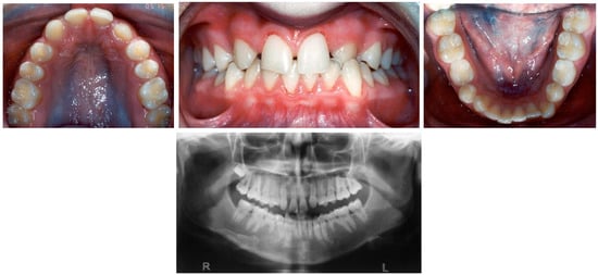
A 14-year-old female presented with a complaint of an unesthetic appearance due to missing lateral incisors and spaces between her maxillary anterior teeth. Her medical and dental histories were non-contributory. Intraoral examination revealed Class I malocclusion in the permanent dentition and maxillary anterior spacing with congenitally missing maxillary lateral incisors, mandibular second premolars, second permanent molars, and retained mandibular left second deciduous molar (Figure 1). Her treatment included orthodontic space closure in the maxillary anterior region, and canine substitution for the missing lateral incisors. The canine crowns were then built up with composite to resemble lateral incisors. Retention was carried out with bonded permanent fixed retainers (Figure 2). A single tooth implant was scheduled later on to replace the missing mandibular second premolars, as she could not afford the financial expense at the completion of the orthodontic treatment.

Figure 1.
Pre-treatment photographs of a 14-year-old female with congenitally missing maxillary lateral incisors, mandibular second premolars, second permanent molars, and retained mandibular left second deciduous molar.

Figure 2.
Post-treatment photographs with canine substitution for the missing lateral incisors.
Case Two:
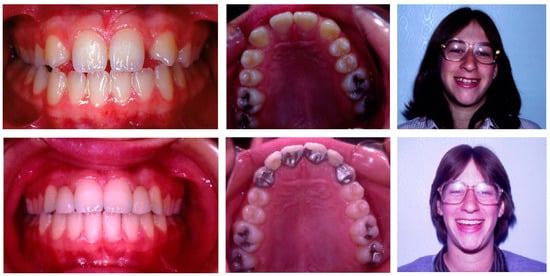
A 16-year-old female was referred for orthodontic treatment. Her intraoral examination revealed Class I dental relationship, with both maxillary lateral incisors congenitally missing and spaces in the maxillary anterior region (Figure 3). Treatment included opening of spaces between the central incisors and canines and constructing a Maryland bridge with two lateral incisors (Figure 3).

Figure 3.
Pre- and post-treatment photographs of a 16-year-old female with missing maxillary lateral incisors treated with spaces opening for Maryland bridge.
Case Three:
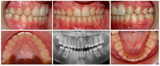
A 14-year-old male was referred for orthodontic treatment. An intraoral examination revealed Class I malocclusion with congenitally missing maxillary lateral incisors, and mandibular second premolars, retained mandibular second deciduous molars, crossbite between the maxillary and mandibular left canines, and minor mandibular anterior crowding (Figure 4). The orthodontic treatment in this case was space closure in the maxillary anterior region where the canines substitute for the missing lateral incisors. The mandible extraction of the retained deciduous second molars was followed by mesial movement of the permanent molars and space closure using mini-plates for anchorage control saving the need for future rehabilitation with implants (Figure 5). Retention was carried out with bonded permanent fixed retainers.

Figure 4.
Pre-treatment photographs of a 14-year-old male with congenitally missing maxillary lateral incisors, and mandibular second premolars, retained mandibular second deciduous molars, cross bite between the maxillary and mandibular left canines, and minor mandibular anterior crowding.

Figure 5.
Post-treatment photographs following space closure of the missing teeth in both arches.
Such treatment of missing posterior teeth is simplified slightly only because of the decreased need to achieve the same degree of esthetics as in the maxillary anterior region.
5. Limitations of the Study
The study sample was limited to orthodontic patients treated at a University Orthodontic Clinic. Thus, the prevalence rate discovered here does not represent the prevalence and patterns of tooth agenesis among the general population, which justify an additional investigation in the future.
6. Conclusions
An overall prevalence of 11% for missing permanent teeth in orthodontically treated individuals was found with a 3:2 female:male ratio, and a higher prevalence in the maxilla than in the mandible.
Maxillary lateral incisors were the most frequently missing teeth followed by the mandibular second premolars. A higher prevalence of bilateral missing pattern both of maxillary lateral incisors and mandibular second premolar was found when compared with unilateral missing teeth.
Author Contributions
Conceptualization, Y.S.; Investigation, Y.S. and T.F.; Data curation, A.M.P.; Visualization, A.M.P. and S.S.; Supervision, Y.S. All authors have read and agreed to the published version of the manuscript.
Funding
This research received no external founding.
Institutional Review Board Statement
The study was conducted in accordance with the Declaration of Helsinki and approved by the Institutional Ethics Committee of Tel Aviv University (No protocol code number).
Informed Consent Statement
Informed consent was obtained from all subjects involved in the study.
Data Availability Statement
The data of the study are available from the corresponding author on request.
Acknowledgments
We thank Amir Shapira (B.Sc) for his valuable help in the preparation of this article.
Conflicts of Interest
The authors declare no conflict of interest.
References
- Garib, D.G.; Alencar, B.M.; Lauris, J.; Baccetti, T. Agenesis of maxillary lateral incisors and associated dental anomalies. Am. J. Orthod. Dentofac. Orthop. 2010, 137, 732.e1–e6, discussion 732–733. [Google Scholar] [CrossRef]
- Peck, S.; Peck, L.; Kataja, M. Prevalence of tooth agenesis and peg-shaped maxillary lateral incisor associated with palatally displaced canine (PDC) anomaly. Am. J. Orthod. Dentofac. Orthop. 1996, 110, 441–443. [Google Scholar] [CrossRef]
- Abu-Hussein, M.; Watted, N.; Yehia, M.; Proff, P.; Iraqi, F. Clinical genetic basis of tooth agenesis. J. Dent. Med. Sci. 2015, 14, 68–77. [Google Scholar]
- Whittington, B.R.; Durward, C.S. Survey of anomalies in primary teeth and their correlation with the permanent dentition. N. Z. Dent. J. 1996, 92, 4–8. [Google Scholar]
- Kapadia, H.; Mues, G.; D’Souza, R. Genes affecting tooth morphogenesis. Orthod. Craniofac. Res. 2007, 10, 105–113. [Google Scholar] [CrossRef]
- Scribante, A.; Sfondrini, M.F.; Cassani, M.; Fraticelli, D.; Beccari, S.; Gandini, P. Sella Turcica bridging and dental anomalies: Is there an association? Int. J. Paediatr. Dent. 2017, 27, 568–573. [Google Scholar] [CrossRef] [PubMed]
- Janes, L.E.; Bazina, M.; Morcos, S.S.; Ukaigwe, A.; Valiathan, M.; Jacobson, R.; Gosain, A.K. The relationship between dental agenesis and maxillary hypoplasia in patients with cleft lip and palate. J. Craniofac. Surg. 2021, 32, 2012–2015. [Google Scholar] [CrossRef] [PubMed]
- Dahlberg, A.A. The dentition of the American Indians. In The Physical Anthropology of the American Indian; The Viking Fund: New York, NY, USA, 1949; pp. 138–176. [Google Scholar]
- Vastardis, H. The genetics of human tooth agenesis: New discoveries for understanding dental anomalies. Am. J. Orthod. Dentofac. Orthop. 2000, 117, 650–656. [Google Scholar] [CrossRef]
- Thesleff, I. Genetic basis of tooth development and dental defects. Acta Odontol. Scand. 2000, 58, 191–194. [Google Scholar] [CrossRef]
- Vieira, A.R.; Meira, R.; Modesto, A.; Murray, J.C. MSX1, PAX9, and TGFA contribute to tooth agenesis in humans. J. Dent. Res. 2004, 83, 723–727. [Google Scholar] [CrossRef]
- Vastardis, H.; Karimbux, N.; Guthua, S.W.; Seidman, J.; Seidman, C.E. A human MSX1 homeodomain missense mutation causes selective tooth agenesis. Nat. Genet. 1996, 13, 417–421. [Google Scholar] [CrossRef]
- Brook, A. A unifying aetiological explanation for anomalies of human tooth number and size. Arch. Oral Biol. 1984, 29, 373–378. [Google Scholar] [CrossRef] [PubMed]
- Grahnen, H. Hypodontia in the permanent dentition. A clinical and genetical investigation. Odontol. Rev. 1956, 7 (Suppl. S3), 1–100. [Google Scholar]
- Butler, P.M. Studies of the mammalian dentition. Differentiation of the post-canine dentition. In Proceedings of the Zoological Society of London; Blackwell Publishing Ltd.: Oxford, UK, 1939; Volume B109, pp. 1–36. [Google Scholar]
- Al-Abdallah, M.; AlHadidi, A.; Hammad, M.; Al-Ahmad, H.; Saleh, R. Prevalence and distribution of dental anomalies: A comparison between maxillary and mandibular tooth agenesis. Am. J. Orthod. Dentofac. Orthop. 2015, 148, 793–798. [Google Scholar] [CrossRef] [PubMed]
- Souza-Silva, B.N.; de Andrade Vieira, W.; de Macedo Bernardino, Í.; Batista, M.J.; Bittencourt, M.A.V.; Paranhos, L.R. Non-syndromic tooth agenesis patterns and their association with other dental anomalies: A retrospective study. Arch. Oral Biol. 2018, 96, 26–32. [Google Scholar] [CrossRef]
- Larmour, C.J.; Mossey, P.A.; Thind, B.S.; Forgie, A.H.; Stirrups, D.R. Hypodontia—A retrospective review of prevalence and etiology. Part I. Quintessence Int. 2005, 36, 263–270. [Google Scholar]
- Brabant, H. Comperison of the characteristics and anomalies of the deciduous and the permanent dentition. J. Dent. Res. 1967, 46, 897–902. [Google Scholar] [CrossRef] [PubMed]
- Jarvinen, S.; Lehtinen, L. Supernumerary and congenitally missing primary teeth in Finish children. An epidemiologic study. Acta Odontol. Scand. 1981, 39, 83–86. [Google Scholar] [CrossRef]
- Abu-Hussein, M.; Watted, N.; Watted, A.; Abu-Hussein, Y.; Yehia, M.; Awadi, O.; Azzaldeen, A. Prevalence of tooth agenesis in orthodontic patients at Arab population in Israel. Int. J. Public Health Res. 2015, 3, 77–82. [Google Scholar]
- Celikoglu, M.; Kazanci, F.; Miloglu, O.; Oztek, O.; Kamak, H.; Ceylan, I. Frequency and charateristics of tooth agenesis among an orthodontic patient population. Med. Oral Pathol. Oral Cir. Bucal 2010, 15, e797–e801. [Google Scholar] [CrossRef]
- Nordgarden, H.; Jensen, J.L.; Storhaug, K. Reported prevalence of congenitally missing teeth in two Norwegian counties. Commun. Dent. Health 2002, 19, 258–261. [Google Scholar]
- Endo, T.; Ozoe, R.; Kubota, M.; Akiyama, M.; Shimooka, S. A survey of hypodontia in Japanese orthodontic patients. Am. J. Orthod. Dentofac. Orthop. 2006, 129, 29–35. [Google Scholar] [CrossRef]
- Gabris, K.; Fabian, G.; Kaan, M.; Rozsa, N.; Tarjan, I. Prevalence of agenesis and hyperdontia in paedodontic and orthodontic patients in Budapest. Commun. Dent. Health 2006, 23, 80–82. [Google Scholar]
- Muller, T.; Hill, I.; Petersen, A.; Blayney, J. A Survey of Congenitally Missing Permanent Teeth. J. Am. Dent. Assoc. 1970, 81, 101–107. [Google Scholar] [CrossRef]
- Serrano, J. Gemination, hypodontia and supernumerary teeth. Oral Surg. Oral Med. Oral Pathol. 1986, 62, 737–738. [Google Scholar] [CrossRef] [PubMed]
- Khan, R.S.; Horrocks, E.N. A Study of Adult Orthodontic Patients and their Treatment. Br. J. Orthod. 1991, 18, 183–194. [Google Scholar] [CrossRef]
- Nattrass, C.; Sandy, J.R. Adult orthodontics: A review. Br. J. Orthod. 1995, 22, 331–337. [Google Scholar] [CrossRef]
- Kokich, V.O. Congenitally missing teeth: Orthodontic management in the adolescent patient. Am. J. Orthod. Dentofac. Orthop. 2002, 121, 594–595. [Google Scholar] [CrossRef]
- Sisman, Y.; Uysal, T.; Gelgor, I.E. Hypodontia. Does the prevalence and distribution pattern differ in orthodontic patients? Eur. J. Dent. 2007, 1, 167–173. [Google Scholar] [CrossRef]
- Symons, A.L.; Stritzel, F.; Stamation, J. Anomalies associated with hypodontia of the permanent lateral incisor and second premolar. J. Clin. Pediatr. Dent. 1993, 17, 109–111. [Google Scholar]
- Gomes, R.R.; Da Fonseca, J.A.C.; Paula, L.M.; Faber, J.; Acevedo, A.C.; Da Fonseca, J.A.C. Prevalence of hypodontia in orthodontic patients in Brasilia, Brazil. Eur. J. Orthod. 2009, 32, 302–306. [Google Scholar] [CrossRef] [PubMed]
- Al Jawad, F.H.A.; Al Yafei, H.; Al Sheeb, M.; Al Emadi, B. Hypodontia prevalence and distribution pattern in a group of Qatari orthodontic and pediatric patients: A retrospective study. Eur. J. Dent. 2015, 9, 267–271. [Google Scholar] [CrossRef] [PubMed]
- Goya, H.A.; Tanaka, S.; Maeda, T.; Akimoto, Y. An orthopantomographic study of hypodontia in permanent teeth of Japanese pediatric patients. J. Oral Sci. 2008, 50, 143–150. [Google Scholar] [CrossRef] [PubMed]
- Zhang, J.; Liu, H.C.; Lyu, X.; Shen, G.H.; Deng, X.X.; Li, W.R.; Zhang, X.X.; Feng, H.L. Prevalence of tooth agenesis in adolescent Chinese populations with or without orthodontics. Chin. J. Dent. Res. 2015, 18, 59–65. [Google Scholar]
- Fekonja, A. Hypodontia in orthodontically treated children. Eur. J. Orthod. 2005, 27, 457–460. [Google Scholar] [CrossRef]
- Alsoleihat, F.; Khraisat, A. Hypodontia: Prevalence and pattern amongst the living Druze population—A Near Eastern genetic isolate. J. Comp. Human Biol. 2014, 65, 201–213. [Google Scholar] [CrossRef]
- Montasser, M.; Taha, M. Prevalence and distribution of dental anomalies in orthodontic patients. Orthod. Art Pract. Dentofac. Enhanc. 2012, 13, 52–59. [Google Scholar]
- Mani, S.A.; Mohsin, W.S.Y.; John, J. Prevalence and patterns of tooth agenesis among Malay children. Southeast Asian J. Trop. Med. Public Health 2014, 45, 490–498. [Google Scholar]
- Tunis, T.S.; Sarne, O.; Hershkovitz, I.; Finkelstein, T.; Pavlidi, A.; Shapira, Y.; Davidovitch, M.; Shpack, N. Dental Anomalies’ Characteristics. Diagnostics 2021, 11, 1161. [Google Scholar] [CrossRef]
- Stamatiou, J.; Symons, A.L. Agenesis of the permanent lateral incisor: Distribution, number and sites. J. Clin. Pediatr. Dent. 1991, 15, 244–246. [Google Scholar]
- Padmanabhan, V.; Rahhal, L.M.K.; Omar Khaled, A.M. Prevalence of bilateral agenesis of mandibular second premolars and maxillary lateral incisors—A retrospective study. EC Dent. Sci. 2020, 19, 89–94. [Google Scholar]
- Ben-Bassat, Y.; Brin, I. Skeletal and dental patterns in patients with severe congenital absence of teeth. Am. J. Orthod. Dentofac. Orthop. 2009, 135, 349–356. [Google Scholar] [CrossRef]
- Øgaard, B.; Krogstad, O. Craniofacial structure and soft tissue profile in patients with severe hypodontia. Am. J. Orthod. Dentofac. Orthop. 1995, 108, 472–477. [Google Scholar] [CrossRef]
- Becker, A. In defense of the guidance theory of palatal canine displacement. Angle Orthod. 1995, 65, 95–98. [Google Scholar] [PubMed]
- Peck, S.; Peck, L.; Kataja, M. The palatally displaced canine as a dental anomaly of genetic origin. Angle Orthod. 1994, 64, 249–256. [Google Scholar]
- Ranta, R. Hypodontia and delayed development of the second premolars in cleft palate children. Eur. J. Orthod. 1983, 5, 145–148. [Google Scholar] [CrossRef] [PubMed]
- Shapira, Y.; Lubit, E.; Kuftinec, M.M. Congenitally missing second premolars in cleft lip and cleft palate children. Am. J. Orthod. Dentofac. Orthop. 1999, 115, 396–400. [Google Scholar] [CrossRef]
- Sharma, P.K.; Sharma, P. Interdisciplinary management of congenitally absent maxillary lateral incisors: Orthodontic/prosthodontic perspectives. Sem. Orthod 2015, 21, 27–37. [Google Scholar] [CrossRef]
- Zachrisson, B.U.; Rosa, M.; Toreskog, S. Congenitally missing maxillary lateral incisors: Canine substitution. Am. J. Orthod. Dentofac. Orthop. 2011, 139, 434–444. [Google Scholar] [CrossRef]
- Kokich, V.O., Jr.; Kinzer, G.A.; Janakievski, J. Congenitally missing maxillary lateral incisors: Restorative replacement. Am. J. Orthod. Dentofac. Orthop. 2011, 139, 441. [Google Scholar] [CrossRef]
- Czochrowska, E.M.; Stenvik, A.; Album, B.; Zachrisson, B.U. Autotransplantation of premolars to replace maxillary incisors: A comparison with natural incisors. Am. J. Orthod. Dentofac. Orthop. 2000, 118, 592–600. [Google Scholar] [CrossRef] [PubMed]
- Czochrowska, E.M.; Stenvik, A.; Bjercke, B.; Zachrisson, B.U. Outcome of tooth transplantation: Survival and success rates 17–41 years posttreatment. Am. J. Orthod. Dentofac. Orthop. 2002, 121, 110–119. [Google Scholar] [CrossRef] [PubMed]
Publisher’s Note: MDPI stays neutral with regard to jurisdictional claims in published maps and institutional affiliations. |
© 2022 by the authors. Licensee MDPI, Basel, Switzerland. This article is an open access article distributed under the terms and conditions of the Creative Commons Attribution (CC BY) license (https://creativecommons.org/licenses/by/4.0/).