
Properties of CAD/CAM 3D Printing Dental Materials and Their Clinical Applications in Orthodontics: Where Are We Now?

 ,
,  , ,
, , 

Abstract
:1. Introduction
2. Three Dimensional Printing Technologies
2.1. Stereolithography (SLA) and Digital Light Processing (DLP)
2.2. Fused Deposition Modeling/Fused Filament Fabrication (FDM/FFF)
2.3. Selective Laser Sintering (SLS)/Melting (SLM) and Electron Beam Melting (EBM)
2.4. Binder Jetting (BJ)
2.5. Material Jetting (MJ)
2.6. Main Features
3. The Digital Workflow
3.1. Intra-Oral Scanning
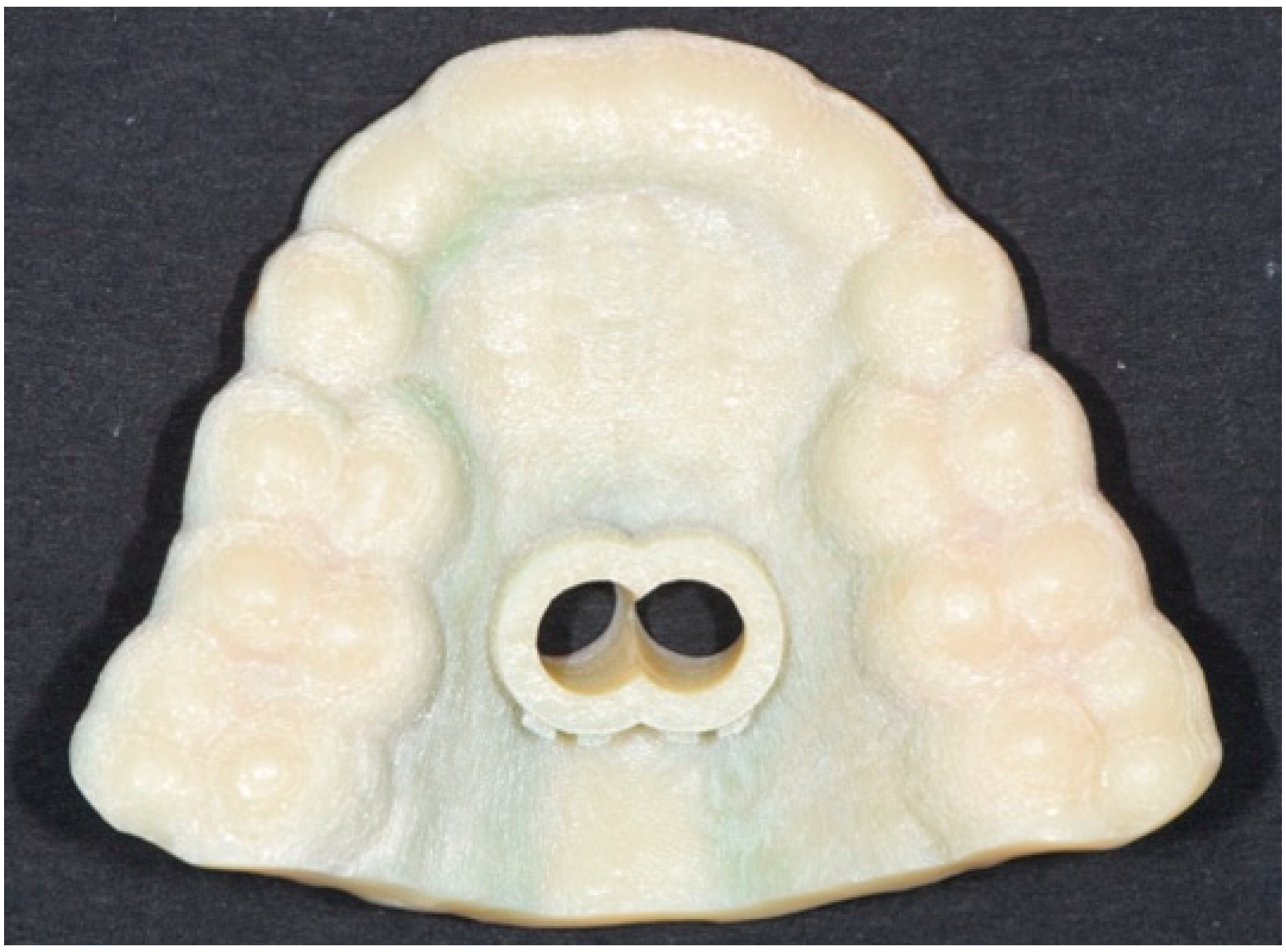
3.2. Elaboration of the Digital Model
3.3. 3D Printing
3.4. Post-Printing Processing
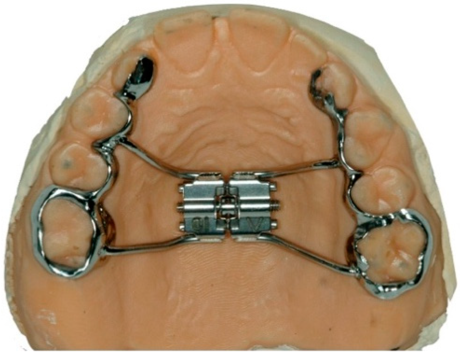
3.5. Production of the Appliance
4. Advantages and Disadvantages of 3D Printing
4.1. Advantages
- -
- Accuracy: while traditional models might undergo technical errors and distortions, 3D-printed models might be more accurate as they involve less steps and no manual labor.
- -
- Comfort to the patient: the scanning process might be more comfortable for the patients than the traditional impression technique.
- -
- Reduction of the times: due to the absence of analogic impressions, clinicians can directly send digital STL files to technicians, resulting in a digital workflow much more rapid compared to the traditional one.
- -
- Management of defects: during the elaboration of the digital model acquired, eventual scan defects can be solved guaranteeing a correct progression towards the printing of a reliable product.
- -
- Reduction of environmental costs: considering that traditional dental impressions are avoided by substituting them with STL file, the digital workflow inevitably reduces the numbers of materials which would require to be specifically disposed of. Accordingly, environmental costs are significantly reduced.
4.2. Disadvantages
- -
- Costs: the expense for the purchase of the 3D printing and hardware are significantly higher compared to the analogic workflow.
- -
- Longer learning curve: specific training is required for clinicians to familiarize themselves with the use of intraoral scanners to acquire a correct scanning guaranteeing a correct result of the entire process.
- -
- The necessity of referral to specific technical laboratories: only laboratories with the available technologies required can be considered by clinicians working with a digital workflow.
- -
- Health risk: specific procedures must be followed when handling uncured resins and cleaning solvents to avoid skin irritations caused by these materials. However, further aspects should be evaluated as regards the eventual toxicity of 3D-printed resins. In particular, monomer resins are recognized as toxic, despite their polymeric form generally are not.
5. Conclusions and Future Perspectives
Author Contributions
Funding
Institutional Review Board Statement
Informed Consent Statement
Conflicts of Interest
References
- Cousley, R.R. Introducing 3D printing in your orthodontic practice. J. Orthod. 2020, 47, 265–272. [Google Scholar] [CrossRef] [PubMed]
- Aragón, M.L.; Pontes, L.F.; Bichara, L.M.; Flores-Mir, C.; Normando, D. Validity and reliability of intraoral scanners compared to conventional gypsum models measurements: A systematic review. Eur. J. Orthod. 2016, 38, 429–434. [Google Scholar] [CrossRef]
- Sfondrini, M.F.; Gandini, P.; Malfatto, M.; Di Corato, F.; Trovati, F.; Scribante, A. Computerized Casts for Orthodontic Purpose Using Powder-Free Intraoral Scanners: Accuracy, Execution Time, and Patient Feedback. BioMed Res. Int. 2018, 2018, 4103232. [Google Scholar] [CrossRef] [PubMed] [Green Version]
- Brown, G.B.; Currier, G.F.; Kadioglu, O.; Kierl, J.P. Accuracy of 3-dimensional printed dental models reconstructed from digital intraoral impressions. Am. J. Orthod. Dentofac. Orthop. 2018, 154, 733–739. [Google Scholar] [CrossRef] [PubMed] [Green Version]
- Beuer, F.; Schweiger, J.; Edelhoff, D. Digital dentistry: An overview of recent develompents for CAD/CAM generated restorations. Br. Dent. J. 2008, 204, 505–511. [Google Scholar] [CrossRef]
- Ligon, S.C.; Liska, R.; Stampfl, J.; Gurr, M.; Mülhaupt, R. Polymers for 3D Printing and Customized Additive Manufacturing. Chem. Rev. 2017, 117, 10212–10290. [Google Scholar] [CrossRef] [Green Version]
- Vidakis, N.; Petousis, M.; Maniadi, A.; Koudoumas, E.; Kenanakis, G.; Romanitan, C.; Tutunaru, O.; Suchea, M.; Kechagias, J. The Mechanical and Physical Properties of 3D-Printed Materials Composed of ABS-ZnO Nanocomposites and ABS-ZnO Microcomposites. Micromachines 2020, 11, 615. [Google Scholar] [CrossRef]
- Skorski, M.; Esenther, J.; Ahmed, Z.; Miller, A.; Hartings, M. The chemical, mechanical, and physical properties of 3D printed materials composed of TiO2 -ABS nanocomposites. Sci. Technol. Adv. Mater. 2016, 17, 89–97. [Google Scholar] [CrossRef] [PubMed] [Green Version]
- Dodziuk, H. Applications of 3D printing in healthcare. Kardiochir. Torakochir. Pol. 2016, 13, 283–293. [Google Scholar] [CrossRef]
- Richert, R.; Goujat, A.; Venet, L.; Viguie, G.; Viennot, S.; Robinson, P.; Farges, J.C.; Fages, M.; Ducret, M. Intraoral Scanner Technologies: A Review to Make a Successful Impression. J. Healthc. Eng. 2017, 2017, 8427595. [Google Scholar] [CrossRef]
- Bhargav, A.; Sanjairaj, V.; Rosa, V.; Feng, L.W.; Fuh Yh, J. Applications of additive manufacturing in dentistry: A review. J. Biomed. Mater. Res. B Appl. Biomater. 2018, 106, 2058–2064. [Google Scholar] [CrossRef]
- Liaw, C.Y.; Guvendiren, M. Current and emerging applications of 3D printing in medicine. Biofabrication 2017, 9, 024102. [Google Scholar] [CrossRef]
- Tack, P.; Victor, J.; Gemmel, P.; Annemans, L. 3D-printing techniques in a medical setting: A systematic literature review. Biomed. Eng. Online 2016, 15, 115. [Google Scholar] [CrossRef] [Green Version]
- Alhnan, M.A.; Okwuosa, T.C.; Sadia, M.; Wan, K.W.; Ahmed, W.; Arafat, B. Emergence of 3D Printed Dosage Forms: Opportunities and Challenges. Pharm. Res. 2016, 33, 1817–1832. [Google Scholar] [CrossRef] [PubMed]
- Kessler, A.; Hickel, R.; Reymus, M. 3D Printing in Dentistry-State of the Art. Oper. Dent. 2020, 45, 30–40. [Google Scholar] [CrossRef]
- Tappa, K.; Jammalamadaka, U. Novel biomaterials used in medical 3D printing techniques. J. Funct. Biomater. 2018, 9, 17. [Google Scholar] [CrossRef] [Green Version]
- Chia, H.N.; Wu, B.M. Recent advances in 3D printing of biomaterials. J. Biol. Eng. 2015, 9, 4. [Google Scholar] [CrossRef] [PubMed] [Green Version]
- Liu, Q.; Leu, M.C.; Schmitt, S.M. Rapid prototyping in dentistry: Technology and application. Int. J. Adv. Manuf. Technol. 2005, 29, 317–335. [Google Scholar] [CrossRef]
- Barazanchi, A.; Li, K.C.; Al-Almeh, B.; Lyons, K.; Waddell, J.N. Additive technology: Update on current materials and applications in dentistry. J. Prosthod. 2017, 26, 156–163. [Google Scholar] [CrossRef] [PubMed]
- Fleming, P.S.; Marinho, V.; Johal, A. Orthodontic measurements on digital study models compared with plaster models: A systematic review. Orthod. Craniofac. Res. 2011, 14, 1–16. [Google Scholar] [CrossRef]
- Al Mortadi, N.; Eggbeer, D.; Lewis, J.; Williams, R.J. CAD/CAM/AM applications in the manufacture of dental appliances. Am. J. Orthod. Dentofac. Orthop. 2012, 142, 727–733. [Google Scholar] [CrossRef] [PubMed]
- ASTM Standard F2792; Standard Terminology for Additive Manufacturing Technologies; ASTM F2792—10e1; ASTM International: West Conshohocken, PA, USA, 2012.
- Bartkowiak, T.; Walkowiak-Sliziuk, A. 3D printing technology in orthodontics–review of current applications. J. Stomatol. 2018, 71, 356–364. [Google Scholar] [CrossRef]
- Osman, R.B.; Alharbi, N.; Wismeijer, D. Build angle: Does it influence the accuracy of 3D-printed dental restorations using digital light-processing technology. Int. J. Prosthodont. 2017, 30, 182–188. [Google Scholar] [CrossRef] [PubMed]
- Taneva, E.; Kusnoto, B.; Evans, C.A. 3D scanning, imaging, and printing in orthodontics. Issues Contemp. Orthod. 2015, 147–188. [Google Scholar]
- Gad, M.M.; Fouda, S.M.; Abualsaud, R.; Alshahrani, F.A.; Al-Thobity, A.M.; Khan, S.Q.; Akhtar, S.; Ateeq, I.S.; Helal, M.A.; Al-Harbi, F.A. Strength and Surface Properties of a 3D-Printed Denture Base Polymer. J. Prosthodont. Off. J. Am. Coll. Prosthodont. 2021. [Google Scholar] [CrossRef]
- Al-Qahtani, A.S.; Tulbah, H.I.; Binhasan, M.; Abbasi, M.S.; Ahmed, N.; Shabib, S.; Farooq, I.; Aldahian, N.; Nisar, S.S.; Tanveer, S.A.; et al. Surface Properties of Polymer Resins Fabricated with Subtractive and Additive Manufacturing Techniques. Polymers 2021, 13, 4077. [Google Scholar] [CrossRef] [PubMed]
- Kim, D.; Shim, J.S.; Lee, D.; Shin, S.H.; Nam, N.E.; Park, K.H.; Shim, J.S.; Kim, J.E. Effects of Post-Curing Time on the Mechanical and Color Properties of Three-Dimensional Printed Crown and Bridge Materials. Polymers 2020, 12, 2762. [Google Scholar] [CrossRef]
- Bayarsaikhan, E.; Lim, J.H.; Shin, S.H.; Park, K.H.; Park, Y.B.; Lee, J.H.; Kim, J.E. Effects of Postcuring Temperature on the Mechanical Properties and Biocompatibility of Three-Dimensional Printed Dental Resin Material. Polymers 2021, 13, 1180. [Google Scholar] [CrossRef]
- Berli, C.; Thieringer, F.M.; Sharma, N.; Müller, J.A.; Dedem, P.; Fischer, J.; Rohr, N. Comparing the mechanical properties of pressed, milled, and 3D-printed resins for occlusal devices. J. Prosthet. Dent. 2020, 124, 780–786. [Google Scholar] [CrossRef]
- Fayyaz Ahamed, S.; Mohnish Kumar, S.; Vijaya Kumar, R.K.; Apros Kanna, A.S.; Indrapriyadharshini, K. Cytotoxic evaluation of directly 3D printed aligners and Invisalign. Eur. J. Mol. Clin. Med. 2020, 7, 1129–1140. [Google Scholar]
- Colombo, M.; Gallo, S.; Poggio, C.; Ricaldone, V.; Arciola, C.R.; Scribante, A. New Resin-Based Bulk-Fill Composites: In Vitro Evaluation of Micro-Hardness and Depth of Cure as Infection Risk Indexes. Materials 2020, 13, 1308. [Google Scholar] [CrossRef] [Green Version]
- Moharamzadeh, K.; Van Noort, R.; Brook, I.M.; Scutt, A.M. Cytotoxicity of resin monomers on human gingival fibroblasts and HaCaT keratinocytes. Dent. Mater. 2007, 23, 40–44. [Google Scholar] [CrossRef]
- Gupta, S.K.; Saxena, P.; Pant, V.A.; Pant, A.B. Release and toxicity of dental resin composite. Toxicol. Int. 2012, 19, 225–234. [Google Scholar] [PubMed] [Green Version]
- Tian, Y.; Chen, C.; Xu, X.; Wang, J.; Hou, X.; Li, K.; Lu, X.; Shi, H.; Lee, E.S.; Jiang, H.B. A Review of 3D Printing in Dentistry: Technologies, Affecting Factors, and Applications. Scanning 2021, 2021, 9950131. [Google Scholar] [CrossRef] [PubMed]
- Graf, S.; Cornelis, M.A.; Hauber Gameiro, G.; Cattaneo, P.M. Computer-aided design and manufacture of hyrax devices: Can we really go digital? Am. J. Orthod. Dentofac. Orthop. 2017, 152, 870–874. [Google Scholar] [CrossRef] [Green Version]
- Graf, S.; Vasudavan, S.; Wilmes, B. CAD-CAM design and 3-dimensional printing of mini-implant retained orthodontic appliances. Am. J. Orthod. Dentofac. Orthop. 2018, 154, 877–882. [Google Scholar] [CrossRef] [PubMed] [Green Version]
- Neumeister, A.; Schultz, L.; Glodecki, C. Investigations on the accuracy of 3D-printed drill guides for dental implantology. Int. J. Comput. Dent. 2017, 20, 35–51. [Google Scholar]
- Osman, R.B.; van der Veen, A.J.; Huiberts, D.; Wismeijer, D.; Alharbi, N. 3D-printing zirconia implants; a dream or a reality? An in-vitro study evaluating the dimensional accuracy, surface topography and mechanical properties of printed zirconia implant and discs. J. Mech. Behav. Biomed. Mater. 2017, 75, 521–528. [Google Scholar] [CrossRef]
- Camardella, L.T.; de Vasconcellos Vilella, O.; Breuning, H. Accuracy of printed dental models made with 2 prototype technologies and different designs of model bases. Am. J. Orthod. Dentofac. Orthop. 2017, 151, 1178–1187. [Google Scholar] [CrossRef] [Green Version]
- Favero, C.S.; English, J.D.; Cozad, B.E.; Wirthlin, J.O.; Short, M.M.; Kasper, F.K. Effect of print layer height and printer type on the accuracy of 3-dimensional printed orthodontic models. Am. J. Orthod. Dentofac. Orthop. 2017, 152, 557–565. [Google Scholar] [CrossRef] [Green Version]
- Hazeveld, A.; Huddleston Slater, J.J.; Ren, Y. Accuracy and reproducibility of dental replica models reconstructed by different rapid prototyping techniques. Am. J. Orthod. Dentofac. Orthop. 2014, 145, 108–115. [Google Scholar] [CrossRef] [PubMed]
- Sherman, S.L.; Kadioglu, O.; Currier, G.F.; Kierl, J.P.; Li, J. Accuracy of digital light processing printing of 3-dimensional dental models. Am. J. Orthod. Dentofac. Orthop. 2020, 157, 422–428. [Google Scholar] [CrossRef] [PubMed]
- Canzi, P.; Magnetto, M.; Marconi, S.; Morbini, P.; Mauramati, S.; Aprile, F.; Avato, I.; Auricchio, F.; Benazzo, M. New frontiers and emerging applications of 3D printing in ENT surgery: A systematic review of the literature. Acta Otorhinolaryngol. Ital. 2018, 38, 286–303. [Google Scholar] [CrossRef] [PubMed]


| Materials | Applications in Dentistry | General Features |
|---|---|---|
| Bio-compatible photopolymer resins | Splint, tray, temporary restoration, gingiva mask | Rigid and deformable polymers certified for contact with skin and mucous tissue for a prescribed time. Materials and machines devoted to the dental field are available in this category (e.g., 3D printed crown and bridge materials → flexural strength: 100–140 MPa; flexural modulus: 0.5–2 GPa; Vickers hardness: 4–20 MPa). Available in SLA and MJ technologies. Photopolymer resins used for 3D printing must be acknowledged by Food and Drug Administration (FDA); their monomeric form is typically quite toxic even though the properly polymerized resin is not. |
| Photopolymer resins | Model, castable surgical guide | Rigid and deformable polymers to be used in SLA or MJ technologies. Wide range of mechanical properties are available, but usually it is not possible to reach high mechanical performance (beside the use of material filled with ceramic or metal powder to be sintered after the printing). E.g., 3D-printed acrylic resin → flexural strength: 63.93–113.16 MPa; Vickers hardness: 25.16–34.62 VHN; surface roughness: 0.12–5.77 µm. |
| Co-Cr | Crowns, implants, partial dentures, hyrax, lingual arch, transpalatal arch, retainers | High density (Co-Cr: 10 g/cm3; Titanium: 4.5 g/cm3), hardness (Vickers hardness Co-Cr: 550–800 MPa; Vickers hardness Titanium: 830–3420 MPa) and elastic modulus higher than bone structures (Co-Cr: 220 GPa; Titanium: 110 GPa; Cortical bone: 18–20 GPa; Trabecular bone: 10–14 GPa). Biocompatible and implantable materials available in SLM/EBM machines. |
| Titanium | ||
| Thermoplastic materials for FDM/FFF printers | Model | Polylactides (PLA) (density: 1.3 g/cm3; elastic modulus: 2.7–16 GPa) |
| Acrylonitrile-butadiene-styrene (ABS) (density: 1.07 g/cm3; elastic modulus: 1.79–3.2 GPa) | ||
| Acrylonitrile Styrene Acrylate (ASA) (density: 1.08 g/cm3; elastic modulus: 2.6 GPa) are rigid materials commonly available in almost all the FDM/FFF machines. | ||
| Thermoplastic Polyurethane (TPU) (density: 1.23 g/cm3; elastic modulus: 0.621–5.50 GPa) is a deformable material with a high elongation at break (it can be used to simulate soft tissue, although available only with high Shore A values—above 50) | ||
| Techno-polymers for FDM/FFF or SLS printers | Implant fixtures | PEEK features an excellent chemical resistance, biocompatibility and good mechanical properties with a slightly lower Young Modulus with respect to bony structures (PPEK: 3.6 GPa; Cortical bone: 18–20 GPa; Trabecular bone: 10–14 GPa). |
Publisher’s Note: MDPI stays neutral with regard to jurisdictional claims in published maps and institutional affiliations. |
© 2022 by the authors. Licensee MDPI, Basel, Switzerland. This article is an open access article distributed under the terms and conditions of the Creative Commons Attribution (CC BY) license (https://creativecommons.org/licenses/by/4.0/).
Share and Cite
Scribante, A.; Gallo, S.; Pascadopoli, M.; Canzi, P.; Marconi, S.; Montasser, M.A.; Bressani, D.; Gandini, P.; Sfondrini, M.F. Properties of CAD/CAM 3D Printing Dental Materials and Their Clinical Applications in Orthodontics: Where Are We Now? Appl. Sci. 2022, 12, 551. https://doi.org/10.3390/app12020551
Scribante A, Gallo S, Pascadopoli M, Canzi P, Marconi S, Montasser MA, Bressani D, Gandini P, Sfondrini MF. Properties of CAD/CAM 3D Printing Dental Materials and Their Clinical Applications in Orthodontics: Where Are We Now? Applied Sciences. 2022; 12(2):551. https://doi.org/10.3390/app12020551
Chicago/Turabian StyleScribante, Andrea, Simone Gallo, Maurizio Pascadopoli, Pietro Canzi, Stefania Marconi, Mona A. Montasser, Davide Bressani, Paola Gandini, and Maria Francesca Sfondrini. 2022. "Properties of CAD/CAM 3D Printing Dental Materials and Their Clinical Applications in Orthodontics: Where Are We Now?" Applied Sciences 12, no. 2: 551. https://doi.org/10.3390/app12020551
APA StyleScribante, A., Gallo, S., Pascadopoli, M., Canzi, P., Marconi, S., Montasser, M. A., Bressani, D., Gandini, P., & Sfondrini, M. F. (2022). Properties of CAD/CAM 3D Printing Dental Materials and Their Clinical Applications in Orthodontics: Where Are We Now? Applied Sciences, 12(2), 551. https://doi.org/10.3390/app12020551