Next Article in Journal
Ultra-Deep Oxidative Desulfurization of Fuel with H2O2 Catalyzed by Mesoporous Silica-Supported Molybdenum Oxide Modified by Ce
Next Article in Special Issue
Effect of Hydrophobic Silica Nanochannel Structure on the Running Speed of a Colloidal Damper
Previous Article in Journal
Smart Rings vs. Smartwatches: Utilizing Motion Sensors for Gesture Recognition
Previous Article in Special Issue
Development of In-Situ Poled Nanofiber Based Flexible Piezoelectric Nanogenerators for Self-Powered Motion Monitoring
 
 
Retraction published on 24 November 2021, see Appl. Sci. 2021, 11(23), 11124.
Article

Biomaterial-Modified Magnetic Nanoparticles γ-Fe2O3, Fe3O4 Used to Improve the Efficiency of Hyperthermia of Tumors in HepG2 Model

School of Mechanical and Aerospace Engineering, ERI, Gyeongsang National University, 501, Jinju-daero, Jinju-si 52828, Korea
*
Author to whom correspondence should be addressed.
Appl. Sci. 2021, 11(5), 2017; https://doi.org/10.3390/app11052017
Submission received: 17 January 2021 / Revised: 12 February 2021 / Accepted: 22 February 2021 / Published: 25 February 2021 / Retracted: 24 November 2021
(This article belongs to the Special Issue Applications of Advanced Nanomaterials)

Abstract

:
The main treatments for cancer recorded to date include chemotherapy, radiotherapy, and surgery. Although we have achieved great success in treating certain types of tumors, there are still many incurable even with the help of modern treatments. Currently, the principles of magnetic-induction hyperthermia in magnetic nanoparticle hyperthermia are considered an effective treatment for cancer cells. As reported in previous articles, these nanoparticles generate a lot of heat that raises the temperatures of tumors, hence treating the cancer cells. The other significant potential of magnetic nanoparticles is the ability to combine heat and drug release for cancer treatment. However, within the biologically safe range of AC magnetic fields, the lack of induction heating power and the high criteria for biocompatibility in superparamagnetic-nanoparticle hyperthermia agents still make up the key challenges for the successful clinical application of magnetic hyperthermia. In this study, two different types of iron oxide nanoparticles (γ-Fe2O3, Fe3O4) were modified with whey protein isolate (WPI) to form bio-modified superparamagnetic nanoparticles with spherical or diamond-shaped structures and diameters between 20 and 100 nm, which demonstrate excellent stability under different conditions. Adriamycin (ADM) has also been successfully loaded onto these nanoparticles and used in this experiment. In vitro and in vivo experimental studies were performed using these WPI-modified nanoparticles on HepG2 tumor models and mice to assess their bioavailability and biological feasibility. The results prove that these WPI-modified nanoparticles perform satisfactorily in conjunction with hyperthermia to cure tumors completely.

1. Introduction

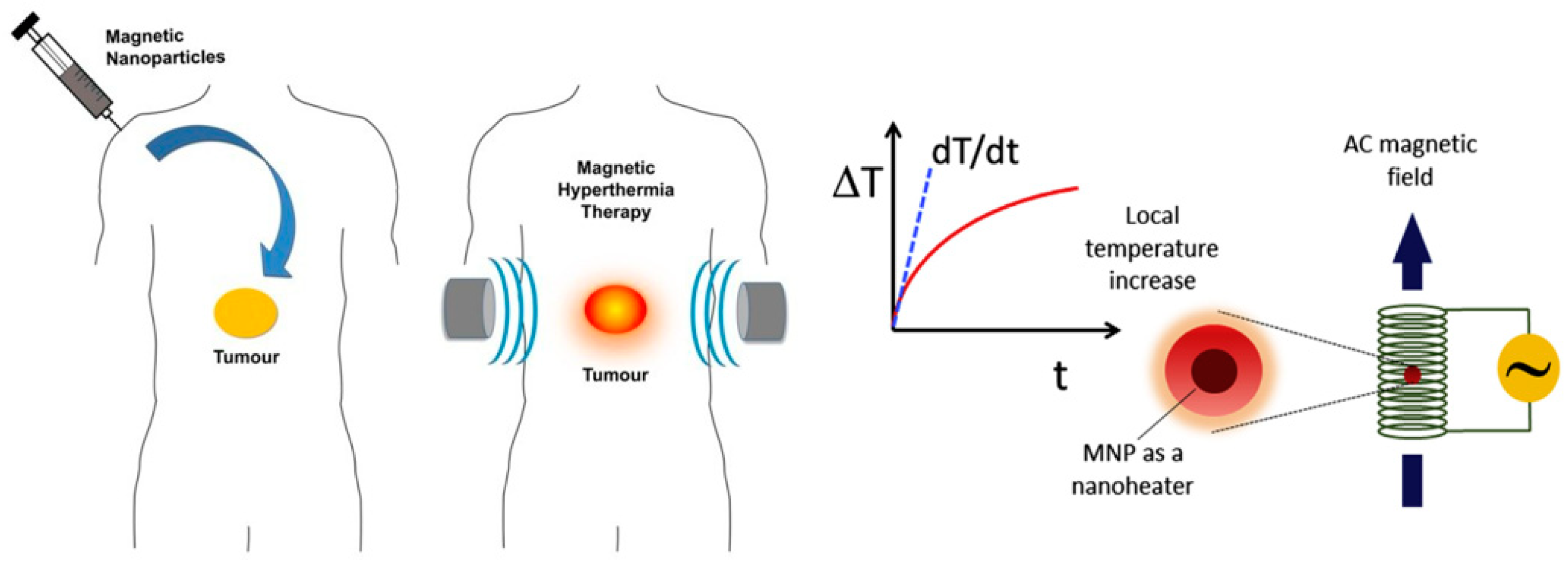
The recent run of industrial development, in particular nanotechnology, has opened-up new possibilities for medical science, especially in the field of magnetic hyperthermia. New superparamagnetic nanoparticle systems of varying nanoscales have been developed and integrated into magnetic hyperthermia (Figure 1) [1,2,3]. The number of patents and products in the field of magnetic hyperthermia is also growing rapidly. A wide range of magnetic hyperthermia technologies are being developed or are already being studied in the clinical stage to provide more effective and safer treatments for clinical applications [4,5,6,7,8,9]. In the field of cancer treatment, one of the fields where magnetic nanotechnologies are most needed, many new superparamagnetic nanoparticles were recently developed, to name a few, Mg0.13Fe2O3 and FASP-Fe3O4, etc. [10,11,12,13,14,15].
Magnetic nanoparticles offer some unique advantages for magnetic hyperthermia. Firstly, the controllability of the morphology and particle size, as well as the sufficiently high AC induction heating power of nanoparticles. The second advantage is based on the capability of nanoparticles to deliver a variety of therapeutic and diagnostic agents, such as peptides, proteins, magnesium, and trace elements, and release active molecules in a controlled manner. Thirdly, nano-carriers can improve the solubility and maximize the stability of drugs [16,17,18,19,20]. Fourthly, through the targeting ability of magnetic nanoparticles, different drug delivery routes and targeted drug delivery can be implemented [21,22,23,24].
This study focuses on developing a new high-biocompatibility superparamagnetic nanoparticle (SNPs), which is expected to be used as a magnetic hyperthermia agent that is not only biologically safe but also boasts high AC induction heating power. The entire development has been a challenge. It was necessary to form superparamagnetic nanoparticles by modifying two well-known magnetic nanoparticles (γ-Fe2O3, Fe3O4) with whey protein isolate (WPI) and applying them to the experiment. From a biological point of view, whey proteins are essential nutrients in life, and WPI can improve the biocompatibility of nanoparticles, promote degradation and metabolism, as well as facilitate the surface modification, drug attachment, and ligand targeting without impeding the magnetism and energy of nanoparticles, these are the reasons why we chose WPI. Subsequently, the potential, granularity, and morphology of both the developed nanoparticles were characterized by using Fourier transform infrared spectroscopy (FTIR), X-ray diffractometer (XRD), transmission electron microscopy (TEM), and scanning electron microscope (SEM). The stability of AC heating power under different ratios and conditions was repeatedly measured and evaluated. At the same time, in order to prove the drug-carrying ability of these nanoparticles, Adriamycin was successfully loaded into these nanoparticles. Adriamycin as an anti-tumor anthracycline antibiotic can be used in mice in vivo experiments. It is slow metabolism during the treatment in mice, showing a significant therapeutic effect. Finally, in vitro and in vivo tests on HepG2 tumor mouse models were carried out to assess the anti-tumor efficiency of the Adriamycin-loaded and WPI-modified superparamagnetic nanoparticles.

2. Materials and Methods

2.1. Materials

The whey protein isolate (WPI) powder was produced by the Department of Food and Nutritional Science of Gyeongsang National University (Jinju, South Korea). The original of γ-Fe2O3 and Fe3O4 particles were purchased from the Korea Institute of Materials Science (Changwon, South Korea). These particles are in powder form and range in size from 100 to 600 nm. The complex enzymes and Adriamycin (ADM) reagent were purchased from Nutrex Biotechnology (Seoul, South Korea). The synthetic media, such as sodium acetate (CH3COONa), (4-aminophenyl) boronic acid, 4.4'-Azobis(4-cyanovaleric acid), tetrabutylammonium bromide(C16H36BrN), methyl sodium(CH3Na), polyethylene glycol(HO(CH2CH2O)nH, hydrazine(N2H4), and polyvinyl pyrrolidone((C6H9NO)n), were supported by the Nano-Information Materials Laboratory of the Department of Materials Engineering and Polymerization Technology of Gyeongsang National University (Jinju, South Korea).

2.2. Theory and Method

2.2.1. Preparation Procedure of WPI

Whey protein concentrates (WPC) was first dissolved in 100 mL deionized water and stirred for 20 min using a magnetic stirrer at 75 °C. An amount of 0.5% complex enzyme was added to the solution, while the temperature and pH of the mixture were adjusted to 45 °C and 7.5, respectively. The reaction went on for 10 h, during which time the temperature and pH remained mostly stable. Finally, the temperature was raised to 75 °C for 20 min before the reaction was stopped. The above solution was cooled to room temperature to obtain the mixed whey protein, then centrifuged at 5500 rpm for 30 min. The supernate was collected and dialyzed in a dialysis bag with a molecular weight cut-off (MWCO) of 7 kDa. Finally, the low-molecular-weight and concentrated whey protein isolate were obtained after vacuum freeze-drying.

2.2.2. Preparation of WPI-SNPs

First, preparation of superparamagnetic nanoparticles followed the steps reported by Prof. Sun Shouheng, preparing the particle-size controlled superparamagnetic nanoparticles of γ-Fe2O3, Fe3O4 using the hydrothermal method [25,26]. WPI (50 mg), (4-aminophenyl) boronic acid (APBA) (30 mg), γ-Fe2O3 (50 mg) were then dispersed into 20 mL distilled water. Then, a certain amount of ACVA (4,4'-Azobis(4-cyanovaleric acid)) (an initiator) was added. The system temperature increased to 70 °C after being exposed to a nitrogen environment. The appearance of milky white color in the reaction system indicated the formation of the WPI-modified superparamagnetic γ-Fe2O3 (WPI-γ-Fe2O3). The resulting suspension was cooled at room temperature, filtered, and dialyzed in distilled water for 24 h with 14 kDa MWCO to remove any aggregates and residual monomers. The same preparation method was also adopted for the WPI-modified superparamagnetic Fe3O4 (WPI-Fe3O4).

2.2.3. ADM Loading of WPI-SNPs

At a pH of 8, 0.5 mg mL−1 of Adriamycin (ADM) was added to the WPI-modified superparamagnetic nanoparticles and mixed with a certain amount of deionized water. At room temperature, it was stirred slowly for 3 h with a light obscuration instrument and then centrifuged at a rate of 6500 rpm for 30 min to separate the Adriamycin-loaded target particles. The loaded Adriamycin was quantified with a microplate reader. The concentration of Adriamycin in the release medium was measured using a molecular device at an excitation wavelength of 400 nm and an emission wavelength of 600 nm. The drug loading content and the drug loading efficiency were evaluated as Equations (1) and (2):
Drug   loading   % = The   weight   of   the   ADM   in   SNPs The   weight   of   the   ADM - loaded   in   SNPs × 100 %
Drug   loading   efficiency   % = The   weight   of   the   ADM   inSNPs The   weight   of   the   feed   in   ADM × 100 %

2.2.4. HepG2 Tumor Inhibition

All experiments involving mouse models were conducted in accordance with guidelines for the use of experimental animals and were approved by the Laboratory Animal Care Agency of Gyeongsang National University. An amount of 0.1 mL kg−1 of HepG2 cells in 0.1 mL saltwater were inoculated on axilla under the left limb of male mice, and mice with tumor volumes of about 200 cubic millimeters were adopted for tests. A primary tumor cell was extracted and harvested from mice for in vitro studies, wrapped in Adriamycin-loaded nanoparticles, and heated by induction heating systems. The other part of mice for experiments in vivo was given intravenous injections at a dose of 1.15 (mg mL1), after which local heating in vivo was conducted using induction heating devices to 46 degrees.

3. Experiment and Results

3.1. Characterization of WPI-SNPs

In this study, WPI was used as the culture medium for modifying nanoparticles, and proteins were hydrolyzed by the complex enzyme to obtain the WPI of low molecular weight in order to improve the solubility. Therefore, the WPI with good solubility was prepared, and then, the WPI-modified superparamagnetic nanoparticles were successfully prepared, which would be used in subsequent experiments. The modified nanoparticles were measured by Fourier transform infrared spectrometer (FTIR) (Table 1). As shown in Figure 2a–d, it is the measured infrared spectral data of the original magnetic nanoparticles (γ-Fe2O3, Fe3O4) and the according to the ratio of 1:1 (WPI = NPs = 50 wt.%) of bio-modified superparamagnetic nanoparticles. Comparing the data at peak (3400, 1637, 1417, 1078 cm−1, etc.) values of the two sets of curves, it is confirmed that surface modification of the nanoparticles was successful. Figure 2e,f show the phase of the γ-Fe2O3, Fe3O4 nanoparticles before and after modification with a WPI by X-ray diffractometer (XRD). Modified nanoparticles phase is shifted, an increase in peak width and staggering of peaks. The results indicate that the nanoparticles are indeed modified by WPI and have an excellent crystalline phase.

3.2. Structural of WPI-SNPs

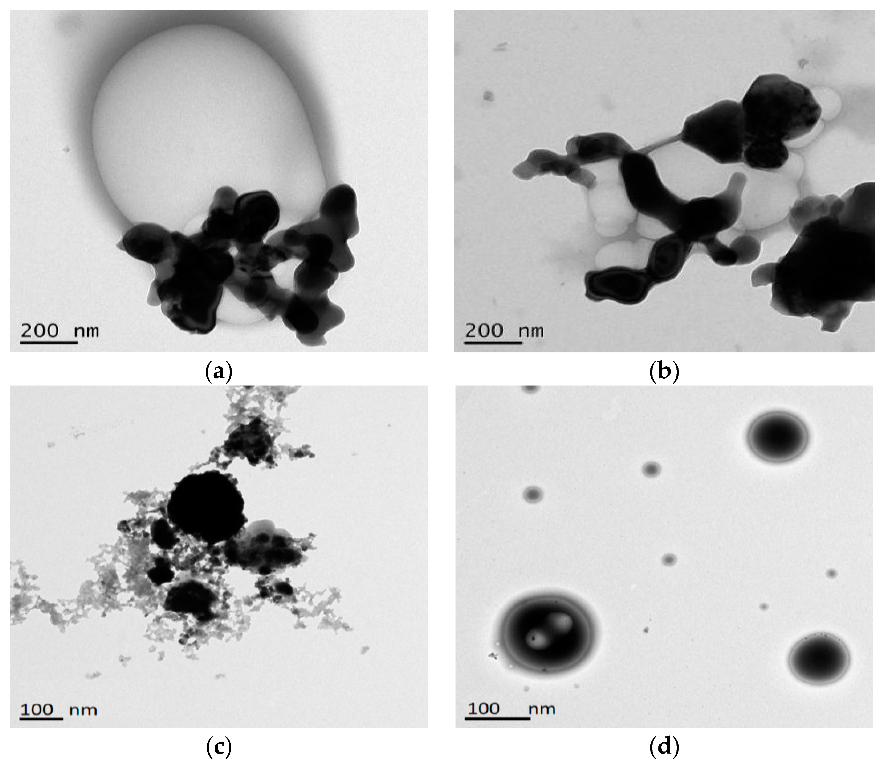
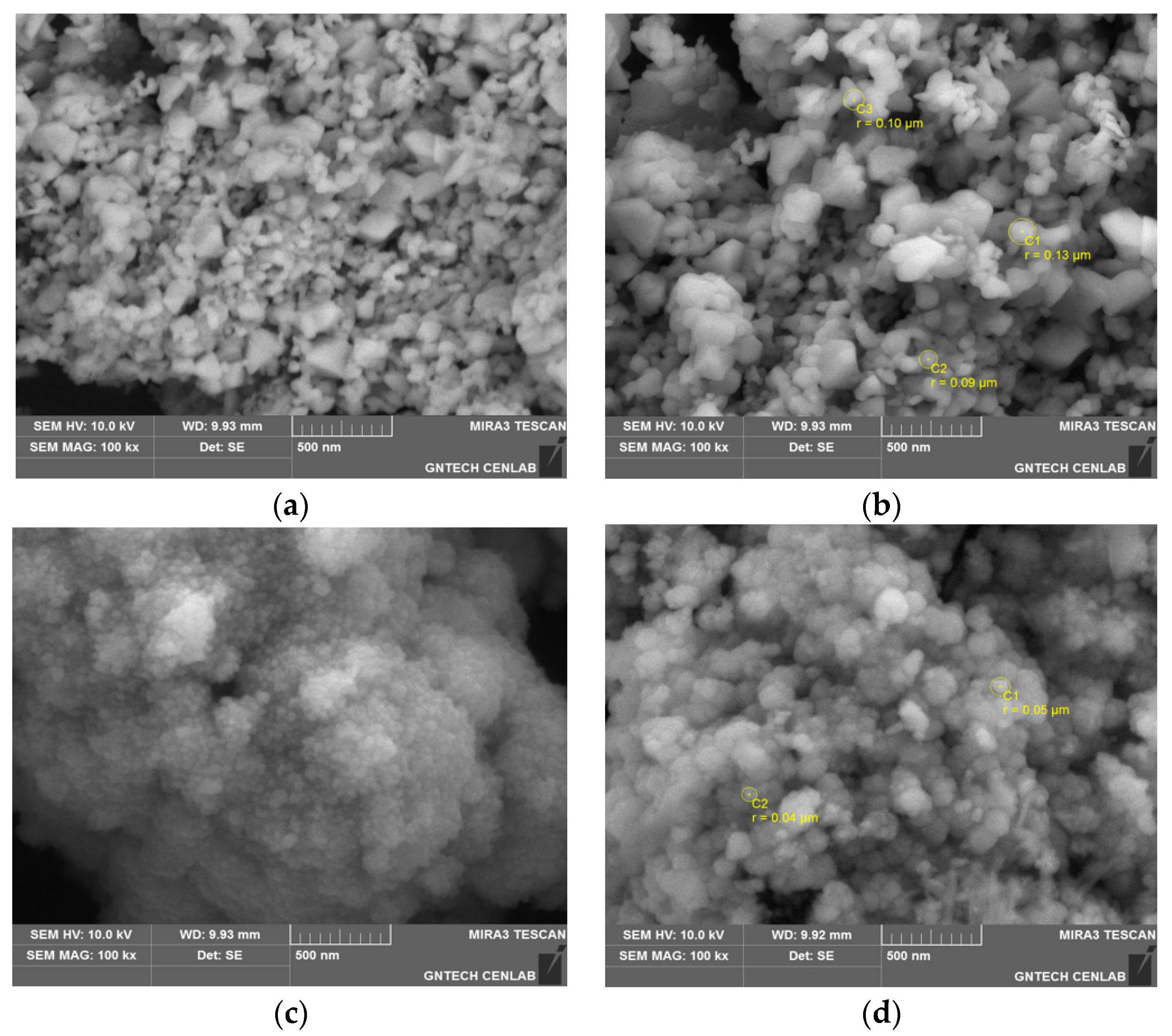
Figure 3 and Figure 4 show the appearance and distribution of original nanoparticles and the bio-modified superparamagnetic nanoparticles (WPI = NPs = 50 wt.%) measured by transmission electron microscopy (TEM) and scanning electron microscope (SEM) (Table 1). From the appearance, the two kinds of nanoparticles have different shapes. The modified γ-Fe2O3 is more like a diamond, while the modified Fe3O4 is more like a sphere. Small molecules surround large molecules, and the smallest nanoparticles reach 20–40 nm diameter by SEM image interception. It can be seen there is no adverse crystal phase of modified nanoparticles, the biomaterials used do not destroy the shape and structure of the original nanoparticles.

3.3. Heating Properties of WPI-SNPs

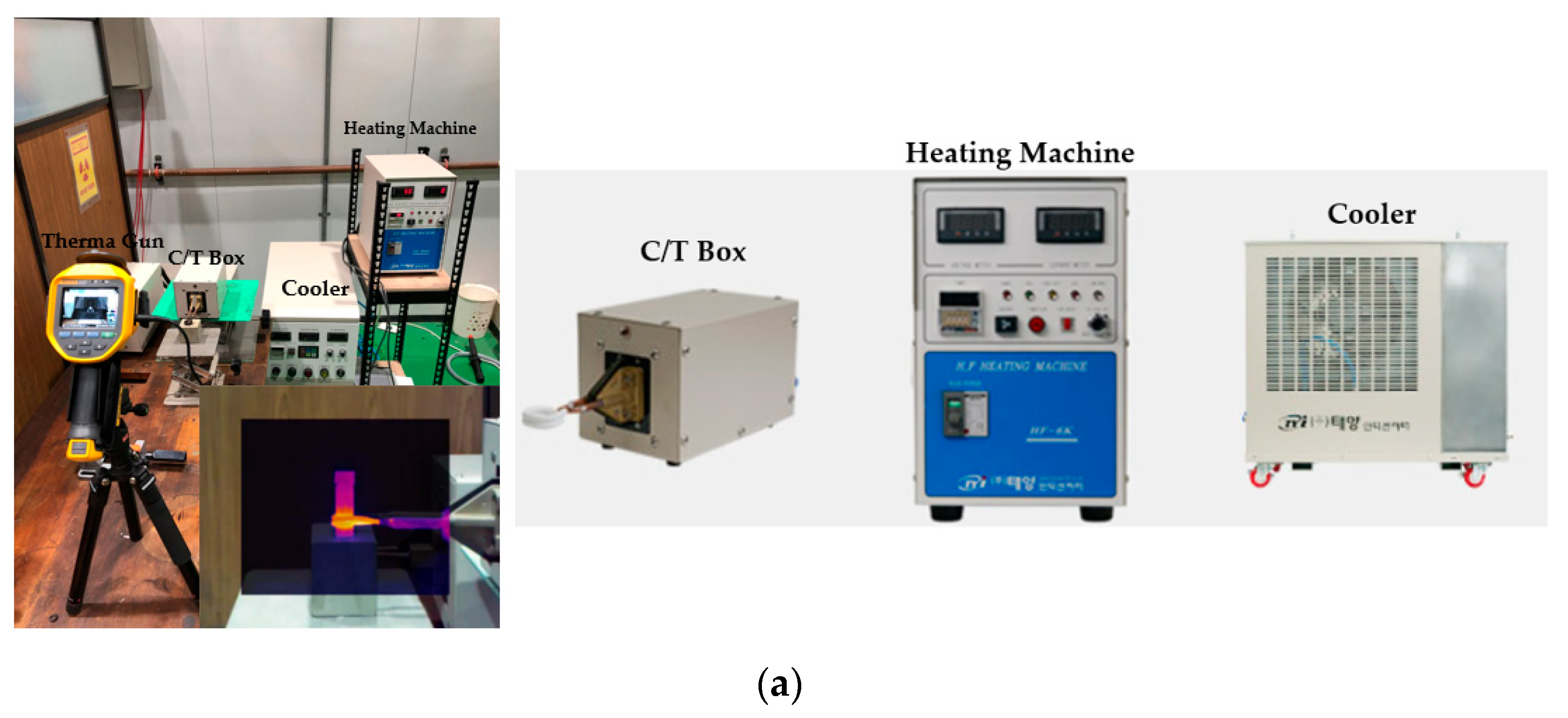
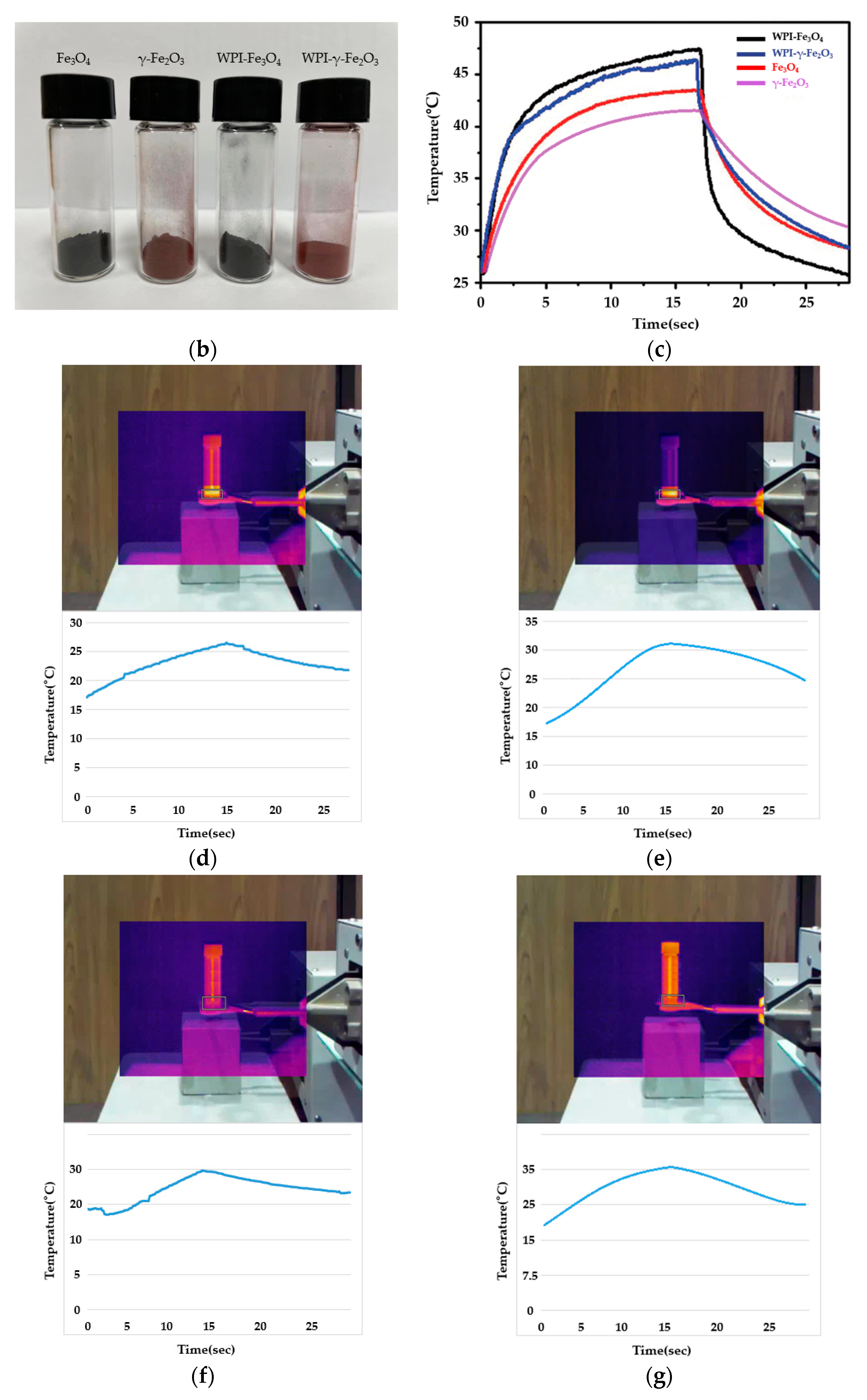
One of the most essential criteria for magnetic hyperthermia is the heating capacity of nanoparticles. In other words, magnetic nanoparticles can treat tumor cells because they can be delivered to target cells and release heat through magnetic induction. According to previous reports, nanoparticles heated to 39–48 °C are effective in treating tumors [27,28,29]. Therefore, induction heating devices were used to test the heating of the modified nanoparticles shown in Figure 5a. The original nanoparticles and the prepared bio-modified nanoparticles (WPI = NPs = 50 wt.%) were placed in a heat-resistant tube at the center of the AC electromagnetic coil and heated, exposing to AC magnetic fields by induction heating devices. Figure 5b,c show the magnetic thermal efficiency was compared with the original nanoparticles (50 mg mL−1) and the modified γ-Fe2O3, Fe3O4 (basic nanoparticle quantifies 50 mg, and then modified with WPI 50 mg) in the same high-power dissipation (6 KW) induction heating for 15 s. The WPI-modified nanoparticles performed well, and the heating temperature was higher than the original nanoparticles. Although the results may be related to the refined superparamagnetic nanoparticles, the biomaterials modification also played a role.
After a number of experiments on concentrations of WPI-nanoparticles, which contained γ-Fe2O3 and Fe3O4 ranged from 50 wt.% to 90 wt.%, under normal power dissipation conditions (3 KW), frequency (100 kHz), and rated time (15 s), we found optimal conditions that WPI-γ-Fe2O3 with γ-Fe2O3 (67 wt.%) and WPI-Fe3O4 with Fe3O4 (85 wt.%) are shown in Figure 5d–g. WPI-γ-Fe2O3(67wt%) can be an easier control of temperature, continuously heated, and slower temperature drop. WPI-Fe3O4(85wt%) can be rapid heating to target temperature in a short time, good thermal destructive power, and faster temperature drop. Based on the above results, two different types of modified superparamagnetic nanoparticles can be selected for different tumor types or different treatment options.
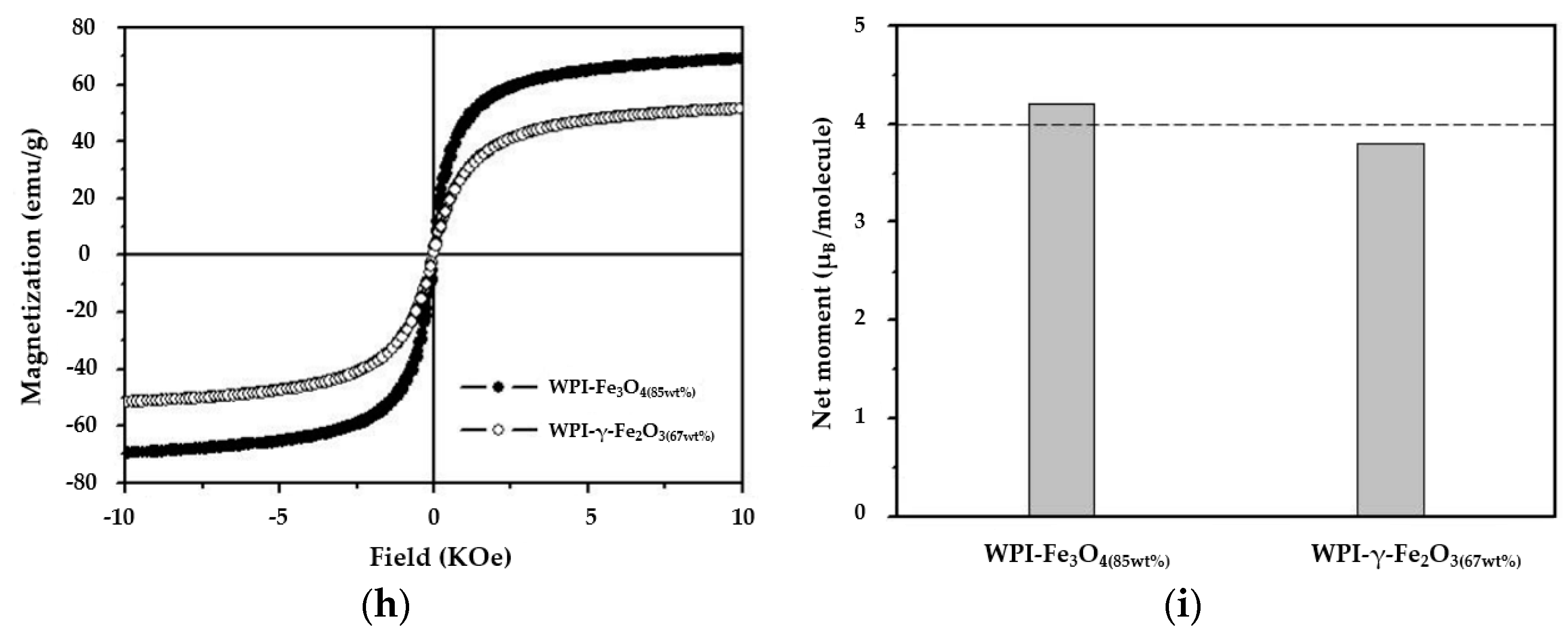
Figure 5h shows Ms value of WPI-γ-Fe2O3(67wt%) and WPI-Fe3O4(85wt%) is 51 (emu/g) and 69 (emu/g). The net magnetic moment, which can be expressed alternatively in terms of μB (Bohr Magnetron) as calculated by the distribution obtained experimentally using Mössbauer spectroscopy, indicated that 3.7 (WPI-γ-Fe2O3(67wt%)) and 4.2 (WPI-Fe3O4(85wt%)) were clearly seen shown in Figure 5i.

3.4. In Vitro Release of WPI-SNPs

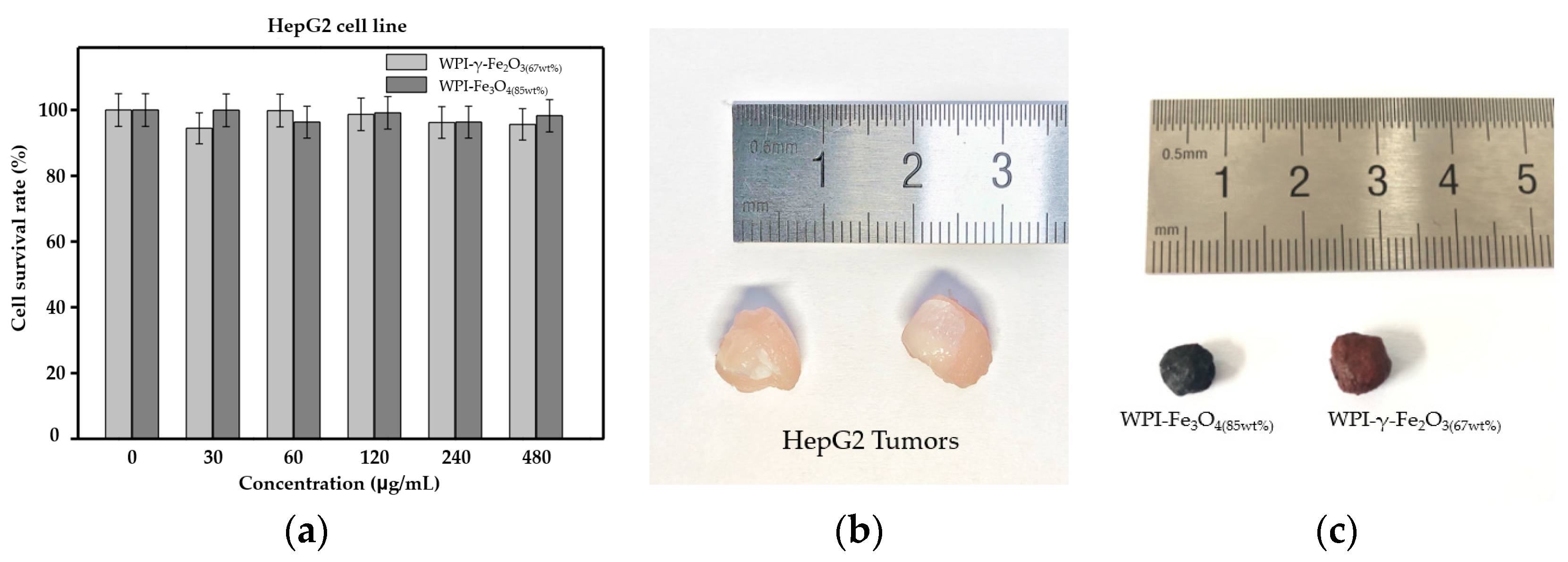
After determining the absorption transmittance, morphological structure, magnetism, AC heating, and other characteristics, Adriamycin was attached to the modified superparamagnetic nanoparticles by electrostatic interaction between nanoparticles and reached an ideal condition of 15% drug loading capacity and 78.53% drug loading efficiency, which also fully explains that the WPI-modified superparamagnetic nanoparticles have a drug loading capacity of higher efficiency. It also supported our subsequent experiments. During conducting in vitro experiments using HepG2, cells were taken from mice, firstly biocompatibility analysis of WPI-γ-Fe2O3, WPI-Fe3O4was performed, in vitro cytotoxicity test of HepG2 cell with WPI-γ-Fe2O3, WPI-Fe3O4 was carried out at the concentration of 30, 60, 120, 240, 480 μg/mL using a Cell Counting Kit-8 analysis. HepG2 cell was treated with WPI-γ-Fe2O3, WPI-Fe3O4 by 24 h. Figure 6a shows the cell survival rate proved that WPI-γ-Fe2O3, WPI-Fe3O4 showed high biocompatibility (non-cytotoxic).
Attempts were made in culture, anatomy, and medicine, and the tumors were placed in the center of the AC electromagnetic coil and exposed to an AC magnetic field (f = 100 kHz, H = 155Oe T (12.33 kA m−1), H · f = 1.23 × 10⁹ A m−1 s−1) for in vitro magnetic hyperthermia by taking out of the subcutaneous tissue of the mice and enveloping it with WPI-modified nanoparticles. The temperature of HepG2 injected with WPI-Fe3O4(85wt%) and WPI-γ-Fe2O3(67wt%) was rapidly increased and stably saturated at ≈48.5 °C during the 15 s of in vitro hyperthermia. Finally, successfully treated HepG2 cells were taken from mice with in vitro hyperthermia. As shown in Figure 6b,c, it is seen that the HepG2 tumor after the hyperthermia not only become smaller but also has the surrounding surface burned and coagulated, losing the biological activity. The more exciting thing that a membrane of tiny lactose components formed around the surface of the tumor through WPI in the heating process, the lactose component is also one of the reasons to increase the heating rate.

3.5. In Vivo Magnetic Hyperthermia

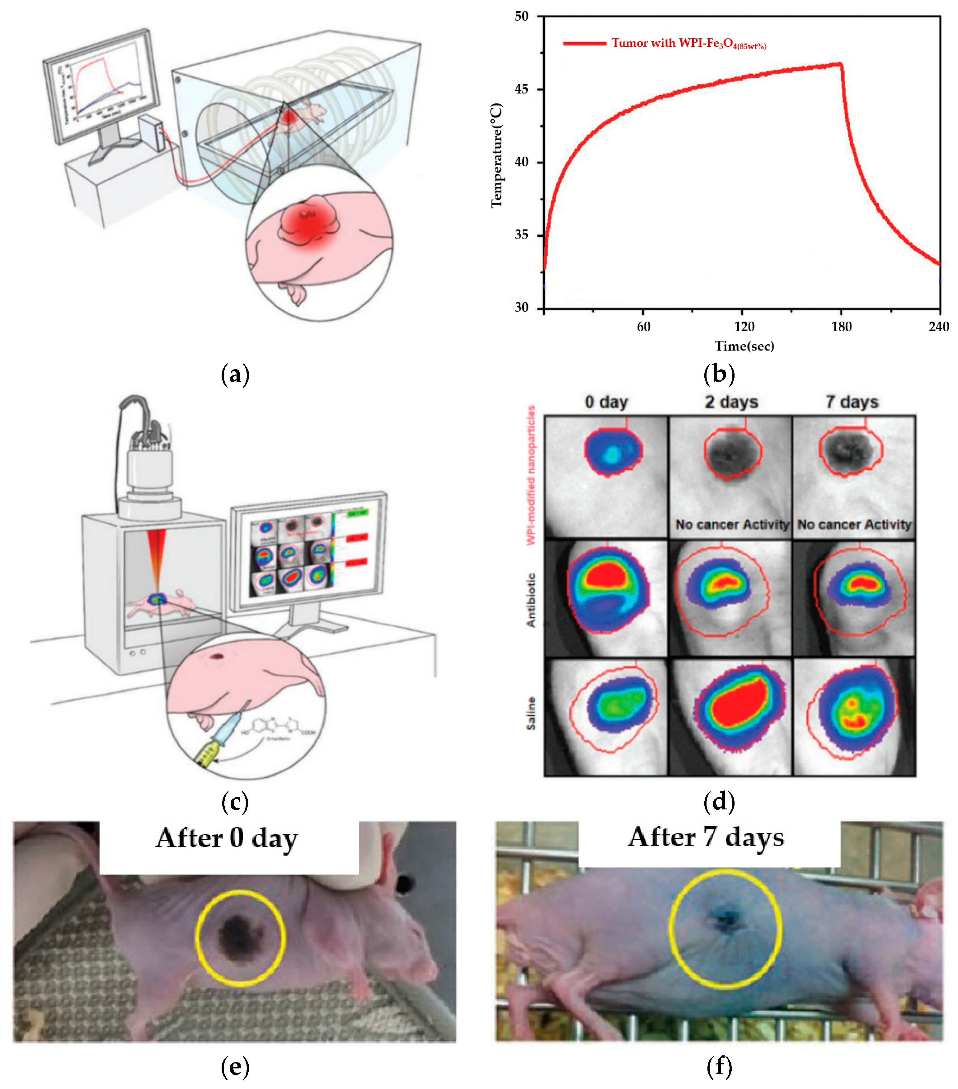
The expense of in vivo magnetic thermotherapy experiments is relatively costly. The most optimal conditions of WPI-Fe3O4(85wt%) to make a nanofluid for in vivo experiments in mouse models have been selected for the therapeutic characteristics of HepG2 tumor cells requiring instantaneous high temperatures to treat tumors. HepG2 cells were kept growing under the skin of mice; nanofluids (1.15 mg mL−1) made of by 100 µL modified superparamagnetic nanoparticles were injected into the tumor through soft tissue surfaces by curved needles, and an optical thermometer was placed in the tumor of the rectal area individual to monitor the temperature during the magnetic hyperthermia process. The mice were placed in the center of the AC electromagnetic coil and exposed to an AC magnetic field (f = 100 kHz, H = 169Oe T (13.62 kA m−1), H · f = 1.36 × 10⁹ A m−1 s−1) for magnetic hyperthermia. The temperature of HepG2 injected with WPI-Fe3O4(85wt%) nanofluids was rapidly increased and stably saturated at ≈46.5 °C during 180 s of hyperthermia, which is expected to be at a thermotherapy temperature (Figure 7a,b). After the magnetic hyperthermia, it can be clearly observed that tumors induced by HepG2 and treated by the WPI-modified superparamagnetic nanoparticles were significantly reduced, damaged by heat, and lost activity (Figure 7c,d). After 7 days of observation after the high-temperature magnetic hyperthermia, no physical damage caused by AC magnetic hyperthermia, tumor regrowth, and serious side effects was observed, thus indicating that AC magnetic hyperthermia applied to tumors was biologically safe for the mice injected (Figure 7e,f). To be clear, due to the imperfection of the experimental equipment and to ensure the integrity, safety, and accuracy of the experiment, we authorized the Korea Institute of Biomaterial to conduct the magnetic hyperthermia experiment in vivo on mice and used high-energy thermal inductance (HETI) technology for periodic observation. In summary, the experimental results confirmed that the use of WPI-modified nanoparticles improved the effectiveness of magnetic hyperthermia in tumor therapy.

4. Conclusions

In this study, the water-soluble low-molecule WPI was prepared by hydrolyzing the WPI. Superparamagnetic nanoparticles (γ-Fe2O3, Fe3O4) prepared by hydrothermal methods were used as tumor-targeting carriers and combined with WPI into bio-modified superparamagnetic nanoparticles, which have an average diameter of 20 to 100 nm, crystal particle morphology, good stability, and biocompatibility under various conditions and were successfully loaded with an ideal load of Adriamycin. Due to the biological properties of the WPI, the efficacy of modified nanoparticles in magnetic hyperthermia was improved. Various chemical and physical analyses were conducted to verify the structure, morphology, biocompatibility, magnetic properties, and induction heating properties of the newly developed nanoparticles. In vitro and in vivo magnetic hyperthermia studies were conducted using HepG2 tumor and HepG2 mouse models to evaluate its clinical feasibility as a magnetic hyperthermia agent. In summary, the research findings fully prove that WPI-modified superparamagnetic nanoparticles have excellent AC induction heating power and biocompatibility in the biologically safe range of AC magnetic field usage. Thus, it should be further popularized as a new magnetic hyperthermia agent for treating tumors.

Author Contributions

Conceptualization, S.Z.; Data curation, S.Z.; Formal analysis, S.Z.; Funding acquisition, S.L.; Investigation, S.Z.; Project administration, S.L.; Validation, S.Z.; Visualization, S.L.; Writing—original draft, S.Z.; Writing—review & editing, S.L. All authors have read and agreed to the published version of the manuscript.

Funding

This research was financially supported by the Brain Korea 21 Plus (BK21+) Project of the GNU Graduate Education and Research Program for Mechanical and Aerospace Engineers.

Institutional Review Board Statement

The study was conducted according to the guidelines of the Declaration of Helsinki, and approved by the Gyeongsang National University Institutional Animal Care Use Committee (GNU2016-121).

Informed Consent Statement

Not applicable.

Data Availability Statement

Not applicable.

Acknowledgments

We are grateful to Nano-Information Materials Laboratory of the Department of Materials Engineering and Polymerization Technology of Gyeongsang National University for providing resources and support to complete this research. We are also grateful to the Food Microbiology Laboratory and the Animal Neuroscience Laboratory of Gyeongsang National University for their guidance and help in research.

Conflicts of Interest

The authors declare no conflict of interest.

References

  1. Lemine, O.M. Magnetic Hyperthermia Therapy Using Hybrid Magnetic Nanostructures; Chapter 7; Elsevier: Riyadh, Saudi Arabia, 2019; pp. 125–138. [Google Scholar]
  2. Rivas, J.; Kolen’ko, Y.V.; Banobre-lopez, M. Magnetic Nanocolloids; Chapter 3; Elsevier: Braga, Portugal, 2016; pp. 75–129. [Google Scholar]
  3. Jeun, M.; Kim, Y.J.; Park, K.H.; Paek, S.H.; Bae, S. Physical Contribution of Néel and Brown Relaxation to Interpreting Intracellular Hyperthermia Characteristics Using Superparamagnetic Nanofluids. J. Nanosci. Nanotechnol. 2013, 13, 5719–5725. [Google Scholar] [CrossRef] [PubMed]
  4. Hilger, I.; Hergt, R.; Kaiser, W.A. Use of magnetic nanoparticle heating in the treatment of breast cancer. IEE Proc.-Nanobiotechnol. 2005, 152, 33–39. [Google Scholar] [CrossRef]
  5. Hergt, R.; Dutz, S.; Roder, M. Effects of size distribution on hysteresis losses of magnetic nanoparticles for hyperthermia. J. Phys. Condens. Matter 2008, 20, 38–50. [Google Scholar] [CrossRef] [PubMed]
  6. Xie, J.; Chen, K.; Lee, H.Y.; Xu, C.; Hsu, A.R.; Peng, S.; Chen, S.; Sun, S. Ultra-Small c(RGDyK)-Coated Fe3O4 Nanoparticles and Their Specific Targeting to Integrin avB3-rich Tumor Cells. J. Am. Chem. Soc. 2008, 130, 7542–7543. [Google Scholar] [CrossRef] [PubMed] [Green Version]
  7. Xu, Z.; Shen, C.; Hou, Y.; Gao, H.; Sun, S. Oleylamine as Both Reducing Agent and Stabilizer in a Facile Synthesis of Magnetite Nanoparticles. Chem. Mater. 2009, 21, 1778–1780. [Google Scholar] [CrossRef]
  8. Cui, X.J.; Dong, L.L.; Zhong, S.L.; Shi, C.; Sun, Y.X.; Chen, P. Sonochemical fabrication of folic acid functionalized multistimuli-responsive magnetic graphene oxide-based nanocapsules for targeted drug delivery. Chem. Eng. J. 2017, 326, 839–848. [Google Scholar] [CrossRef]
  9. Li, S.; Zhang, J.; Deng, C.; Meng, F.H.; Yu, L.; Zhong, Z.Y. Redox-sensitive and intrinsically fuorescent photoclick hyaluronic acid nanogels for traceable and targeted delivery of cytochrome c to breast tumor in mice. ACS Appl. Mater. Interfaces 2016, 8, 21155–21162. [Google Scholar] [CrossRef]
  10. Kale, S.S.; Burga, R.A.; Sweeney, E.E.; Zun, Z.; Sze, R.W.; Tuesca, A.; Subramony, A.; Fernandes, R. Composite iron oxide-Prussian blue nanoparticles for magnetically guided T1-weighted magnetic resonance imaging and photothermal therapy of tumors. Int. J. Nanomed. 2017, 12, 6413–6418. [Google Scholar] [CrossRef] [PubMed] [Green Version]
  11. Kandasamy, G.; Sudame, A.; Luthra, T.; Saini, K.; Maity, D. Functionalized Hydrophilic Superparamagnetic Iron Oxide Nanoparticles for Magnetic Fluid Hyperthermia Application in Liver Cancer Treatment. ACS Omega 2018, 3, 3991–4005. [Google Scholar] [CrossRef]
  12. Ji, H.; Zhou, S.; Fu, Y.; Wang, Y.; Mi, J.; Lu, T.; Wang, X.; Lü, C. Size-controllable preparation and antibacterial mechanism of thermo-responsive copolymer-stabilized silver nanoparticles with high antimicrobial activity. Mater. Sci. Eng. C. 2020, 110, 110735. [Google Scholar] [CrossRef] [PubMed]
  13. Song, Z.; Wu, Y.; Wang, H.; Han, H. Synergistic antibacterial effects of curcumin modified silver nanoparticles through ROS-mediated pathways. Mater. Sci. Eng. C. 2019, 99, 255–263. [Google Scholar] [CrossRef]
  14. Hajizadeh, H.; Peighambardoust, S.J.; Peighambardoust, S.H.; Peressini, D. Physical, mechanical, and antibacterial characteristics of bio-nanocomposite films loaded with Ag-modified SiO2 and TiO2 nanoparticles. J. Food Sci. 2020, 85, 1193–1202. [Google Scholar] [CrossRef] [PubMed]
  15. Jordan, A.; Scholz, R.; Maier-Hauff, K.; Johannsen, M.; Wust, P.; Nadobny, J.; Schirra, H.; Schmidt, H.; Loening, E.; Felix, R.; et al. Presentation of a new magnetic field therapy system for the treatment of human solid tumors with magnetic fluid hyperthermia. J. Magn. Magn. 2001, 225, 118–126. [Google Scholar] [CrossRef] [Green Version]
  16. Kandasamy, G.; Sudame, A.; Bhati, P.; Chakrabarty, A.; Kale, S.N.; Maity, D. Systematic magnetic fluid hyperthermia studies of carboxyl functionalized hydrophilic superparamagnetic iron oxide nanoparticles based ferrofluids. J. Colloid Interface Sci. 2018, 514, 534–543. [Google Scholar] [CrossRef]
  17. Saeedi, M.; Vahidi, O.; Bonakdar, S. Synthesis and characterization of glycyrrhizic acid coated iron oxide nanoparticles for hyperthermia applications. Mater. Sci. Eng. C. Mater. Biol. Appl. 2017, 77, 1060–1067. [Google Scholar] [CrossRef]
  18. Lahiri, B.B.; Ranoo, S.; Philip, J. Magnetic hyperthermia study in water based magnetic fluids containing TMAOH coated Fe3O4 using infrared thermography. Infrared Phys. Technol. 2017, 80, 71–82. [Google Scholar] [CrossRef]
  19. Oenbrink, G.; Jurgenlimke, P.; Gabel, D. Accumulation of porphyrins in cells: Influence of hydrophobicity aggregation and protein binding. Photochem. Photobiol. 1988, 48, 451–456. [Google Scholar] [CrossRef] [PubMed]
  20. Martinkova, P.; Brtnicky, M.; Kynicky, J.; Pohanka, M. Iron Oxide Nanoparticles: Innovative Tool in Cancer Diagnosis and Therapy. Adv. Healthc. Mater. 2017, 7, 1700932. [Google Scholar] [CrossRef] [PubMed]
  21. Basini, M.; Guerrini, A.; Cobianchi, M.; Orsini, F.; Bettega, D.; Avolio, M.; Innocenti, C.; Sangregorio, C.; Lascialfari, A.; Arosio, P. Tailoring the magnetic core of organic-coated iron oxides nanoparticles to influencetheir contrast efficiency for Magnetic Resonance Imaging. J. Alloys Compd. 2019, 770, 58–66. [Google Scholar] [CrossRef]
  22. Peddis, D.; Cannas, C.; Musinu, A.; Ardu, A.; Orrù, F.; Fiorani, D.; Laureti, S.; Rinaldi, D.; Muscas, G.; Concas, G.; et al. Beyond the Effect of Particle Size: Influence of CoFe2O4 Nanoparticle Arrangements on Magnetic Properties. Chem. Mater. 2013, 25, 2005–2013. [Google Scholar] [CrossRef]
  23. Omelyanchik, A.; Salvador, M.; D’Orazio, F.; Mameli, V.; Cannas, C.; Fiorani, D.; Musinu, A.; Rivas, M.; Rodionova, V.; Varvaro, G.; et al. Magnetocrystalline and Surface Anisotropy in CoFe2O4 Nanoparticles. Nanomaterials 2020, 10, 1288. [Google Scholar] [CrossRef]
  24. Xie, H.; Zhu, Y.; Jiang, W.; Zhou, Q.; Yang, H.; Gu, N.; Zhang, Y.; Xu, H.; Xu, H.; Yang, X. Lactoferrin-conjugated superparamagnetic iron oxide nanoparticles as a specific MRI contrast agent for detection of brain glioma in vivo. Biomaterials 2011, 32, 495–502. [Google Scholar] [CrossRef]
  25. Sun, S.; Zeng, H. Size-controlled synthesis of magnetite nanoparticles. J. Am. Chem. Soc. 2002, 124, 8204–8205. [Google Scholar] [CrossRef] [PubMed]
  26. Sun, S.; Zeng, H.; Robinson, D.B.; Raoux, S.; Rice, P.M.; Wang, S.X.; Li, G. Monodisperse MFe2O4 (M = Fe, Co, Mn) Nanoparticles. J. Am. Chem. Soc. 2004, 126, 273–279. [Google Scholar] [CrossRef] [PubMed]
  27. Kumar, C.; Mohammad, F. Magnetic nanomaterials for hyperthermia-based therapy and controlled drug delivery. Adv. Drug Deliv. Rev. 2011, 63, 789–808. [Google Scholar] [CrossRef] [Green Version]
  28. Ho, D.; Sun, X.L.; Sun, S.H. Monodisperse magnetic nanoparticles for theranostic applications. Accounts Chem. Res. 2011, 44, 875–882. [Google Scholar] [CrossRef] [PubMed] [Green Version]
  29. Hildebrandt, B.; Wust, P.; Ahlers, O.; Dieing, A.; Sreenivasa, G.; Kerner, T.; Felix, R.; Riess, H. The cellular and molecular basis of hyperthermia. Crit. Rev. Oncol. Hemat. 2002, 43, 33–56. [Google Scholar] [CrossRef]
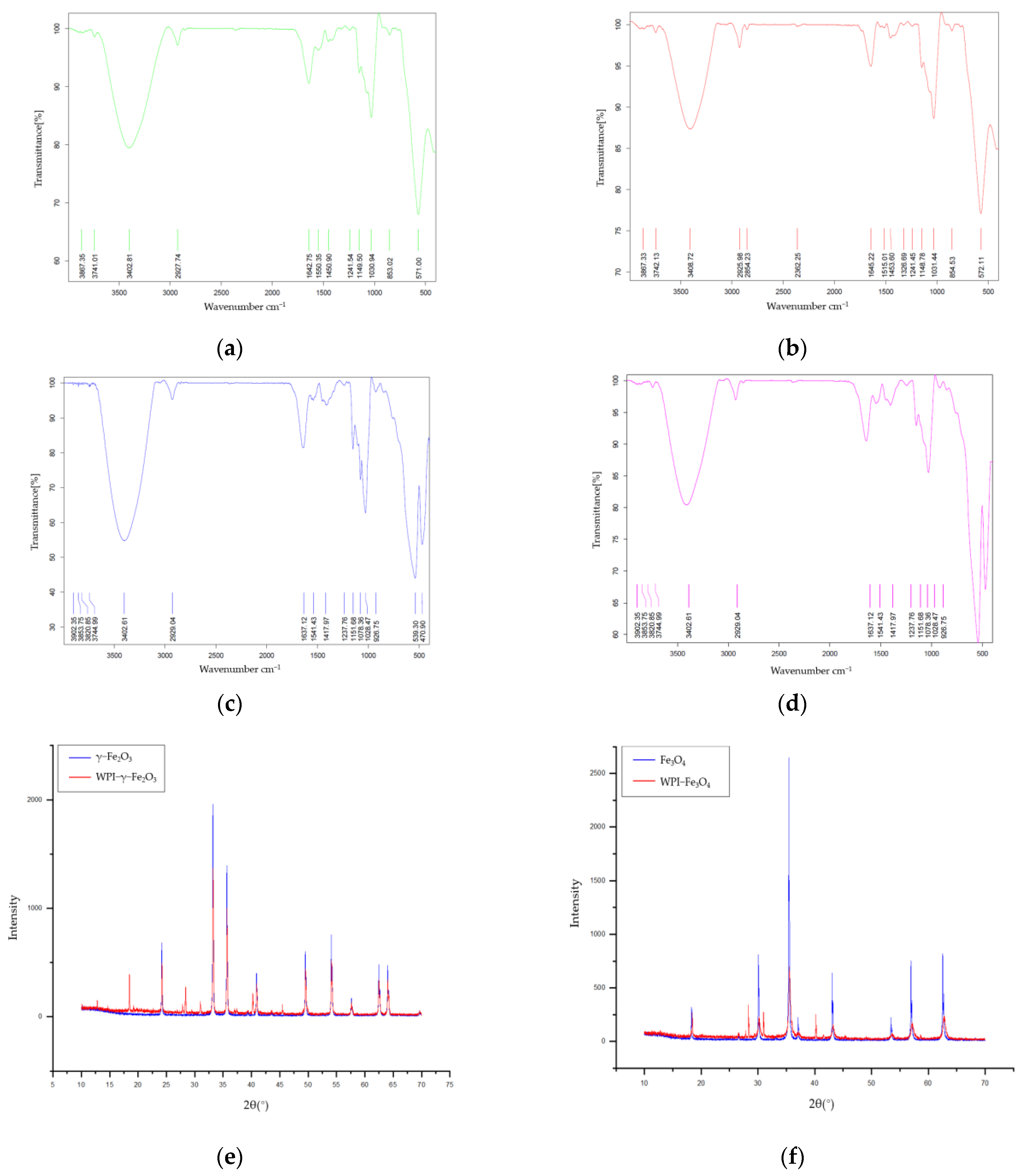
Figure 1. Magnetic nanoparticles hyperthermia [1,2].
Applsci 11 02017 g001
Figure 2. Characterization of WPI−SNPs: (a) FTIR spectra result of the original γ−Fe2O3; (b) FTIR spectra result of the modified WPI−γ−Fe2O3; (c) FTIR spectra result of the original Fe3O4; (d) FTIR spectra result of the modified WPI−Fe3O4; (e) XRD comparison result of γ−Fe2O3 and WPI−γ−Fe2O3; (f) XRD comparison result of Fe3O4 and WPI−Fe3O4.
Applsci 11 02017 g002
Figure 3. TEM image of (a) γ−Fe2O3; (b) WPI−γ−Fe2O3; (c) Fe3O4; (d) WPI−Fe3O4.
Applsci 11 02017 g003
Figure 4. SEM image of (a) γ−Fe2O3; (b) WPI−γ−Fe2O3; (c) Fe3O4; (d) WPI−Fe3O4.
Applsci 11 02017 g004
Figure 5. (a) Induction heating devices and ThermalGun that measure temperature; (b,c) induction heating results of WPI-γ-Fe2O3 and WPI-Fe3O4 compared to the γ-Fe2O3 and Fe3O4; (d) heating results of WPI-γ-Fe2O3(50wt%); (e) heating results of WPI-γ-Fe2O3(67wt%); (f) heating results of WPI-Fe3O4(50wt%); (g) heating results of WPI-Fe3O4(85wt%); (h) hysteresis curve result of WPI-γ-Fe2O3(67wt%) and WPI-Fe3O4(85wt%); (i) the net magnetic moment of WPI-γ-Fe2O3(67wt%) and WPI-Fe3O4(85wt%).
Applsci 11 02017 g005aApplsci 11 02017 g005bApplsci 11 02017 g005c
Figure 6. In vitro release of WPI-SNPs: (a) cells survival rate results of WPI-γ-Fe2O3(67wt%) and WPI-Fe3O4(85wt%) with HepG2 cell line; (b) HepG2 tumors were taken from mice for in vitro experiments; (c) HepG2 tumor after in vitro magnetic hyperthermia with WPI-γ-Fe2O3(67wt%) and WPI-Fe3O4(85wt%).
Applsci 11 02017 g006
Figure 7. (a) Model of in vivo magnetic hyperthermia with mice carrying HepG2 tumors; (b) results of tumor temperature during in vivo magnetic hyperthermia; (c) model of high-energy thermal inductance (HETI) equipment for check result of tumor scale after in vivo magnetic hyperthermia; (d) set result of tumor scale: the first row using in vivo magnetic hyperthermia with WPI-Fe2O3(85wt%). The middle row using treatment with Adriamycin. The bottom row using treatment with normal saline; (e) the survival curve of mice at 0 day after magnetic hyperthermia; (f) the survival curve of mice at 7 days after magnetic hyperthermia.
Applsci 11 02017 g007
Table 1. Instrumentation.
FTIRXRDTEMSEMInduction Heater
Spectral range: 7800~100 cm−1Measuring circle diameter: 600 mmLaB6, high-voltage range: 20 to 120 kVResolution: 0.5 nm at 15 kV with BD; 0.8 nm at 1 kV with BD; 1.0 nm at 15 kV in LowVac380 H × 230 W × 385 L
Resolution: 0.09 cm−1Max. usable angular range: −60~+158Point resolution: 0.34 nmLanding energy: 20 eV~30 keVPower dissipation: 6 KW
Scan speed: 65 scan/secX-Ray Generator: 60 kV, 2.2 kWLine resolution: 0.20 nmEDS resolution: 127 eV at Mn Ka, ta 130,000 cpsFrequency: 100 KHz–600 KHz
S/N ratio: 55,000:1Maximum Rotating Speed: 120 rpmGatan US1000 digital CCD cameraHigh-vacuum heating stage to 1100 °C/ EDS compatibility to 500 °CPower supply: single-phase 220 VAC, 50/60 Hz
Publisher’s Note: MDPI stays neutral with regard to jurisdictional claims in published maps and institutional affiliations.