Review

Pedobarography: A Review on Methods and Practical Use in Foot Disorders

by
Jacek Lorkowski
1,2,
Karolina Gawronska
3 and
Mieczyslaw Pokorski
4,5,*
1
Clinical Department of Orthopedics, Traumatology and Sports Medicine of the Central Clinical Hospital of the Ministry of the Internal Affairs and Administration, 137 Woloska Street, 02-507 Warsaw, Poland
2
Faculty of Health Sciences, Medical University of Mazovia, 8 Rydygiera Street, 01-793 Warsaw, Poland
3
Rehabilitation Center of the Central Clinical Hospital of the Ministry of the Internal Affairs and Administration, 137 Woloska Street, 02-507 Warsaw, Poland
4
Institute of Health Sciences, Opole University, 68 Katowicka Street, 45-060 Opole, Poland
5
Faculty of Health Sciences, The Jan Długosz University in Częstochowa, 4/8 Waszyngtona Street, 42-200 Częstochowa, Poland
*
Author to whom correspondence should be addressed.
Appl. Sci. 2021, 11(22), 11020; https://doi.org/10.3390/app112211020
Submission received: 19 October 2021 / Revised: 10 November 2021 / Accepted: 18 November 2021 / Published: 21 November 2021
(This article belongs to the Special Issue Advances in Technology of Brain-Computer Interface)

Abstract

:
Pedobarographic examination is a non-invasive method that enables the quantitative and qualitative evaluation of plantar pressure distribution, notably the plantar pressure distribution, referring to the function of the entire musculoskeletal system. This is a scoping review that aims to update knowledge on the practical use of pedobarography in foot disorders. We also attempted to systematize the methodological principles of conducting the pedobarographic examination. We searched Medline/PubMed, Embase, Web of Science, and the Cochrane Database of Systematic Reviews for the articles on the methodology of pedobarography. The search encompassed clinical trials, randomized controlled trials, meta-analyses, and reviews published in English between January 1982 and February 2021. The literature distinguishes three different types of examinations: static, postural, and dynamic. The rationale for each is presented. The review pointedly shows the unique use of pedobarography for the quantitative and qualitative evaluations of the plantar pressure distribution. It also points to the need for enhancing the awareness among medical professionals of the method and advantages it provides for patient management. Shortcomings of the method are discussed of which the difficulty in establishing the cause-and-effect relationship of foot disorders is the most disturbing as it limits the comparative verification of results of different studies. There also appears a need for developing standardized algorithmic protocols and recommendations to seamlessly perform pedobarography in clinical settings, which would help make wider use of this valuable tool.

1. Introduction

Foot disorders are an underestimated widespread ailment, particularly in developed societies. Although often neglected as being of little medical importance, foot disorders may be crippling at any age, adversely affect the quality of life, and hinder the musculoskeletal development and socioemotional development of children [1,2]. Examples are the flat foot or platypodia, one of the commonest disorders [3,4], and more serious conditions such as a discrepancy in leg length, causing a differential overload of both legs, which destabilize posture and may lead to disability [5]. The diagnostic methods for foot disorders are not standardized or unified, appear uncertain and troublesome, and are often guided empirically or by healers’ experience rather than being evidence-based.
Recent technological developments, including artificial intelligence, nanotechnology, and medical engineering supported by image analysis, have enabled musculoskeletal diagnostics based on biomechanics rather than the anatomy alone [6,7]. The pedobarography is an examination of lower limbs, foremost of plantar pressure distribution pattern, providing a graphic illustration of results on standing and walking [8,9,10]. Marey and Demeny, who pioneered in the scanning and evaluation of the plantar foot surface, laid the foundation for contemporary pedobarography in 1880 [11]. Beely [12] later provided the first well-documented data on the plantar surface. He evaluated footprints using a linen bag with a quick-drying plaster inside. In 1901, Seitz [13] proposed the capillary blood flow at the plantar surface as a pressure change indicator. The study considered the first podoscopy trial. Patients were instructed to stand on a glass plate so that plantar skin color changes could be monitored with a mirror. Despite the promising results, progress in foot research was limited. Further studies on the plantar pressure distribution were performed by Elftman [14] who investigated the ‘sole’ print in patients walking on a rubber mat. In the 1940s, the so-called Harris’s mat was employed in gait studies [15]. A breakthrough in the studies on the foot came together with the emergence of automated measuring devices. In 1947, Schwartz and Heat [16] used piezoelectric sensors to evaluate the plantar surface pressure during gait, presumedly marking the beginning of the pedobarographic era. Cavanagh and Ae [17] introduced quantitative evaluation of dynamic pressure distribution under the feet while walking in shoes, the method was a predecessor of contemporary pedobarography. It was improved by Hennig et al. [18] who evaluated the contact pressure between the plantar surface and the shoe insole and Péruchon et al. [19] who assessed foot-pressure distribution in multisensory artificial soles composed of barosensitive cells during walking conditions. Subsequently, Bassett et al. [20] introduced electronic pedometers, the devices that were highly accurate for recording walking-related activity. It should be noted that this achievement was preceded by an almost three-decade-long work-out of pedometer-like devices that were progressively improved by the Japanese scientists, as reported by Hatano [21]. Plantography and podoscopy are prototypes of pedobarography. These examinations provide static and qualitative information about the contact loads of individual fragments of the sole with the ground. The pedobarography is the only examination allowing for a quantitative evaluation. The use of the method is limited due to the lack of unified protocols and guidelines, and heterogeneity of studies. A major hindrance is the scarcity and diversity of evidence-based information. The methodological information is mostly brief and included in the articles’ fragments that deal with clinical conditions and the management of disorders. Therefore, this is a scoping review that aims to update knowledge on the use of pedobarography in foot disorders. We also attempted to systematize the methodological principles of conducting the pedobarographic examination based on the available literature.

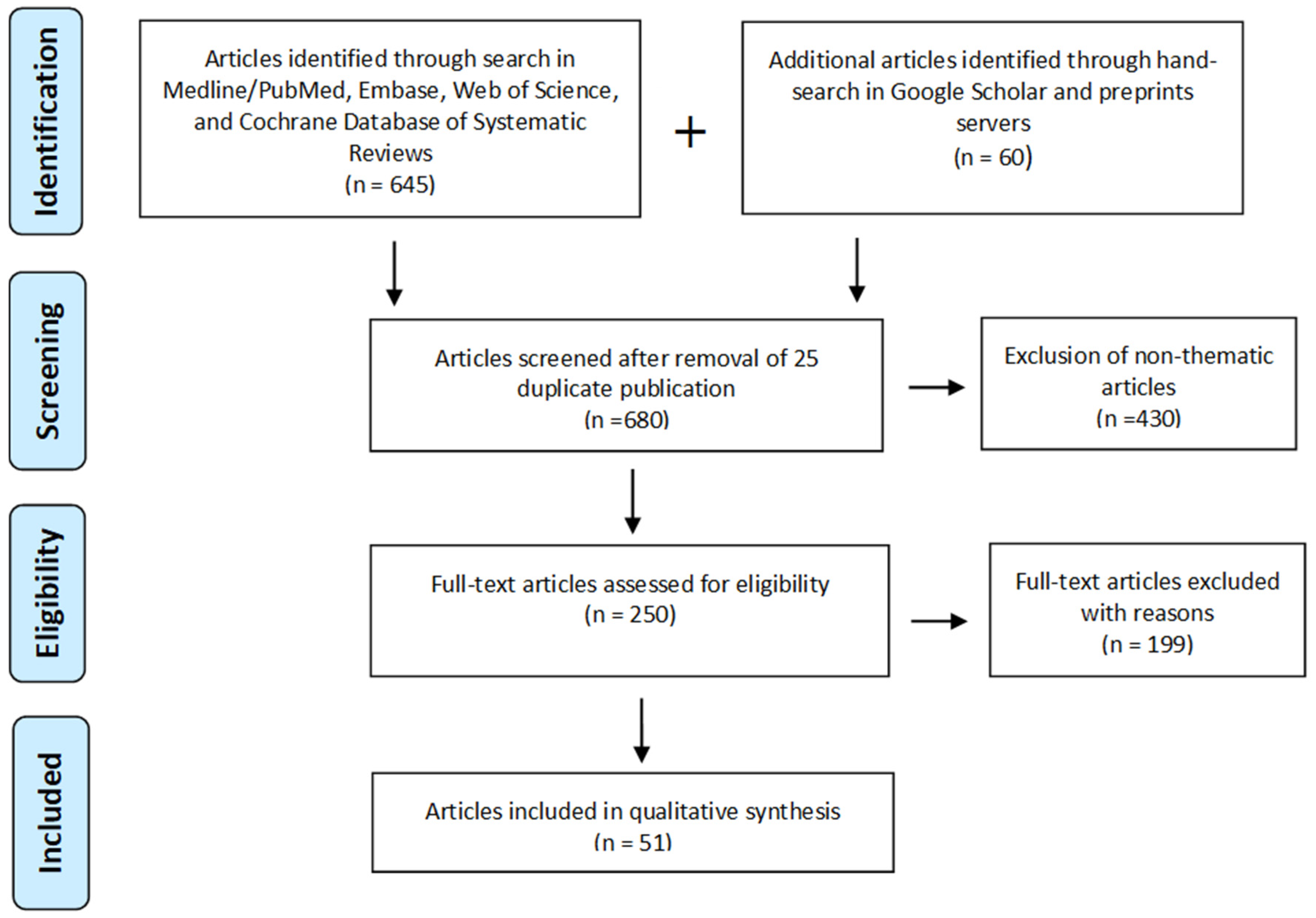
2. Material and Methods

Literature Search

We searched Medline/PubMed, Embase, Web of Science, and the Cochrane Database of Systematic Reviews for articles on the methodology of pedobarography. The search encompassed clinical trials, controlled trials, meta-analyses, and reviews published only in English between January 1982 and February 2021. The following sets of search commands were used:
  • Methodology AND Pedobarography or Plantar Pressure Distribution or Foot Pressure Distribution or Underfoot Pressure Distribution;
  • Examination AND Pedobarography or Plantar Pressure Distribution or Foot Pressure Distribution or Underfoot Pressure Distribution;
  • Procedure AND Pedobarography or Plantar Pressure Distribution or Foot Pressure Distribution or Underfoot Pressure Distribution.
Additionally, a hand search was conducted in Google Scholar, which displays results by relevance, using the keywords ‘Pedobarography’, ‘Plantar pressure distribution’, ‘Foot pressure distribution’, or ‘Underfoot pressure distribution’. This search encompassed 20 top results and was complemented by another 20 top results reported by the preprint servers of Preprints.org and Medrxiv. The rationale for choosing the first 20 positions was that the search algorithms of these engines are set in such a way that the higher the number of openings the higher the position of a referenced item appearing on the list. Therefore, articles that rank higher are presumed to be most frequently accessed by readers and judged as the most relevant and weighing in on the field in question. Search details of the search are depicted in Figure 1.
Inclusion criteria were as follows:
  • Peer-reviewed journal articles and published conference papers;
  • Studies describing the procedure for performing a pedobarographic examination;
  • Publications focusing on pedobarographic systems and masking foot regions.
Exclusion criteria were the studies without a methodological component on the pedobarographic examination, case reports, operating manuals, comments, and editorials on published pedobarographic articles, patient education handouts, or newspaper articles.

3. Results

Overall, the initial screening search yielded 705 articles based on titles and abstracts. Among them, we identified 25 duplicate articles defined as publishing the same or very similar scientific content more than once by the same author group. These articles were excluded. Out of the remaining 680 articles, 430 were discarded as they brought nothing to the topic of the pedobarographic methodology being strictly clinically oriented. The remaining 250 articles were judged eligible for further scrutiny. Out of them, 199 were excluded failing to fully meet inclusion criteria due to a dearth of data, or partial description of pedobarographic procedures, or the lack of scientific precision in performance. Those articles contained null or fleeting methodological meaning for the pedobarography focusing on clinical disorders and treatment. In the end, 51 articles were deemed suitable for further in-depth evaluation (Figure 1). Most articles covered more than one thread. The research quality, relevance, and the rigor of methods of the selected articles were based on screening of the main body text of the articles performed independently by three assessors, the authors of this article, and reaching at least a two-assessor unanimous opinion. The methods of pedobarographic examinations were extracted from a range of pathologies presented in different groups of patients as no article entirely methodological was found. Several categories relating to pedobarography emerged (Table 1).
The first topic category contains articles on pedobarographic systems based on various measurement techniques. The second consists of pedobarographic examination models, parameters tested, and graphical illustration of results. Masking of foot areas and their classification is another category. Conducting foot examination comes next, followed by limitations and potential improvements that would help eliminate shortcomings of the procedure. The final category of articles covers additional issues such as the general definition and types of pedobarography.

4. Discussion

Pedobarographic examination enables the quantitative and qualitative evaluations of the plantar pressure distribution. It offers a reliably repeatable measurement of pressures on each square centimeter of the foot sole with a graphical illustration in the form of a load map [8,9,22,23]. The literature distinguishes three types of examinations: static, postural, and dynamic. Static pedobarography describes plantar pressure distribution at a given time while ‘dynamics of standing’ or changes during a person’s situational posture are tested with postural pedobarography. The forces acting on the plantar surface of the foot during gait are determined by dynamic pedobarography that captures the foot propulsion phase [24,25,26].

4.1. Pedobarographic Measurements

The pressure distribution is determined using a measuring platform or mat on which the person stands or walks during the test. Sensor signals are transmitted to a PC to create an image of the plantar surface as a pressure map expressed in g/cm2. The credibility of results is influenced by the exact horizontal positioning of the platform. Data such as age, body weight, and height, and the foot length of a patient are entered into the computer memory according to the operating instruction of the specific system [27,28].
Currently, over 40 measurement systems, both static and dynamic, are used to record the plantar surface pressure (e.g., Footscan, F-scan, Pedar-X, EMED, PEL, Electrodynogram, Musgrave Footprint System, IngVaL, or Gaitview). The systems have methodological and practical differences that make comparisons difficult. For instance, F-scan and Pedar-X systems accept footwear during the examination as they require in-shoe sensors in contrast to barefoot trials in other baropodometers. The methods include optic, piezoelectric, resistive, capacitive techniques based on digital signal inputs [18,29,30,31,32,33,34,35,36]. The image of the plantar pressure distribution can be presented in several ways. Standardly, pressures are displayed using the full color range from blue (low pressure) to red (high pressure) as a 2D or 3D image. Additionally, the pressure distribution can be presented as ‘iso-clamps’ using three colors usually corresponding to the three pressure ranges: 25–50%, 50–75%, and >75% of the maximum pressure. Pressures below 25% are invisible in this option. The reference values for ‘iso-clamps’ can be adapted case-by-case depending on the foot region. When evaluating pressures in the hindfoot region, it is advisable to set higher reference values than those for the forefoot. The percentage visualization also is possible. Pressure points are then expressed as a percentage of the maximum pressure, and the graph is presented as an image composed of a series of numerical values. This method of the findings illustration seems optimal when pressure values are read off ‘manually’ as well as for the interpretation and verification of results depicted by a range of colors [27,37].
The following parameters can be evaluated with static pedobarography: average and maximum pressures (g/cm2) and total support surface for each foot (cm2). The postural examination evaluates the average and maximum pressures, total support region for each foot, and the contact area of the plantar surface with the ground at specific moments of standing. Dynamic pedobarography evaluates these variables at specific moments in gait phases. Additionally, it provides the foot contact pattern, plantar pressure distribution such as peak pressure time or pressure-time integrals, and the center of pressure (CoP) trajectories [4,38]. Hellstrom et al. [39] have proposed a division into the spatiotemporal variables (step and stride length, stride width, step time, speed, ground contact duration, and cadence) and the force and pressure variables (peak forces and pressures, mean plantar pressure, CoP, speed of CoP shift, contact region, force–time integral, arch index, and the foot posture).
Before the examination, the foot sole should be divided into regions by choosing one of the existing classifications based on the regional anatomic differences in the plantar pressure [40,41]. In some studies, the examination is limited to a part of the foot sole, e.g., the forefoot [42,43,44], or the anatomic region, e.g., hindfoot [45], leaving out other parts. The division of the plantar surface is made individually for each patient since the foot length must be considered. A commonly used plantar surface division in children has been described by Bowen et al. [46]. This division considers five regions: the hindfoot, lateral midfoot, medial midfoot, lateral forefoot, and medial forefoot. For comparison, a division proposed by Cavanagh [47] considers 10 regions: the medial heel (MH), lateral heel (LH), medial midfoot (MM), and lateral midfoot (LM); the regions under the first metatarsal head (1st MET), second metatarsal head (2nd MET), 3rd–5th metatarsal heads (Lateral MET), hallux (H), second toe (2nd Toe), and 3rd–5th toes (Lateral toes). Foot masking is performed according to the protocols supported by the automated masking software, e.g., Research Foot [48]. The auto masking algorithms precisely define foot regions in healthy feet. The precision is upheld in gait but reduced in foot deformities, for instance, halluxes [49].
The pedobarographic examination is performed in sequence. The static measurement comes first. The patient is instructed to stand barefoot in an upright position keeping the symmetry of the body and gazing straight ahead. The test starts with an introductory trial to familiarize the patient with the procedure and to calibrate the equipment. Then, the active measurement begins and is repeated until three reproducible results are obtained. In the static model, a single image of the plantar pressure distribution at a specific point in time during the patient’s stance is recorded. The postural or dynamic examination follows. The postural model enables the evaluation of the dynamics of standing. It consists of a series of static tests that show the distribution of plantar surface pressures over time, which is presented as an average of all measurements [50]. In the dynamic model, patients are instructed to walk barefoot at the natural walking speed while taking steps of the same length. Concerning the test on a mat, it is best performed with a starting slide step designated at the usual patient’s length. The patient walks a minimum of three steps before and after contacting the platform located in the middle of the walkway. It is essential to ensure that patients do not aim for the platform as they walk and are unaware of the measurement time. Three to five trials are performed and patients who report fatigue are permitted to rest for 2–5 min in-between. The test results are controlled for age, height, weight, and foot length [51,52,53]. In the case of the insole-based system, patients are fitted with proper insoles having embedded sensors. Tightly fitting insole pads must be used. During testing, the patient is instructed to walk approximately 15 m before returning to the starting point [27]. Hellstrom et al. [39] have opted for a long straight 60 m distance while performing a test using the Ingval System, disregarding the data of the first and last 5 m corresponding to acceleration and deceleration phases. A shorter walk distance has been proposed by Rome et al. [48] during the in-shoe-based examination performed according to a seven-stride protocol. The first and last strides are discarded, with the remaining being averaged in each trial. In a study by Lee et al. [54] using an F-scan in-shoe pressure measurement, patients were asked to walk 10 m along the walkway.

4.2. Difficulties and Ambiguities concerning Pedobarography

A pedobarography system is a complex computerized setup equipped with pricey sensors. For instance, the F-Scan System has sensitive in-soles whose structure is based on load-sensitive resistive membranes (force-sensing resistor—FSR). These sensors last for approximately 30 gait cycles [55,56]. The pressure exerted on the boundary of FSR increases the output and shortens the sensor’s lifetime. To extend the durability of sensors, improvements must be made to the insole to protect the sensor’s edge [39]. Another drawback of the embedded sensors is that the footwear itself may modify the measurement [57,58]. While walking barefoot, the pressures are higher in the metatarsal, hindfoot, and lateral part of the forefoot with lower pressures observed under the toes, compared to the test performed in the shoes [59]. Chen et al. [60] has also reported that the pressure distribution varies depending on the type of insoles and shoes. To minimize the distortion of results caused by the footwear, pedobarography should be best performed using two different types of footwear [27]. A solution to this problem could be to use the Pedar-X in-sole system inside the anti-skid socks to minimize the influence of footwear on the plantar pressure [61].
Pedobarography is a tedious and time-consuming method, which rules out its standard use in the clinic. The tests must be preceded by instructive, often numerous training trials. Additionally, the in-soles are calibrated according to the manufacturer’s instructions and the zero-setting procedure is required before data acquisition. The average time required for a dynamic pedobarography examination using the in-shoe system is 25 min [27].
A disadvantage of pedobarography for diagnostic use lies in a variety of plantar regional divisions. For instance, Lee et al. [54] have distinguished the following regions: anterior masks (1st–5th toes and 1st–5th metatarsals), posterior masks (medial heel and lateral heel), medial masks (medial heel, 1st metatarsal, and 1st toe), and the lateral masks (lateral heel, 2nd–5th metatarsals, and 2nd–5th toes). The classification proposed by Lorkowski [62] is a modification of Cavanagh and Ae’s [17] by adding a T region between hind-foot and mid-foot. Different plantar divisions point to the lack of a consensual classification and make the comparison of various tests unreliable in the diagnostics of foot pathologies.
Difficulties in the evaluation of dynamic pedobarography are usually caused by over-emphasizing one of the gait phases, especially the stance phase. Consequently, to the excessive patient’s concentration on this phase, the maximum pressure in the hindfoot region becomes higher, which hinders the interpretation of results while walking. Burnfield et al. [63] have noticed that the gait velocity may increase pressures arising in the heel and forefoot regions. Dynamic pedobarography is particularly difficult to perform in pre-school children who despite correctly designated starting slide and repeated trials, often bypass the platform or step partway on it [64]. Yuan et al. [51] and Xu et al. [53] have contended that the platform surface should be covered with an overlayer of ethylene-vinyl acetate (EVA) to ensure that the patient is unaware of the measurement time.
The diagnostic pedobarography is also hindered by plantar pressure differences in the same patient during an examination. The variability is due mainly to the functional state of the musculoskeletal system but may also be caused by disparities in whole-body movements [65]. Quaney et al. [66] and Hughes [67] have demonstrated that joint stiffness is responsible for higher foot pressures while walking. However, Cavanagh et al. [47] point out that structural characteristics account for only about one-third of the variability observed in healthy people during dynamic pedobarography. Difficulties in gait evaluation may also arise in pregnancy which affects the plantar pressure distribution due to bodyweight asymmetry. The maximum pressure is greater under each foot region in pregnancy, compared to non-pregnant women [68].
The purpose of this scoping review was to explore the available literature on pedobarography, focusing on the different modes of tests. The exploration was spurred by a relative rarity and ambiguity of relevant information and on the other side, an increasing value of this technique for the evaluation of plantar pressure distribution that is indispensable in the modern management of foot disorders. We also present the evolution of the technique over time. It is difficult to generalize pedobarographic findings due mostly to the qualitative type of research so that a summary answer concerning discrete clinical foot disorders cannot be formulated. Neither can the cause-and-effect relationship be substantiated in most studies reviewed. Another limitation stems from insufficient instructions for use of pedobarographic test devices. All that narrows the volume of the available literature referring to the unambiguously verified methods of examination. Comparative studies are also subject to the inherent risk of bias. Despite these limitations, we believe that in this review we have demonstrated the contemporary status of pedobarography, the methodological side of testing, and the use of pedobarography in quantitative evaluation of plantar pressure distribution in the diagnostics and management of foot ailments. There appears a need for developing standardized protocols and recommendations to seamlessly perform the pedobarographic examination and make wider use of this valuable tool.

Author Contributions

Conceptualization, J.L. and K.G.; Methodology, J.L., K.G. and M.P.; Formal Analysis: J.L. and K.G.; Original Draft Preparation: J.L. and K.G.; Writing—Review and Editing: M.P.; Supervision: M.P. All authors have read and agreed to the published version of the manuscript.

Funding

This research received no external funding.

Institutional Review Board Statement

Ethical review and approval were waived for this study since it does not contain any studies with human participants or animals performed by any of the authors.

Informed Consent Statement

Not applicable.

Data Availability Statement

Publicly available datasets were analyzed in this study. This data can be found here: https://www.nlm.nih.gov/bsd/pmresources.html, accessed on 1 March 2021; https://www.embase.com/landing?status=grey, accessed on 30 April 2021; https://clarivate.com/webofsciencegroup/solutions/web-of-science/, accessed on 10 July 2021; https://www.cochranelibrary.com/cdsr/about-cdsr, accessed on 10 July 2021.

Conflicts of Interest

The authors declare no conflict of interest concerning this article.

References

  1. López-López, D.; Pérez-Ríos, M.; Ruano-Ravina, A.; Losa-Iglesias, M.E.; Becerro-de-Bengoa-Vallejo, R.; Romero-Morales, C.; Calvo-Lobo, C.; Navarro-Flores, E. Impact of quality of life related to foot problems: A case-control study. Sci. Rep. 2021, 11, 14515. [Google Scholar] [CrossRef]
  2. López-López, D.; Becerro-de-Bengoa-Vallejo, R.; Losa-Iglesias, M.E.; Palomo-López, P.; Rodríguez-Sanz, D.; Brandariz-Pereira, J.M.; Calvo-Lobo, C. Evaluation of foot health related quality of life in individuals with foot problems by gender: A cross-sectional comparative analysis study. BMJ Open 2018, 8, e023980. [Google Scholar] [CrossRef] [Green Version]
  3. Chang, C.H.; Yang, W.T.; Wu, C.P.; Chang, L.W. Would foot arch development in children characterize a body maturation process? A prospective longitudinal study. Biomed. J. 2021, in press. [Google Scholar] [CrossRef] [PubMed]
  4. Hösl, M.; Böhm, H.; Oestreich, C.; Dussa, C.U.; Schäfer, C.; Döderlein, L.; Nader, S.; Fenner, V. Self-perceived foot function and pain in children and adolescents with flexible flatfeet—Relationship between dynamic pedobarography and the foot function index. Gait Posture 2020, 77, 225–230. [Google Scholar] [CrossRef] [PubMed]
  5. Pereiro-Buceta, H.; Becerro-de-Bengoa-Vallejo, R.; Losa-Iglesias, M.E.; López-López, D.; Navarro-Flores, E.; Martínez-Jiménez, E.M.; Martiniano, J.; Calvo-Lobo, C. The Effect of Simulated Leg-Length Discrepancy on the Dynamic Parameters of the Feet during Gait-Cross-Sectional Research. Healthcare 2021, 9, 932. [Google Scholar] [CrossRef] [PubMed]
  6. Galbusera, F.; Cina, A.; Panico, M.; Albano, D.; Messina, C. Image-based biomechanical models of the musculoskeletal system. Eur. Radiol. Exp. 2020, 4, 49. [Google Scholar] [CrossRef]
  7. Lorkowski, J.; Grzegorowska, O.; Pokorski, M. Artificial intelligence in the healthcare system: An overview. In Best Practice in Health Care; Springer: Cham, Switzerland, 2021; Volume 1335, pp. 1–10. [Google Scholar]
  8. Ozkan, H.; Ege, T.; Koca, K.; Can, N.; Yurttas, Y.; Tunay, S. Pedobarographic measurements after repair of Achilles tendon by minimal invasive surgery. Acta Orthop. Belg. 2016, 82, 271–274. [Google Scholar]
  9. Skopljak, A.; Muftic, M.; Sukalo, A.; Masic, I.; Zunic, L. Pedobarography in diagnosis and clinical application. Acta Inform. Med. 2014, 22, 374–378. [Google Scholar] [CrossRef] [Green Version]
  10. Abboud, R.J. Relevant foot biomechanics. Curr. Orthop. 2002, 16, 165–179. [Google Scholar] [CrossRef] [Green Version]
  11. Lefebvre, T.; Malthête, J. Lettres d’Etienne-Jules Marey à Georges Demenÿ, 1880–1894; AFRHC: Paris, France, 2002; ISBN 10: 2913758002. [Google Scholar]
  12. Beely, F. Zur Mechanik des Stehens. Arch. Klin. Chir. 1882, 27, 457–468. [Google Scholar]
  13. Seitz, L. Die vordeven stutzpunkte des fusses under normalen and pathologische verhaltnissen. Z. Orthop. Chir. 1901, 8, 37–38. [Google Scholar]
  14. Elftman, H. A cinematic study of the distribution of pressure in the human foot. Anat. Rec. 1934, 59, 481–491. [Google Scholar] [CrossRef]
  15. Yamamoto, H.; Muneta, T.; Asahina, S.; Furuya, K. Forefoot pressures during walking in feet afflicted with hallux valgus. Clin. Orthop. Relat. Res. 1996, 323, 247–253. [Google Scholar] [CrossRef]
  16. Schwartz, R.P.; Heat, A.L. The definition of human locomotion on the basis of measurement. J. Bone Jt. Surg. 1947, 1, 203–214. [Google Scholar]
  17. Cavanagh, P.R.; Ae, M. A technique for the display of pressure distributions beneath the foot. J. Biomech. 1980, 13, 69–75. [Google Scholar] [CrossRef]
  18. Hennig, E.M.; Cavanagh, P.R.; Albert, H.T.; Macmillan, N.H. A piezoelectric method of measuring the vertical contact stress beneath the human foot. J. Biomed. Eng. 1982, 4, 213–222. [Google Scholar] [CrossRef]
  19. Péruchon, E.; Jullian, J.M.; Rabischong, P. Wearable unrestraining footprint analysis system. Applications to human gait study. Med. Biol. Eng. Comput. 1989, 27, 557–565. [Google Scholar] [CrossRef]
  20. Bassett, D.R.; Ainsworth, B.E.; Leggett, S.R.; Mathien, C.A.; Main, J.A.; Hunter, D.C.; Duncan, G.E. Accuracy of five electronic pedometers for measuring distance walked. Med. Sci. Sports Exerc. 1996, 28, 1071–1077. [Google Scholar] [CrossRef]
  21. Hatano, Y. Use of the pedometer for promoting daily walking exercise. J. Int. Comm. Health Phys. Educ. Recreat. 1993, 29, 4–8. [Google Scholar]
  22. Ramanathan, A.K.; Kiran, P.; Arnold, G.P.; Wang, W.; Abboud, R.J. Repeatability of the Pedar-X in-shoe pressure measuring system. Foot Ankle Surg. 2010, 16, 70–73. [Google Scholar] [CrossRef]
  23. Gurney, J.K.; Marshall, P.W.; Rosenbaum, D.; Kersting, U.G. Test-retest reliability of dynamic plantar loading and foot geometry measures in diabetics with peripheral neuropathy. Gait Posture 2013, 37, 135–137. [Google Scholar] [CrossRef] [PubMed]
  24. Hagen, L.; Pape, J.P.; Kostakev, M.; Peterlein, C.D. Pedobarographic changes during first month after subtalar extra-articular screw arthroereisis (SESA) operation of juvenile flexible flatfoot. Arch. Orthop. Trauma Surg. 2020, 140, 313–320. [Google Scholar] [CrossRef]
  25. Güven, M.; Kocadal, O.; Akman, B.; Poyanlı, O.S.; Kemah, B.; Atay, E.F. Proximal femoral nail shows better concordance of gait analysis between operated and uninjured limbs compared to hemiarthroplasty in intertrochanteric femoral fractures. Injury 2016, 47, 1325–1331. [Google Scholar] [CrossRef] [PubMed]
  26. Lorkowski, J.; Grzegorowska, O.; Kotela, I. The use of pedobarographic examination to biomechanical evaluation of foot and ankle joint in adult—Own experience. Ortop. Traumatol. Rehabil. 2015, 17, 207–213. [Google Scholar] [CrossRef] [Green Version]
  27. Gurney, J.K.; Kersting, U.G.; Rosenbaum, D.; Dissanayake, A.; York, S.; Grech, R.; Ng, A.; Milne, B.; Stanley, J.; Sarfati, D. Pedobarography as a clinical tool in the management of diabetic feet in New Zealand: A feasibility study. J. Foot Ankle Res. 2017, 10, 24. [Google Scholar] [CrossRef] [PubMed] [Green Version]
  28. Lorkowski, J.; Grzegorowska, O.; Kotela, I. The use of pedobarographic examination in children—Own experience and review of literature. Fizjoterapia Pol. 2015, 14, 46–51. [Google Scholar]
  29. Stolz, B.; Grim, C.; Lutter, C.; Gelse, K.; Schell, M.; Swoboda, B.; Carl, H.D.; Hotfiel, T. Assessing foot loads in continuous passive motion (CPM) and active knee joint motion devices. Sportverletz Sportschaden 2021, 35, 18–23. [Google Scholar] [CrossRef]
  30. Xiang, L.; Mei, Q.; Fernandez, J.; Gu, Y. A biomechanical evaluation of the acute hallux abduction manipulation intervention. Gait Posture 2020, 76, 210–217. [Google Scholar] [CrossRef]
  31. Hellstrom, P.A.R.; Folke, M. Carried weight affects walking speed monitoring with the IngVaL system. Stud. Health Technol. Inform. 2019, 261, 317–320. [Google Scholar]
  32. Vilarinho, D.; Theodosiou, A.; Leitão, C.; Leal-Junior, A.G.; Domingues, M.F.; Kalli, K.; André, P.; Antunes, P.; Marques, C. POFBG-embedded cork insole for plantar pressure monitoring. Sensors 2017, 17, 2924. [Google Scholar] [CrossRef] [Green Version]
  33. Fishco, W.D.; Ellis, M.B.; Cornwall, M.W. Influence of a metatarsus adductus foot type on plantar pressures during walking in adults using a pedobarograph. J. Foot Ankle Surg. 2015, 54, 449–453. [Google Scholar] [CrossRef]
  34. Kim, Y.T.; Lee, J.S. Normal pressures and reliability of the Gaitview system in healthy adults. Prosthet. Orthot. Int. 2012, 36, 159–164. [Google Scholar] [CrossRef] [Green Version]
  35. Morgan, S.; Ng, A.; Clough, T. The long-term outcome of silastic implant arthroplasty of the first metatarsophalangeal joint: A retrospective analysis of one hundred and eight feet. Int. Orthop. 2012, 36, 1865–1869. [Google Scholar] [CrossRef] [Green Version]
  36. Reed, L.; Bennett, P.J. Changes in foot function with the use of Root and Blake orthoses. J. Am. Podiatr. Med. Assoc. 2001, 91, 184–193. [Google Scholar] [CrossRef]
  37. Lorkowski, J.; Mazur, T. Application of the pedobarography in hallux valgus diagnosis. Scr. Period 2000, 3, 409–413. [Google Scholar]
  38. Park, E.S.; Kim, H.W.; Park, C.I.; Rha, D.; Park, C.W. Dynamic foot pressure measurements for assessing foot deformity in persons with spastic cerebral palsy. Arch. Phys. Med. Rehabil. 2006, 87, 703–709. [Google Scholar] [CrossRef]
  39. Hellstrom, P.A.R.; Åkerberg, A.; Ekström, M.; Folke, M. Evaluation of the IngVaL pedobarography system for monitoring of walking speed. Healthc. Inform. Res. 2018, 24, 118–124. [Google Scholar] [CrossRef] [PubMed] [Green Version]
  40. Kernozek, T.; Roehrs, T.; McGarvey, S. Analysis of plantar loading parameters pre and post-surgical intervention for hallux valgus. Clin. Biomech. 1997, 12, S18–S19. [Google Scholar] [CrossRef]
  41. Blomgren, M.; Turan, I.; Agadir, M. Gait analysis in hallux valgus. J. Foot Surg. 1991, 30, 70–71. [Google Scholar] [PubMed]
  42. Konings-Pijnappels, A.P.M.; Tenten-Diepenmaat, M.; Dahmen, R.; Verberne, S.K.; Dekker, J.; Twisk, J.W.R.; Roorda, L.D.; van der Leeden, M. Forefoot pathology in relation to plantar pressure distribution in patients with rheumatoid arthritis: A cross-sectional study in the Amsterdam foot cohort. Gait Posture 2019, 68, 317–322. [Google Scholar] [CrossRef] [PubMed]
  43. Bryant, A.; Singer, K.; Tinley, P. Comparison of the reliability of plantar pressure measurements using the two-step and midgait methods of data collection. Foot Ankle Int. 1999, 20, 646–650. [Google Scholar] [CrossRef]
  44. Stess, R.M.; Jensen, S.R.; Mirmiran, R. The role of dynamic plantar pressures in diabetic foot ulcers. Diabetes Care 1997, 20, 855–858. [Google Scholar] [CrossRef]
  45. Schaff, P.S.; Cavanagh, P.R. Shoes for the insensitive foot: The effect of a “rocker bottom” shoe modification on plantar pressure distribution. Foot Ankle 1990, 11, 129–140. [Google Scholar] [CrossRef] [PubMed]
  46. Bowen, T.R.; Miller, F.; Castagno, P.; Richards, J.; Lipton, G. A method of dynamic foot-pressure measurement for the evaluation of pediatric orthopaedic foot deformities. J. Pediatr. Orthop. 1998, 18, 789–793. [Google Scholar] [CrossRef] [PubMed]
  47. Cavanagh, P.R.; Rodgers, M.M.; Iiboshi, A. Pressure distribution under symptom-free feet during barefoot standing. Foot Ankle 1987, 7, 262–276. [Google Scholar] [CrossRef] [PubMed]
  48. Rome, K.; Survepalli, D.G.; Lobo, M.; Dalbeth, N.; McQueen, F.; McNair, P.J. Evaluating intratester reliability of manual masking of plantar pressure measurements associated with chronic gout. J. Am. Podiatr. Med. Assoc. 2011, 101, 424–429. [Google Scholar] [CrossRef]
  49. Ellis, S.J.; Stoecklein, H.; Yu, J.C.; Syrkin, G.; Hillstrom, H.; Deland, J.T. The accuracy of an automasking algorithm in plantar pressure measurements. HSS J. 2011, 7, 57–63. [Google Scholar] [CrossRef] [Green Version]
  50. Lorkowski, J.; Lorkowska, B.; Skawina, A. Underfoot pressure distribution of patients with spinal problems. Acta Kinesiol. Univ. Tart. 2001, 6, 152–155. [Google Scholar]
  51. Yuan, X.N.; Liang, W.D.; Zhou, F.H.; Li, H.T.; Zhang, L.X.; Zhang, Z.Q.; Li, J.J. Comparison of walking quality variables between incomplete spinal cord injury patients and healthy subjects by using a footscan plantar pressure system. Neural Regen. Res. 2019, 14, 354–360. [Google Scholar]
  52. Chae, Y.H.; Kim, J.S.; Kang, Y.; Kim, H.Y.; Yi, T.I. Clinical and biomechanical effects of low-dye taping and figure-8 modification of low-dye taping in patients with heel pad atrophy. Ann. Rehabil. Med. 2018, 42, 222–228. [Google Scholar] [CrossRef] [Green Version]
  53. Xu, C.; Yan, Y.B.; Zhao, X.; Wen, X.X.; Shang, L.; Huang, L.Y.; Lei, W. Pedobarographic analysis following Pemberton’s pericapsular osteotomy for unilateral developmental dysplasia of the hip: An observational study. Medicine 2015, 94, e932. [Google Scholar] [CrossRef]
  54. Lee, S.H.; Lee, H.J.; Chang, W.H.; Choi, B.O.; Lee, J.; Kim, J.; Ryu, G.H.; Kim, Y.H. Gait performance and foot pressure distribution during wearable robot-assisted gait in elderly adults. J. Neuroeng. Rehabil. 2017, 14, 123. [Google Scholar] [CrossRef] [Green Version]
  55. Nicolopoulos, C.S.; Anderson, E.G.; Solomonidis, S.E.; Giannoudis, P.V. Evaluation of the gait analysis FSCAN pressure system: Clinical tool or toy? Foot 2000, 10, 124–130. [Google Scholar] [CrossRef]
  56. Woodburn, J.; Helliwell, P. Observations on the F-Scan in-shoe pressure measuring system. Clin. Biomech. 1997, 12, S16. [Google Scholar] [CrossRef]
  57. Payne, C.; Turner, D.; Miller, K. Determinants of plantar pressures in the diabetic foot. J. Diabetes Complicat. 2002, 16, 277–283. [Google Scholar] [CrossRef]
  58. Foti, T.; Davids, J.R.; Bagley, A. A biomechanical analysis of gait during pregnancy. J. Bone Jt. Surg. Am. 2000, 82, 625–632. [Google Scholar] [CrossRef]
  59. Soames, R.W.; Stott, J.R.; Goodbody, A.; Blake, C.D.; Brewerton, D.A. Measurement of pressure under the foot during function. Med. Biol. Eng. Comput. 1982, 20, 489–495. [Google Scholar] [CrossRef] [PubMed]
  60. Chen, H.; Nigg, B.M.; Hulliger, M.; de Koning, J. Influence of sensory input on plantar pressure distribution. Clin. Biomech. 1995, 10, 271–274. [Google Scholar] [CrossRef]
  61. Bacarin, T.A.; Sacco, I.C.; Hennig, E.M. Plantar pressure distribution patterns during gait in diabetic neuropathy patients with a history of foot ulcers. Clinics 2009, 64, 113–120. [Google Scholar] [CrossRef] [PubMed] [Green Version]
  62. Lorkowski, J. Methodology of pedobarographic examination—Own experiences and review of literature. Przegl. Lek. 2006, 63 (Suppl. 5), 23–27. (In Polish) [Google Scholar]
  63. Burnfield, J.M.; Few, C.D.; Mohamed, O.S.; Perry, J. The influence of walking speed and footwear on plantar pressures in older adults. Clin. Biomech. 2004, 19, 78–84. [Google Scholar] [CrossRef] [PubMed]
  64. Kellis, E. Plantar pressure distribution during barefoot standing, walking and landing in preschool boys. Gait Posture 2001, 14, 92–97. [Google Scholar] [CrossRef]
  65. Sadeghi, H.; Allard, P.; Prince, F.; Labelle, H. Symmetry and limb dominance in able-bodied gait: A review. Gait Posture 2000, 12, 34–45. [Google Scholar] [CrossRef]
  66. Quaney, B.; Meyer, K.; Cornwall, M.W.; McPoil, T.G. A comparison of the dynamic pedobarograph and EMED systems for measuring dynamic foot pressures. Foot Ankle Int. 1995, 16, 562–566. [Google Scholar] [CrossRef] [PubMed]
  67. Hughes, J. The clinical use of pedobarography. Acta Orthop. Belg. 1993, 59, 10–16. [Google Scholar]
  68. Elsayed, E.; Devreux, I.; Embaby, H.; Alsayed, A.; Alshehri, M. Changes in foot plantar pressure in pregnant women. J. Back Musculoskelet. Rehabil. 2017, 30, 863–867. [Google Scholar] [CrossRef]
Figure 1. Flow-chart diagram of the literature search on pedobarographic examinations.
Applsci 11 11020 g001
Table 1. Main topics covered in pedobarography-related articles reviewed.
Articles (n)
Pedobarographic systems10
Pedobarographic measurements 6
Masking foot regions9
Conducting the examination13
Types of pedobarographic assessments3
Basic information/definitions4
Limitations in pedobarographic examination24
The contents of some articles overlap different topics making the total number greater than the 51 articles reviewed.
Publisher’s Note: MDPI stays neutral with regard to jurisdictional claims in published maps and institutional affiliations.

