Article

Non-Thermal O2 Plasma Efficacy on C. albicans and Its Effect on Denture Base Resin Color

1
Laboratoire-Biomatériaux et Inflammation en Site Osseux-EA 4691, Université de Reims Champagne Ardenne, SFR CAP-Santé, FED 4231, 51100 Reims, France
2
UFR Odontologie de Reims, Université de Reims Champagne Ardenne, 2 rue du Général Koenig, 51100 Reims, France
3
Pôle de Médecine Bucco-Dentaire de Reims, Centre Hospitalier Universitaire de Reims, 45 rue Cognacq-Jay, 51100 Reims, France
4
Ecole nationale Supérieure d’Ingénieurs de Reims, Université de Reims Champagne Ardenne, 3 Esplanade Roland Garros, FED 4231, 51100 Reims, France
5
Laboratoire-ItheMM-EA 7548, Université de Reims Champagne Ardenne, Chemin des Rouliers, FED 4231, 51100 Reims, France
6
Aurora Cold Plasma Sterilisation, 70 rue du Docteur Yersin, 59120 Loos, France
*
Author to whom correspondence should be addressed.
Appl. Sci. 2021, 11(21), 10367; https://doi.org/10.3390/app112110367
Submission received: 14 September 2021 / Revised: 26 October 2021 / Accepted: 31 October 2021 / Published: 4 November 2021
(This article belongs to the Special Issue Recent Advances in Plasma Technology and Applications)

Abstract

:

Featured Application

Development of a new non-thermal O2 plasma process for future dentures sterilization related to denture stomatitis.

Abstract

Denture stomatitis is a disease involving C. albicans, which can affect elderly and immuno-compromised people. To avoid any recurrence of this pathology, it is necessary to treat patients regularly and disinfect dentures. However, the denture cleansers’ efficacy is not optimal and often leads to adverse color effects on the denture base resins. The aim of this study was to investigate the efficacy of a low-pressure non-thermal O2 plasma (NTP) treatment on C. albicans seeded on ProBase®Hot resin (Ivoclar Vivadent). The viability reduction of C. albicans was assessed by colony forming units (CFU) analysis and by scanning electron microscopy (SEM). The effect of repeated treatments on the resin color was evaluated by spectrophotometry. The resin samples were placed in a sealed bag in which O2 plasma was generated in low-pressure conditions. The results showed that a 120-min O2 NTP treatment led to a 6-log reduction of C. albicans viability (p < 0.05) and to yeasts’ major alterations observed by SEM. Furthermore, significant slight color changes of the resin (∆E00 = 1.33) were noted only after six plasma treatments (p < 0.05). However, the denture aesthetic was preserved, as the color changes were not perceptible and remained below the acceptability threshold (∆E00 < 4).

1. Introduction

The human oral microbiota includes hundreds of microorganisms that contribute to a well-balanced buccal environment [1]. Candida albicans figures among the most common yeasts identified in approximately 50% of healthy adults [2]. This microorganism is an opportunistic pathogen that can lead to different forms of oral fungal infections especially denture stomatitis (DS) [3,4].
DS is a common disorder that can affect a variable number of denture wearers, ranging from 11% up to 75% of them [5,6]. The characteristics of this disease are essentially an inflammatory process and erythema of the palatal oral mucosa associated with Candida species, especially C. albicans, which adhere and form biofilms on the oral mucosa and denture surfaces [7,8]. Its common symptoms are “bleeding, swelling, burning or other painful sensations, halitosis, unpleasant taste and dryness in the mouth” [9]. Moreover, this disorder could potentially enhance the risk of systemic infections, particularly in elderly subjects [10,11]. This disease involves different etiologies such as autoimmune disorders, systemic diseases, salivary dysfunctions, trauma, poor denture hygiene and wearing denture overnight [3,7,8]. The DS therapeutic strategies to overcome this fungal infection must therefore take into consideration both patients and dentures. Thus, a topical and/or systemic antifungal treatment must be administered to patients who must also improve their oral and dental hygiene [8,11,12]. However, an increase in antifungal-resistant Candida strains has been reported in recent years [12,13,14]. This can lead to poorer clinical outcomes for patients and create a higher risk of systemic infections [15]. Consequently, regular maintenance of dentures is essential. Nowadays, there are many guidelines all over the world which usually recommend to daily brush dentures and to remove them at night [16,17]. In addition, many disinfectants are proposed to clean dentures regularly in order to reduce or prevent the formation of Candida biofilm on them. These disinfectant solutions include a wide variety of chemicals such as sodium hypochlorite, alkaline peroxides, chlorhexidine, plant extracts, etc. [18,19,20,21,22] However, prolonged use of these molecules is necessary to avoid any recurrence of DS [6,12]. However, numerous studies reveal that these disinfection methods can alter the properties of dentures base resins such as flexural strength [19,23,24], hardness [20,25,26], surface roughness [25,26,27,28,29,30] and especially the color stability [18,19,21,24,25,27,29,31,32]. Indeed, the main color change results in resin bleaching [16,19,21,24,25,27,29,30,31]. Those adverse effects can have consequences on denture wearers’ aesthetics and comfort, and therefore on their social life.
To overcome those issues, many alternative disinfection methods have been proposed such as microwaves [33,34] or ultraviolet (UV) radiation [35]. Cold plasma, or non-thermal process, has been developed as a new disinfection method in the medical field for the last decade. Plasma is an ionized gas consisting of ions, electrons, UVs, photons, neutral species and reactive oxygen and nitrogen species (RONS) that have shown their efficacy to inactivate various micro-organisms such as bacteria, spores, yeasts or viruses [36,37,38,39,40]. Many techniques using non-thermal plasma [41,42,43,44] differ from one another in terms of operating pressure (low or atmospheric pressure), and operating conditions (electrical current, gas nature and flow rate). A few years ago, a low-pressure plasma process was developed, which could preserve the sterile state of items after the end of treatment [37,38]. In dentistry, this process could be used by dentists or by care home staff for denture maintenance.
The aim of this study was to assess whether a non-thermal O2 plasma process could inactivate a C. albicans strain on a heat-cured polymethyl methacrylate (PMMA) resin for denture base without significantly modifying its color. The null hypotheses were that this non-thermal plasma process would not affect C. albicans viability and not alter the resin color.

2. Material and Methods

2.1. Samples Preparation

2.1.1. Microbiological Assay

  • 55 × 25 mm glass microscope slides (Ghäasel, Rogo-Sampaic, Wissous, France) (n = 27) were sterilized by high-pressure saturated steam at 134 °C for 18 min.
  • 55 × 25 × 1.8 mm polymethyl methacrylate (PMMA) resin samples (ProBase®Hot 36 P-V, Ivoclar Vivadent, Saint-Jorioz, France) (n = 27) were prepared following the manufacturer’s instructions, cleaned by exposure to 70% alcohol and by mechanical agitation in distilled water with a soft sonication for 5 min, then dried.
Glass and resin samples were distributed into three groups (n = 9 per group): (1) control (C): untreated samples; (2) 120-min low-pressure exposure (LP); (3) 120-min non-thermal O2 plasma cycle (P1).

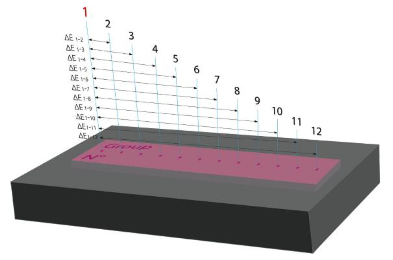
2.1.2. Visual and Spectrophotometry Assay

A total of 48 resin samples of 64 × 10 × 1.8 mm (ProBase®Hot 36 P-V, Ivoclar Vivadent) were produced. After the polymerization, one side of the samples was polished by brushing with decreasing the size of pumice particles. The samples distribution and the associated treatment is summarized in Table 1. The samples were distributed in four groups (n = 12 per group): (1) Group C: control (untreated samples); (2) Group LP: 120-min low-pressure exposure; (3) Group P1: one 120-min non-thermal O2 plasma exposure; (4) Group P6: six successive 120-min non-thermal plasma O2 exposures. Regarding this last group, the samples were kept in distilled water for 48 h then dried between each plasma exposure. For each group, each sample was observed by three qualified observers and analyzed by spectrophotometry.

2.2. Efficacy of Non-Thermal O2 Plasma against C. albicans

2.2.1. Candida albicans Growth Conditions

Candida albicans ATCC® 10231TM was provided from American Type Culture Collection (Manassas, VA, USA). The strain was sequentially subcultured three times in specific Sabouraud medium. The final suspension was made by transferring C. albicans colonies from Sabouraud Dextrose Agar (SDA) to a fresh Sabouraud solution (300 mL) for 6 h at 37 °C. Yeasts from the exponential phase were centrifuged at 5000× g for 10 min at 20 °C, and washed in phosphate-buffered saline (PBS) at pH 7 to achieve a concentration of approximately 1010 colony forming units (CFU)/mL. Forty microliters of this yeast suspension were spread on glass or resin samples and then dried for 15 min before plasma exposure.

2.2.2. Sterilization by Non-Thermal O2 Plasma

The prototype consisted of a 35-L stainless-steel vacuum chamber and enabled to generate a non-thermal O2 plasma (O2 NTP) inside a sealed bag (SüdPack®, Medica, Germany) under low-pressure as previously described [38,45]. Three contaminated glass or resin samples (P1) were placed inside the bag which was sealed and then set on the radio-frequency (RF) polarization plate. When the low-pressure reached 8 × 10⁻⁵ mbar (8 × 10⁻³ Pa), O2 gas was injected (1 sccm) through the first Tyvek® membrane of the bag and the excess gas was released through the second Tyvek® membrane into the vacuum chamber. Then, the discharge was induced by the RF polarization (100 W), and the plasma was densified inside the bag by the magnetic field (10 Gauss) for 120 min. At the end of the process, the vacuum chamber was returned to atmospheric pressure. Untreated samples (group C) were used as controls. Each experiment was independently performed three times.

2.2.3. Counts on Sabouraud Dextrose Agar

Glass and resin samples were submitted to mechanical agitation in 50 mL phosphate-buffer saline (PBS) (30 s of vortex, 12 min in an ultrasonic bath (VWR TH USC 300 THD, 45 KHz), then 30 s of vortex) to remove yeasts and obtain homogenous suspensions. Yeasts dilutions were seeded on SDA. After a 48-h incubation at 37 °C, the colonies were counted with the Scan R 1200 (Interscience, Saint-Nom-la-Bretèche, France). The results were expressed in log CFU/mL.

2.2.4. Scanning Electron Microscopy

Contaminated glass and resin samples before or after low-pressure or NTP treatments were fixed with 2.5% glutaraldehyde (Sigma-Aldrich, St. Louis, MO, USA) in PBS for 1 h at room temperature, and rinsed twice with PBS for 10 min. The samples were than dehydrated in graded series of ethanol/water solutions at 50%, 70%, 90%, and 100% (twice) and covered with hexamethyldisilazane solution (Sigma-Aldrich, St. Louis, MO, USA) [46]. The samples were desiccated overnight at room temperature then coated with gold/paladium on a Jeol Ion Sputter JFC-1100 and observed on a Jeol JSM-7900F Scanning Electron Microscope (PICT URCA Platform, Reims, France). Images were acquired from secondary electrons at a primary beam energy of 2 kV.

2.3. Non-Thermal O2 Plasma Effects on Resin Color

2.3.1. Visual Inspection

Three observers qualified as having superior color discrimination competency evaluated the homogeneity of whole samples. They were selected by a color vision deficiency test (Farnsworth-Munsell 100 hue test) (ISO/TR 28642:2016). Visual inspection of samples of all groups (C, LP, P1 and P6) was performed in a dark room in a standard viewing cabinet illuminated with D65 standard artificial daylight (color temperature: 6500 K) (ISO/TR 28642:2016). The samples were positioned on the floor of the cabinet which was painted neutral gray, with 0° illuminating geometry. All observers’ heads were fixed at a 45° angle superior to the horizontal plane at a distance of 30 cm (ISO/TR 28642:2016) [47,48]. Each observer determined the perceptibility threshold (“Can I see a difference in color?”) and then the acceptability threshold of the color change for each group (“Is this difference in color acceptable?”).

2.3.2. Spectrophotometry

The resin color change (∆E) was evaluated and analyzed with the CIELab color system by the formula ΔE00 (ISO 11664-6) [47,48].
Δ E 00 = ( Δ L k L S L ) 2 + ( Δ C k C S C ) 2   + ( Δ H k H S H ) 2 + R T   ( Δ C k C S C )   ( Δ H k H S H )  
A black silicon mold was used to accurately reposition the resin samples. Measurements were performed on the polished side, using a spectrophotometer (eXactTM Standard, X-Rite PANTONE®) which set-up was: standard illuminant D65, 45/0° optical geometry, CIE 1964 10° observer, and 2-mm2 surface measurement (Color iControl software, X-Rite).
Beforehand, the color acceptability threshold was experimentally evaluated. Regarding each sample (n = 48), a first point was defined as the sample color standard (Figure 1). From this standard point, eleven measurements were taken every 0.5 mm and were each compared to the standard point to determine ∆E. The results met the color acceptability threshold for acrylic denture base resins recently determined by Ren et al. (ΔE00 < 4.00) [47].
Then, for each group, twelve measurements per sample were performed before and after 3 weeks of resin natural aging (group C) or after each treatment (LP, P1 and P6), determining the values of the CIELab color coordinates (L* (lightness), a* (redness-greenness) and b* (yellowness-blueness)). ∆E00 was determined using the sample color standard previously measured.

2.4. Statistical Analysis

Regarding the microbiological assay, the Shapiro-Wilk test revealed a non-normal distribution for CFU counts. Then, a pair-wise comparison was performed between the independent groups using the non-parametric Wilcoxon-Mann-Whitney test (Prism5, GraphPad Software, San Diego, CA, USA). A P value less than 0.05 was considered to be “statistically significant”.
As for the spectrophotometry assay, the Shapiro-Wilk test revealed a non-normal distribution. The independent groups were compared with a Kruskall-Wallis test followed by a Wilcoxon rank sum test (Rstudio Software, 1.41717, 2021, RStudio, Inc., Boston, MA, USA). A P value less than 0.05 was considered to be “statistically significant”.

3. Results

3.1. Efficacy of Non-Thermal O2 Plasma against C. albicans

The antifungal efficacy of non-thermal O2 plasma (O2 NTP) treatment was evaluated on C. albicans while considering the potential impact of low-pressure (LP) on C. albicans viability.

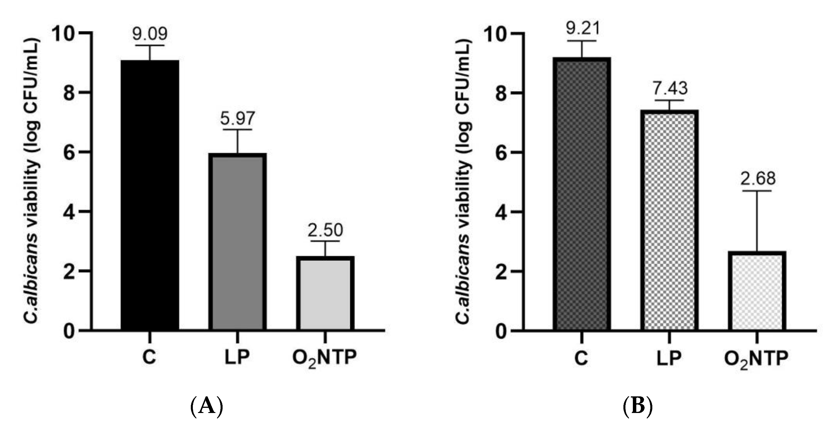
3.1.1. Low-Pressure Effect on C. albicans Viability

During the process, the samples were subjected to low pressure prior to O2 NTP treatment. To differentiate the low-pressure efficacy from O2 NTP, some contaminated samples were subjected only to the low pressure without injection of gas for a 120-min period. At the end of the treatment, the vacuum level decreased up to 2 × 10⁻⁵ mbar in 120 min. When glass samples were contaminated with C. albicans, the viability decreased up to 5.97 log compared to the control (9.09 log) (p < 0.0001) (Figure 2A). When resin samples were contaminated with C. albicans, the viability decreased by up to 7.43 log compared to the control (9.21 log) (p < 0.0001) (Figure 2B).

3.1.2. Efficacy of O2 NTP Treatment on C. albicans Viability

During the process of sterilization, the samples were subjected to O2 NTP for 120 min. When glass slides were contaminated with C. albicans, the viability decreased up to 2.50 log compared to the control (9.09 log) (p < 0.0001) (Figure 2A). This viability decreased up to 2.68 log when C. albicans was seeded on resin samples (p < 0.0001) (Figure 2B). Thus, the O2 NTP treatment had antifungal efficacy (≥ 6-log reduction). A significant reduction was observed between the low pressure and the O2 NTP treatments, whatever the material (glass or resin) (p < 0.0001).

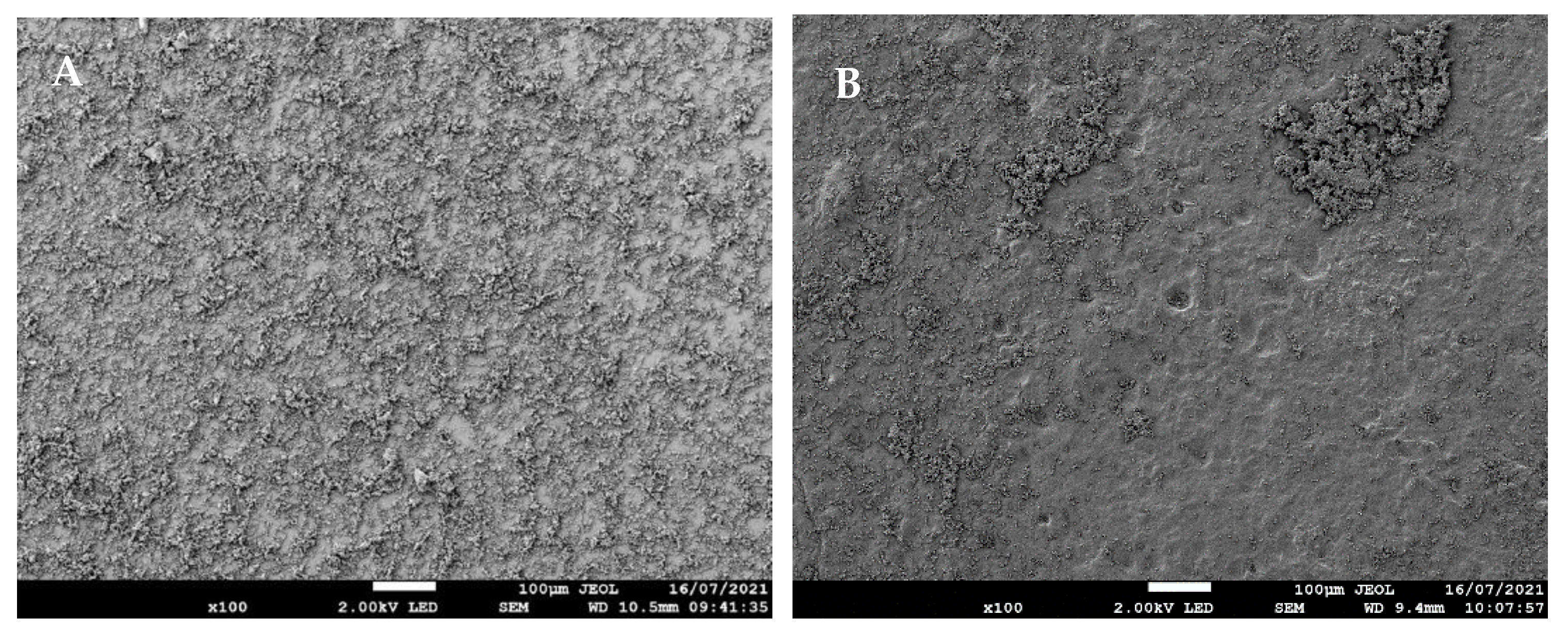
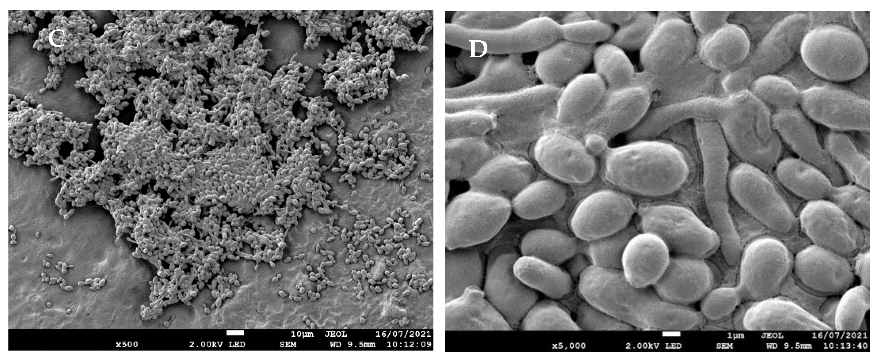
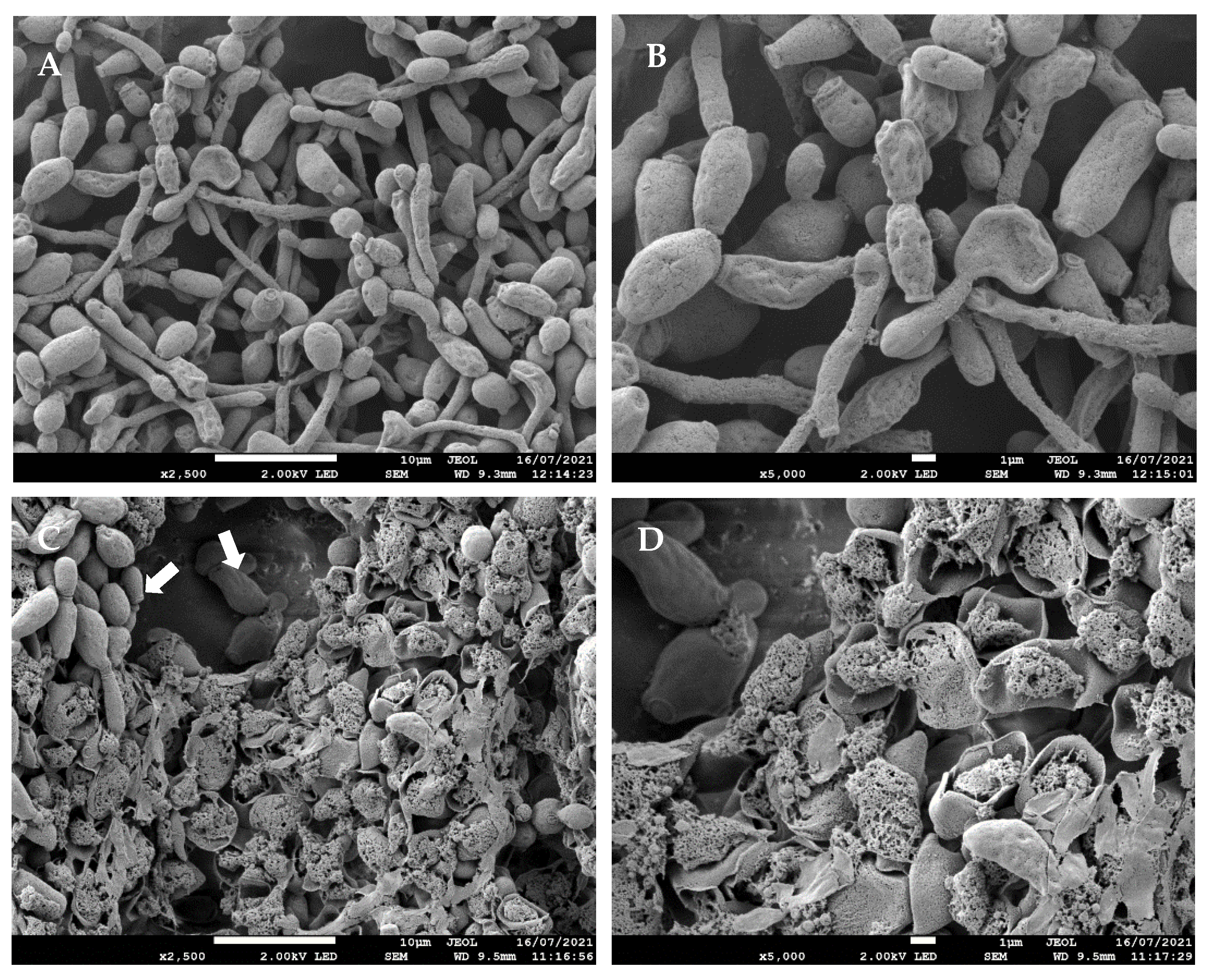
3.1.3. Effects of O2 NTP Treatment on C. albicans Morphology Determined by SEM

Figure 3 shows C. albicans adhesion on glass and unpolished resin samples before O2 NTP treatments. A homogeneous spread of C. albicans was observed on the glass slide (Figure 3A) while it was heterogeneous on the resin sample (Figure 3B). Indeed, we observed yeast monolayers sometimes located in depressions, and multilayers clusters on resin sample (Figure 3B,C). These clusters included different forms of yeasts: mainly blastospores that appeared as smooth ovoid or spherical cells, some pseudohyphae and hyphae forms exhibiting elongated shape (Figure 3D).
Figure 4 shows the effect of low-pressure and 120-min O2 NTP treatments on C. albicans seeded on resin sample. Low-pressure treatment did not seem to lead to major alterations of yeasts morphology (Figure 4A,B). Only a few cells were damaged, characterized by a rough surface (Figure 4B). A 120-min O2 NTP exposure led to major alterations of a large proportion of yeasts with a disruption of membranes and loss of intracellular components (Figure 4C,D). However, some yeasts located in the deepest layers of cluster seemed to be preserved and undamaged (Figure 4C).

3.2. Low-Pressure and O2 NTP Treatments Effect on the Resin COLOR

3.2.1. Visual Aspect

Each observer determined the perceptibility and acceptability thresholds of the color changes for all samples after the different treatments. No perceptible color modifications were noted after LP, P1 and P6 treatments compared to the control (group C).

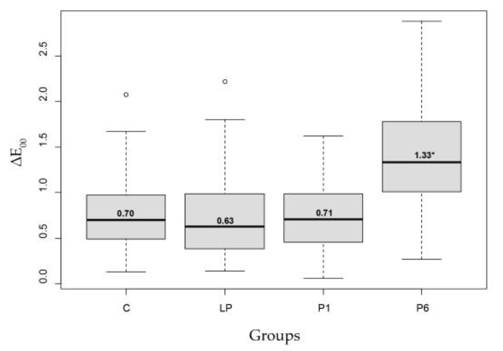
3.2.2. Spectrophotometry

Table 2 summarizes the resin color change (ΔE) after each treatment. ΔE of group C represented the resin natural aging, and consequently was determined after the evaluation of the other groups (LP, P1 and P6). Then, after three weeks the median value of ΔE was 0.70 with a maximum value of 2.07. After a 120-min LP treatment, the median ∆E value was 0.63 with a maximum of 2.22. Similarly, after one 120-min O2 NTP (P1) the ∆E was 0.71 with a maximum value of 1.62. No significant modification of ΔE was noted in groups LP and P1 compared to group C (p > 0.05) (Figure 5). Concerning group P6, a significant increase of ΔE up to 1.33 with a maximum value of 2.88 was observed (p < 0.05) (Figure 5). However, all the results remained under the acceptability threshold (ΔE < 4.00) [47].
Table 3 summarizes the values of the CIELab color coordinates (L* (lightness), a* (redness-greenness) and b* (yellowness-blueness)) evaluated before and after each treatment. Concerning group C, the median lightness value (L*) slightly increased from 34.77 (±0.98) to 35.10 (±1.10) after 3 weeks of natural aging. On the opposite, a slight decrease was observed regarding the color coordinate a* (from 8.36 (±0.65) to 8.25 (±0.66)) and the color coordinate b* (from 1.55 (±0.40) to 1.46 (±0.44)) (p > 0.05). Similarly, after LP and one O2 NTP treatments (P1), no significant variation of lightness and of a* and b* color coordinates were noted (p > 0.05). Regarding group P6, the increase of lightness was significant (from 32.95 (±1.02) to 35.31 (±1.27) (p < 0.05). In contrast to groups C, LP and P1, a significant increase of the color coordinate a* (from 8.23 (±0.60) to 8.80 (±0.65)) and the color coordinate b* (from 0.98 (±0.30) to 1.45 (±0.43)) were noted (p < 0.05).

4. Discussion

The aim of this study was to evaluate the efficacy of a non-thermal O2 plasma (O2 NTP) as an alternative method for the disinfection or sterilization of denture base resin. The null hypotheses were rejected since O2 NTP reduced C. albicans viability and slightly altered the color stability of the resin for denture base.
DS is a common disease in elderly and immuno-depressed people, and mainly occurs under imbalance of the oral microbiota entailing local factors such as wearing dentures [2,3,4,5,6,12]. The focus in the treatment of denture stomatitis is to eliminate the source of pathogens which contaminate dentures [3,8,11,12]. However, it is essential to obtain a safe disinfection or sterilization protocol without any harmful effect on the physical and mechanical properties of resins for denture base [16].
Nowadays, there are different mechanical or chemical techniques to decontaminate dentures [12,16,49,50]. Alkaline peroxide is the most common molecule among chemical cleansers [22]. However, Uludamar et al. [51] highlighted that the 15-min treatment recommended in some manufacturer’s instructions (Fittydent, Polident, Corega) is not sufficient for denture in vivo decontamination. They noted that only Fittydent brand allows an antifungal activity after a 60-min immersion. Moreover, these cleansers must be used correctly due to their possible toxicity on oral and oropharyngeal tissues as the degradation of the different agents leads to hydrogen peroxide [16,22]. Many other molecules or cleansers have been tested or recommended in guidelines, such as chlorhexidine, chlorine dioxide, sodium hypochloride, and plant extracts [17,50,51]. However, if some agents reduce C. albicans viability, other molecules are not efficient [22]. Therefore, the development of new techniques could be an alternative to those chemical antiseptics. Hence, plasma processes have been tested for one decade on resin for denture base. Some studies dealt with the effect of atmospheric plasma on C. albicans biofilms developed on PMMA [52,53]. They evaluated atmospheric plasma using argon (Ar) or Ar/O2 mixture as working gas. Matthes et al. [52] obtained a 4.12-log reduction of C. albicans viability over 10 min using a dielectric barrier discharge whereas Wang et al. [53] reached a 6-log reduction over 8 min using a plasma jet. These results vary due to the different plasma sources and the different methodologies used for evaluation. In previous studies [37,38,46], we have developed a low-pressure NTP process that treats prepackaged items. This process has already proved efficacy against S. aureus, P. aeruginosa and B. subtilis spores. In this study, this approach was completed by investigating C. albicans inactivation using O2 as the working gas. We demonstrated that low pressure without plasma generated only a small decrease of C. albicans viability (glass: 3.12-log reduction, resin: 1.78-log reduction). Thus, the direct effect of low pressure could not solely be responsible for the C. albicans inactivation. Moreover, when low pressure was applied without plasma, the pressure in the vacuum chamber decreased up to 2 × 10⁻⁵ mbar over 120 min. This pressure is lower compared to the pressure during O2 plasma treatment (2.50 × 10⁻⁴ mbar). Hence, the results of low pressure on C. albicans were probably overstated. Regarding O2 NTP treatment in low pressure condition, a 120-min treatment led to a significant reduction (>6 log) of C. albicans viability. Consequently, the antifungal activity was essentially due to the plasma process.
The efficacy of the different atmospheric- and low-pressure plasma devices highlights the role of the various generated species during the plasma process. Different hypotheses involve essentially UVs, reactive oxygen and nitrogen species (RONS), ion and electron species [41,42,44,54]. These species have enough energy to break covalent bonds and initiate various chemical reactions in C. albicans cells. Thus, UVs are well known to damage DNA cells by dimerization of the thymine base [55,56]. RONS cause photodesorption or etching of the cell wall, peroxidation of lipids and ergosterol, the latter being a critical sterol of the cell membrane [57]. This leads to an increase in membrane permeability favoring leakage of cytoplasm and intracellular components, and ultimately to lethal cellular damage [40,53,58,59]. Our SEM images highlighted the rupture of the cell membrane and the leakage of intracellular components after a 120-min O2 NTP, which is in accordance with the previous hypotheses.
After NTP O2 treatment, the reduction of C. albicans viability was similar whatever the sample material. However, the results were less reproducible when C. albicans was seeded on resin samples, which was probably due to the different affinity of C. albicans for the two materials. Indeed, the SEM analyses highlighted that the contaminated glass surface was characterized by large areas of yeast monolayers and small clusters. Conversely, the yeasts’ repartition was heterogeneous on resin samples, with many thick clusters and some non-contaminated areas (Figure 3). The resin hydrophobicity and surface roughness could explain this specific adhesion of C. albicans [6,7,60]. Indeed, destroying those clusters was more difficult due to their thickness and the presence of an extracellular matrix. Hence, the deeper yeast layers were better protected from NTP [37,40,61]. According to many authors [38,40,53], increasing the treatment time or producing intracellular ROS in biofilm [62] would favor the destructuration of those clusters. Despite the clusters on resin surface, our NTP plasma led to a significant 6-log viability reduction after a 120-min treatment in low working pressure. This efficacy could be explained by the generation of vacuum ultraviolet (VUV) photons in low-pressure plasma conditions only [54,63]. These VUV photons are highly energetic due to their short wavelength (from 100 to 200 nm). Thus, VUV photons and previously described species may act together and have a considerable impact on biofilms. However, a better comprehension of the mechanism of active plasma species could help to optimize the treatment.
Concerning the resin color stability, chemical cleansers used to decontaminate dentures lead to adverse effects when repeated or prolonged treatments are applied [18,19,21,24,25,27,29,31,32]. The most frequent resin color changes are bleaching using sodium hypochlorite [16,19,21,27,29] and alkaline peroxides [19,24,25,27,30,31]. In the same way, one in vitro study highlighted resin staining after prolonged treatment using lemongrass extract [18].
The evaluation of color change is related to ISO technical report (TR) 28642:2016, which defined two color distance metrics ∆E*ab and ∆E00. It only defines a perceptibility threshold (PT) of ΔE*ab = 1.2 and an acceptability threshold (AT) of ΔE*ab = 2.7. This technical report does not concern the color change of resins for denture base, but only dental ceramics, dental resins or silicone elastomers. The previous authors mainly used ∆E*ab to evaluate the effect of the different cleansers on resin color. However, those studies presented many discrepancies with respect to devices and methodologies, which led to disagreements upon PT and AT values [48]. Hence, Ren et al. [47,48] carried out a study using ∆E00 to specifically determine the PT and the AT of color changes, after accelerated aging of an acrylic denture base resin: PT value was 1.71∆E00 and AT value was 4.00∆E00. Like Ren et al., we investigated the possible adverse effects of our O2 NTP process on the color of ProBase®Hot 36 P-V resin (Ivoclar Vivadent), using ∆E00. The spectrophotometry and the visual analyses showed a slight variation of the resin color within each sample in all groups. This could involve the presence of red fibers in the resin that mimic blood vessels within the mucosa, improving the denture aesthetics. As a result, biases may exist during spectrophotometric measurements when repositioning the sample before and after treatments. The spectrophotometry analysis showed no significant color change (∆E) of the resin when exposed to 120-min low-pressure or one O2 NTP treatment. However, a significant increase of ∆E occurred after six repeated treatments (1.33, p < 0.05). This significant color change involved a significant increase of the lightness (L*) and a higher proportion of red and yellow color components (a* and b*), which slightly increased the color chroma. However, ∆E value was under the acceptability threshold for acrylic denture base resins (<4.00) recently determined by Ren et al. [47,48]. Moreover, this color modification was not perceptible by any of the three qualified observers. Hence, our process would not have unacceptable adverse effects on the resin color and would preserve its aesthetic appearance for patients.
This study shows promising results; nevertheless, there are still some limitations concerning the shape of the samples, which is not representative of dentures, and the non-evaluation of all resin properties. Consequently, further investigations should take into account the possible adverse effects of this process on different physical (dimensional stability, wettability) and mechanical (roughness, hardness and toughness) resin properties after repeated treatments. Moreover, future assays would assess the efficacy of this process on contaminated dentures.

5. Conclusions

In our study, we evaluated a low-pressure O2 NTP as a method to clean a denture-base resin contaminated with C. albicans. Our microbial investigation showed an antifungal efficacy. Therefore, a daily brush supplemented with a regular plasma treatment would reduce the risk of recurrent denture stomatitis by complete elimination of candida on the denture surfaces. Furthermore, our process could maintain the sterile state of dentures until delivery to patients without needing desorption time. This repeated treatment would not cause any perceptible adverse effects on the resin color, thus preserving the denture wearers’ aesthetics and social life.

6. Patents/Brevets

“Device for cold plasma sterilization of an object, such as a medical device, particularly an implant, and method using this device.” Popot J.M., Gelle M.P. EP2618851B1.

Author Contributions

C.M.: Conceptualization, Methodology, Investigation, Writing-Original Draft, Visualization. S.O.: Methodology, Investigation. M.M.: Investigation. F.L.B.: Methodology. F.V.: Investigation. M.-P.G.: Conceptualization, Methodology, Writing-Original Draft, Visualization, Supervision, Project administration. All authors have read and agreed to the published version of the manuscript.

Funding

This research received no external funding.

Institutional Review Board Statement

Not applicable.

Informed Consent Statement

Not applicable.

Acknowledgments

The authors thank Marjorie Cantener for her assistance in proofreading the English language. They also thank PICT-URCA platform (University of Reims Champagne-Ardenne) for imaging core facilities.

Conflicts of Interest

The authors declare no conflict of interest.

References

  1. Mosaddad, S.A.; Tahmasebi, E.; Yazdanian, A.; Rezvani, M.B.; Seifalian, A.; Yazdanian, M.; Tebyanian, H. Oral Microbial Biofilms: An Update. Eur. J. Clin. Microbiol. Infect. Dis. 2019, 38, 2005–2019. [Google Scholar] [CrossRef]
  2. Millsop, J.W.; Fazel, N. Oral Candidiasis. Clin. Dermatol. 2016, 34, 487–494. [Google Scholar] [CrossRef]
  3. Salerno, C.; Pascale, M.; Contaldo, M.; Esposito, V.; Busciolano, M.; Milillo, L.; Guida, A.; Petruzzi, M.; Serpico, R. Candida-Associated Denture Stomatitis. Med. Oral. Patol. Oral. Cir. Bucal. 2011, 16, e139–e143. [Google Scholar] [CrossRef] [PubMed]
  4. Vila, T.; Sultan, A.S.; Montelongo-Jauregui, D.; Jabra-Rizk, M.A. Oral Candidiasis: A Disease of Opportunity. J. Fungi 2020, 6, 15. [Google Scholar] [CrossRef] [Green Version]
  5. Susewind, S.; Lang, R.; Hahnel, S. Biofilm Formation and Candida Albicans Morphology on the Surface of Denture Base Materials. Mycoses 2015, 58, 719–727. [Google Scholar] [CrossRef] [PubMed] [Green Version]
  6. Ohshima, T.; Ikawa, S.; Kitano, K.; Maeda, N. A Proposal of Remedies for Oral Diseases Caused by Candida: A Mini Review. Front. Microbiol. 2018, 9, 1522. [Google Scholar] [CrossRef]
  7. Gendreau, L.; Loewy, Z.G. Epidemiology and Etiology of Denture Stomatitis: Denture Stomatitis. J. Prosthodont. 2011, 20, 251–260. [Google Scholar] [CrossRef] [PubMed]
  8. Hilgert, J.B.; Giordani, J.M.D.A.; de Souza, R.F.; Wendland, E.M.D.R.; D’Avila, O.P.; Hugo, F.N. Interventions for the Management of Denture Stomatitis: A Systematic Review and Meta-Analysis. J. Am. Geriatr. Soc. 2016, 64, 2539–2545. [Google Scholar] [CrossRef]
  9. Webb, B.C.; Thomas, C.J.; Willcox, M.D.P.; Harty, D.W.S.; Knox, K.W. Candida-Associated Denture Stoma Titis. Aetiology and Management: A Review. Part 2. Oral Diseases Caused by Candida Species. Aust. Dent. J. 1998, 43, 160–166. [Google Scholar] [CrossRef]
  10. Yarborough, A.; Cooper, L.; Duqum, I.; Mendonça, G.; McGraw, K.; Stoner, L. Evidence Regarding the Treatment of Denture Stomatitis: Treatment of Denture Stomatitis. J. Prosthodont. 2016, 25, 288–301. [Google Scholar] [CrossRef]
  11. Passariello, C.; Di Nardo, F.; Polimeni, A.; Di Nardo, D.; Testarelli, L. Probiotic Streptococcus Salivarius Reduces Symptoms of Denture Stomatitis and Oral Colonization by Candida Albicans. Appl. Sci. 2020, 10, 3002. [Google Scholar] [CrossRef]
  12. Emami, E.; Kabawat, M.; Rompre, P.H.; Feine, J.S. Linking Evidence to Treatment for Denture Stomatitis: A Meta-Analysis of Randomized Controlled Trials. J. Dent. 2014, 42, 99–106. [Google Scholar] [CrossRef]
  13. Spettel, K.; Barousch, W.; Makristathis, A.; Zeller, I.; Nehr, M.; Selitsch, B.; Lackner, M.; Rath, P.-M.; Steinmann, J.; Willinger, B. Analysis of Antifungal Resistance Genes in Candida Albicans and Candida Glabrata Using next Generation Sequencing. PLoS ONE 2019, 14, e0210397. [Google Scholar] [CrossRef]
  14. Pereira, R.; Santos Fontenelle, R.O.; Brito, E.H.S.; Morais, S.M. Biofilm of Candida Albicans: Formation, Regulation and Resistance. J. Appl. Microbiol. 2021, 131, 11–22. [Google Scholar] [CrossRef]
  15. Pristov, K.E.; Ghannoum, M.A. Resistance of Candida to Azoles and Echinocandins Worldwide. Clin. Microbiol. Infect. 2019, 25, 792–798. [Google Scholar] [CrossRef] [PubMed]
  16. Felton, D.; Cooper, L.; Duqum, I.; Minsley, G.; Guckes, A.; Haug, S.; Meredith, P.; Solie, C.; Avery, D.; Deal Chandler, N. Evidence-Based Guidelines for the Care and Maintenance of Complete Dentures: A Publication of the American College of Prosthodontists. J. Prosthodont. 2011, 20, S1–S12. [Google Scholar] [CrossRef]
  17. Bartlett, D.; Carter, N.; de Baat, C.; Duyck, J.; Goffin, G.; Müller, F.; Kawai, Y. White Paper on Optimal Care and Maintenance of Full Dentures for Oral and General Health; 2018. Available online: https://www.dentalhealth.org/denturecareguidelines (accessed on 21 August 2021).
  18. Madeira, P.L.B.; Carvalho, L.T.; Paschoal, M.A.B.; de Sousa, E.M.; Moffa, E.B.; dos Santos da Silva, M.A.; de Jesus Rodolfo Tavarez, R.; Gonçalves, L.M. In Vitro Effects of Lemongrass Extract on Candida Albicans Biofilms, Human Cells Viability, and Denture Surface. Front. Cell. Infect. Microbiol. 2016, 6, 71. [Google Scholar] [CrossRef] [PubMed] [Green Version]
  19. Peracini, A.; Machado Andrade, I.; Oliveira, V.C.; Macedo, A.P.; Silva-Lovato, C.H.; Oliveira Pagnano, V.; Watanabe, E.; Oliveira, P.H.F. Antimicrobial Action and Long-Term Effect of Overnight Denture Cleansers. Am. J. Dent. 2017, 30, 101–108. [Google Scholar]
  20. Kurt, A.; Erkose-Genc, G.; Uzun, M.; Sarı, T.; Isik-Ozkol, G. The Effect of Cleaning Solutions on a Denture Base Material: Elimination of Candida Albicans and Alteration of Physical Properties: Effect of Cleaning Solutions on Denture Properties. J. Prosthodont. 2018, 27, 577–583. [Google Scholar] [CrossRef]
  21. Zago, P.M.W.; dos Santos Castelo Branco, S.J.; de Albuquerque Bogéa Fecury, L.; Carvalho, L.T.; Rocha, C.Q.; Madeira, P.L.B.; de Sousa, E.M.; de Siqueira, F.S.F.; Paschoal, M.A.B.; Diniz, R.S.; et al. Anti-Biofilm Action of Chenopodium Ambrosioides Extract, Cytotoxic Potential and Effects on Acrylic Denture Surface. Front. Microbiol. 2019, 10, 1724. [Google Scholar] [CrossRef] [Green Version]
  22. Rocha, G.D.S.R.; Neves Duarte, T.; de Oliveira, C.G.; Nampo, F.K.; de Paula, R.S. Chemical Cleaning Methods for Prostheses Colonized by Candida Spp.: A Systematic Review. J. Prosthet. Dent. 2020, 124, 653–658. [Google Scholar] [CrossRef]
  23. Savabi, O.; Attar, K.; Nejatidanesh, F.; Goroohi, H.; Badrian, H. Effect of Different Chemical Disinfectants on the Flexural Strength of Heat-Polymerized Acrylic Resins. Eur. J. Prosthodont. Restor. Dent. 2013, 21, 105–108. [Google Scholar] [CrossRef] [PubMed]
  24. Shah, V.; Shah, D.; Chauhan, C.; Doshi, P.; Kumar, A. Evaluation of Flexural Strength and Color Stability of Different Denture Base Materials Including Flexible Material after Using Different Denture Cleansers. J. Indian Prosthodont. Soc. 2015, 15, 367–373. [Google Scholar] [CrossRef]
  25. Durkan, R.; Ayaz, E.A.; Bagis, B.; Gurbuz, A.; Ozturk, N.; Korkmaz, F.M. Comparative Effects of Denture Cleansers on Physical Properties of Polyamide and Polymethyl Methacrylate Base Polymers. Dent. Mater. J. 2013, 32, 367–375. [Google Scholar] [CrossRef] [PubMed] [Green Version]
  26. Ozyilmaz, O.Y.; Akin, C. Effect of Cleansers on Denture Base Resins’ Structural Properties. J. Appl. Biomater. Funct. Mater. 2019, 17, 228080001982779. [Google Scholar] [CrossRef] [PubMed] [Green Version]
  27. Paranhos, H.D.F.O.; Peracini, A.; Pisani, M.X.; Oliveira, V.D.C.; de Souza, R.F.; Silva-Lovato, C.H. Color Stability, Surface Roughness and Flexural Strength of an Acrylic Resin Submitted to Simulated Overnight Immersion in Denture Cleansers. Braz. Dent. J. 2013, 24, 152–156. [Google Scholar] [CrossRef] [Green Version]
  28. Carvalho, C.F.; Vanderlei, A.D.; Marocho, S.M.S.; Pereira, S.M.B.; Nogueira, L.; Paes-Junior, T.J.A. Effect of Disinfectant Solutions on a Denture Base Acrylic Resin. Acta Odontol. Latinoam. 2012, 25, 6. [Google Scholar]
  29. Fernandes, F.H.C.N.; Orsi, I.A.; Villabona, C.A. Effects of the Peracetic Acid and Sodium Hypochlorite on the Colour Stability and Surface Roughness of the Denture Base Acrylic Resins Polymerised by Microwave and Water Bath Methods: Effects of the Peracetic Acid and Sodium Hypochlorite on Denture Base Acrylic Resins. Gerodontology 2013, 30, 18–25. [Google Scholar] [CrossRef]
  30. Fergus, C.; Santos, M.; Soo, S.; Petridis, H. The Effect of Different Chemical Intra-Oral Prostheses Cleansers on the Surface Properties of Parylene-Coated PMMA. Dent. Mater. J. 2017, 36, 129–134. [Google Scholar] [CrossRef] [Green Version]
  31. Moon, A.; Powers, J.M.; Kiat-amnuay, S. Color Stability of Denture Teeth and Acrylic Base Resin Subjected Daily to Various Consumer Cleansers: Color Stability of Denture Teeth and Resin Exposed to Cleansers. J. Esthet. Restor. Dent. 2014, 26, 247–255. [Google Scholar] [CrossRef] [Green Version]
  32. Lohitha, K.; Prakash, M.; Gopinadh, A.; Sai Sankar, A.; Sandeep, C.; Sreedevi, B. Color Stability of Heat-Cure Acrylic Resin Subjected to Simulated Short-Term Immersion in Fast-Acting Denture Cleansers. Ann. Med. Health Sci. Res. 2016, 6, 291–295. [Google Scholar] [CrossRef]
  33. Senna, P.M.; Jose Da Silva, W.; Faot, F.; Antoninha Del Bel Cury, A. Microwave Disinfection: Cumulative Effect of Different Power Levels on Physical Properties of Denture Base Resins. J. Prosthodont. 2011, 20, 606–612. [Google Scholar] [CrossRef]
  34. Polychronakis, N.; Polyzois, G.; Lagouvardos, P.; Andreopoulos, A.; Ngo, H.C. Long-Term Microwaving of Denture Base Materials: Effects on Dimensional, Color and Translucency Stability. J. Appl. Oral. Sci. 2018, 26, e20170536. [Google Scholar] [CrossRef] [PubMed]
  35. Binns, R.; Li, W.; Wu, C.D.; Campbell, S.; Knoernschild, K.; Yang, B. Effect of Ultraviolet Radiation on Candida Albicans Biofilm on Poly(Methylmethacrylate) Resin. J. Prosthodont. 2020, 29, 686–692. [Google Scholar] [CrossRef] [PubMed]
  36. Alkawareek, M.Y.; Algwari, Q.T.; Laverty, G.; Gorman, S.P.; Graham, W.G.; O’Connell, D.; Gilmore, B.F. Eradication of Pseudomonas Aeruginosa Biofilms by Atmospheric Pressure Non-Thermal Plasma. PLoS ONE 2012, 7, e44289. [Google Scholar] [CrossRef] [Green Version]
  37. Ben Belgacem, Z.; Carré, G.; Boudifa, M.; Charpentier, E.; Cawe, B.; Gellé, M.P. Effectiveness of Non-Thermal O 2 –N 2 Plasma on P. Aeruginosa Multilayer Biofilms Cultured on Hydroxyapatite. IRBM 2016, 37, 68–74. [Google Scholar] [CrossRef]
  38. Ben Belgacem, Z.; Carré, G.; Charpentier, E.; Le-Bras, F.; Maho, T.; Robert, E.; Pouvesle, J.-M.; Polidor, F.; Gangloff, S.C.; Boudifa, M.; et al. Innovative Non-Thermal Plasma Disinfection Process inside Sealed Bags: Assessment of Bactericidal and Sporicidal Effectiveness in Regard to Current Sterilization Norms. PLoS ONE 2017, 12, e0180183. [Google Scholar] [CrossRef] [Green Version]
  39. Filipić, A.; Gutierrez-Aguirre, I.; Primc, G.; Mozetič, M.; Dobnik, D. Cold Plasma, a New Hope in the Field of Virus Inactivation. Trends Biotechnol. 2020, 38, 1278–1291. [Google Scholar] [CrossRef] [PubMed]
  40. Jungbauer, G.; Moser, D.; Müller, S.; Pfister, W.; Sculean, A.; Eick, S. The Antimicrobial Effect of Cold Atmospheric Plasma against Dental Pathogens—A Systematic Review of In-Vitro Studies. Antibiotics 2021, 10, 211. [Google Scholar] [CrossRef] [PubMed]
  41. Moisan, M.; Barbeau, J.; Moreau, S.; Pelletier, J.; Tabrizian, M.; Yahia, L. Low-Temperature Sterilization Using Gas Plasmas: A Review of the Experiments and an Analysis of the Inactivation Mechanisms. Int. J. Pharm. 2001, 226, 1–21. [Google Scholar] [CrossRef]
  42. Moreau, M.; Orange, N.; Feuilloley, M.G.J. Non-Thermal Plasma Technologies: New Tools for Bio-Decontamination. Biotechnol. Adv. 2008, 26, 610–617. [Google Scholar] [CrossRef] [PubMed]
  43. Koban, I.; Matthes, R.; Hübner, N.-O.; Welk, A.; Meisel, P.; Holtfreter, B.; Sietmann, R.; Kindel, E.; Weltmann, K.-D.; Kramer, A.; et al. Treatment of Candida Albicans Biofilms with Low-Temperature Plasma Induced by Dielectric Barrier Discharge and Atmospheric Pressure Plasma Jet. New J. Phys. 2010, 12, 073039. [Google Scholar] [CrossRef]
  44. Fiebrandt, M.; Lackmann, J.-W.; Stapelmann, K. From Patent to Product? 50 Years of Low-Pressure Plasma Sterilization. Plasma Process. Polym. 2018, 15, 1800139. [Google Scholar] [CrossRef] [Green Version]
  45. Popot, J.M.; Gelle, M.P. Device for Cold Plasma Sterilization of an Object, Such as a Medical Device, Particularly an Implant, and Method Using This. Device. Patent WO/2012/038669, 23 March 2012. [Google Scholar]
  46. Carré, G.; Charpentier, E.; Audonnet, S.; Terryn, C.; Boudifa, M.; Doliwa, C.; Belgacem, Z.B.; Gangloff, S.C.; Gelle, M.-P. Contribution of Fluorescence Techniques in Determining the Efficiency of the Non-Thermal Plasma Treatment. Front. Microbiol. 2018, 9, 2171. [Google Scholar] [CrossRef] [Green Version]
  47. Ren, J.; Lin, H.; Huang, Q.; Liang, Q.; Zheng, G. Color Difference Threshold Determination for Acrylic Denture Base Resins. BME 2015, 26, S35–S43. [Google Scholar] [CrossRef] [PubMed] [Green Version]
  48. Ren, J.; Lin, H.; Huang, Q.; Zheng, G. Determining Color Difference Thresholds in Denture Base Acrylic Resin. J. Prosthet. Dent. 2015, 114, 702–708. [Google Scholar] [CrossRef]
  49. Lee, H.-E.; Li, C.-Y.; Chang, H.-W.; Yang, Y.-H.; Wu, J.-H. Effects of Different Denture Cleaning Methods to Remove Candida Albicans from Acrylic Resin Denture Based Material. J. Dent. Sci. 2011, 6, 216–220. [Google Scholar] [CrossRef] [Green Version]
  50. Rodrigues, C.F.; Rodrigues, M.E.; Henriques, M.C.R. Promising Alternative Therapeutics for Oral Candidiasis. CMC 2019, 26, 2515–2528. [Google Scholar] [CrossRef]
  51. Uludamar, A.; Özkan, Y.K.; Kadir, T.; Ceyhan, I. In Vivo Efficacy of Alkaline Peroxide Tablets and Mouthwashes on Candida Albicans in Patients with Denture Stomatitis. J. Appl. Oral Sci. 2010, 18, 291–296. [Google Scholar] [CrossRef] [PubMed] [Green Version]
  52. Matthes, R.; Jablonowski, L.; Koban, I.; Quade, A.; Hübner, N.-O.; Schlueter, R.; Weltmann, K.-D.; von Woedtke, T.; Kramer, A.; Kocher, T. In Vitro Treatment of Candida Albicans Biofilms on Denture Base Material with Volume Dielectric Barrier Discharge Plasma (VDBD) Compared with Common Chemical Antiseptics. Clin. Oral. Investig. 2015, 19, 2319–2326. [Google Scholar] [CrossRef] [PubMed]
  53. Wang, G.M.; Sun, P.P.; Pan, H.; Ye, G.P.; Sun, K.; Zhang, J.; Pan, J.; Fang, J. Inactivation of Candida Albicans Biofilms on Polymethyl Methacrylate and Enhancement of the Drug Susceptibility by Cold Ar/O2 Plasma Jet. Plasma Chem. Plasma Process. 2016, 36, 383–396. [Google Scholar] [CrossRef]
  54. Halfmann, H.; Denis, B.; Bibinov, N.; Wunderlich, J.; Awakowicz, P. Identification of the Most Efficient VUV/UV Radiation for Plasma Based Inactivation of Bacillus Atrophaeus Spores. J. Phys. D Appl. Phys. 2007, 40, 5907–5911. [Google Scholar] [CrossRef]
  55. Lackmann, J.-W.; Schneider, S.; Edengeiser, E.; Jarzina, F.; Brinckmann, S.; Steinborn, E.; Havenith, M.; Benedikt, J.; Bandow, J.E. Photons and Particles Emitted from Cold Atmospheric-Pressure Plasma Inactivate Bacteria and Biomolecules Independently and Synergistically. J. R. Soc. Interface 2013, 10, 20130591. [Google Scholar] [CrossRef] [Green Version]
  56. Lackmann, J.-W.; Bandow, J.E. Inactivation of Microbes and Macromolecules by Atmospheric-Pressure Plasma Jets. Appl. Microbiol. Biotechnol. 2014, 98, 6205–6213. [Google Scholar] [CrossRef]
  57. Suchodolski, J.; Muraszko, J.; Bernat, P.; Krasowska, A. A Crucial Role for Ergosterol in Plasma Membrane Composition, Localisation, and Activity of Cdr1p and H+-ATPase in Candida Albicans. Microorganisms 2019, 7, 378. [Google Scholar] [CrossRef] [PubMed] [Green Version]
  58. Ebrahimi-Shaghaghi, F.; Noormohammadi, Z.; Atyabi, S.-M.; Razzaghi-Abyaneh, M. Inhibitory Effects of Cold Atmospheric Plasma on the Growth, Virulence Factors and HSP90 Gene Expression in Candida Albicans. Arch. Biochem. Biophys. 2021, 700, 108772. [Google Scholar] [CrossRef] [PubMed]
  59. Misra, N.N.; Yadav, B.; Roopesh, M.S.; Jo, C. Cold Plasma for Effective Fungal and Mycotoxin Control in Foods: Mechanisms, Inactivation Effects, and Applications: Cold Plasma for Effective Funga. Compr. Rev. Food Sci. Food Saf. 2019, 18, 106–120. [Google Scholar] [CrossRef] [PubMed] [Green Version]
  60. Yodmongkol, S.; Chantarachindawong, R.; Thaweboon, S.; Thaweboon, B.; Amornsakchai, T.; Srikhirin, T. The Effects of Silane-SiO2 Nanocomposite Films on Candida Albicans Adhesion and the Surface and Physical Properties of Acrylic Resin Denture Base Material. J. Prosthet. Dent. 2014, 112, 1530–1538. [Google Scholar] [CrossRef]
  61. Handorf, O.; Weihe, T.; Bekeschus, S.; Graf, A.C.; Schnabel, U.; Riedel, K.; Ehlbeck, J. Nonthermal Plasma Jet Treatment Negatively Affects the Viability and Structure of Candida Albicans SC5314 Biofilms. Appl. Environ. Microbiol. 2018, 84, e01163-18. [Google Scholar] [CrossRef] [Green Version]
  62. Xu, Z.; Shen, J.; Zhang, Z.; Ma, J.; Ma, R.; Zhao, Y.; Sun, Q.; Qian, S.; Zhang, H.; Ding, L.; et al. Inactivation Effects of Non-Thermal Atmospheric-Pressure Helium Plasma Jet on Staphylococcus Aureus Biofilms: Inactivation Effects of He APPJ on S. Aureus Biofilms. Plasma Process. Polym. 2015, 12, 827–835. [Google Scholar] [CrossRef]
  63. Golda, J.; Biskup, B.; Layes, V.; Winzer, T.; Benedikt, J. Vacuum Ultraviolet Spectroscopy of Cold Atmospheric Pressure Plasma Jets. Plasma Process. Polym. 2020, 17, 1900216. [Google Scholar] [CrossRef] [Green Version]
Figure 1. Evaluation of ∆E in accordance with the acceptability threshold determined by Ren et al. [47]. The sample was placed on a black silicon mold for repositioning. The first measurement was defined as the sample color standard. From this standard point, eleven measurements were taken every 0.5 mm and were each compared to the standard point to determine ∆E.
Applsci 11 10367 g001
Figure 2. Efficacy of non-thermal O2 plasma on C. albicans viability. (A) Contaminated glass slides. (B) Contaminated resin samples. C: Control (untreated samples); LP: 120-min low-pressure exposure; P1: 120-min non-thermal O2 plasma (O2 NTP) exposure.
Applsci 11 10367 g002
Figure 3. Scanning electron microscopic (SEM) images of C. albicans on glass and resin samples before O2 NTP treatment. (A) C. albicans repartition on glass slide (G × 100; scale bar = 100 µm); (B) C. albicans repartition on resin sample (G × 100; scale bar = 10 µm); (C) C. albicans clusters on resin sample (G × 500; scale bar = 10 µm); (D) Blastospores, pseudohyphae and hyphae at the top of the clusters were embedded in extracellular polymeric matrix (G × 5000; scale bar = 1 µm).
Applsci 11 10367 g003aApplsci 11 10367 g003b
Figure 4. SEM images of C. albicans seeded on resin samples after 120-min LP and O2 NTP treatments. (A,B) C. albicans exposed to low-pressure: (A) Most of the yeasts seemed to be preserved (G × 2500; scale bar = 10 µm); (B) Some alterations such as a rough surface could be observed (G × 5000; scale bar = 1 µm); (C,D) C. albicans exposed to O2 NTP treatment: (C) Most of the yeasts were damaged and only some yeasts appearing deeper in the cluster seemed to remain intact (G × 2500; scale bar = 10 µm). (D) Rupture of the yeasts membrane with exposed intracellular components (G × 5000; scale bar = 1 µm).
Applsci 11 10367 g004
Figure 5. Box plots. ∆E00 of groups C, LP, P1 and P6. P6 showed a significant color change compared to the other groups (C, LP, P1); * significant difference according to Kruskall-Wallis and Wilcoxon rank sum tests (p < 0.05).
Applsci 11 10367 g005
Table 1. Samples distribution for visual and spectrophotometry assay.
GroupTreatmentNumber of Samples
CControlUntreated samples (resin natural aging)n = 12
LPLow-pressure120 minn = 12
P1O2 NTP120 minn = 12
P6O2 NTP120 min × 6
(between each treatment, the samples were immerged in distilled water for 48 h)
n = 12
C: control, LP: 120-min low-pressure exposure; P1: 120-min non-thermal O2 plasma (O2 NTP) treatment; P6: six 120-min non-thermal O2 plasma treatments.
Table 2. Resin color changes (ΔE00) in the evaluated groups.
∆E00Min1st Qu.MedianMean3st Qu.Max
C0.130.490.700.750.972.07
LP0.140.380.630.730.992.22
P10.060.450.710.750.981.62
P60.271.011.33 *1.401.772.88
C: resin natural aging; LP: 120-min low-pressure (LP) treatment; P1: one 120-min O2 NTP treatment; P6: six 120-min O2 NTP treatments: * significant difference according to Kruskall-Wallis and Wilcoxon rank sum tests (p < 0.05).
Table 3. Color coordinates changes of resin in the evaluated groups.
L*a*b*
GroupsMeasurementMedian (±SD)minmaxMedian (±SD)minmaxMedian (±SD)minmax
Cinitial34.77 (±0.98)32.4337.298.36 (±0.65)6.8510.051.55 (±0.40)0.782.96
at 3 weeks35.10 (±1.10)32.8137.558.25 (±0.66)6.65 9.731.46 (±0.44)0.632.98
LPbefore34.26 (±0.82)32.5136.448.22 (±0.60)7.1310.041.39 (±0.28)0.582.29
after34.97 (±0.79)33.0838.088.12 (±0.56)6.87 9.581.32 (±0.26)0.722.26
P1before32.74 (±1.07)30.5535.758.03 (±0.59)6.02 9.970.91 (±0.22)0.291.62
after33.58 (±1.23)31.0636.997.98 (±0.61)6.22 9.710.82 (±0.29)0.001.51
P6before32.95 (±1.02)30.7036.038.23 (±0.60)7.07 9.490.98 (±0.30)0.381.78
after35.31 (±1.27) *31.7539.948.80 (±0.65) *7.1310.311.45 (±0.43) *0.332.48
L* (lightness), a* (redness-greenness), and b* (yellowness-blueness) values before and after LP exposure and 120-min O2 NTP treatments (P1 and P6). C represented the resin natural aging; (n = 12 samples per group, n = 12 measurements per resin samples); P6 showed a significant increase of lightness and a significant decrease of a* and b* color coordinates; * significant difference according to Kruskall-Wallis and Wilcoxon rank sum tests (p < 0.05).
Publisher’s Note: MDPI stays neutral with regard to jurisdictional claims in published maps and institutional affiliations.

