
High-Grade B-Cell Lymphoma Not Otherwise Specified (HGBL, NOS) in the Maxillary Sinus Mimicking Periapical Inflammation: Case Report and Review of the Literature

 ,
,  ,
, 
Abstract
:1. Introduction
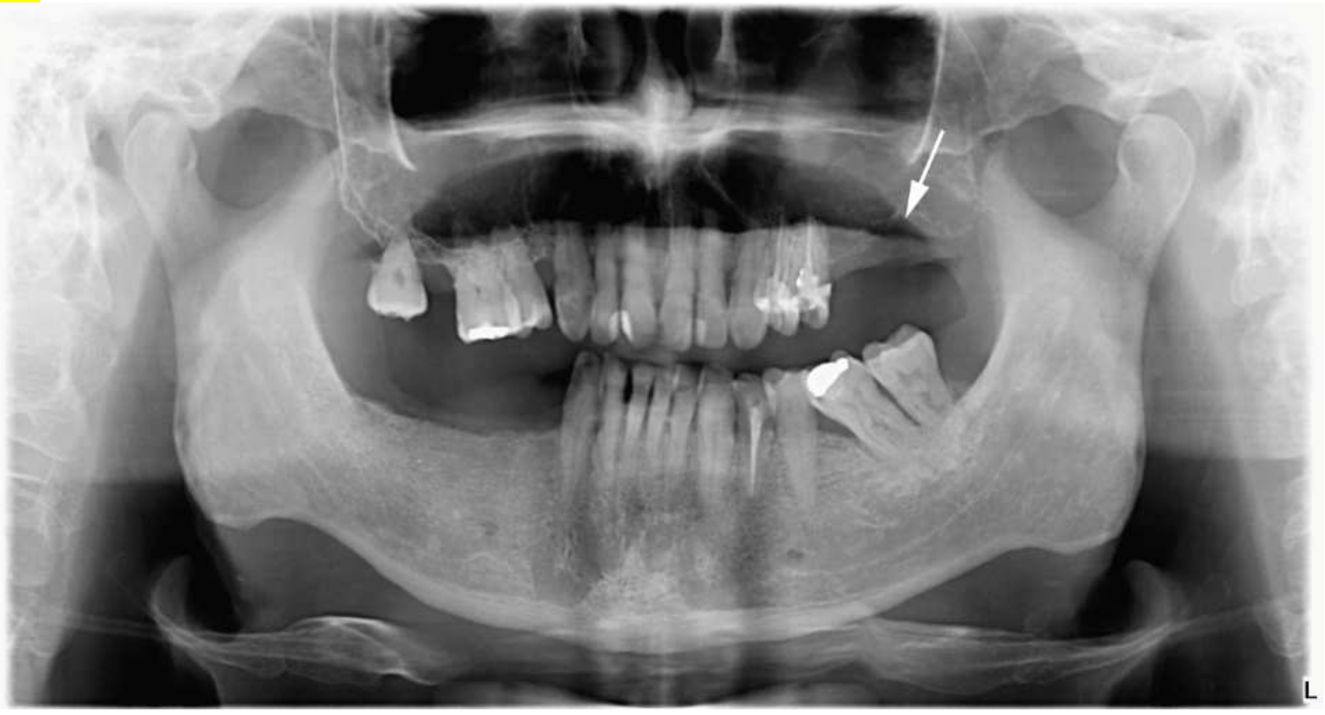
1.1. Case Report
1.2. Histology, Immune-Histochemistry, Fluorescence In Situ Hybridization (FISH)
1.3. Treatment
1.4. Discussion
Author Contributions
Funding
Institutional Review Board Statement
Informed Consent Statement
Data Availability Statement
Acknowledgments
Conflicts of Interest
Abbreviations
| HGBL, NOS | high-grade B-cell lymphoma not otherwise specified; NHL: non-Hodgkin lymphoma |
| DLBCL | diffuse large B-cell lymphoma |
| DLBCL, NOS | diffuse large B-cell lymphoma not otherwise specified |
| DEL | double expressed lymphomas |
| MOD | mesial-occlusal-distal |
| OP | orthopantomography |
| MRI | magnetic resonance imaging |
| DWIBS | diffusion-weighted whole-body imaging with background body signal suppression |
| ADC | apparent diffusion coefficient |
| FISH | fluorescence in situ hybridization |
| R-CHOP | chemotherapy combination |
| PET-CT | positron emission tomography and computed tomography |
References
- Rodrigues-Fernandes, C.I.; de Souza, L.L.; Santos-Costa, S.F.D.; Pontes, H.A.R.; de Almeida, O.P.; Vargas, P.A.; Henao, J.R.; Rahimi, S.; Brennan, P.A.; Fonseca, F.P. Clinicopathological analysis of oral diffuse large B-cell lymphoma, NOS: A systematic review. J. Oral Pathol. Med. 2019, 48, 185–191. [Google Scholar] [CrossRef] [PubMed]
- Walter, C.; Ziebart, T.; Sagheb, K.; Rahimi-Nedjat, R.K.; Manz, A.; Hess, G. Malignant lymphomas in the head and neck region—A retrospective, single-center study over 41 years. Int. J. Med. Sci. 2015, 12, 141–145. [Google Scholar] [CrossRef] [PubMed] [Green Version]
- Das, J.; Ray, S.; Sen, S.; Chandy, M. Extranodal involvement in lymphoma—A Pictorial Essay and Retrospective Analysis of 281 PET/CT studies. Asia Ocean. J. Nucl. Med. Biol. 2014, 2, 42–56. [Google Scholar] [PubMed]
- MacDonald, D.; Li, T.; Leung, S.F.; Curtin, J.; Yeung, A.; Martin, M.A. Extranodal lymphoma arising within the maxillary alveolus: A case report. Oral Surg. Oral Med. Oral Pathol. Oral Radiol. 2017, 124, e233–e238. [Google Scholar] [CrossRef]
- Silva, T.; Ferreira, C.; Leite, G.; Pontes, J.; Antunes, H. Oral manifestations of lymphoma: A systematic review. Ecancermedicalscience 2016, 10, 665. [Google Scholar] [CrossRef] [Green Version]
- Kamulegeya, A.; Kalyanyama, B.M. Oral maxillofacial neoplasms in an East African population a 10 year retrospective study of 1863 cases using histopathological reports. BMC Oral Health 2008, 8, 19. [Google Scholar] [CrossRef] [Green Version]
- Nagafuji, H.; Yokoi, H.; Ohara, A.; Fujiwara, M.; Takayama, N.; Saito, K. Primary diffuse large B-cell lymphoma of the frontal sinus: A case report and literature review. Radiol. Case Rep. 2018, 13, 635–639. [Google Scholar] [CrossRef] [PubMed]
- Wolvius, E.B.; van der Valk, P.; van der Wal, J.E.; van Diest, P.J.; Huijgens, P.C.; van der Waal, I.; Snow, G.B. Primary extranodal non-Hodgkin lymphoma of the oral cavity. An analysis of 34 cases. Eur. J. Cancer B Oral Oncol. 1994, 30, 121–125. [Google Scholar] [CrossRef]
- Doumas, S.; Sakkas, L.; Panayiotidis, P.; Wozniak, G.; Vlychou, M.; Vassilopoulos, G. Favorable outcome in non-Hodgkin lymphoma of the maxillary sinus treated with R-CHOP. Arch. Med. Sci. 2014, 10, 406–409. [Google Scholar] [CrossRef] [Green Version]
- Iguchi, H.; Wada, T.; Matsushita, N.; Oishi, M.; Yamane, H. Anatomic distribution of hematolymphoid malignancies in the head and neck: 7 years of experience with 122 patients in a single institution. Acta Oto-Laryngol. 2012, 132, 1224–1231. [Google Scholar] [CrossRef] [PubMed]
- Steven, H.; Swerdlow, E.C.; Harris, N.L.; Jaffe, E.S.; Pileri, S.A.; Stein, J.T.H.; Arber, D.A.; Hasserjian, R.P.; Michelle, M.; Beau, A.O.L.; et al. WHO Classification of Tumours of Haematopoietic and Lymphoid Tissues, 4th ed.; IARC: Lyon, France, 2017. [Google Scholar]
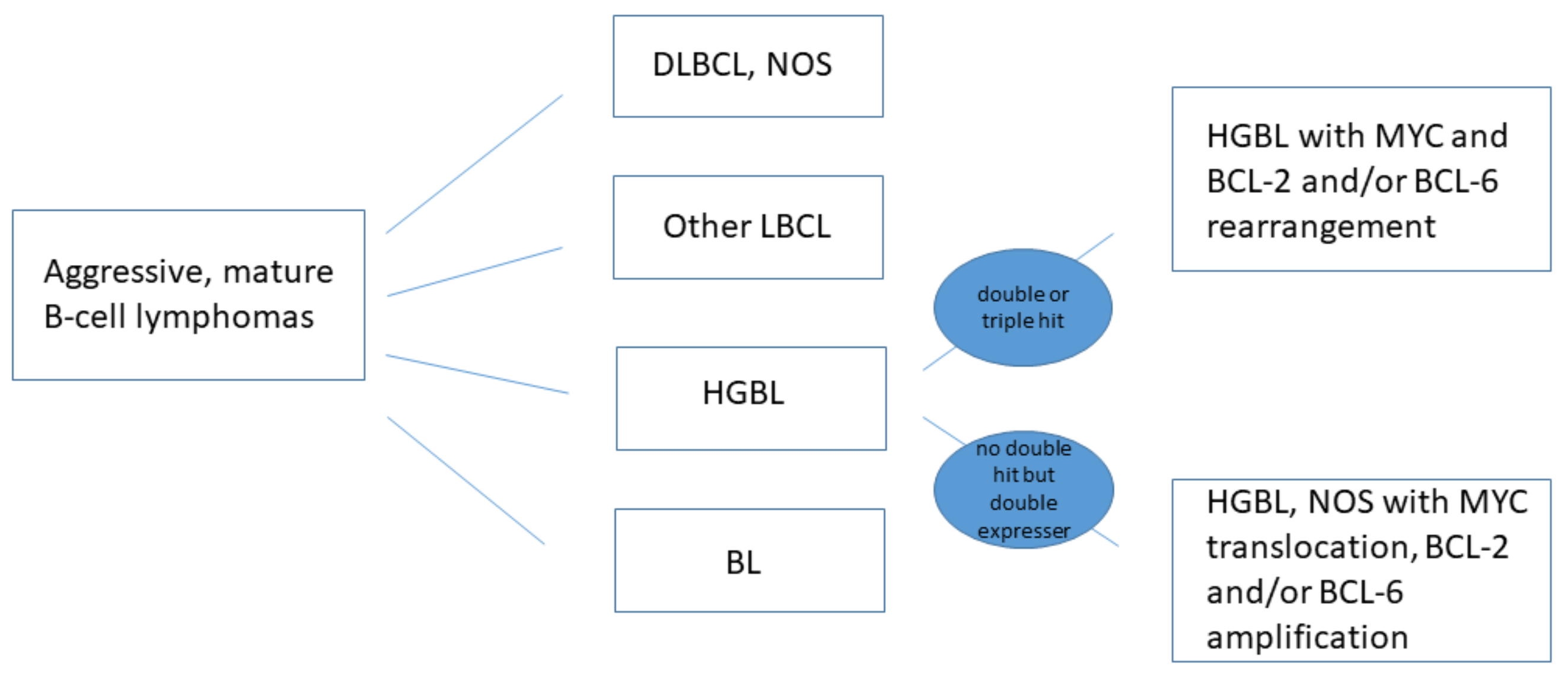
- Grimm, K.E.; O’Malley, D.P. Aggressive B cell lymphomas in the 2017 revised WHO classification of tumors of hematopoietic and lymphoid tissues. Ann. Diagn. Pathol. 2019, 38, 6–10. [Google Scholar] [CrossRef] [PubMed]
- Jaffe, E.S.; Pittaluga, S. Aggressive B-cell lymphomas: A review of new and old entities in the WHO classification. Hematol. Am. Soc. Hematol. Educ. Program. 2011, 2011, 506–514. [Google Scholar] [CrossRef]
- Ahn, J.Y.; Seo, Y.H.; Park, P.W.; Kim, K.H.; Park, M.J.; Jeong, J.H.; Park, S.H.; Song, Y.H. A case of B-cell lymphoma, unclassifiable, with features intermediate between diffuse large B-cell lymphoma and Burkitt lymphoma in a Korean child. Ann. Lab. Med. 2012, 32, 162–166. [Google Scholar] [CrossRef] [Green Version]
- Bemis, T.; Ioanitescu, J.; Mackovick, L.; Hammad, A.; Rubin, J. A Case of Double Expresser Diffuse Large B Cell Lymphoma Treated with R-CODOX-M/R-IVAC. Case Rep. Oncol. 2019, 12, 595–602. [Google Scholar] [CrossRef] [PubMed]
- Rosenthal, A.; Younes, A. High grade B-cell lymphoma with rearrangements of MYC and BCL2 and/or BCL6: Double hit and triple hit lymphomas and double expressing lymphoma. Blood Rev. 2017, 31, 37–42. [Google Scholar] [CrossRef] [PubMed]
- Szumera-Cieckiewicz, A.; Rymkiewicz, G.; Grygalewicz, B.; Jesionek-Kupnicka, D.; Gruchala, A.; Ziarkiewicz-Wroblewska, B.; Galazka, K.; Reszec, J.; Borg, K.; Prochorec-Sobieszek, M. Comprehensive histopathological diagnostics of aggressive B-cell lymphomas based on the updated criteria of the World Health Organisation’s 2017 classification. Pol. J. Pathol. 2018, 69, 1–19. [Google Scholar] [CrossRef] [PubMed]
- Kikuchi, K.; Inoue, H.; Miyazaki, Y.; Ide, F.; Matsuki, E.; Shigematu, H.; Okamoto, S.; Sakashita, H.; Kusama, K. Adult sporadic burkitt lymphoma of the oral cavity: A case report and literature review. J. Oral Maxillofac. Surg. 2012, 70, 2936–2943. [Google Scholar] [CrossRef] [PubMed]
- Jesionek-Kupnicka, D.; Braun, M.; Robak, T.; Kuncman, W.; Kordek, R. A large single-institution retrospective analysis of aggressive B-cell lymphomas according to the 2016/2017 WHO classification. Adv. Clin. Exp. Med. 2019, 28, 1359–1365. [Google Scholar] [CrossRef] [Green Version]
- Oprea, C.; Cainap, C.; Azoulay, R.; Assaf, E.; Jabbour, E.; Koscielny, S.; Lapusan, S.; Vanel, D.; Bosq, J.; Ribrag, V. Primary diffuse large B-cell non-Hodgkin lymphoma of the paranasal sinuses: A report of 14 cases. Br. J. Haematol. 2005, 131, 468–471. [Google Scholar] [CrossRef]
- Bischin, A.M.; Dorer, R.; Aboulafia, D.M. Transformation of Follicular Lymphoma to a High-Grade B-Cell Lymphoma with MYC and BCL2 Translocations and Overlapping Features of Burkitt Lymphoma and Acute Lymphoblastic Leukemia: A Case Report and Literature Review. Clin. Med. Insights Blood Disord. 2017, 10, 5421. [Google Scholar] [CrossRef] [Green Version]
- Chen, B.-J.; Fend, F.; Campo, E.; Quintanilla-Martinez, L. Aggressive B-cell lymphomas—From morphology to molecular pathogenesis. Ann. Lymphoma 2019, 3. Available online: https://aol.amegroups.com/article/view/4949 (accessed on 22 September 2021).
- Li, J.; Liu, X.; Yao, Z.; Zhang, M. High-Grade B-Cell Lymphomas, Not Otherwise Specified: A Study of 41 Cases. Cancer Manag. Res. 2020, 12, 1903–1912. [Google Scholar] [CrossRef] [Green Version]
- de Castro, M.S.; Ribeiro, C.M.; de Carli, M.L.; Pereira, A.A.C.; Sperandio, F.F.; de Almeida, O.P.; Hanemann, J.A.C. Fatal primary diffuse large B-cell lymphoma of the maxillary sinus initially treated as an infectious disease in an elderly patient: A clinicopathologic report. Gerodontology 2018, 35, 59–62. [Google Scholar] [CrossRef]
- Gill, D.S.; Cunliffe, D.; Ali, A.; Sternberg, A. Malignant lymphoma of the maxillary sinus masquerading as an odontogenic infection: Report of a case. Dent. Update 2000, 27, 132–134. [Google Scholar] [CrossRef] [PubMed]
- Mensch, K.; Szarka, K.; Mensch, H.; Dobai, A.; Magyar, Z.; Pacurar, M.; Vartolomei, A.C.; Manuc, D.; Nagy, C.D. PCR technique assisting the early diagnosis of human papillomavirus a retrospective clinical study. Rev. Chim. 2018, 69, 2781–2787. [Google Scholar] [CrossRef]
- Hariram, S.M.; Malkunje, L.R.; Singh, N.; Das, S.; Mehta, G. Ameloblastoma of the anterior mandible. Natl. J. Maxillofac. Surg. 2014, 5, 47–50. [Google Scholar] [CrossRef] [Green Version]
- Brody, A.; Zalatnai, A.; Csomo, K.; Belik, A.; Dobo-Nagy, C. Difficulties in the diagnosis of periapical translucencies and in the classification of cemento-osseous dysplasia. BMC Oral Health 2019, 19, 139. [Google Scholar] [CrossRef]
- Kawaguchi, M.; Kato, H.; Tomita, H.; Mizuta, K.; Aoki, M.; Hara, A.; Matsuo, M. Imaging Characteristics of Malignant Sinonasal Tumors. J. Clin. Med. 2017, 6, 116. [Google Scholar] [CrossRef] [Green Version]
- MacDonald, D.; Lim, S. Extranodal lymphoma arising within the maxillary alveolus: A systematic review. Oral Radiol. 2018, 34, 113–126. [Google Scholar] [CrossRef] [Green Version]
- Adwani, D.; Arora, R.; Bhattacharya, A.; Bhagat, B. Non-Hodgkin’s lymphoma of maxillary sinus: An unusual presentation. Ann. Maxillofac. Surg. 2013, 3, 95–97. [Google Scholar] [CrossRef] [Green Version]
- Dolan, J.M.; DeGraft-Johnson, A.; McDonald, N.; Ward, B.B.; Phillips, T.J.; Munz, S.M. Maxillary and Mandibular Non-Hodgkin Lymphoma with Concurrent Periapical Endodontic Disease: Diagnosis and Management. J. Endod. 2017, 43, 1744–1749. [Google Scholar] [CrossRef] [PubMed]
- Longo, F.; De Maria, G.; Esposito, P.; Califano, L. Primary non-Hodgkin’s lymphoma of the mandible. Report of a case. Int. J. Oral Maxillofac. Surg. 2004, 33, 801–803. [Google Scholar] [CrossRef] [PubMed]
- Pereira, D.L.; Fernandes, D.T.; Santos-Silva, A.R.; Vargas, P.A.; de Almeida, O.P.; Lopes, M.A. Intraosseous Non-Hodgkin Lymphoma Mimicking a Periapical Lesion. J. Endod. 2015, 41, 1738–1742. [Google Scholar] [CrossRef] [PubMed]


Publisher’s Note: MDPI stays neutral with regard to jurisdictional claims in published maps and institutional affiliations. |
© 2021 by the authors. Licensee MDPI, Basel, Switzerland. This article is an open access article distributed under the terms and conditions of the Creative Commons Attribution (CC BY) license (https://creativecommons.org/licenses/by/4.0/).
Share and Cite
Brody, A.; Dobo-Nagy, C.; Mensch, K.; Oltyan, Z.; Csomor, J.; Pacurar, M.; Dobai, A. High-Grade B-Cell Lymphoma Not Otherwise Specified (HGBL, NOS) in the Maxillary Sinus Mimicking Periapical Inflammation: Case Report and Review of the Literature. Appl. Sci. 2021, 11, 8803. https://doi.org/10.3390/app11198803
Brody A, Dobo-Nagy C, Mensch K, Oltyan Z, Csomor J, Pacurar M, Dobai A. High-Grade B-Cell Lymphoma Not Otherwise Specified (HGBL, NOS) in the Maxillary Sinus Mimicking Periapical Inflammation: Case Report and Review of the Literature. Applied Sciences. 2021; 11(19):8803. https://doi.org/10.3390/app11198803
Chicago/Turabian StyleBrody, Andrea, Csaba Dobo-Nagy, Karoly Mensch, Zsuzsanna Oltyan, Judit Csomor, Mariana Pacurar, and Adrienn Dobai. 2021. "High-Grade B-Cell Lymphoma Not Otherwise Specified (HGBL, NOS) in the Maxillary Sinus Mimicking Periapical Inflammation: Case Report and Review of the Literature" Applied Sciences 11, no. 19: 8803. https://doi.org/10.3390/app11198803
APA StyleBrody, A., Dobo-Nagy, C., Mensch, K., Oltyan, Z., Csomor, J., Pacurar, M., & Dobai, A. (2021). High-Grade B-Cell Lymphoma Not Otherwise Specified (HGBL, NOS) in the Maxillary Sinus Mimicking Periapical Inflammation: Case Report and Review of the Literature. Applied Sciences, 11(19), 8803. https://doi.org/10.3390/app11198803