
Dose Estimation by Geant4-Based Simulations for Cone-Beam CT Applications: A Systematic Review


 and
and
Abstract
1. Introduction
- RQ1: What are the most significant current publications estimating radiation dose using Geant4 in CBCT applications?
- RQ2: Which are the most common CBCT applications that use Geant4 simulations to estimate the dose and how have they changed over the years?
- RQ3: How advances in Geant4-based CBCT dose estimator may help to evaluate the risk of developing stochastic effects?
- RQ4: What are the current research trends and disciplines for prospective research on this topic?
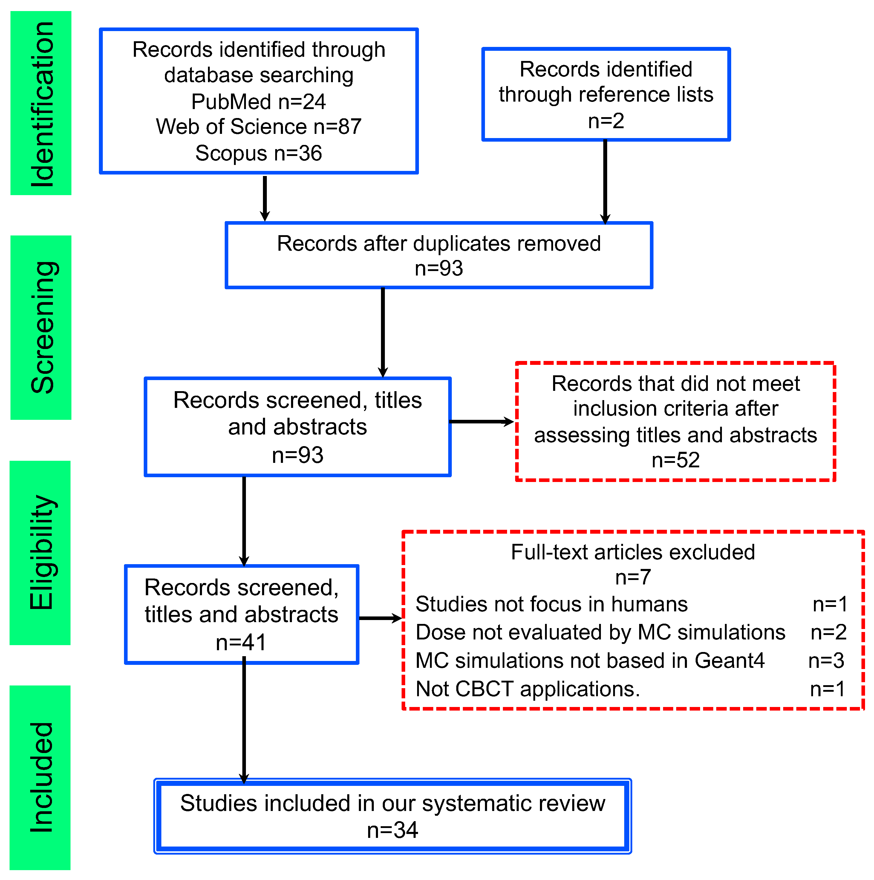
2. Methodology
2.1. Literature Search
- Geant4 OR GATE OR GAMOS to include all the toolkit based on these codes.
- Cone beam computed tomography according to medical subject headings (MeSH).
- Cone beam computed tomography as a generic term that refers to the medical imaging technique with a conic/pyramidal X-ray beam of radiation.
- Radiation dosage and radiation protection according to medical subject headings (MeSH).
- Dose OR Dosimetry as a generic term including all types of doses described by the International Commission on Radiological Protection (ICRP) [38].
2.2. Inclusion and Exclusion Criteria
- not medical use;
- papers that do not focus on CBCT;
- studies that do not focus on humans; and
- papers that do not estimate the dose using Geant4-based toolkits.
2.3. Data Extraction and Analysis
3. Results and Discussion
3.1. RQ1: What Are the Most Significant Current Publications Estimating Radiation Dose Using Geant4 in CBCT Applications?
3.2. RQ2: Which Are the Most Common CBCT Applications That Use Geant4 Simulations to Estimate the Dose, and How Have They Changed over the Years?
3.3. RQ3: How Advances in Geant4-Based CBCT Dose Estimator May Help to Evaluate the Risk of Developing Stochastic Effects?
3.4. RQ4: What Are the Current Research Trends and Disciplines for Prospective Research on This Topic?
4. Conclusions
Supplementary Materials
Author Contributions
Funding
Institutional Review Board Statement
Informed Consent Statement
Acknowledgments
Conflicts of Interest
References
- Pfeiffer, D.; Pfeiffer, F.; Rummeny, E. Advanced X-ray Imaging Technology. Recent Results Cancer Res. 2020, 216, 3–30. [Google Scholar] [CrossRef]
- Jaffray, D.A.; Siewerdsen, J.H. Cone-beam computed tomography with a flat-panel imager: Initial performance characterization. Med. Phys. 2000, 27, 1311–1323. [Google Scholar] [CrossRef]
- Jaffray, D.A.; Siewerdsen, J.H.; Wong, J.W.; Martinez, A.A. Flat-panel cone-beam computed tomography for image-guided radiation therapy. Int. J. Radiat. Oncol. Biol. Phys. 2002, 53, 1337–1349. [Google Scholar] [CrossRef]
- Orth, R.C.; Wallace, M.J.; Kuo, M.D. C-arm cone-beam CT: General principles and technical considerations for use in interventional radiology. J. Vasc. Interv. Radiol. JVIR 2008, 19, 814–820. [Google Scholar] [CrossRef]
- Scarfe, W.C.; Farman, A.G.; Sukovic, P. Clinical applications of cone-beam computed tomography in dental practice. J. Can. Dent. Assoc. 2006, 72, 75–80. [Google Scholar]
- Ding, G.X.; Coffey, C.W. Radiation dose from kilovoltage cone beam computed tomography in an image-guided radiotherapy procedure. Int. J. Radiat. Oncol. Biol. Phys. 2009, 73, 610–617. [Google Scholar] [CrossRef] [PubMed]
- Cho, P.S.; Johnson, R.H.; Griffin, T.W. Cone-beam CT for radiotherapy applications. Phys. Med. Biol. 1995, 40, 1863–1883. [Google Scholar] [CrossRef] [PubMed]
- Chetty, I.J.; Curran, B.; Cygler, J.E.; DeMarco, J.J.; Ezzell, G.; Faddegon, B.A.; Kawrakow, I.; Keall, P.J.; Liu, H.; Ma, C.M.; et al. Report of the AAPM Task Group No. 105: Issues associated with clinical implementation of Monte Carlo-based photon and electron external beam treatment planning. Med. Phys. 2007, 34, 4818–4853. [Google Scholar] [CrossRef] [PubMed]
- Cheng, Y.C.; Tsai, S.H.; Cheng, Y.; Chen, J.H.; Chai, J.W.; Chen, C.C.C. Percutaneous Transthoracic Lung Biopsy: Comparison between C-Arm Cone-Beam CT and Conventional CT Guidance. Transl. Oncol. 2015, 8, 258–264. [Google Scholar] [CrossRef] [PubMed]
- Ding, G.X.; Alaei, P.; Curran, B.; Flynn, R.; Gossman, M.; Mackie, T.R.; Miften, M.; Morin, R.; Xu, X.G.; Zhu, T.C. Image guidance doses delivered during radiotherapy: Quantification, management, and reduction: Report of the AAPM Therapy Physics Committee Task Group 180. Med. Phys. 2018, 45, e84–e99. [Google Scholar] [CrossRef]
- Alaei, P.; Spezi, E. Imaging dose from cone beam computed tomography in radiation therapy. Phys. Med. 2015, 31, 647–658. [Google Scholar] [CrossRef]
- Bryce-Atkinson, A.; de Jong, R.; Bel, A.; Aznar, M.C.; Whitfield, G.; van Herk, M. Evaluation of Ultra-low-dose Paediatric Cone-beam Computed Tomography for Image-guided Radiotherapy. Clin. Oncol. R. Coll. Radiol. 2020, 32, 835–844. [Google Scholar] [CrossRef] [PubMed]
- Tyndall, D.A.; Rathore, S. Cone-beam CT diagnostic applications: Caries, periodontal bone assessment, and endodontic applications. Dent. Clin. N. Am. 2008, 52, 825–841. [Google Scholar] [CrossRef]
- Venskutonis, T.; Plotino, G.; Juodzbalys, G.; Mickevičienė, L. The importance of cone-beam computed tomography in the management of endodontic problems: A review of the literature. J. Endod. 2014, 40, 1895–1901. [Google Scholar] [CrossRef] [PubMed]
- Barghan, S.; Tetradis, S.; Mallya, S. Application of cone beam computed tomography for assessment of the temporomandibular joints. Aust. Dent. J. 2012, 57 (Suppl. 1), 109–118. [Google Scholar] [CrossRef]
- O’Connell, A.; Conover, D.L.; Zhang, Y.; Seifert, P.; Logan-Young, W.; Lin, C.F.L.; Sahler, L.; Ning, R. Cone-Beam CT for Breast Imaging: Radiation Dose, Breast Coverage, and Image Quality. Am. J. Roentgenol. 2010, 195, 496–509. [Google Scholar] [CrossRef]
- Chen, Y.; Liu, B.; O’Connor, J.M.; Didier, C.S.; Glick, S.J. Characterization of scatter in cone-beam CT breast imaging: Comparison of experimental measurements and Monte Carlo simulation. Med. Phys. 2009, 36, 857–869. [Google Scholar] [CrossRef]
- Uhlig, J.; Uhlig, A.; Biggemann, L.; Fischer, U.; Lotz, J.; Wienbeck, S. Diagnostic accuracy of cone-beam breast computed tomography: A systematic review and diagnostic meta-analysis. Eur. Radiol. 2019, 29, 1194–1202. [Google Scholar] [CrossRef] [PubMed]
- O’Connell, A.M.; Karellas, A.; Vedantham, S.; Kawakyu-O’Connor, D.T. Newer Technologies in Breast Cancer Imaging: Dedicated Cone-Beam Breast Computed Tomography. Semin. Ultrasound CT MR 2018, 39, 106–113. [Google Scholar] [CrossRef] [PubMed]
- Chen, B.; Ning, R. Cone-beam volume CT breast imaging: Feasibility study. Med. Phys. 2002, 29, 755–770. [Google Scholar] [CrossRef]
- Wienbeck, S.; Lotz, J.; Fischer, U. Review of clinical studies and first clinical experiences with a commercially available cone-beam breast CT in Europe. Clin. Imaging 2017, 42, 50–59. [Google Scholar] [CrossRef] [PubMed]
- Lindfors, K.K.; Boone, J.M.; Nelson, T.R.; Yang, K.; Kwan, A.L.C.; Miller, D.F. Dedicated breast CT: Initial clinical experience. Radiology 2008, 246, 725–733. [Google Scholar] [CrossRef]
- Thacker, S.C.; Glick, S.J. Normalized glandular dose (DgN) coefficients for flat-panel CT breast imaging. Phys. Med. Biol. 2004, 49, 5433–5444. [Google Scholar] [CrossRef] [PubMed]
- Poludniowski, G.; Allinson, N.M.; Evans, P.M. Proton radiography and tomography with application to proton therapy. Br. J. Radiol. 2015, 88, 20150134. [Google Scholar] [CrossRef] [PubMed]
- Amblard, R.; Floquet, V.; Angellier, G.; Hannoun-Lévi, J.M.; Hérault, J. Proton imaging applications for proton therapy: State of the art. Cancer Radiother. 2015, 19, 136–139. [Google Scholar] [CrossRef]
- Johnson, R.P. Review of medical radiography and tomography with proton beams. Rep. Prog. Physics. Phys. Soc. 2018, 81, 16701. [Google Scholar] [CrossRef]
- Giacometti, V.; Hounsell, A.R.; McGarry, C.K. A review of dose calculation approaches with cone beam CT in photon and proton therapy. Phys. Med. 2020, 76, 243–276. [Google Scholar] [CrossRef]
- Bongrand, A.; Koumeir, C.; Villoing, D.; Guertin, A.; Haddad, F.; Métivier, V.; Poirier, F.; Potiron, V.; Servagent, N.; Supiot, S.; et al. A Monte Carlo Determination of Dose and Range Uncertainties for Preclinical Studies with a Proton Beam. Cancers 2021, 13, 1889. [Google Scholar] [CrossRef]
- Nasseh, I.; Al-Rawi, W. Cone Beam Computed Tomography. Dent. Clin. N. Am. 2018, 62, 361–391. [Google Scholar] [CrossRef]
- Rehani, M.M.; Berry, M. Radiation doses in computed tomography. The increasing doses of radiation need to be controlled. BMJ 2000. [Google Scholar] [CrossRef] [PubMed]
- Strauss, K.J.; Kaste, S.C. The ALARA (as low as reasonably achievable) concept in pediatric CT intelligent dose reduction: Multidisciplinary conference organized by the Society of Pediatric Radiology. Pediatr Radiol. 2002, 32, 217–313. [Google Scholar] [CrossRef]
- Tsapaki, V.; Aldrich, J.E.; Sharma, R.; Staniszewska, M.A.; Krisanachinda, A.; Rehani, M.; Hufton, A.; Triantopoulou, C.; Maniatis, P.N.; Papailiou, J.; et al. Dose reduction in CT while maintaining diagnostic confidence: Diagnostic reference levels at routine head, chest, and abdominal CT–IAEA-coordinated research project. Radiology 2006, 240, 828–834. [Google Scholar] [CrossRef]
- Brenner, D.J.; Hall, E.J. Computed tomography—An increasing source of radiation exposure. N. Engl. J. Med. 2007, 357, 2277–2284. [Google Scholar] [CrossRef] [PubMed]
- McCollough, C.H. Computed Tomography Technology-and Dose-in the 21st Century. Health Phys. 2019, 116, 157–162. [Google Scholar] [CrossRef] [PubMed]
- Schauer, D.A.; Linton, O.W. Ncrp Report No. 160, Ionizing Radiation Exposure of the Population of the United States, Medical Exposure—Are We Doing Less with More, and Is There A Role for Health Physicists? Health Physics 2009, 97, 1–5. [Google Scholar] [CrossRef]
- Muhammad, N.A.; Kayun, Z.; Abu Hassan, H.; Wong, J.H.D.; Ng, K.H.; Karim, M.K.A. Evaluation of Organ Dose and Image Quality Metrics of Pediatric CT Chest-Abdomen-Pelvis (CAP) Examination: An Anthropomorphic Phantom Study. Appl. Sci. 2021, 11, 2047. [Google Scholar] [CrossRef]
- Allan, J.M. Genetic susceptibility to radiogenic cancer in humans. Health Phys. 2008, 95, 677–686. [Google Scholar] [CrossRef]
- Valentin, J. Avoidance of radiation injuries from medical interventional procedures. Ann. ICRP 2000, 30, 7–67. [Google Scholar] [CrossRef]
- Bhargavan-Chatfield, M.; Morin, R.L. The ACR Computed Tomography Dose Index Registry: The 5 million examination update. J. Am. Coll. Radiol. JACR 2013, 10, 980–983. [Google Scholar] [CrossRef]
- Shope, T.B.; Gagne, R.M.; Johnson, G.C. A method for describing the doses delivered by transmission X-ray computed tomography. Med. Phys. 1981, 8, 488–495. [Google Scholar] [CrossRef]
- McCollough, C.H. It is time to retire the computed tomography dose index (CTDI) for CT quality assurance and dose optimization. Against the proposition. Med. Phys. 2006, 33, 1190–1191. [Google Scholar] [CrossRef] [PubMed]
- McCollough, C.H.; Leng, S.; Yu, L.; Cody, D.D.; Boone, J.M.; McNitt-Gray, M.F. CT dose index and patient dose: They are not the same thing. Radiology 2011. [Google Scholar] [CrossRef] [PubMed]
- Kim, S.; Yoshizumi, T.T.; Toncheva, G.; Yoo, S.; Yin, F.F. Comparison of radiation doses between cone beam CT and multi detector CT: TLD measurements. Radiat. Prot. Dosim. 2008, 132, 339–345. [Google Scholar] [CrossRef]
- Council, N.R. Health Risks from Exposure to Low Levels of Ionizing Radiation: BEIR VII Phase 2; The National Academy of Sciences: Washington, DC, USA, 2006; pp. 1–406. [Google Scholar] [CrossRef]
- Pearce, M.S.; Salotti, J.A.; Little, M.P.; McHugh, K.; Lee, C.; Kim, K.P.; Howe, N.L.; Ronckers, C.M.; Rajaraman, P.; Sir Craft, A.W.; et al. Radiation exposure from CT scans in childhood and subsequent risk of leukaemia and brain tumours: A retrospective cohort study. Lancet 2012, 380, 499–505. [Google Scholar] [CrossRef]
- Mullenders, L.; Atkinson, M.; Paretzke, H.; Sabatier, L.; Bouffler, S. Assessing cancer risks of low-dose radiation. Nat. Rev. Cancer 2009, 46, 596–604. [Google Scholar] [CrossRef]
- Kim, S.; Yoshizumi, T.T.; Frush, D.P.; Toncheva, G.; Yin, F.F. Radiation dose from cone beam CT in a pediatric phantom: Risk estimation of cancer incidence. Am. J. Roentgenol. 2010, 194, 186–190. [Google Scholar] [CrossRef] [PubMed]
- Jarry, G.; Graham, S.A.; Moseley, D.J.; Jaffray, D.J.; Siewerdsen, J.H.; Verhaegen, F. Characterization of scattered radiation in kV CBCT images using Monte Carlo simulations. Med. Phys. 2006, 33, 4320–4329. [Google Scholar] [CrossRef]
- Landry, G.; Hua, C.H. Current state and future applications of radiological image guidance for particle therapy. Med. Phys. 2018, 45, e1086–e1095. [Google Scholar] [CrossRef]
- Newhauser, W.D.; Durante, M. Assessing the risk of second malignancies after modern radiotherapy. Nat. Rev. Cancer 2011, 11, 438–448. [Google Scholar] [CrossRef]
- Jeong, H.; Rah, J.E.; Hwang, U.J.; Yoo, S.H.; Min, B.J.; Lee, S.Y.; Yoon, M.; Shin, D.H.; Park, S.Y.; Lee, S.B.; et al. Estimation of the secondary cancer risk induced by diagnostic imaging radiation during proton therapy. J. Radiol. Prot. 2011, 31, 477–487. [Google Scholar] [CrossRef]
- Zhang, Y.; Wu, H.; Chen, Z.; Knisely, J.P.S.; Nath, R.; Feng, Z.; Bao, S.; Deng, J. Concomitant Imaging Dose and Cancer Risk in Image Guided Thoracic Radiation Therapy. Int. J. Radiat. Oncol. Biol. Phys. 2015, 93, 523–531. [Google Scholar] [CrossRef] [PubMed]
- Marcu, L.G. Photons—Radiobiological issues related to the risk of second malignancies. Phys. Med. 2017, 42, 213–220. [Google Scholar] [CrossRef]
- Trott, K.R. Special radiobiological features of second cancer risk after particle radiotherapy. Phys. Med. 2017, 42, 221–227. [Google Scholar] [CrossRef] [PubMed]
- ICRP. 2000 Annual Report of the International Commission on Radiological Protection; ICRP: Ottawa, ON, Canada, 2000; pp. 1–30. [Google Scholar]
- Kase, K.R. Radiation protection principles of NCRP. Health Phys. 2004, 87, 251–257. [Google Scholar] [CrossRef]
- Rogers, D.W. The role of Monte Carlo simulation of electron transport in radiation dosimetry. Int. J. Radiat. Appl. Instrum. 1991, 42, 965–974. [Google Scholar] [CrossRef]
- Ma, C.M.; Jiang, S.B. Monte Carlo modelling of electron beams from medical accelerators. Phys. Med. Biol. 1999, 44, R157–R189. [Google Scholar] [CrossRef] [PubMed]
- Andreo, P. Monte Carlo simulations in radiotherapy dosimetry. Radiat. Oncol. 2018, 13, 121. [Google Scholar] [CrossRef]
- Lewis, R.D.; Ryde, S.J.; Seaby, A.W.; Hancock, D.A.; Evans, C.J. Use of Monte Carlo computation in benchmarking radiotherapy treatment planning system algorithms. Phys. Med. Biol. 2000, 45, 1755–1764. [Google Scholar] [CrossRef]
- Chow, J.C.L. Depth Dose Enhancement on Flattening-Filter-Free Photon Beam: A Monte Carlo Study in Nanoparticle-Enhanced Radiotherapy. Appl. Sci. 2020, 10, 7052. [Google Scholar] [CrossRef]
- Nazemi, E.; Six, N.; Iuso, D.; De Samber, B.; Sijbers, J.; De Beenhouwer, J. Monte-Carlo-Based Estimation of the X-ray Energy Spectrum for CT Artifact Reduction. Appl. Sci. 2021, 11, 3145. [Google Scholar] [CrossRef]
- Verhaegen, F.; Seuntjens, J. Monte Carlo modelling of external radiotherapy photon beams. Phys. Med. Biol. 2003, 48, R107–R164. [Google Scholar] [CrossRef]
- Kawrakow, I.; Mainegra Hing, E.; Tessier, F.; Walters, B.R.B. The EGSnrc Code System: Monte Carlo Simulation of Electron and Photon Transport. 2021. Available online: https://nrc-cnrc.github.io/EGSnrc/doc/pirs701-egsnrc.pdf (accessed on 15 April 2021).
- Briesmeister, J.F. MCNP: A General Monte Carlo N-Particle Transport Code; Los Alamos National Laboratory: Santa Fe, NM, USA, 2000.
- Salvat, F.; Fernandez-Varea, J.M.; Acosta, E.; Sempau, J. Penelope—A Code System for Monte Carlo Simulation of Electron and Photon Transport; Nuclear Energy Agency: Paris, France, 2001. [Google Scholar]
- Tapiovaara, M.; Lakkisto, M.; Servomaa, A. PCXMC A PC-Based Monte Carlo Program for Calculating Patient Doses in Medical X-ray Examinations; Technical Report; IAEA: Vienna, Austria, 1997. [Google Scholar]
- Agostinelli, S.; Allison, J.; Amako, K.; Apostolakis, J.; Araujo, H.; Arce, P.; Asai, M.; Axen, D.; Banerjee, S.; Barrand, G.; et al. GEANT4—A simulation toolkit. Nucl. Instrum. Methods Phys. Res. 2003, 506, 250–303. [Google Scholar] [CrossRef]
- Allison, J.; Amako, K.; Apostolakis, J.; Araujo, H.; Arce Dubois, P.; Asai, M.; Barrand, G.; Capra, R.; Chauvie, S.; Chytracek, R.; et al. Geant4 developments and applications. IEEE Trans. Nucl. Sci. 2006, 53, 270–278. [Google Scholar] [CrossRef]
- Jahnke, L.; Fleckenstein, J.; Wenz, F.; Hesser, J. GMC: A GPU implementation of a Monte Carlo dose calculation based on Geant4. Phys. Med. Biol. 2012, 57, 1217–1229. [Google Scholar] [CrossRef]
- Bert, J.; Perez-Ponce, H.; El Bitar, Z.; Jan, S.; Boursier, Y.; Vintache, D.; Bonissent, A.; Morel, C.; Brasse, D.; Visvikis, D. Geant4-based Monte Carlo simulations on GPU for medical applications. Phys. Med. Biol. 2013, 58, 5593–5611. [Google Scholar] [CrossRef]
- Sarrut, D.; Bardiès, M.; Boussion, N.; Freud, N.; Jan, S.; Létang, J.M.; Loudos, G.; Maigne, L.; Marcatili, S.; Mauxion, T.; et al. A review of the use and potential of the GATE Monte Carlo simulation code for radiation therapy and dosimetry applications. Med. Phys. 2014, 41, 64301. [Google Scholar] [CrossRef] [PubMed]
- Boone, J.M.; Shah, N.; Nelson, T.R. A comprehensive analysis of coefficients for pendant-geometry cone-beam breast computed tomography. Med. Phys. 2004, 31, 226–235. [Google Scholar] [CrossRef] [PubMed]
- Jan, S.; Santin, G.; Strul, D.; Staelens, S.; Assié, K.; Autret, D.; Avner, S.; Barbier, R.; Bardiès, M.; Bloomfield, P.M.; et al. GATE: A simulation toolkit for PET and SPECT. Phys. Med. Biol. 2004, 49, 4543–4561. [Google Scholar] [CrossRef] [PubMed]
- Jan, S.; Benoit, D.; Becheva, E.; Carlier, T.; Cassol, F.; Descourt, P.; Frisson, T.; Grevillot, L.; Guigues, L.; Maigne, L.; et al. GATE V6: A major enhancement of the GATE simulation platform enabling modelling of CT and radiotherapy. Phys. Med. Biol. 2011, 56, 881–901. [Google Scholar] [CrossRef]
- Papadimitroulas, P. Dosimetry applications in GATE Monte Carlo toolkit. Phys. Med. 2017, 41, 136–140. [Google Scholar] [CrossRef]
- Townsend, D. In Proceedings of the 2011 IEEE Nuclear Science Symposium and Medical Imaging Conference (NSS/MIC 2011), Valencia, Spain, 23–29 October 2011.
- Arce, P.; Ignacio, J.; Harkness, L.; Pérez-astudillo, D.; Cañadas, M.; Rato, P.; Prado, M.D.; Abreu, Y.; Lorenzo, G.D.; Kolstein, M.; et al. Nuclear Instruments and Methods in Physics Research A G AMOS: A framework to do G EANT 4 simulations in different physics fi elds with an user-friendly interface. Nucl. Inst. Methods Phys. Res. 2014, 735, 304–313. [Google Scholar] [CrossRef]
- Arce, P.; Rato, P.; Canadas, M.; Lagares, J.I. GAMOS: A Geant4-based easy and flexible framework for nuclear medicine applications. In Proceedings of the 2008 IEEE Nuclear Science Symposium Conference Record, Dresden, Germany, 19–25 October 2008; pp. 3162–3168. [Google Scholar] [CrossRef]
- Karaoglu, A.; Arce, P.; Obradors, D.; Lagares, J.I.; Unak, P. Calculation by GAMOS/Geant4 simulation of cellular energy distributions from alpha and lithium-7 particles created by BNCT. Appl. Radiat. Isot. 2018, 132, 206–211. [Google Scholar] [CrossRef]
- Auditore, L.; Amato, E.; Italiano, A.; Arce, P.; Campennì, A.; Baldari, S. Internal dosimetry for TARE therapies by means of GAMOS Monte Carlo simulations. Phys. Med. 2019, 64, 245–251. [Google Scholar] [CrossRef]
- Bongrand, A.; Busato, E.; Force, P.; Martin, F.; Montarou, G. Use of short-lived positron emitters for in-beam and real-time β(+) range monitoring in proton therapy. Phys. Med. 2020, 69, 248–255. [Google Scholar] [CrossRef] [PubMed]
- Arce, P.; Lagares, J.I.; Azcona, D. A utility to read automatically DICOM format data for GAMOS/Geant4 simulation. Phys. Med. 2016, 32, 256. [Google Scholar] [CrossRef]
- Antoniadi, A.M.; Du, Y.; Guendouz, Y.; Wei, L.; Mazo, C.; Becker, B.A.; Mooney, C. Current Challenges and Future Opportunities for XAI in Machine Learning-Based Clinical Decision Support Systems: A Systematic Review. Appl. Sci. 2021, 11, 5088. [Google Scholar] [CrossRef]
- Longo, U.G.; De Salvatore, S.; Candela, V.; Zollo, G.; Calabrese, G.; Fioravanti, S.; Giannone, L.; Marchetti, A.; De Marinis, M.G.; Denaro, V. Augmented Reality, Virtual Reality and Artificial Intelligence in Orthopedic Surgery: A Systematic Review. Appl. Sci. 2021, 11, 3253. [Google Scholar] [CrossRef]
- Lucchese, A.; Bonini, C.; Noviello, M.; Lupo Stanghellini, M.T.; Greco, R.; Peccatori, J.; Biella, A.; Tassi, E.; Beretta, V.; Ciceri, F.; et al. The Effect of Removable Orthodontic Appliances on Oral Microbiota: A Systematic Review. Appl. Sci. 2021, 11, 2881. [Google Scholar] [CrossRef]
- Chen, C. CiteSpace II: Detecting and visualizing emerging trends and transient patterns in scientific literature. J. Am. Soc. Inf. Sci. Technol. 2006, 57, 359–377. [Google Scholar] [CrossRef]
- White, H.D.; McCain, K.W. Visualizing a discipline: An author co-citation analysis of information science, 1972–1995. J. Am. Soc. Inf. Sci. 1998, 49, 327–355. [Google Scholar] [CrossRef]
- Aria, M.; Cuccurullo, C. Bibliometrix: An R-tool for comprehensive science mapping analysis. J. Inf. 2017, 11, 959–975. [Google Scholar] [CrossRef]
- Tseng, H.W.; Karellas, A.; Vedantham, S. Radiation dosimetry of a clinical prototype dedicated cone-beam breast CT system with offset detector. Med. Phys. 2021, 48, 1079–1088. [Google Scholar] [CrossRef]
- Cho, S.; Lim, S.; Kim, C.; Wi, S.; Kwon, T.; Youn, W.S.; Lee, S.H.; Kang, B.S.; Cho, S. Enhancement of soft-tissue contrast in cone-beam CT using an anti-scatter grid with a sparse sampling approach. Phys. Med. 2020, 70, 1–9. [Google Scholar] [CrossRef] [PubMed]
- Principi, S.; Wang, A.; Maslowski, A.; Wareing, T.; Jordan, P.; Schmidt, T.G. Deterministic linear Boltzmann transport equation solver for patient-specific CT dose estimation: Comparison against a Monte Carlo benchmark for five realistic scanner configurations and patient models. Med. Phys. 2020, 47, 6470–6483. [Google Scholar] [CrossRef]
- Shi, M.; Myronakis, M.; Jacobson, M.; Ferguson, D.; Williams, C.; Lehmann, M.; Baturin, P.; Huber, P.; Fueglistaller, R.; Lozano, I.V.; et al. GPU-accelerated Monte Carlo simulation of MV-CBCT. Phys. Med. Biol. 2020, 65, 235042. [Google Scholar] [CrossRef] [PubMed]
- Lee, H.; Lee, S.; Cha, J.G.; Baek, T.; Yang, K.M. Postmortem Computed Tomography and Computed Tomography Angiography: Cardiothoracic Imaging Applications in Forensic Medicine. J. Thorac. Imaging 2019, 34, 286–298. [Google Scholar] [CrossRef] [PubMed]
- Jia, S.; Gao, H.; Zhang, L.; Xing, Y.; Zheng, J. Dental and maxillofacial cone beam computed tomography absorbed dose distribution calculation by GEANT4. In Proceedings of the SPIE Medical Imaging, San Diego, CA, USA, 16–21 February 2019; Volume 10948. [Google Scholar] [CrossRef]
- Wang, A.; Maslowski, A.; Wareing, T.; Star-Lack, J.; Schmidt, T.G. A fast, linear Boltzmann transport equationsolver for computed tomography dose calculation (Acuros CTD). Med. Phys. 2019, 46, 925–933. [Google Scholar] [CrossRef]
- Ardenfors, O.; Henry, T.; Gudowska, I.; Poludniowski, G.; Dasu, A. Comparison of Beam Characteristicsand Organ Doses for a Proton Gantry-Mounted CBCT System Modelled with MCNP6 and GATE. Med. Phys. 2018, 45, E430. [Google Scholar]
- Ardenfors, O.; Henry, T.; Gudowska, I.; Poludniowski, G.; Dasu, A. Organ doses from a proton gantry-mounted cone-beam computed tomography system characterized with MCNP6 and GATE. Phys. Med. 2018, 53, 56–61. [Google Scholar] [CrossRef]
- Gholami, S.; Longo, F.; Nedaie, H.A.; Berti, A.; Mousavi, M.; Meigooni, A.S. Application of Geant4 Monte Carlo simulation in dose calculations for small radiosurgical fields. Med. Dosim. 2018, 43, 214–223. [Google Scholar] [CrossRef]
- Maslowski, A.; Wang, A.; Sun, M.; Wareing, T.; Davis, I.; Star-Lack, J. Acuros CTS: A fast, linear Boltzmann transport equation solver for computed tomography scatter—Part I: Core algorithms and validation. Med. Phys. 2018, 45, 1899–1913. [Google Scholar] [CrossRef] [PubMed]
- Leotta, S.; Amato, E.; Settineri, N.; Basile, E.; Italiano, A.; Auditore, L.; Santacaterina, A.; Pergolizzi, S. Patient Dose in Image Guided Radiotherapy: Monte Carlo Study of the CBCT Dose Contribution. Atti Accad. Peloritana Dei-Pericolanti-Cl. Sci. Fis. Mat. Nat. 2018, 96. [Google Scholar] [CrossRef]
- Son, K.; Chang, J.; Lee, H.; Kim, C.; Lee, T.; Cho, S.; Park, S.; Kim, J.S. Optimal dose reduction algorithm using an attenuation-based tube current modulation method for cone-beam CT imaging. PLoS ONE 2018, 13, e0192933. [Google Scholar] [CrossRef] [PubMed]
- Zhu, J.; Penfold, S.N. Europium-155 as a source for dual energy cone beam computed tomography in adaptive proton therapy: A simulation study. Med. Phys. 2017, 44, 5143–5152. [Google Scholar] [CrossRef]
- Benhalouche, S.; Bert, J.; Boussion, N.; Autret, A.; Pradier, O.; Visvikis, D. GATE Monte-Carlo Simulation of an MV-CBCT Flat Panel for Synergistic Imaging and Dosimetric Applications in Radiotherapy. IEEE Trans. Radiat. Plasma Med. Sci. 2017, 39, 4529–4544. [Google Scholar] [CrossRef]
- Myronakis, M.; Star-Lack, J.; Baturin, P.; Rottmann, J.; Morf, D.; Wang, A.; Hu, Y.H.; Shedlock, D.; Berbeco, R.I. A novel multilayer MV imager computational model for component optimization. Med. Phys. 2017, 44, 4213–4222. [Google Scholar] [CrossRef] [PubMed]
- Sakata, D.; Haga, A.; Kida, S.; Imae, T.; Takenaka, S.; Nakagawa, K. Effective atomic number estimation using kV-MV dual-energy source in LINAC. Phys. Med. 2017, 39, 9–15. [Google Scholar] [CrossRef]
- Son, K.; Kim, J.S.; Lee, H.; Cho, S. Imaging dose of human organs from KV-CBCT in image-guided radiation therapy. Radiat. Prot. Dosim. 2017, 175, 194–200. [Google Scholar] [CrossRef]
- Marchant, T.E.; Joshi, K.D. Comprehensive Monte Carlo study of patient doses from cone-beam CT imaging in radiotherapy. J. Radiol. Prot. 2017, 37, 13–30. [Google Scholar] [CrossRef]
- Xu, M.; Cheng, X.; Cheng, X.; Lan, X.; Chen, S.; Ji, J. Areas of breast tissue covered in cone beam breast CT imaging. Exp. Ther. Med. 2017, 13, 913–916. [Google Scholar] [CrossRef] [PubMed]
- Shi, L.; Vedantham, S.; Karellas, A.; Zhu, L. Library based X-ray scatter correction for dedicated cone beam breast CT. Med. Phys. 2016, 43, 4529–4544. [Google Scholar] [CrossRef] [PubMed]
- Baldacci, F.; Mittone, A.; Bravin, A.; Coan, P.; Delaire, F.; Ferrero, C.; Gasilov, S.; Létang, J.M.; Sarrut, D.; Smekens, F.; et al. A track length estimator method for dose calculations in low-energy X-ray irradiations: Implementation, properties and performance. Z. Fur Med. Phys. 2015, 25, 36–47. [Google Scholar] [CrossRef]
- Choi, J.H.; Constantin, D.; Ganguly, A.; Girard, E.; Morin, R.L.; Dixon, R.L.; Fahrig, R. Practical dose point-based methods to characterize dose distribution in a stationary elliptical body phantom for a cone-beam C-arm CT system. Med. Phys. 2015, 42, 4920–4932. [Google Scholar] [CrossRef] [PubMed]
- Brochu, F.M.; Burnet, N.G.; Jena, R.; Plaistow, R.; Parker, M.A.; Thomas, S.J. Geant4 simulation of the Elekta XVI kV CBCT unit for accurate description of potential late toxicity effects of image-guided radiotherapy. Phys. Med. Biol. 2014, 59, 7601–7608. [Google Scholar] [CrossRef] [PubMed]
- Li, X.; Zhang, D.; Yang, J.; Liu, B. A study of the short- to long-phantom dose ratios for CT scanning without table translation. Med. Phys. 2014, 41, 091912. [Google Scholar] [CrossRef]
- Hansen, D.C.; Petersen, J.B.B.; Bassler, N.; Sørensen, T.S. Improved proton computed tomography by dual modality image reconstruction. Med. Phys. 2014, 41, 031904. [Google Scholar] [CrossRef]
- Son, K.; Cho, S.; Kim, J.S.; Han, Y.; Ju, S.G.; Choi, D.H. Evaluation of radiation dose to organs during kilovoltage cone-beam computed tomography using Monte Carlo simulation. J. Appl. Clin. Med. Phys. 2014, 15, 295–302. [Google Scholar] [CrossRef]
- Bartzsch, S.; Oelfke, U. A new concept of pencil beam dose calculation for 40–200 keV photons using analytical dose kernels. Med. Phys. 2013, 40, 111714. [Google Scholar] [CrossRef]
- Lanconelli, N.; Mettivier, G.; Lo Meo, S.; Russo, P. Investigation of the dose distribution for a cone beam CT system dedicated to breast imaging. Phys. Med. 2013, 29, 379–387. [Google Scholar] [CrossRef][Green Version]
- Fleckenstein, J.; Jahnke, L.; Lohr, F.; Wenz, F.; Hesser, J. Development of a Geant4 based Monte Carlo Algorithm to evaluate the MONACO VMAT treatment accuracy. Z. Fur Med. Phys. 2013, 23, 33–45. [Google Scholar] [CrossRef]
- Vedantham, S.; Shi, L.; Karellas, A.; Noo, F. Dedicated breast CT: Radiation dose for circle-plus-line trajectory. Med. Phys. 2012, 39, 1530–1541. [Google Scholar] [CrossRef]
- Sechopoulos, I.; Feng, S.; D’Orsi, C. Dosimetric characterization of a dedicated breast computed tomography clinical prototype. Med. Phys. 2010, 37, 4110–4120. [Google Scholar] [CrossRef] [PubMed]
- Chen, L.; Shen, Y.; Lai, C.J.; Han, T.; Zhong, Y.; Ge, S.; Liu, X.; Wang, T.; Yang, W.T.; Whitman, G.J.; et al. Dual resolution cone beam breast CT: A feasibility study. Med. Phys. 2009, 36, 4007–4014. [Google Scholar] [CrossRef]
- Chen, L.; Shaw, C.C.; Altunbas, M.C.; Lai, C.J.; Liu, X.; Han, T.; Wang, T.; Yang, W.T.; Whitman, G.J. Feasibility of volume-of-interest (VOI) scanning technique in cone beam breast CT—A preliminary study. Med. Phys. 2008, 35, 3482–3490. [Google Scholar] [CrossRef]
- ICRP International Commission on Radiological Protection. The 2007 Recommendations of the International Commission on Radiological Protection; ICRP International Commission on Radiological Protection: Ottawa, ON, Canada, 2017; pp. 1–337. [Google Scholar]
- Nations, U.; Committee, S.; Radiation, A. UNSCEAR 2012 Report. Report to the General Assembly; UNSCEAR: New York, NY, USA, 2012. [Google Scholar]
- Kan, M.W.K.; Leung, L.H.T.; Wong, W.; Lam, N. Radiation dose from cone beam computed tomography for image-guided radiation therapy. Int. J. Radiat. Oncol. Biol. Phys. 2008, 70, 272–279. [Google Scholar] [CrossRef]
- Verellen, D.; De Ridder, M.; Linthout, N.; Tournel, K.; Soete, G.; Storme, G. Innovations in image-guided radiotherapy. Nat. Rev. Cancer 2007, 7, 949–960. [Google Scholar] [CrossRef]
- DeSantis, C.; Ma, J.; Bryan, L.; Jemal, A. Breast cancer statistics, 2013. CA Cancer J. Clin. 2014, 64, 52–62. [Google Scholar] [CrossRef]
- Wienbeck, S.; Uhlig, J.; Luftner-Nagel, S.; Zapf, A.; Surov, A.; von Fintel, E.; Stahnke, V.; Lotz, J.; Fischer, U. The role of cone-beam breast-CT for breast cancer detection relative to breast density. Eur. Radiol. 2017, 27, 5185–5195. [Google Scholar] [CrossRef]
- O’Connell, A.M.; Kawakyu-O’Connor, D. Dedicated Cone-beam Breast Computed Tomography and Diagnostic Mammography: Comparison of Radiation Dose, Patient Comfort, And Qualitative Review of Imaging Findings in BI-RADS 4 and 5 Lesions. J. Clin. Imaging Sci. 2012, 2, 7. [Google Scholar] [CrossRef]
- He, N.; Wu, Y.P.; Kong, Y.; Lv, N.; Huang, Z.M.; Li, S.; Wang, Y.; Geng, Z.J.; Wu, P.H.; Wei, W.D. The utility of breast cone-beam computed tomography, ultrasound, and digital mammography for detecting malignant breast tumors: A prospective study with 212 patients. Eur. J. Radiol. 2016, 85, 392–403. [Google Scholar] [CrossRef]
- Lee, H.; Lee, J. A Deep Learning-Based Scatter Correction of Simulated X-ray Images. Electronics 2019, 8, 944. [Google Scholar] [CrossRef]
- Song, Y.; Chen, X.; Hao, T.; Liu, Z.; Lan, Z. Exploring two decades of research on classroom dialogue by using bibliometric analysis. Comput. Educ. 2019, 137, 12–31. [Google Scholar] [CrossRef]
- van Eck, N.J.; Waltman, L. Software survey: VOSviewer, a computer program for bibliometric mapping. Scientometrics 2010, 84, 523–538. [Google Scholar] [CrossRef]
- Tseng, H.H.; Luo, Y.; Ten Haken, R.K.; El Naqa, I. The Role of Machine Learning in Knowledge-Based Response-Adapted Radiotherapy. Front. Oncol. 2018, 8, 266. [Google Scholar] [CrossRef]
- Schmidhuber, J. Deep learning in neural networks: An overview. Neural Netw. 2015, 61, 85–117. [Google Scholar] [CrossRef]
- Xu, L.; Ren, J.S.J.; Liu, C.; Jia, J. Deep Convolutional Neural Network for Image Deconvolution. In Advances in Neural Information Processing Systems; Ghahramani, Z., Welling, M., Cortes, C., Lawrence, N., Weinberger, K.Q., Eds.; Curran Associates, Inc.: Red Hook, NY, USA, 2014; Volume 27. [Google Scholar]
- Avanzo, M.; Trianni, A.; Botta, F.; Talamonti, C.; Stasi, M.; Iori, M. Artificial Intelligence and the Medical Physicist: Welcome to the Machine. Appl. Sci. 2021, 11, 1691. [Google Scholar] [CrossRef]
- Chartrand, G.; Cheng, P.M.; Vorontsov, E.; Drozdzal, M.; Turcotte, S.; Pal, C.J.; Kadoury, S.; Tang, A. Deep Learning: A Primer for Radiologists. Radiographics 2017, 37, 2113–2131. [Google Scholar] [CrossRef]
- Ronneberger, O.; Fischer, P.; Brox, T. U-Net: Convolutional Networks for Biomedical Image Segmentation. arXiv 2015, arXiv:1505.04597v1. [Google Scholar]
- Cagni, E.; Botti, A.; Micera, R.; Galeandro, M.; Sghedoni, R.; Orlandi, M.; Iotti, C.; Cozzi, L.; Iori, M. Knowledge-based treatment planning: An inter-technique and inter-system feasibility study for prostate cancer. Phys. Med. Eur. Med. Phys. 2017, 36, 38–45. [Google Scholar] [CrossRef] [PubMed]
- Götz, T.I.; Lang, E.W.; Schmidkonz, C.; Kuwert, T.; Ludwig, B. Dose voxel kernel prediction with neural networks for radiation dose estimation. Z. Med. Phys. 2021, 31, 23–36. [Google Scholar] [CrossRef]
- Götz, T.I.; Schmidkonz, C.; Chen, S.; Al-Baddai, S.; Kuwert, T.; Lang, E.W. A deep learning approach to radiation dose estimation. Phys. Med. Biol. 2020, 65, 35007. [Google Scholar] [CrossRef] [PubMed]
- Lou, B.; Doken, S.; Zhuang, T.; Wingerter, D.; Gidwani, M.; Mistry, N.; Ladic, L.; Kamen, A.; Abazeed, M.E. An image-based deep learning framework for individualizing radiotherapy dose. Lancet Digit. Health 2019, 1, e136–e147. [Google Scholar] [CrossRef]
- Nguyen, D.; Long, T.; Jia, X.; Lu, W.; Gu, X.; Iqbal, Z.; Jiang, S. A feasibility study for predicting optimal radiation therapy dose distributions of prostate cancer patients from patient anatomy using deep learning. Sci. Rep. 2019, 9, 1076. [Google Scholar] [CrossRef] [PubMed]


| Indexing Terms | Items (n) |
|---|---|
| Web of Science | 235.997 |
| #1 Geant4 OR GATE OR GAMOS | 15.035 |
| #2 Cone Beam Computed Tomography OR CBCT | 1.32 |
| #3 Radiation Dosage OR Dose* OR Dosimetry OR Radiation protection | 4.814 |
| #4 #1 AND #2 AND #3 | 87 |
| Scopus | 324.712 |
| #1 Geant4 OR GATE OR GAMOS | 23.097 |
| #2 Cone Beam Computed Tomography OR CBCT | 2.46 |
| #3 Radiation Dosage OR Dose* OR Dosimetry OR Radiation protection | 7.584 |
| #4 #1 AND #2 AND #3 | 36 |
| PubMed | 23.473 |
| #01 Geant4 OR GATE OR GAMOS | 14.283 |
| #02 Cone beam computed tomography | 9.704 |
| #03 Cone beam computed tomography [MeSH] | 959.263 |
| #04 Dose* | 109.722 |
| #05 Radiation Dosage | 62.177 |
| #06 Radiation dosage [MeSH] | 30.678 |
| #07 Radiation protection | 12.216 |
| #08 Radiation protection [MeSH] | 14.283 |
| #09 #2 OR #3 OR | 1.02 |
| #10 #4 OR #5 OR #6 OR #7 OR #8 OR #9 | 1.140 |
| #11 #9 AND #10 AND #1 | 24 |
| Results | |
|---|---|
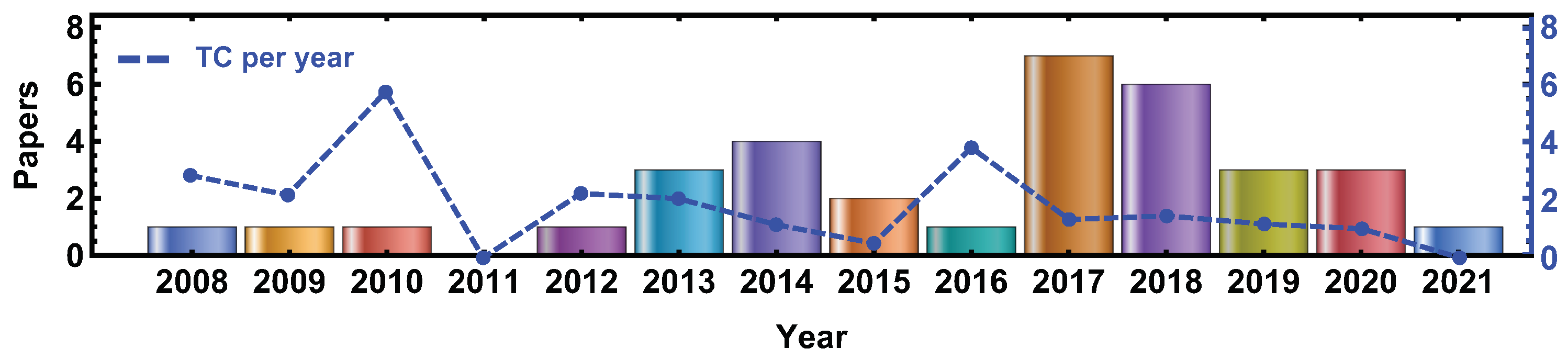
| Timespan | 2008–2021 |
| Sources (journals, books, etc.) | 14 |
| Documents | 34 |
| Average years from publication | 4.97 |
| Average citations per documents | 9.56 |
| Average citations per year per doc | 1.27 |
| References | 839 |
| Articles | 32 |
| Conference papers | 1 |
| Meeting abstracts | 1 |
| Keywords plus | 132 |
| Author’s keywords | 107 |
| Authors | 150 |
| Authors of single-authored documents | 0 |
| Authors of multi-authored documents | 150 |
| Documents per author | 0.23 |
| Authors per document | 4.41 |
| Co-Authors per documents | 5.62 |
| Collaboration index | 4.41 |
| Reference | Applications | Year |
|---|---|---|
| Tseng, H. W. et al. [90] | Cone-Beam Breast CT | 2021 |
| Cho, S. et al. [91] | Novel method to improve quality of CBCT images | 2020 |
| Principi S. et al. [92] | Comparison between LBTE methods against Geant4 benchmark | 2020 |
| Shi M. et al. [93] | GPU-based for proton simulation | 2020 |
| Lee, H. et al. [94] | Deep Learning-based scatter correction method | 2019 |
| Jia, S et al. [95] | Dental and maxillofacial CBCT | 2019 |
| Wang A. et al. [96] | Comparison between Acuros CTD against Geant4 | 2019 |
| Ardenfors, O. et al. [97] | Proton Gantry-Mounted CBCT | 2018 |
| Ardenfors, O. et al. [98] | Proton Gantry-Mounted CBCT | 2018 |
| Gholami, S. et al. [99] | Stereotactic Radiosurgery with small radiation fields | 2018 |
| Maslowski, A. et al. [100] | Comparison between Acuros CTD against Geant4 | 2018 |
| Leotta, S. et al. [101] | Image-Guided RadioTherapy | 2018 |
| Son, K. et al. [102] | Image-Guided RadioTherapy | 2018 |
| Zhu, J. et al. [103] | Adaptive Radiotherapy | 2017 |
| Benhalouche, S. et al. [104] | Simulated Mega-Voltage CBCT | 2017 |
| Myronaki, M. et al. [105] | Optimization of a Multilayer Imager Mega-Voltage CBCT | 2017 |
| Sakata, D. et al. [106] | Dual-Energy Computed Tomography | 2017 |
| Son, K. et al. [107] | Image-Guided RadioTherapy | 2017 |
| Marchant, T.E. et al. [108] | Radiotherapy | 2017 |
| Xu, M. et al. [109] | Cone-Beam Breast CT | 2017 |
| Shi, L. et al. [110] | Cone-Beam Breast CT | 2016 |
| Baldacci, F. et al. [111] | TLE method for low-energy X-ray irradiations | 2015 |
| Choi, J.H. et al. [112] | New dose point measurement method validated by Geant4 | 2015 |
| Brochu, F.M. et al. [113] | Radiotherapy | 2014 |
| Li, X. et al. [114] | Perfusion and Interventional CBCT | 2014 |
| Hansen, D.C. et al. [115] | Proton Computed Tomography | 2014 |
| Son, K. et al. [116] | Image-Guided RadioTherapy | 2014 |
| Bartzsch, S. et al. [117] | Image-Guided RadioTherapy | 2013 |
| Lanconelli, N. et al. [118] | Cone-Beam Breast CT | 2013 |
| Fleckenstein, J. et al. [119] | Volumetric Modulated Arc Therapy | 2013 |
| Vedantham, S. et al. [120] | Cone-Beam Breast CT | 2012 |
| Sechopoulos, I. et al. [121] | Cone-Beam Breast CT | 2010 |
| Chen L. et al. [122] | Cone-Beam Breast CT | 2009 |
| Chen L. et al. [123] | Cone-Beam Breast CT | 2008 |
| Paper | TC | TC per Year | LC | LC % |
|---|---|---|---|---|
| Sechopoulos I. et al., 2010, Med. Phys. [121] | 63 | 5.25 | 4 | 6.35 |
| Chen L. et al., 2008, Med. Phys. [123] | 37 | 2.64 | 0 | 0.0 |
| Chen L.et al., 2009, Med. Phys. [122] | 26 | 2.0 | 0 | 0.0 |
| Vedantham S. et al., 2012, Med. Phys. [120] | 20 | 2.0 | 20 | 10.0 |
| Shi L. et al., 2016, Med. Phys. [110] | 19 | 3.17 | 0 | 0.0 |
| Bartzsch S. et al., 2013, Med. Phys. [117] | 19 | 2.11 | 0 | 0.0 |
| Fleckenstein J. et al., 2013, Z Med. Phys. [119] | 19 | 2.11 | 0 | 0.0 |
| Maslowski A. et al., 2018, Med. Phys. [100] | 15 | 3.75 | 2 | 13.33 |
| Myronakis M. et al., 2017, Med. Phys. [105] | 14 | 2.80 | 1 | 7.14 |
| Marchant, T.E. et al., 2014 Med. Phys. [108] | 11 | 2.20 | 0 | 0.00 |
| Hansen D.C., 2014, Med. Phys. [115] | 11 | 1.4 | 0 | 0.0 |
Publisher’s Note: MDPI stays neutral with regard to jurisdictional claims in published maps and institutional affiliations. |
© 2021 by the authors. Licensee MDPI, Basel, Switzerland. This article is an open access article distributed under the terms and conditions of the Creative Commons Attribution (CC BY) license (https://creativecommons.org/licenses/by/4.0/).
Share and Cite
Cabanas, A.M.; Arriagada-Benítez, M.; Ubeda, C.; Meseguer-Ruiz, O.; Arce, P. Dose Estimation by Geant4-Based Simulations for Cone-Beam CT Applications: A Systematic Review. Appl. Sci. 2021, 11, 6136. https://doi.org/10.3390/app11136136
Cabanas AM, Arriagada-Benítez M, Ubeda C, Meseguer-Ruiz O, Arce P. Dose Estimation by Geant4-Based Simulations for Cone-Beam CT Applications: A Systematic Review. Applied Sciences. 2021; 11(13):6136. https://doi.org/10.3390/app11136136
Chicago/Turabian StyleCabanas, Ana M., Mauricio Arriagada-Benítez, Carlos Ubeda, Oliver Meseguer-Ruiz, and Pedro Arce. 2021. "Dose Estimation by Geant4-Based Simulations for Cone-Beam CT Applications: A Systematic Review" Applied Sciences 11, no. 13: 6136. https://doi.org/10.3390/app11136136
APA StyleCabanas, A. M., Arriagada-Benítez, M., Ubeda, C., Meseguer-Ruiz, O., & Arce, P. (2021). Dose Estimation by Geant4-Based Simulations for Cone-Beam CT Applications: A Systematic Review. Applied Sciences, 11(13), 6136. https://doi.org/10.3390/app11136136