Combining 3D Structured Light Imaging and Spine X-ray Data Improves Visualization of the Spinous Lines in the Scoliotic Spine


Featured Application
Abstract
1. Introduction
2. Materials and Methods
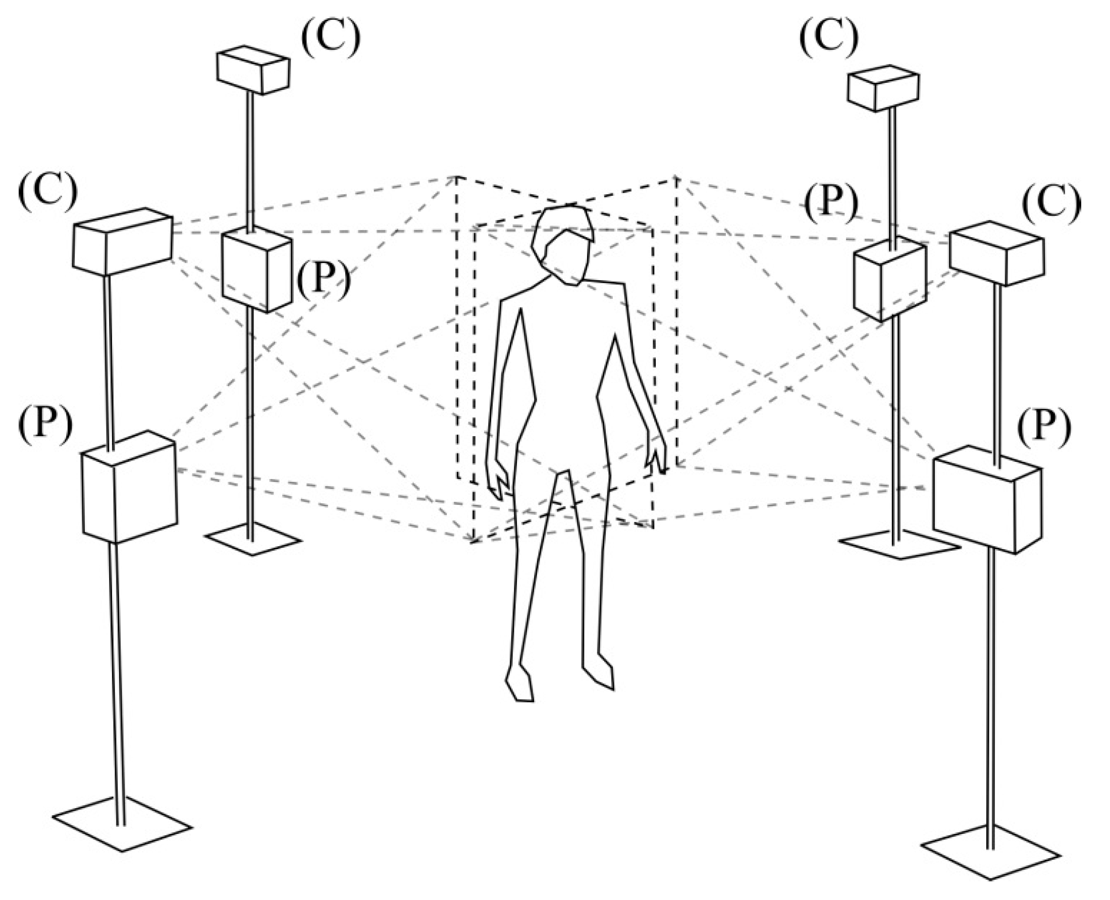
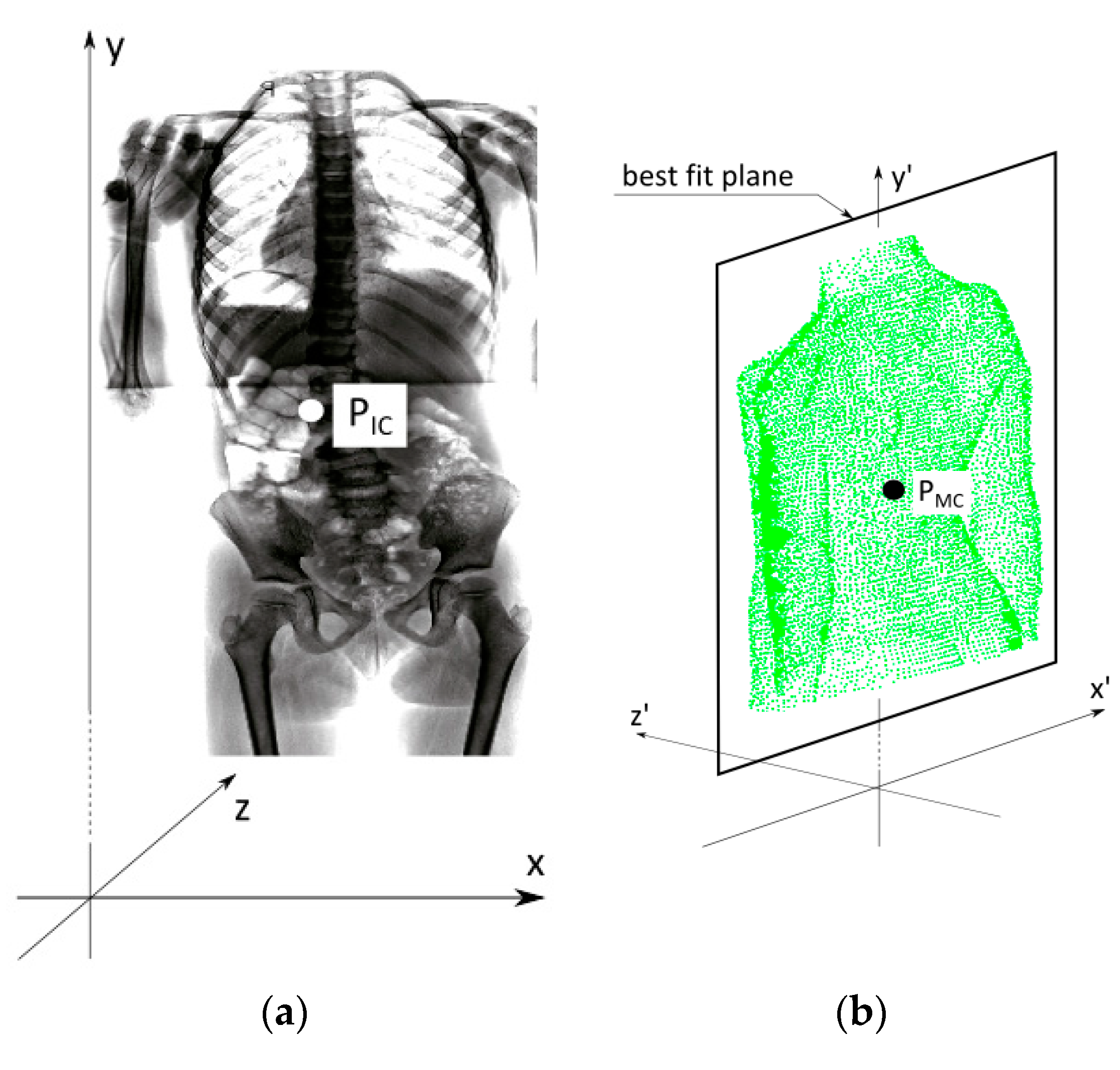
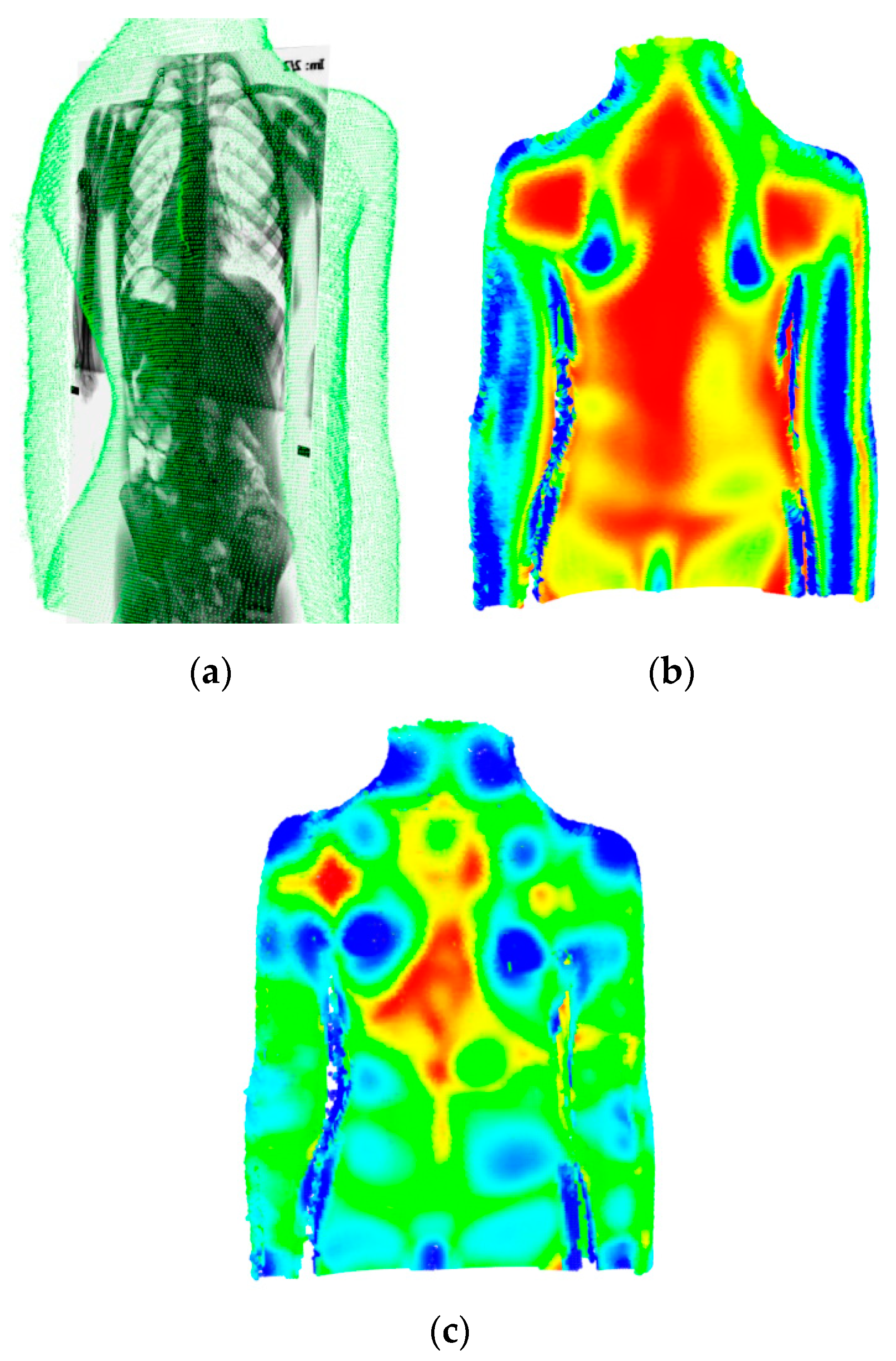
2.1. Data Merging
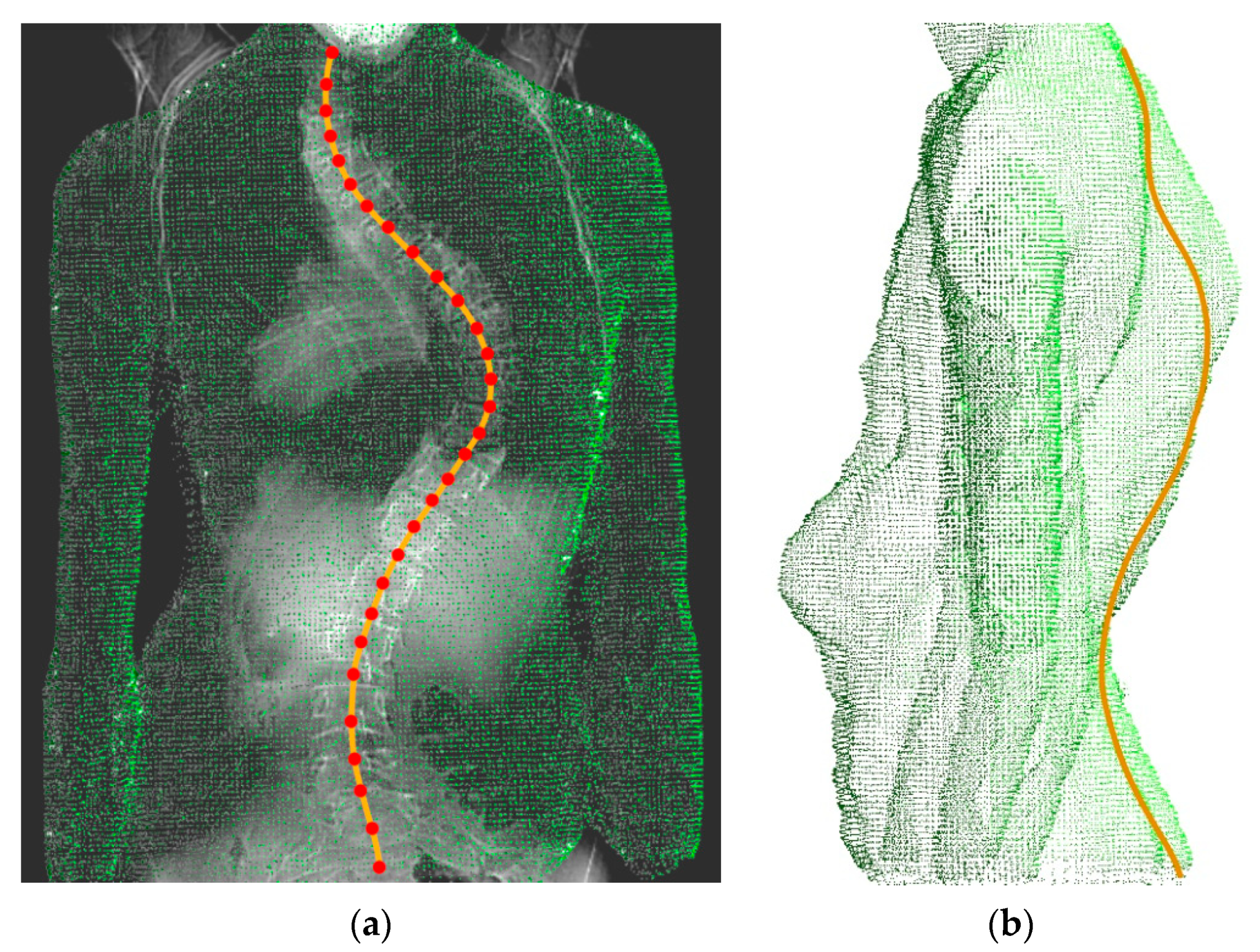
2.2. Data Analysis
2.3. Comparison of the Results
2.4. Statistical Analysis
3. Results
4. Discussion
5. Conclusions
Author Contributions
Funding
Institutional Review Board Statement
Informed Consent Statement
Data Availability Statement
Acknowledgments
Conflicts of Interest
References
- Wall, B.F.; Hart, D. Revised radiation doses for typical X-ray examinations. Report on a recent review of doses to patients from medical X-ray examinations in the UK by NRPB. National Radiological Protection Board. Br. J. Radiol. 1997, 70, 437–439. [Google Scholar] [CrossRef] [PubMed]
- Larson, A.N.; Schueler, B.A.; Dubousset, J. Radiation in Spine Deformity: State-of-the-Art Reviews. Spine Deform. 2019, 7, 386–394. [Google Scholar] [CrossRef] [PubMed]
- Nash, C.L., Jr.; Gregg, E.C.; Brown, R.H.; Pillai, K. Risks of exposure to X-rays in patients undergoing long-term treatment for scoliosis. J. Bone Jt. Surg. Am. Vol. 1979, 61, 371–374. [Google Scholar] [CrossRef]
- Ronckers, C.M.; Land, C.E.; Miller, J.S.; Stovall, M.; Lonstein, J.E.; Doody, M.M. Cancer mortality among women frequently exposed to radiographic examinations for spinal disorders. Radiat. Res. 2010, 174, 83–90. [Google Scholar] [CrossRef]
- Ronckers, C.M.; Doody, M.M.; Lonstein, J.E.; Stovall, M.; Land, C.E. Multiple diagnostic X-rays for spine deformities and risk of breast cancer. Cancer Epidemiol. Biomark. Prev. 2008, 17, 605–613. [Google Scholar] [CrossRef]
- Ardran, G.M.; Coates, R.; Dickson, R.A.; Dixon-Brown, A.; Harding, F.M. Assessment of scoliosis in children: Low dose radiographic technique. Br. J. Radiol. 1980, 53, 146–147. [Google Scholar] [CrossRef]
- Simony, A.; Hansen, E.J.; Christensen, S.B.; Carreon, L.Y.; Andersen, M.O. Incidence of cancer in adolescent idiopathic scoliosis patients treated 25 years previously. Eur. Spine J. 2016, 25, 3366–3370. [Google Scholar] [CrossRef]
- Novosad, J.; Eng, B.; Cheriet, F.; Delorme, S.; Poirier, S.; Beausejour, M.; Labelle, H. Self-calibration of biplanar radiographs for a retrospective comparative study of the 3D correction of adolescent idiopathic scoliosis. Stud. Health Technol. Inform. 2002, 91, 272–275. [Google Scholar]
- Legaye, J.; Saunier, P.; Dumas, R.; Vallee, C. Correction for patient sway in radiographic biplanar imaging for three-dimensional reconstruction of the spine: In vitro study of a new method. Acta Radiol. 2009, 50, 781–790. [Google Scholar] [CrossRef] [PubMed]
- Ilharreborde, B.; Steffen, J.S.; Nectoux, E.; Vital, J.M.; Mazda, K.; Skalli, W.; Obeid, I. Angle measurement reproducibility using EOS three-dimensional reconstructions in adolescent idiopathic scoliosis treated by posterior instrumentation. Spine 2011, 36, E1306–E1313. [Google Scholar] [CrossRef] [PubMed]
- Glaser, D.A.; Doan, J.; Newton, P.O. Comparison of 3-dimensional spinal reconstruction accuracy: Biplanar radiographs with EOS versus computed tomography. Spine 2012, 37, 1391–1397. [Google Scholar] [CrossRef] [PubMed]
- Ilharreborde, B.; Dubousset, J.; Skalli, W.; Mazda, K. Spinal penetration index assessment in adolescent idiopathic scoliosis using EOS low-dose biplanar stereoradiography. Eur. Spine J. 2013, 22, 2438–2444. [Google Scholar] [CrossRef] [PubMed]
- Courvoisier, A.; Drevelle, X.; Vialle, R.; Dubousset, J.; Skalli, W. 3D analysis of brace treatment in idiopathic scoliosis. Eur. Spine J. 2013, 22, 2449–2455. [Google Scholar] [CrossRef] [PubMed]
- Ilharreborde, B.; Vidal, C.; Skalli, W.; Mazda, K. Sagittal alignment of the cervical spine in adolescent idiopathic scoliosis treated by posteromedial translation. Eur. Spine J. 2013, 22, 330–337. [Google Scholar] [CrossRef]
- Amzallag-Bellenger, E.; Uyttenhove, F.; Nectoux, E.; Moraux, A.; Bigot, J.; Herbaux, B.; Boutry, N. Idiopathic scoliosis in children and adolescents: Assessment with a biplanar X-ray device. Insights Imaging 2014, 5, 571–583. [Google Scholar] [CrossRef]
- Ilharreborde, B.; Ferrero, E.; Alison, M.; Mazda, K. EOS microdose protocol for the radiological follow-up of adolescent idiopathic scoliosis. Eur. Spine J. 2016, 25, 526–531. [Google Scholar] [CrossRef]
- Dubousset, J.; Charpak, G.; Dorion, I.; Skalli, W.; Lavaste, F.; Deguise, J.; Kalifa, G.; Ferey, S. A new 2D and 3D imaging approach to musculoskeletal physiology and pathology with low-dose radiation and the standing position: The EOS system. Bull. Acad. Natl. Med. 2005, 189, 287–297, discussion 297–300. [Google Scholar]
- Hoan, N.N.; Majewski, S.; Charpak, G.; Policarpo, A.J. An efficient, gaseous detector with good low-energy resolution for (less than or equal to 50 keV) imaging. J. Nucl. Med. 1979, 20, 335–340. [Google Scholar]
- Ng, S.Y.; Bettany-Saltikov, J. Imaging in the Diagnosis and Monitoring of Children with Idiopathic Scoliosis. Open Orthop. J. 2017, 11, 1500–1520. [Google Scholar] [CrossRef]
- Pedersen, P.H.; Vergari, C.; Alzakri, A.; Vialle, R.; Skalli, W. A reduced micro-dose protocol for 3D reconstruction of the spine in children with scoliosis: Results of a phantom-based and clinically validated study using stereo-radiography. Eur. Radiol. 2019, 29, 1874–1881. [Google Scholar] [CrossRef]
- Hui, S.C.; Pialasse, J.P.; Wong, J.Y.; Lam, T.P.; Ng, B.K.; Cheng, J.C.; Chu, W.C. Radiation dose of digital radiography (DR) versus micro-dose X-ray (EOS) on patients with adolescent idiopathic scoliosis: 2016 SOSORT-IRSSD “John Sevastic Award” Winner in Imaging Research. Scoliosis Spinal Disord. 2016, 11, 46. [Google Scholar] [CrossRef] [PubMed]
- Luo, T.D.; Stans, A.A.; Schueler, B.A.; Larson, A.N. Cumulative Radiation Exposure With EOS Imaging Compared With Standard Spine Radiographs. Spine Deform. 2015, 3, 144–150. [Google Scholar] [CrossRef]
- McKenna, C.; Wade, R.; Faria, R.; Yang, H.; Stirk, L.; Gummerson, N.; Sculpher, M.; Woolacott, N. EOS 2D/3D X-ray imaging system: A systematic review and economic evaluation. Health Technol. Assess. 2012, 16, 1–188. [Google Scholar] [CrossRef] [PubMed]
- Brink, R.C.; Wijdicks, S.P.J.; Tromp, I.N.; Schlosser, T.P.C.; Kruyt, M.C.; Beek, F.J.A.; Castelein, R.M. A reliability and validity study for different coronal angles using ultrasound imaging in adolescent idiopathic scoliosis. Spine J. 2018, 18, 979–985. [Google Scholar] [CrossRef] [PubMed]
- Ahmad, O.; Lombaert, H.; Parent, S.; Labelle, H.; Cheriet, F. Spectral Shape Analysis of Human Torsos: Application to the Evaluation of Scoliosis Surgery Outcome. IEEE J. Biomed. Health Inform. 2018, 22, 1552–1560. [Google Scholar] [CrossRef]
- Melhem, E.; Assi, A.; El Rachkidi, R.; Ghanem, I. EOS((R)) biplanar X-ray imaging: Concept, developments, benefits, and limitations. J. Child. Orthop. 2016, 10, 1–14. [Google Scholar] [CrossRef]
- Assi, K.C.; Grenier, S.; Parent, S.; Labelle, H.; Cheriet, F. A physically based trunk soft tissue modeling for scoliosis surgery planning systems. Comput. Med Imaging Graph. 2015, 40, 217–228. [Google Scholar] [CrossRef][Green Version]
- Mahboub-Ahari, A.; Hajebrahimi, S.; Yusefi, M.; Velayati, A. EOS imaging versus current radiography: A health technology assessment study. Med J. Islamic Repub. Iran 2016, 30, 331. [Google Scholar]
- Duong, L.; Cheriet, F.; Labelle, H. Automatic detection of scoliotic curves in posteroanterior radiographs. IEEE Trans. Biomed. Eng. 2010, 57, 1143–1151. [Google Scholar] [CrossRef]
- Rigo, M.D.; Villagrasa, M.; Gallo, D. A specific scoliosis classification correlating with brace treatment: Description and reliability. Scoliosis 2010, 5, 1. [Google Scholar] [CrossRef]
- Michonski, J.; Glinkowski, W.; Witkowski, M.; Sitnik, R. Automatic recognition of surface landmarks of anatomical structures of back and posture. J. Biomed. Opt. 2012, 17, 056015. [Google Scholar] [CrossRef] [PubMed]
- Glinkowski, W.M.; Tomasik, P.; Walesiak, K.; Gluszak, M.; Krawczak, K.; Michonski, J.; Czyzewska, A.; Zukowska, A.; Sitnik, R.; Wielgos, M. Posture and low back pain during pregnancy-3D study. Ginekol. Pol. 2016, 87, 575–580. [Google Scholar] [CrossRef] [PubMed]
- Michonski, J.; Walesiak, K.; Pakula, A.; Glinkowski, W.; Sitnik, R. Monitoring of spine curvatures and posture during pregnancy using surface topography-case study and suggestion of method. Scoliosis Spinal Disord. 2016, 11, 31. [Google Scholar] [CrossRef] [PubMed]
- Firth, N.C.; Brown, N.; Blagg, J. Plane of best fit: A novel method to characterize the three-dimensionality of molecules. J. Chem. Inf. Modeling 2012, 52, 2516–2525. [Google Scholar] [CrossRef]
- Shannon, T.M. Development of an apparatus to evaluate Adolescent Idiopathic Scoliosis by dynamic surface topography. Stud. Health Technol. Inform. 2008, 140, 121–127. [Google Scholar]
- Berryman, F.; Pynsent, P.; Fairbank, J.; Disney, S. A new system for measuring three-dimensional back shape in scoliosis. Eur. Spine J. 2008, 17, 663–672. [Google Scholar] [CrossRef]
- Drerup, B. Rasterstereographic measurement of scoliotic deformity. Scoliosis 2014, 9, 22. [Google Scholar] [CrossRef]
- Patias, P.; Grivas, T.B.; Kaspiris, A.; Aggouris, C.; Drakoutos, E. A review of the trunk surface metrics used as Scoliosis and other deformities evaluation indices. Scoliosis 2010, 5, 12. [Google Scholar] [CrossRef]
- Drerup, B.; Hierholzer, E. Automatic localization of anatomical landmarks on the back surface and construction of a body-fixed coordinate system. J. Biomech. 1987, 20, 961–970. [Google Scholar] [CrossRef]
- Barsky, B.A.; Bartels, R.H.; Beatty, J.C. An Introduction to Splines for Use in Computer Graphics and Geometric Modeling. Available online: https://books.google.pl/books?hl=en&lr=&id=9bQ0f8sYqaAC&oi=fnd&pg=PA1&dq=Bartels,+R.H.%3B+Beatty,+J.C.%3B+Barsky,+B.A.:+An+Introduction+to+Splines+for+Use+in+Computer+Graphics+%26+Geometric+Modeling.+Morgan+Kaufmann+Publishers+1987.&ots=yEiSx4797R&sig=d5EPvK6pgAaLFRWWAZPmmtxrrQE&redir_esc=y#v=onepage&q&f=false (accessed on 20 November 2020).
- The Visualization Toolkit Open Source Project on Open HUB, Online. Available online: https://www.openhub.net/p/vtk (accessed on 20 November 2020).
- Lotfi, N.; Chauhan, G.S.; Gardner, A.; Berryman, F.; Pynsent, P. The relationship between measures of spinal deformity and measures of thoracic trunk rotation. J. Spine Surg. 2020, 6, 555–561. [Google Scholar] [CrossRef]
- Ghaneei, M.; Komeili, A.; Li, Y.; Parent, E.C.; Adeeb, S. 3D Markerless asymmetry analysis in the management of adolescent idiopathic scoliosis. BMC Musculoskelet. Disord. 2018, 19, 385. [Google Scholar] [CrossRef] [PubMed]
- Knott, P.; Sturm, P.; Lonner, B.; Cahill, P.; Betsch, M.; McCarthy, R.; Kelly, M.; Lenke, L.; Betz, R. Multicenter Comparison of 3D Spinal Measurements Using Surface Topography With Those From Conventional Radiography. Spine Deform. 2016, 4, 98–103. [Google Scholar] [CrossRef] [PubMed]
- Komeili, A.; Westover, L.; Parent, E.C.; El-Rich, M.; Adeeb, S. Monitoring for idiopathic scoliosis curve progression using surface topography asymmetry analysis of the torso in adolescents. Spine J. 2015, 15, 743–751. [Google Scholar] [CrossRef] [PubMed]
- Fortin, C.; Feldman, D.E.; Cheriet, F.; Labelle, H. Validity of a quantitative clinical measurement tool of trunk posture in idiopathic scoliosis. Spine 2010, 35, E988–E994. [Google Scholar] [CrossRef]
- Parent, E.C.; Damaraju, S.; Hill, D.L.; Lou, E.; Smetaniuk, D. Identifying the best surface topography parameters for detecting idiopathic scoliosis curve progression. Stud. Health Technol. Inform. 2010, 158, 78–82. [Google Scholar]
- Kotwicki, T.; Negrini, S.; Grivas, T.B.; Rigo, M.; Maruyama, T.; Durmala, J.; Zaina, F.; Members of the international Society on Scoliosis, O.; Rehabilitation, T. Methodology of evaluation of morphology of the spine and the trunk in idiopathic scoliosis and other spinal deformities-6th SOSORT consensus paper. Scoliosis 2009, 4, 26. [Google Scholar] [CrossRef]
- Goldberg, C.J.; Grove, D.; Moore, D.P.; Fogarty, E.E.; Dowling, F.E. Surface topography and vectors: A new measure for the three dimensional quantification of scoliotic deformity. Stud. Health Technol. Inform. 2006, 123, 449–455. [Google Scholar]
- Goldberg, C.J.; Kaliszer, M.; Moore, D.P.; Fogarty, E.E.; Dowling, F.E. Surface topography, Cobb angles, and cosmetic change in scoliosis. Spine 2001, 26, E55–E63. [Google Scholar] [CrossRef]
- Ono, T. Trunk deformity in scoliosis studied by surface measurement. Nihon Seikeigeka Gakkai Zasshi 1995, 69, 915–926. [Google Scholar]
- Dawson, E.G.; Kropf, M.A.; Purcell, G.; Kabo, J.M.; Kanim, L.E.; Burt, C. Optoelectronic evaluation of trunk deformity in scoliosis. Spine 1993, 18, 326–331. [Google Scholar] [CrossRef]
- Glinkowski, W.; Michonski, J.; Glinkowska, B.; Zukowska, A.; Sitnik, R.; Gorecki, A. Telediagnostic 3D school screening of back curvatures and posture using structured light method-pilot study. Stud. Health Technol. Inform. 2012, 176, 291–294. [Google Scholar] [PubMed]
- Takasaki, H. Moire topography. Appl. Opt. 1970, 9, 1467–1472. [Google Scholar] [CrossRef] [PubMed]
- Buendia, M.; Salvador, R.; Cibrian, R.; Laguia, M.; Sotoca, J.M. Determination of the object surface function by structured light: Application to the study of spinal deformities. Phys. Med. Biol. 1999, 44, 75–86. [Google Scholar] [CrossRef] [PubMed]
- Sitnik, R. Four-dimensional measurement by a single-frame structured light method. Appl. Opt. 2009, 48, 3344–3354. [Google Scholar] [CrossRef]
- Drerup, B. The measurement of the kyphotic angle by contact free registration of back-shape (author’s transl). Zeitschrift fur Orthopadie und ihre Grenzgebiete 1982, 120, 64–70. [Google Scholar] [CrossRef]
- Andonian, A.T. Detection of stimulated back muscle contractions by moire topography. J. Biomech. 1984, 17, 653–661. [Google Scholar] [CrossRef]
- Horst, M.; Albrecht, D.; Drerup, B. Objective determination of the shape of the anterior chest wall using moire topography. Method and development of dimension-free indices for the evaluation of funnel chest. Zeitschrift fur Orthopadie und ihre Grenzgebiete 1985, 123, 357–364. [Google Scholar] [CrossRef]
- Ashizawa, K.; Kuki, T.; Kusumoto, A.; Tsutsumi, E.; Kato, S. Approximate surface development of the left side half-trunk by a free-formed model. Am. J. Phys. Anthropol. 1988, 76, 165–173. [Google Scholar] [CrossRef]
- Stokes, I.A.; Moreland, M.S. Concordance of back surface asymmetry and spine shape in idiopathic scoliosis. Spine 1989, 14, 73–78. [Google Scholar] [CrossRef]
- Batouche, M.; Benlamri, R.; Kholladi, M.K. A computer vision system for diagnosing scoliosis using moire images. Comput. Biol. Med. 1996, 26, 339–353. [Google Scholar] [CrossRef]
- Wong, H.K.; Balasubramaniam, P.; Rajan, U.; Chng, S.Y. Direct spinal curvature digitization in scoliosis screening--a comparative study with Moire contourgraphy. J. Spinal Disord. 1997, 10, 185–192. [Google Scholar] [CrossRef] [PubMed]
- Liu, X.C.; Thometz, J.G.; Lyon, R.M.; McGrady, L. Effects of trunk position on back surface-contour measured by raster stereophotography. Am. J. Orthop. 2002, 31, 402–406. [Google Scholar] [PubMed]
- Hill, D.L.; Berg, D.C.; Raso, V.J.; Lou, E.; Durdle, N.G.; Mahood, J.K.; Moreau, M.J. Evaluation of a laser scanner for surface topography. Stud. Health Technol. Inform. 2002, 88, 90–94. [Google Scholar] [PubMed]
- Jacobi, U.; Chen, M.; Frankowski, G.; Sinkgraven, R.; Hund, M.; Rzany, B.; Sterry, W.; Lademann, J. In vivo determination of skin surface topography using an optical 3D device. Skin Res. Technol. 2004, 10, 207–214. [Google Scholar] [CrossRef] [PubMed]
- Minguez, M.F.; Buendia, M.; Cibrian, R.M.; Salvador, R.; Laguia, M.; Martin, A.; Gomar, F. Quantifier variables of the back surface deformity obtained with a noninvasive structured light method: Evaluation of their usefulness in idiopathic scoliosis diagnosis. Eur. Spine J. 2007, 16, 73–82. [Google Scholar] [CrossRef] [PubMed]
- Zubovic, A.; Davies, N.; Berryman, F.; Pynsent, P.; Quraishi, N.; Lavy, C.; Bowden, G.; Wilson-Macdonald, J.; Fairbank, J. New method of Scoliosis Deformity Assessment: ISIS2 System. Stud. Health Technol. Inform. 2008, 140, 157–160. [Google Scholar] [CrossRef]
- Ghaneei, M.; Ekyalimpa, R.; Westover, L.; Parent, E.C.; Adeeb, S. Customized k-nearest neighbourhood analysis in the management of adolescent idiopathic scoliosis using 3D markerless asymmetry analysis. Comput. Methods Biomech. Biomed. Eng. 2019, 1–10. [Google Scholar] [CrossRef]
- Komeili, A.; Westover, L.; Parent, E.C.; El-Rich, M.; Adeeb, S. Correlation Between a Novel Surface Topography Asymmetry Analysis and Radiographic Data in Scoliosis. Spine Deform. 2015, 3, 303–311. [Google Scholar] [CrossRef]
- Komeili, A.; Westover, L.M.; Parent, E.C.; Moreau, M.; El-Rich, M.; Adeeb, S. Surface topography asymmetry maps categorizing external deformity in scoliosis. Spine J. 2014, 14, 973–983 e972. [Google Scholar] [CrossRef]
- Glinkowski, W.; Sitnik, R.; Witkowski, M.; Kocon, H.; Bolewicki, P.; Gorecki, A. Method of pectus excavatum measurement based on structured light technique. J Biomed Opt 2009, 14, 044041. [Google Scholar] [CrossRef]
- Newton, P.O.; Osborn, E.J.; Bastrom, T.P.; Doan, J.D.; Reighard, F.G. The 3D Sagittal Profile of Thoracic Versus Lumbar Major Curves in Adolescent Idiopathic Scoliosis. Spine Deform. 2019, 7, 60–65. [Google Scholar] [CrossRef] [PubMed]
- Boice, J.D., Jr. Carcinogenesis—A synopsis of human experience with external exposure in medicine. Health Phys. 1988, 55, 621–630. [Google Scholar] [CrossRef] [PubMed]
- Hoffman, D.A.; Lonstein, J.E.; Morin, M.M.; Visscher, W.; Harris, B.S., 3rd; Boice, J.D., Jr. Breast cancer in women with scoliosis exposed to multiple diagnostic x rays. J. Natl. Cancer Inst. 1989, 81, 1307–1312. [Google Scholar] [CrossRef] [PubMed]
- Boice, J.D., Jr. Radiation and breast carcinogenesis. Med Pediatric Oncol. 2001, 36, 508–513. [Google Scholar] [CrossRef]
- Kim, H.S.; Ishikawa, S.; Ohtsuka, Y.; Shimizu, H.; Shinomiya, T.; Viergever, M.A. Automatic scoliosis detection based on local centroids evaluation on moire topographic images of human backs. IEEE Trans. Med. Imaging 2001, 20, 1314–1320. [Google Scholar] [CrossRef]
- Stokes, I.A. Three-dimensional terminology of spinal deformity. A report presented to the Scoliosis Research Society by the Scoliosis Research Society Working Group on 3-D terminology of spinal deformity. Spine 1994, 19, 236–248. [Google Scholar] [CrossRef]
- Berryman, F.; Pynsent, P.; Fairbank, J. Measuring the rib hump in scoliosis with ISIS2. Stud. Health Technol. Inform. 2008, 140, 65–67. [Google Scholar]
- McArdle, F.J.; Griffiths, C.J.; Macdonald, A.M.; Gibson, M.J. Monitoring the thoracic sagittal curvature in kyphoscoliosis with surface topography: A trend analysis of 57 patients. Stud. Health Technol. Inform. 2002, 91, 199–203. [Google Scholar]
- Klos, S.S.; Liu, X.C.; Lyon, R.M.; Tassone, J.C.; Thometz, J.G. Reliability of a functional classification system in the monitoring of patients with idiopathic scoliosis. Spine 2007, 32, 1662–1666. [Google Scholar] [CrossRef]
- Betsch, M.; Wild, M.; Rath, B.; Tingart, M.; Schulze, A.; Quack, V. Radiation-free diagnosis of scoliosis: An overview of the surface and spine topography. Orthopade 2015, 44, 845–851. [Google Scholar] [CrossRef]
- Lenke, L.G.; Edwards, C.C., 2nd; Bridwell, K.H. The Lenke classification of adolescent idiopathic scoliosis: How it organizes curve patterns as a template to perform selective fusions of the spine. Spine 2003, 28, S199–S207. [Google Scholar] [CrossRef] [PubMed]


| Patient | Max (x) | Max (z) | Max | Mean (x) | Mean (z) | Mean |
|---|---|---|---|---|---|---|
| PT1 | 4.17 | 2.40 | 4.34 | 2.54 | 1.01 | 2.84 |
| PT2 | 14.20 | 1.70 | 14.20 | 0.59 | 7.90 | 7.94 |
| PT3 | 12.35 | 1.48 | 12.36 | 6.34 | 0.64 | 6.40 |
| PT4 | 16.35 | 8.13 | 17.83 | 4.91 | 1.58 | 5.31 |
| PT5 | 18.67 | 9.19 | 18.90 | 8.20 | 2.72 | 8.82 |
| PT6 | 11.50 | 1.91 | 11.50 | 6.79 | 0.74 | 6.87 |
| PT7 | 1.23 | 4.38 | 4.45 | 0.38 | 1.38 | 1.50 |
| PT8 | 5.00 | 4.84 | 6.95 | 0.66 | 2.80 | 2.97 |
| PT9 | 0.88 | 6.01 | 6.03 | 0.34 | 1.46 | 1.55 |
| PT10 | 2.88 | 7.78 | 7.83 | 0.65 | 3.22 | 3.35 |
| PT11 | 4.65 | 6.64 | 6.65 | 0.77 | 3.27 | 3.50 |
| PT12 | 1.99 | 9.00 | 9.21 | 0.51 | 1.73 | 1.89 |
Publisher’s Note: MDPI stays neutral with regard to jurisdictional claims in published maps and institutional affiliations. |
© 2020 by the authors. Licensee MDPI, Basel, Switzerland. This article is an open access article distributed under the terms and conditions of the Creative Commons Attribution (CC BY) license (http://creativecommons.org/licenses/by/4.0/).
Share and Cite
Paśko, S.; Glinkowski, W. Combining 3D Structured Light Imaging and Spine X-ray Data Improves Visualization of the Spinous Lines in the Scoliotic Spine. Appl. Sci. 2021, 11, 301. https://doi.org/10.3390/app11010301
Paśko S, Glinkowski W. Combining 3D Structured Light Imaging and Spine X-ray Data Improves Visualization of the Spinous Lines in the Scoliotic Spine. Applied Sciences. 2021; 11(1):301. https://doi.org/10.3390/app11010301
Chicago/Turabian StylePaśko, Sławomir, and Wojciech Glinkowski. 2021. "Combining 3D Structured Light Imaging and Spine X-ray Data Improves Visualization of the Spinous Lines in the Scoliotic Spine" Applied Sciences 11, no. 1: 301. https://doi.org/10.3390/app11010301
APA StylePaśko, S., & Glinkowski, W. (2021). Combining 3D Structured Light Imaging and Spine X-ray Data Improves Visualization of the Spinous Lines in the Scoliotic Spine. Applied Sciences, 11(1), 301. https://doi.org/10.3390/app11010301