Correction: Levicar et al. Methods of Radiographic Measurements of Heart and Left Atrial Size in Dogs with and without Myxomatous Mitral Valve Disease: Intra- and Interobserver Agreement and Practicability of Different Methods. Animals 2022, 12, 2531


 , ,
, , 
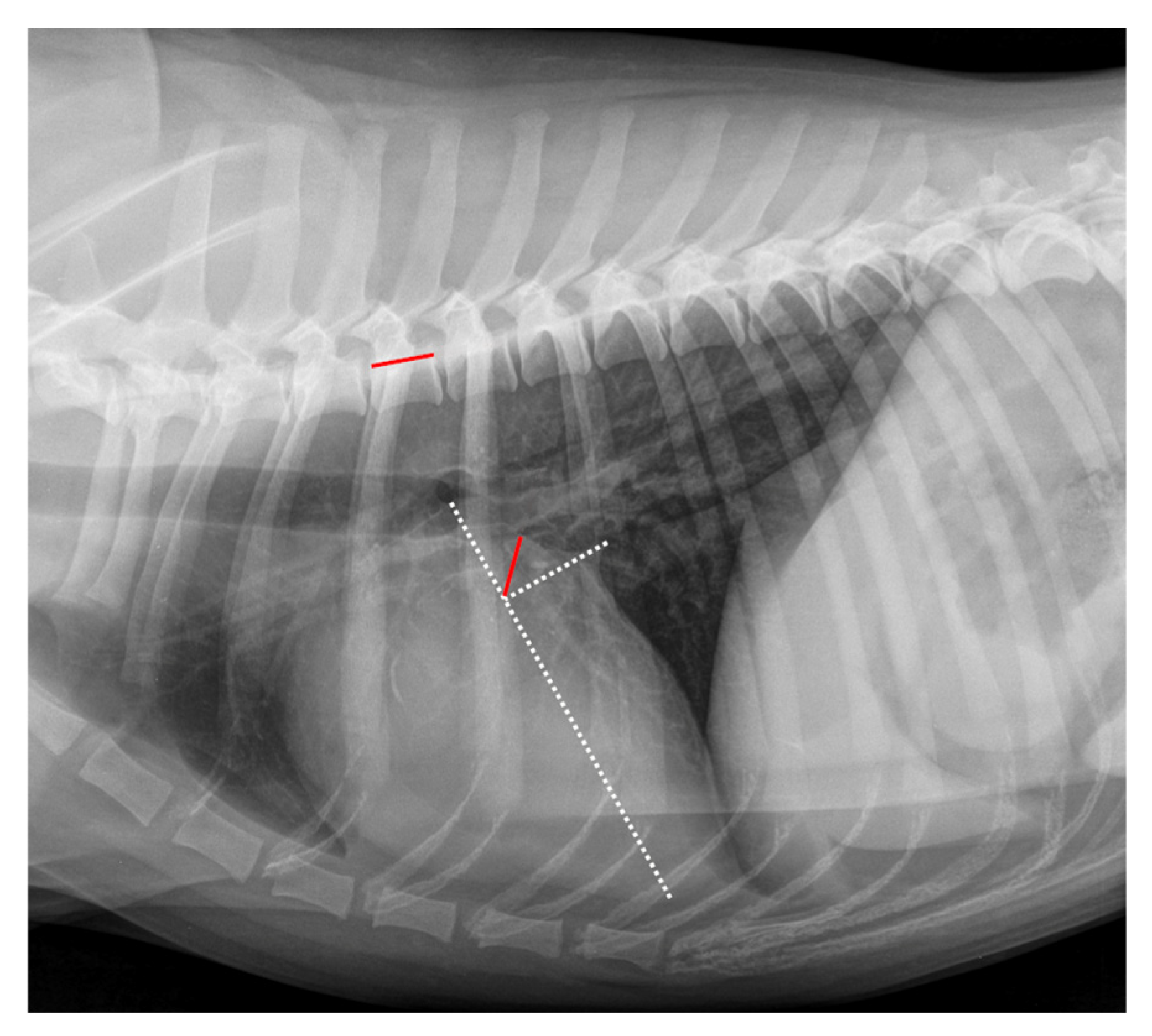
Error in Figure 2
Reference
- Levicar, C.; Nolte, I.; Granados-Soler, J.L.; Freise, F.; Raue, J.F.; Bach, J.-P. Methods of Radiographic Measurements of Heart and Left Atrial Size in Dogs with and without Myxomatous Mitral Valve Disease: Intra- and Interobserver Agreement and Practicability of Different Methods. Animals 2022, 12, 2531. [Google Scholar] [CrossRef] [PubMed]
Disclaimer/Publisher’s Note: The statements, opinions and data contained in all publications are solely those of the individual author(s) and contributor(s) and not of MDPI and/or the editor(s). MDPI and/or the editor(s) disclaim responsibility for any injury to people or property resulting from any ideas, methods, instructions or products referred to in the content. |
© 2025 by the authors. Licensee MDPI, Basel, Switzerland. This article is an open access article distributed under the terms and conditions of the Creative Commons Attribution (CC BY) license (https://creativecommons.org/licenses/by/4.0/).
Share and Cite
Levicar, C.; Nolte, I.; Granados-Soler, J.L.; Freise, F.; Raue, J.F.; Bach, J.-P. Correction: Levicar et al. Methods of Radiographic Measurements of Heart and Left Atrial Size in Dogs with and without Myxomatous Mitral Valve Disease: Intra- and Interobserver Agreement and Practicability of Different Methods. Animals 2022, 12, 2531. Animals 2025, 15, 1082. https://doi.org/10.3390/ani15081082
Levicar C, Nolte I, Granados-Soler JL, Freise F, Raue JF, Bach J-P. Correction: Levicar et al. Methods of Radiographic Measurements of Heart and Left Atrial Size in Dogs with and without Myxomatous Mitral Valve Disease: Intra- and Interobserver Agreement and Practicability of Different Methods. Animals 2022, 12, 2531. Animals. 2025; 15(8):1082. https://doi.org/10.3390/ani15081082
Chicago/Turabian StyleLevicar, Charanthorn, Ingo Nolte, José Luis Granados-Soler, Fritjof Freise, Jonathan Friedemann Raue, and Jan-Peter Bach. 2025. "Correction: Levicar et al. Methods of Radiographic Measurements of Heart and Left Atrial Size in Dogs with and without Myxomatous Mitral Valve Disease: Intra- and Interobserver Agreement and Practicability of Different Methods. Animals 2022, 12, 2531" Animals 15, no. 8: 1082. https://doi.org/10.3390/ani15081082
APA StyleLevicar, C., Nolte, I., Granados-Soler, J. L., Freise, F., Raue, J. F., & Bach, J.-P. (2025). Correction: Levicar et al. Methods of Radiographic Measurements of Heart and Left Atrial Size in Dogs with and without Myxomatous Mitral Valve Disease: Intra- and Interobserver Agreement and Practicability of Different Methods. Animals 2022, 12, 2531. Animals, 15(8), 1082. https://doi.org/10.3390/ani15081082