
C-Reactive Protein as a Diagnostic Marker in Dogs: A Review

Abstract
Simple Summary
Abstract
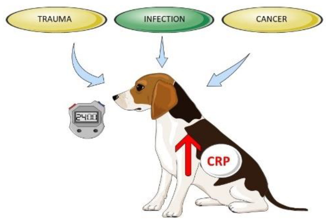
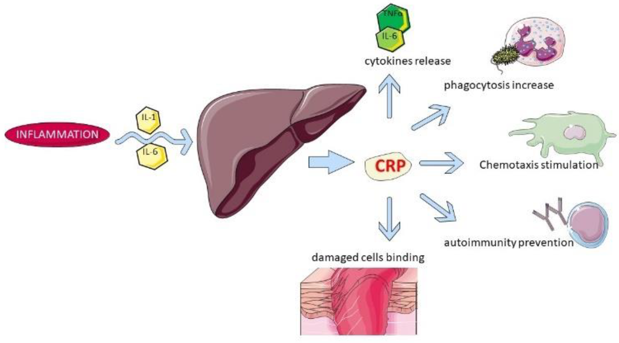
1. Introduction
2. Clinical Resources Available in Veterinary Medicine
3. CRP Concentrations in Healthy Dogs
4. Physical Exercise
5. Bacterial and Viral Etiology Diseases
6. Parasitic Etiology Diseases
7. Surgery
8. Orthopedic Diseases
9. Autoimmune Diseases
10. Neoplasia
11. Other Diseases
12. Conclusions
Author Contributions
Funding
Institutional Review Board Statement
Informed Consent Statement
Data Availability Statement
Acknowledgments
Conflicts of Interest
References
- Casella, S.; Fazio, F.; Russo, C.; Giudice, E.; Piccione, G. Acute phase proteins response in hunting dogs. J. Vet. Diagn. Investig. 2013, 25, 577–580. [Google Scholar] [CrossRef] [PubMed]
- Ceron, J.J.; Eckersall, P.D.; Martinez-Subiela, S. Acute Phase Proteins in dogs and cats: Current knowledge and future perspectives. Vet. Clin. Pathol. 2005, 34, 85–99. [Google Scholar] [CrossRef] [PubMed]
- McClure, V.; van Schoor, M.; Thompson, P.N.; Kjelgaard-Hansen, M.; Goddard, A. Evaluation of the use of serum C-reactive protein concentration to predict outcome in puppies infected with canine parvovirus. J. Am. Vet. Med. Assoc. 2013, 243, 361–366. [Google Scholar] [CrossRef] [PubMed]
- Jasensky, A.K.; Bondzio, A.; Murugaiyan, J.; Siebert, U.; Roesler, U.; Kohn, B.; Einspanier, R. Characterization of the native C-reactive protein (cCRP) and the corresponding liver mRNA in dogs. Biochem. Biophys. Res. Commun. 2014, 452, 462–467. [Google Scholar] [CrossRef]
- Paul, C.; Hansson, L.-O.; Seierstad, S.L.; Kriz, K. Canine C-Reactive Protein—A Clinical Guide, 1st ed.; LifeAssays AB: Lund, Sweden, 2011; p. 3. [Google Scholar]
- Fransson, B.A.; Karlstam, E.; Bergstrom, A.; Lagerstedt, A.S.; Park, J.S.; Evans, M.A.; Ragle, C.A. C-reactive Protein in the Differentiation of Pyometra From Cystic Endometrial Hyperplasia/Mucometra in Dogs. J. Am. Anim. Hosp. Assoc. 2004, 40, 391–399. [Google Scholar] [CrossRef]
- Keany, K.M.; Fosgate, G.T.; Perry, S.M.; Stroup, S.T.; Steiner, J.M. Serum concentrations of canine pancreatic lipase immunoreactivity and C-reactive protein for monitoring disease progression in dogs with acute pancreatitis. J. Vet. Intern. Med. 2021, 35, 2187–2195. [Google Scholar] [CrossRef]
- Torrente, C.; Manzanilla, E.G.; Bosch, L.; Fresno, L.; Rivera Del Alamo, M.; Andaluz, A.; Saco, Y.; Ruiz de Gopegui, R. Plasma iron, C-reactive protein, albumin, and plasma fibrinogen concentrations in dogs with systemic inflammatory response syndrome. J. Vet. Emerg. Crit. Care 2015, 25, 611–619. [Google Scholar] [CrossRef]
- Nakamura, M.; Takahashi, M.; Ohno, K.; Koshino, A.; Nakashima, K.; Setoguchi, A.; Fujino, Y.; Tsujimoto, H. C-Reactive Protein Concentration in Dogs with Various Diseases. J. Vet. Med. Sci. 2008, 70, 127–131. [Google Scholar] [CrossRef]
- Canonne, A.M.; Menard, M.; Maurey, C.; Benchrekroun, G.; Fernandes Rodrigues, N.; Billen, F.; Clecx, C. Comparison of C-reactive protein concentrations in dogs with Bordetella bronchiseptica infection and aspiration bronchopneumonia. J. Vet. Intern. Med. 2021, 35, 1519–1524. [Google Scholar] [CrossRef] [PubMed]
- Griebsch, C.; Arndt, G.; Raila, J.; Schweigert, F.J.; Kohn, B. C-reactive protein concentration in dogs with primary immune-mediated hemolytic anemia. Vet. Clin. Pathol. 2009, 38, 421–425. [Google Scholar] [CrossRef]
- Ferreira, R.F.; Dittrich, R.L.; Zimmermann, I.B.; Ljubic, B.B.; Mrljak, V.; Eckersall, P.D. Differential acute-phase protein responses in dogs seropositive or seronegative for Neospora caninum. Parasitol. Res. 2021, 120, 3529–3535. [Google Scholar] [CrossRef] [PubMed]
- Yamamoto, S.; Shida, T.; Honda, M.; Ashida, Y.; Rikihisa, Y.; Odakura, M.; Hayashi, S.; Nomura, M.; Isayama, Y. Serum C-reactive protein and immune responses in dogs inoculated with Bordetella bronchiseptica (phase I cells). Vet. Res. Commun. 1994, 18, 347–357. [Google Scholar] [CrossRef] [PubMed]
- Martínez-Subiela, S.; Tecles, F.; Eckersall, P.D.; Cerón, J.J. Serum concentrations of acute phase proteins in dogs with leishmaniasis. Vet. Rec. 2002, 150, 241–244. [Google Scholar] [CrossRef] [PubMed]
- Ndung’u, J.M.; Eckersall, P.D.; Jennings, F.W. Elevation of the concentration of acute phase proteins in dogs infected with Trypanosoma brucei. Acta Trop. 1991, 49, 77–86. [Google Scholar] [CrossRef]
- Matijatko, V.; Mrljak, V.; Kis, I.; Kucer, N.; Forsek, J.; Zivicnjak, T.; Romić, Z.; Simec, Z.; Ceron, J.J. Evidence of an acute phase response in dogs naturally infected with Babesia canis. Vet. Parasitol. 2007, 144, 242–250. [Google Scholar] [CrossRef]
- Asawakarn, S.; Sirisawadi, S.; Kunnasut, N.; Kamkong, P.; Taweethavonsawat, P. Serum protein profiles and C-reactive protein in natural canine filariasis. Vet. World 2021, 14, 860–864. [Google Scholar] [CrossRef]
- Nye, G.; Liebel, F.X.; Harcourt-Brown, T. C-reactive protein in dogs with suspected bacterial diskospondylitis: 16 cases (2010-2019). Vet. Rec. Open 2020, 7, e000386. [Google Scholar] [CrossRef]
- Mitchell, K.D.; Kruth, S.A.; Wood, R.D.; Jefferson, B. Serum acute phase protein concentrations in dogs with autoimmune hemolytic anemia. J. Vet. Intern. Med. 2009, 23, 585–591. [Google Scholar] [CrossRef]
- Grobman, M.; Outi, H.; Rindt, H.; Reinero, C. Serum Thymidine Kinase 1, Canine-C-Reactive Protein, Haptoglobin, and Vitamin D Concentrations in Dogs with Immune-Mediated Hemolytic Anemia, Thrombocytopenia, and Polyarthropathy. J. Vet. Intern. Med. 2017, 31, 1430–1440. [Google Scholar] [CrossRef]
- Zilli, J.; Olszewska, A.; Farke, D.; Schmidt, M.J. Successful surgical and medical treatment of a severe, acute epidural bleed in a young dog due to steroid responsive meningitis-arteritis. Acta Vet. Scand. 2021, 63, 27. [Google Scholar] [CrossRef] [PubMed]
- de la Fuente, C.; Monreal, L.; Cerón, J.; Pastor, J.; Viu, J.; Añor, S. Fibrinolytic activity in cerebrospinal fluid of dogs with different neurological disorders. J. Vet. Intern. Med. 2012, 26, 1365–1373. [Google Scholar] [CrossRef] [PubMed]
- McCann, T.; Ridyard, A.E.; Simpson, J.W. Evaluation of the Utility of C-Reactive Protein in the Diagnosis of Chronic Gastrointestinal Disease in Dogs. In Proceedings of the British Small Animal Veterinary Congress, Birmingham, England, 3–6 April 2008. [Google Scholar]
- Lee, J.H.; Kim, H.S.; Lee, D.; Yun, T.; Koo, Y.; Chae, Y.; Kang, J.H.; Kang, B.T.; Yang, M.P.; Kim, H. Clinical signs, duodenal histopathological grades, and serum high-mobility group box 1 concentrations in dogs with inflammatory bowel disease. J. Vet. Intern. Med. 2021, 35, 2205–2214. [Google Scholar] [CrossRef] [PubMed]
- Holm, J.L.; Rozanski, L.; Freeman, L.M.; Webster, C.R.L. C-reactive protein concentrations in canine acute pancreatitis. J. Vet. Emerg. Crit. Care 2004, 14, 183–186. [Google Scholar] [CrossRef]
- Boal, S.; Carreira, M.L. Serum and synovial fluid C-reactive protein level variations in dogs with degenerative joint disease and their relationships with physiological parameters. Vet. Res. Commun. 2015, 39, 163–169. [Google Scholar] [CrossRef]
- Kjelgaard-Hansen, M.; Kristensen, A.T.; Jensen, A.L. Evaluation of a commercially available enzyme-linked immunosorbent assay (ELISA) for the determination of C-reactive protein in canine serum. J. Vet. Med. Ser. A 2003, 50, 164–168. [Google Scholar] [CrossRef] [PubMed]
- Covin, M.A.; Gomez, R.R.; Suchodolski, J.S.; Steiner, J.M.; Lidbury, J.A. Analytical validation of a point-of-care test and an automated immunoturbidimetric assay for the measurement of canine C-reactive protein in serum. Can. J. Vet. Res. 2021, 85, 285–292. [Google Scholar]
- Hillström, A.; Hagman, R.; Tvedten, H.; Kjelgaard-Hansen, M. Validation of a commercially available automated canine-specific immunoturbidimetric method for measuring canine C-reactive protein. Vet. Clin. Pathol. 2014, 43, 235–243. [Google Scholar] [CrossRef]
- Hindenberg, S.; Klenner-Gastreich, S.; Kneier, N.; Zielinsky, S.; Gommeren, K.; Bauer, N.; Moritz, A. Evaluation of a species-specific C-reactive protein assay for the dog on the ABX Pentra 400 clinical chemistry analyzer. BMC Vet. Res. 2017, 13, 146. [Google Scholar] [CrossRef] [PubMed]
- Jasensky, A.K.; Klenner, S.; Einspanier, R.; Kohn, B. Evaluation of three different point-of-care tests for quantitative measurement of canine C-reactive protein. Vet. Clin. Pathol. 2015, 44, 205–214. [Google Scholar] [CrossRef]
- Favrot, C.; Fischer, N.; Rostaher, A.; Olivry, T. Evaluation of Plasma C-Reactive Protein as a Biomarker in Dogs with Atopic -Dermatitis Receiving Allergen-Specific Immunotherapy: A Pilot Study. Schweiz. Arch. Tierheilkd. 2021, 163, 67–72. [Google Scholar] [CrossRef]
- Nielsen, L.; Toft, N.; Eckersall, P.D.; Mellor, D.J.; Morris, J.S. Serum C-reactive protein concentration as an indicator of remission status in dogs with multicentric lymphoma. J. Vet. Intern. Med. 2007, 21, 1231–1236. [Google Scholar] [CrossRef]
- Kjelgaard-Hansen, M.; Jensen, A.L.; Kristensen, A.T. Evaluation of a commercially available human C-reactive protein (CRP) turbidometric immunoassay for determination of canine serum CRP concentration. Vet. Clin. Pathol. 2003, 32, 81–87. [Google Scholar] [CrossRef]
- Klenner, S.; Bauer, N.; Moritz, A. Evaluation of three automated human immunoturbidimetric assays for the detection of C-reactive protein in dogs. J. Vet. Diagn. Investig. 2010, 22, 544–552. [Google Scholar] [CrossRef]
- Hindenberg, S.; Keßler, M.; Zielinsky, S.; Langenstein, J.; Moritz, A.; Bauer, N. Evaluation of a novel quantitative canine species-specific point-of-care assay for C-reactive protein. BMC Vet. Res. 2018, 14, 99. [Google Scholar] [CrossRef] [PubMed]
- Yamamoto, S.; Shida, T.; Okimura, T.; Otabe, K.; Honda, M.; Ashida, Y.; Furukawa, E.; Sarikaputi, M.; Naiki, M. Determination of C-reactive protein in serum and plasma from healthy dogs and dogs with pneumonia by ELISA and slide reversed passive latex agglutination test. Vet. Q. 1994, 16, 74–77. [Google Scholar] [CrossRef] [PubMed]
- Tagata, K.; Yokoyama, S.; Ginbo, T.; Honda, M.; Okimura, T.; Odakura, M.; Nomura, M.; Yamamoto, S. Quantitative capillary reversed passive latex agglutination test for C-reactive protein (CRP) in the dog. Vet. Res. Commun. 1996, 20, 21–30. [Google Scholar] [CrossRef]
- Riley, R.F.; Zontine, W. Further observations on the properties of dog C-reactive protein and the C-reactive protein response in the dog. J. Lab. Clin. Med. 1972, 80, 698–703. [Google Scholar]
- Martínez-Subiela, S.; Cerón, J.J. Effects of hemolysis, lipemia, hyperbilirrubinemia, and anticoagulants in canine C-reactive protein, serum amyloid A, and ceruloplasmin assays. Can. Vet. J. 2005, 46, 625–629. [Google Scholar]
- Dossus, L.; Becker, S.; Achaintre, D.; Kaaks, R.; Rinaldi, S. Validity of multiplex-based assays for cytokine measurements in serum and plasma from “non-diseased” subjects: Comparison with ELISA. J. Immunol. Methods 2009, 31, 125–132. [Google Scholar] [CrossRef]
- Yamashita, K.; Fujinaga, T.; Miyamoto, T. Canine acute phase response: Relationship between serum cytokine activity and acute phase protein in dogs. J. Vet. Med Sci. 1994, 56, 487–492. [Google Scholar] [CrossRef]
- Eckersall, P.D.; Conner, J.G.; Parton, H. An enzyme-linked immunosorbent assay for canine C-reactive protein. Vet. Rec. 1989, 124, 490–491. [Google Scholar] [CrossRef]
- Martínez-Subiela, S.; Ginel, P.; Ceron, J.J. Effects of different glucocorticoid treatments on serum acute phase proteins in dogs. Vet. Rec. 2004, 154, 814–817. [Google Scholar] [CrossRef] [PubMed]
- Otabe, K.; Sugimoto, T.; Jinbo, T.; Honda, M.; Kitao, S.; Hayashi, S.; Shimizu, M.; Yamamoto, S. Physiological levels of C-reactive protein in normal canine sera. Vet. Rec. Commun. 1998, 22, 77–85. [Google Scholar] [CrossRef] [PubMed]
- Yamamoto, S.; Tagata, K.; Nagahata, H.; Ishikawa, Y.; Morimatsu, M.; Naiki, M. Isolation of canine Creactive protein and characterization of its properties. Vet. Immunol. Immunopathol. 1992, 30, 329–339. [Google Scholar] [CrossRef]
- Kjelgaard-Hansen, M. Comments on measurement of C-reactive protein in dogs. Vet. Clin. Pathol. 2010, 39, 402–403. [Google Scholar] [CrossRef] [PubMed]
- Cerón, J.J. Acute phase proteins, saliva and education in laboratory science: An update and some reflections. BMC Vet. Res. 2019, 15, 197. [Google Scholar] [CrossRef]
- Hekman, J.P.; Karas, A.Z.; Sharp, C.R. Psychogenic Stress in Hospitalized Dogs: Cross Species Comparisons, Implications for Health Care, and the Challenges of Evaluation. Animals 2014, 4, 331–347. [Google Scholar] [CrossRef]
- Carney, P.C.; Ruaux, C.G.; Suchodolski, J.S.; Steiner, J.M. Biological variability of C-reactive protein and specific canine pancreatic lipase immunoreactivity in apparently healthy dogs. J. Vet. Intern. Med. 2011, 25, 825–830. [Google Scholar] [CrossRef]
- Wong, V.M.; Kidney, B.A.; Snead, E.C.; Myers, S.L.; Jackson, M.L. Serum C-reactive protein concentrations in healthy Miniature Schnautzer dogs. Vet. Clin. Pathol. 2011, 40, 380–383. [Google Scholar] [CrossRef]
- Gan, S.I.; Edwards, A.L.; Symonds, C.J.; Beck, P.L. Hypertriglyceridemia-induced pancreatitis: A case-based review. World J. Gastroenterol. 2006, 12, 7197–7202. [Google Scholar] [CrossRef]
- Kuribayashi, T.; Shimad, T.; Matsumoto, M.; Kawato, K.; Honjyo, T.; Fukuyama, M.; Yamamoto, Y.; Yamamoto, S. Determination of serum C-reactive protein (CRP) in healthy beagle dogs of various ages and pregnant beagle dogs. Exp. Anim. 2003, 52, 387–390. [Google Scholar] [CrossRef] [PubMed]
- Kimura, T.; Kotani, K. Perinatal veterinary medicine-related evaluation in hematological and serum biochemical profiles of experimental beagles throughout pregnancy and parturition. Anim. Model. Exp. Med. 2018, 1, 282–294. [Google Scholar] [CrossRef] [PubMed]
- Hayashi, S.; Jinbo, T.; Iguchi, K.; Shimizu, M.; Shimada, T.; Nomura, M.; Ishida, Y.; Yamamoto, S. A Comparison of the Concentrations of C-reactive Protein and a1-Acid Glycoprotein in the Serum of Young and Adult Dogs with Acute Inflammation. Vet. Res. Commun. 2001, 25, 117–126. [Google Scholar] [CrossRef]
- Witkowska-Piłaszewicz, O.; Bąska, P.; Czopowicz, M.; Żmigrodzka, M.; Szczepaniak, J.; Szarska, E.; Winnicka, A. Changes in Serum Amyloid A (SAA) Concentration in Arabian Endurance Horses During First Training Season. Animals 2019, 9, 330. [Google Scholar] [CrossRef] [PubMed]
- Tharwat, M.; Al-Sobayil, F.; Buczinski, S. Influence of racing on the serum concentrations of acute-phase proteins and bone metabolism biomarkers in racing greyhounds. Vet. J. 2014, 202, 372–377. [Google Scholar] [CrossRef] [PubMed]
- Goldírová, K.; Fialkovičová, M.; Benková, M.; Tóthová, C.; Harčárová, M. The Influence of Short Duration Exercise on the Concentration of C-Reactive Protein and Selected Haematological and Biochemical Parameters in the Blood of German Shepherd Dogs. FoliaVet 2017, 61, 35–43. [Google Scholar] [CrossRef][Green Version]
- Wakshlag, J.J.; Stokol, T.; Geske, S.M.; Greger, C.E.; Angle, C.T.; Gillette, R.L. Evaluation of exercise-induced changes in concentrations of C-reactive protein and serum biochemical values in sled dogs completing a long-distance endurance race. Am. J. Vet. Res. 2010, 71, 1207–1213. [Google Scholar] [CrossRef] [PubMed]
- Yazwinski, M.; Milizio, J.G.; Wakshlag, J.J. Assessment of serum myokines and markers of inflammation associated with exercise in endurance racing sled dogs. J. Vet. Intern. Med. 2013, 27, 371–376. [Google Scholar] [CrossRef] [PubMed]
- Fergestad, M.E.; Jahr, T.H.; Krontveit, R.I.; Skancke, E. Serum concentration of gastrin, cortisol and C-reactive protein in a group of Norwegian sled dogs during training and after endurance racing: A prospective cohort study. Acta Vet. Scand. 2016, 58, 24. [Google Scholar] [CrossRef]
- Frye, C.W.; Mann, S.; Joseph, J.L.; Hansen, C.; Sass, B.; Wakshlag, J.J. Serum Biochemistry and Inflammatory Cytokines in Racing Endurance Sled Dogs With and Without Rhabdomyolysis. Front. Vet. Sci. 2018, 5, 145. [Google Scholar] [CrossRef]
- Coventry, B.J.; Ashdown, M.L.; Quinn, M.A.; Markovic, S.N.; Yatomi-Clarke, S.L.; Robinson, A.P. CRP identifies homeostatic immune oscillations in cancer patients: A potential treatment targeting tool? J. Transl. Med. 2009, 7, 102. [Google Scholar] [CrossRef] [PubMed]
- The Many Types of Shock. Available online: https://www.cliniciansbrief.com/article/many-types-shock-hypovolemic-cardiogenic-distributiveobstructive-hypoxic-metabolic (accessed on 9 October 2022).
- Hindenberg, S.; Bauer, N.; Moritz, A. Extremely high canine C-reactive protein concentrations > 100 mg/L—Prevalence, etiology and prognostic significance. BMC Vet. Res. 2020, 16, 147. [Google Scholar] [CrossRef] [PubMed]
- Cals, J.W.; Ebell, M.H. C-reactive protein: Guiding antibiotic prescribing decisions at the point of care. Br. J. Gen. Pract. 2018, 68, 112–113. [Google Scholar] [CrossRef] [PubMed]
- Ruggerone, B.; Scavone, D.; Troìa, R.; Giunti, M.; Dondi, F.; Paltrinieri, S. Comparison of Protein Carbonyl (PCO), Paraoxonase-1 (PON1) and C-Reactive Protein (CRP) as Diagnostic and Prognostic Markers of Septic Inflammation in Dogs. Vet. Sci. 2021, 8, 93. [Google Scholar] [CrossRef]
- Gebhardt, C.; Hirschberger, J.; Rau, S.; Arndt, G.; Krainer, K.; Schweigert, F.J.; Brunnberg, L.; Kaspers, B.; Kohn, B. Use of C-reactive protein to predict outcome in dogs with systemic inflammatory response syndrome or sepsis. J. Vet. Emerg. Crit. Care 2009, 19, 450–458. [Google Scholar] [CrossRef]
- Viitanen, S.J.; Laurila, H.P.; Lilja-Maula, L.I.; Melamies, M.A.; Rantala, M.; Rajamäki, M.M. Serum C-Reactive Protein as a Diagnostic Biomarker in Dogs with Bacterial Respiratory Diseases. J. Vet. Intern. Med. 2014, 28, 84–91. [Google Scholar] [CrossRef]
- Viitanen, S.J.; Lappalainen, A.K.; Christensen, M.B.; Sankari, S.; Rajamäki, M.M. The Utility of Acute-Phase Proteins in the Assessment of Treatment Response in Dogs With Bacterial Pneumonia. J. Vet. Intern. Med. 2017, 31, 124–133. [Google Scholar] [CrossRef]
- Paltrinieri, S.; Ibba, F.; Barbè, F.; Rossi, G. Influence of domperidone supplementation on short-term changes in C-reactive protein and paraoxonase-1 in dogs with leishmaniasis undergoing meglumine antimoniate and allopurinol therapy. Vet. Clin. Pathol. 2020, 49, 618–623. [Google Scholar] [CrossRef]
- Zheng, W.B.; Zou, Y.; He, J.J.; Liu, G.H.; Hu, M.H.; Zhu, X.Q. Proteomic alterations in the plasma of Beagle dogs induced by Toxocara canis infection. J. Proteom. 2021, 232, 104049. [Google Scholar] [CrossRef]
- Mylonakis, M.E.; Ceron, J.J.; Leontides, L.; Siarkou, V.I.; Martinez, S.; Tvarijonaviciute, A.; Koutinas, A.F.; Harrus, S. Serum acute phase proteins as clinical phase indicators and outcome predictors in naturally occurring canine monocytic ehrlichiosis. J. Vet. Intern. Med. 2011, 25, 811–817. [Google Scholar] [CrossRef]
- Kjelgaard-Hansen, M.; Jensen, A.L.; Houser, G.A.; Jessen, L.R.; Kristensen, A.T. Use of serum C-reactive protein as an early marker of inflammatory activity in canine type II immune-mediated polyarthritis: Case report. Acta Vet. Scand. 2006, 48, 9. [Google Scholar] [CrossRef] [PubMed][Green Version]
- Freeman, L.J.; Rahmani, E.Y.; Sherman, S.; Chiorean, M.; Selzer, D.J.; Constable, P.D.; Snyder, P.W. Oophorectomy by natural orifice transluminal endoscopic surgery: Feasibility study in dogs. Gastrointest. Endosc. 2009, 69, 1321–1332. [Google Scholar] [CrossRef] [PubMed]
- Nevill, B.; Leisewitz, A.; Goddard, A.; Thompson, P. An evaluation of changes over time in serum creatinine kinase activity and C-reactive protein concentration in dogs undergoing hemilaminectomy or ovariohysterectomy. J. South Afr. Vet. Assoc. 2010, 81, 22–26. [Google Scholar] [CrossRef][Green Version]
- Löfqvist, K.; Kjelgaard-Hansen, M.; Nielsen, M.B.M. Usefulness of C-reactive protein and serum amyloid A in early detection of postoperative infectious complications to tibial plateau leveling osteotomy in dogs. Acta Vet. Scand. 2018, 60, 30. [Google Scholar] [CrossRef] [PubMed]
- Ahn, S.; Bae, H.; Kim, J.; Kim, S.; Park, J.; Kim, S.K.; Jung, D.I.; Yu, D. Comparison of clinical and inflammatory parameters in dogs with pyometra before and after ovariohysterectomy. Can. J. Vet. Res. 2021, 85, 271–278. [Google Scholar] [PubMed]
- Kjelgaard-Hansen, M.; Strom, H.; Mikkelsen, L.F.; Eriksen, T.; Jensen, A.L.; Luntang-Jensen, M. Canine serum C-reactive protein as a quantitative marker of the inflammatory stimulus of aseptic elective soft tissue surgery. Vet. Clin. Pathol. 2013, 42, 342–345. [Google Scholar] [CrossRef]
- Tvarijonaviciute, A.; Martínez-Subiela, S.; Carrillo-Sanchez, J.D.; Tecles, F.; Ceron, J.J. Effects of Orchidectomy in Selective Biochemical Analytes in Beagle Dogs. Reprod. Domest. Anim. 2011, 46, 957–963. [Google Scholar] [CrossRef]
- Michelsen, J.; Heller, J.; Wills, F.; Noble, G.K. Effect of surgeon experience on postoperative plasma cortisol and C-reactive protein concentrations after ovariohysterectomy in the dog: A randomised trial. Aust. Vet. J. 2012, 90, 474–478. [Google Scholar] [CrossRef]
- Christensen, M.B.; Eriksen, T.; Kjelgaard-Hansen, M. C-reactive protein: Quantitative marker of surgical trauma and post-surgical complications in dogs: A systematic review. Acta Vet. Scand. 2015, 57, 71. [Google Scholar] [CrossRef]
- Sibanda, S.; Hughes, J.M.; Pawson, P.E.; Kelly, G.; Bellenger, C.R. The effects of preoperative extradural bupivacaine and morphine on the stress response in dogs undergoing femoro-tibial joint surgery. Vet. Anaesth. Analg. 2009, 36, 246–257. [Google Scholar] [CrossRef]
- Saunders, A.B.; Hanzlicek, A.S.; Martinez, E.A.; Stickney, M.J.; Steiner, J.M.; Suchodolski, J.S.; Fosgate, G.T. Assessment of cardiac troponin I and C-reactive protein concentrations associated with anesthetic protocols using sevoflurane or a combination of fentanyl, midazolam, and sevoflurane in dogs. Vet. Anaesth. Analg. 2009, 36, 449–456. [Google Scholar] [CrossRef]
- Foreman, M.; Vettorato, E.; Caine, A.; Monti, P.; Cherubini, G.B.; Eminaga, S. Serum C-reactive protein in dogs with paraplegia secondary to acute intervertebral disc extrusion. J. Vet. Intern. Med. 2021, 35, 1857–1864. [Google Scholar] [CrossRef] [PubMed]
- Jervan, M.; Szlosek, D.A.; Friis, H.; Coyne, M.J.; DeNicola, D.; Johnsen, O.H. Characterization of C-reactive protein in dogs undergoing medial patellar luxation surgery. PLoS ONE 2020, 15, e0231445. [Google Scholar] [CrossRef]
- Trub, S.A.; Bush, W.W.; Paek, M.; Cuff, D.E. Use of C-reactive protein concentration in evaluation of diskospondylitis in dogs. J. Vet. Intern. Med. 2021, 35, 209–216. [Google Scholar] [CrossRef]
- Smith, S.A. Hemostatic complications of canine IMHA. In Proceedings of the 25th ACVIM Forum, Seattle, DC, USA, 6–9 July 2007. [Google Scholar]
- Andersen-Ranberg, E.; Berendt, M.; Gredal, H. Biomarkers of non-infectious inflammatory CNS diseases in dogs—Where are we now? Part I: Meningoencephalitis of unknown origin. Vet. J. 2021, 273, 105678. [Google Scholar] [CrossRef] [PubMed]
- Andersen-Ranberg, E.; Berendt, M.; Gredal, H. Biomarkers of non-infectious inflammatory CNS diseases in dogs: Where are we now? Part 2—Steroid responsive meningitis-arteritis. Vet. J. 2021, 273, 105692. [Google Scholar] [CrossRef]
- Lowrie, M.; Penderis, J.; Eckersall, P.D.; McLaughlin, M.; Mellor, D.; Anderson, T.J. The role of acute phase proteins in diagnosis and management of steroid-responsive meningitis arteritis in dogs. Vet. J. 2009, 182, 125–130. [Google Scholar] [CrossRef]
- Severo, J.S.; Santana, A.E.; Aoki, V.; Michalany, N.S.; Mantovani, M.M.; Larsson, C.E., Jr.; Larsson, C.E. Evaluation of C-reactive protein as an inflammatory marker of pemphigus foliaceus and superficial pyoderma in dogs. Vet. Dermatol. 2018, 29, 128-e51. [Google Scholar] [CrossRef] [PubMed]
- Szalai, A.J. C-reactive protein (CRP) and autoimmune disease: Facts and conjectures. Clin. Dev. Immunol. 2004, 11, 221–226. [Google Scholar] [CrossRef]
- Tecles, F.; Caldín, M.; Zanella, A.; Membiela, F.; Tvarijonaviciute, A.; Subiela, S.M.; Cerón, J.J. Serum acute phase protein concentrations in female dogs with mammary tumors. J. Vet. Diagn. Investig. 2009, 21, 214–219. [Google Scholar] [CrossRef]
- Jergens, A.E.; Schreiner, C.A.; Frank, D.E.; Niyo, Y.; Ahrens, F.E.; Eckersall, P.D.; Benson, T.J.; Evans, R.A. A scoring index for disease activity in canine inflammatory bowel disease. J. Vet. Intern. Med. 2003, 17, 291–297. [Google Scholar] [CrossRef] [PubMed]
- Galezowski, A.M.; Snead, E.C.; Kidney, B.A.; Jackson, M.L. C-reactive protein as a prognostic indicator in dogs with acute abdomen syndrome. J. Vet. Diagn. Investig. 2010, 22, 395–401. [Google Scholar] [CrossRef] [PubMed]
- Bjørnkjær-Nielsen, K.A.; Bjørnvad, C.R. Corticosteroid treatment for acute/acute-on-chronic experimental and naturally occurring pancreatitis in several species: A scoping review to inform possible use in dogs. Acta Vet. Scand. 2021, 63, 28. [Google Scholar] [CrossRef]
- Brunner, A.; Schuller, S.; Hettlich, B.; Marti, E.; Lehmann, A.; Peters, L.M.; Adamik, K.N. Kinetics of Plasma Cytokines, Angiopoietin-2, and C-Reactive Protein in Dogs With Gastric Dilatation Volvulus. Front. Vet. Sci. 2021, 16, 652479. [Google Scholar] [CrossRef] [PubMed]


| Disease/Pathology | Noted CRP Level |
|---|---|
| Bordetella bronchoseptica infection | 720 μg/mL [13] 20 μg/mL [10] |
| Aspiration bronchopneumonia | Symptomatic 65.03 μg/mL [10] |
| Leishmaniosis (mean) | Asymptomatic 30.08 μg/mL [14] |
| Trypanosoma brucei | >160 μg/mL [15] |
| Babesia canis | >200 μg/mL [16] |
| Dirofilaria immitis | 69.9 μg/mL [17] |
| Parvovirus enteritis | Survivors—100.6 μg/mL Non-survivors—146.3 μg/mL [3] |
| Discospondylitis | 100.7 μg/mL [18] |
| Immune-mediated hemolytic anemia-related systemic inflammatory response syndrome | Up to 435.1 μg/mL on day of admission [19] |
| Immune-mediated hemolytic anemia | 11.70 ± 48.18 μg/mL [20] |
| Immune-mediated thrombocytopenia | 11.55 ± 26.55 [20] |
| Immune-mediated polyarthritis | 1.90 ± 7.00 μg/mL [20] |
| Steroid-responsive meningitis arthritis | 85–327.1 μg/mL [19,20,21,22] |
| Inflammatory bowel disease | 13.6 ± 7.6 μg/mL [23] 1.53–67.69 μg/mL [24] |
| Dietary responsive diarrhea | 11.5 ± 3.9 μg/mL [23] |
| Antibiotic responsive diarrhea | 13.8 ± 1.7 μg/mL [23] |
| Spontaneous acute pancreatitis | 56.1 ± 12.7 ug/mL [25] |
| Pyometra | 200.28 ± 93.51 μg [6] |
| Cystic endometrial hyperplasia | 53.51 ± 66.24 μg/mL [6] |
| Type of Test | Validation | Random-Access Availability | Duration | Prozone Effect | Lipemia and Hemolysis Interference | Inter-Assay Reliability (CV) |
|---|---|---|---|---|---|---|
| ELISA | [27,28] | − | Longer than acceptable for POCT | + | + [2] | 7.5–29% [27] |
| Immuno-turbidimetric | Canine-specific [29,30] POCT [27,28,31] | + | Short time | + | − [32] | <5% [32,33] <10% for all POCT, all 3 POCT correlated with the results from the ELISA reference method (0.97) but still did not achieve an acceptable coefficient [28,31] |
| Human-specific validated for use in canines [34,35] | + | Short time | − | Not stated | Every batch of reagents has to be calibrated for valid results; CV <10% [35] | |
| Colloidal gold immunochromatography | Species-specific rabbit anti-dog-CRP antibodies [36] | + | Short time | − | No interference in clinically relevant ranges of up to 800 mg/L bilirubin, 4 g/L hemoglobin and 8 g/L soybean oil (Intralipid) [36] | moderate to high CRP concentrations, ≤11%; ≤28% at low concentrations [36] |
| Capillary reversed passive latex agglutination test | [37] | − | Longer than acceptable for POCT | − | Not stated | 10.28–12.40% [38] |
Publisher’s Note: MDPI stays neutral with regard to jurisdictional claims in published maps and institutional affiliations. |
© 2022 by the authors. Licensee MDPI, Basel, Switzerland. This article is an open access article distributed under the terms and conditions of the Creative Commons Attribution (CC BY) license (https://creativecommons.org/licenses/by/4.0/).
Share and Cite
Malin, K.; Witkowska-Piłaszewicz, O. C-Reactive Protein as a Diagnostic Marker in Dogs: A Review. Animals 2022, 12, 2888. https://doi.org/10.3390/ani12202888
Malin K, Witkowska-Piłaszewicz O. C-Reactive Protein as a Diagnostic Marker in Dogs: A Review. Animals. 2022; 12(20):2888. https://doi.org/10.3390/ani12202888
Chicago/Turabian StyleMalin, Katarzyna, and Olga Witkowska-Piłaszewicz. 2022. "C-Reactive Protein as a Diagnostic Marker in Dogs: A Review" Animals 12, no. 20: 2888. https://doi.org/10.3390/ani12202888
APA StyleMalin, K., & Witkowska-Piłaszewicz, O. (2022). C-Reactive Protein as a Diagnostic Marker in Dogs: A Review. Animals, 12(20), 2888. https://doi.org/10.3390/ani12202888