Abstract
Postharvest diseases represent a critical challenge for global agriculture, resulting in substantial economic losses and threatening worldwide food security. Species of the genus Colletotrichum stand out among the main phytopathogens for being responsible for up to 40% of postharvest losses in various crops, including Capsicum species. This study evaluated the antifungal activity of two Streptomyces strains isolated from Amazonian sediments against different Colletotrichum species, with a focus on C. scovillei, the causal agent of anthracnose in Capsicum chinense fruits. Multilocus phylogenetic analyses indicated that strain APUR 32.5 possibly represents a new species, while MPUR 40.3 was identified as Streptomyces murinus. Both strains exhibited in vitro antifungal activity against seven Colletotrichum species, with inhibition percentages ranging from 56.3% to 88.6%. In fruit bioassays, S. murinus MPUR 40.3 reduced the incidence of anthracnose by 95%, while Streptomyces sp. APUR 32.5 achieved a 39.25% reduction. Scanning electron microscopy revealed complementary mechanisms of antifungal action, with MPUR 40.3 acting during the early infection stages through germination tube lysis, while APUR 32.5 targeted established mycelial structures through hyphal degradation. Additionally, both strains demonstrated plant growth-promoting capacity and exhibited biotechnologically relevant characteristics, including production of hydrolytic enzymes, siderophores, and phosphate solubilization ability. These results highlight the biotechnological promise of these Amazonian isolates as multifunctional agents for the sustainable management of anthracnose in Capsicum peppers.
1. Introduction
Postharvest diseases threaten global food security, causing annual losses that exceed 1.3 billion tons and generating approximately US $200 billion in economic damage worldwide [1,2]. In developing countries, where storage infrastructure and disease management are often inadequate, reductions in yield can be as high as 78% in fruits, 54% in vegetables, and 32% in cereals, directly impacting farmers’ livelihoods and food availability [3,4]. In this context, phytopathogens emerge as a critical determinant that compromises global agricultural production, accounting for an estimated 30% to 40% reduction in the potential crop yield in various cultures.
The genus Colletotrichum encompasses phytopathogenic species that are capable of causing disease during both preharvest and postharvest stages, and affect numerous economically important plant species [5,6,7]. These pathogens exhibit genetic mechanisms that favor fungicide-resistant genotype selection under selective pressure, making long-term control particularly challenging [8,9,10]. The intensive use of fungicides creates directional selection pressure, which represents one of the main obstacles for sustainable disease management and highlights the need for integrated strategies that minimize resistance development while preserving the efficacy of control measures [11].
Anthracnose is among the most destructive diseases that affect pepper (Capsicum spp.) production in tropical and subtropical regions, and causes substantial losses during both field cultivation and postharvest storage [12]. Colletotrichum scovillei is responsible for the most destructive and prevalent manifestation of this disease in pepper crops. Although the first official report from Brazil of C. scovillei as the causal agent dates from 2014, recent evidence indicates systematic underestimation of its occurrence in pepper-producing regions [13,14]. This situation is further complicated by the growing evidence showing C. scovillei isolates with reduced fungicide sensitivity, resulting from intensive usage of chemical compounds [12,15]. This complex epidemiological scenario, characterized by widespread pathogen distribution and emerging resistant populations, demands alternative control strategies that are effective, sustainable, and capable of reducing resistance selection risk [16].
Among the biocontrol agents that are capable of sustainable phytopathogen management, Streptomyces bacteria distinguish themselves through their exceptional biosynthetic capacity and ecological versatility. These species produce vast arsenals of bioactive secondary metabolites and extracellular enzymes with antimicrobial activity, accounting for approximately 75–80% of all commercially used natural bioactive compounds, with over 100,000 documented antibiotics [17,18]. The identification of 279 new natural metabolites between 2015 and 2020 demonstrates the genus’s capacity as a source of biotechnological innovation for agricultural applications [18]. In agricultural contexts, Streptomyces performs multiple beneficial functions simultaneously: biological control of phytopathogens, promotion of plant growth, and soil health maintenance [19,20,21,22]. This multifunctionality is particularly relevant for developing microbial inoculants, as demonstrated by commercial biofungicides based on S. griseoviridis and S. lydicus. In addition to applications in biocontrol, these bacteria also contribute to nutrient cycling through complex organic residue degradation and participate in biofertilizer production, offering integrated approaches for sustainable agricultural systems [23,24].
Given the increasing use of agrochemicals in contemporary agriculture and the associated environmental impacts, the development of sustainable alternatives that can be effectively incorporated into agricultural practices becomes imperative [1,2,14].
The Amazon basin, a global hotspot of biodiversity, is characterized by its unique ecological pressures and unparalleled microbial diversity. It is suggested that this selective environment drives the evolution of microorganisms, such as Streptomyces, towards the production of novel and highly potent bioactive compounds, which may offer innovative solutions for agricultural challenges [25,26]. Therefore, we hypothesize that Streptomyces strains isolated from these underexplored Amazonian sediments may possess enhanced or distinct biocontrol mechanisms and plant growth-promoting capabilities. These traits could provide valuable alternatives for sustainable management of phytopathogens like Colletotrichum scovillei.
The present study investigated the antifungal activity and plant growth-promoting capabilities of two Amazonian Streptomyces strains (APUR 32.5 and MPUR 40.3) against Colletotrichum species, with a focus on C. scovillei as the main causal agent of pepper fruit anthracnose during the postharvest phases. To this end, our methodology included comprehensive in vitro antifungal assays, postharvest biocontrol evaluations on Capsicum chinense fruits, with mechanistic insights from scanning electron microscopy, and assessments of plant growth-promoting potential. We further characterized biotechnologically relevant traits like hydrolytic enzyme production, siderophore secretion, and phosphate solubilization, in addition to evaluating their ecophysiological versatility.
2. Materials and Methods
2.1. Sample Collection
Sample collection was carried out in December 2018 from sediments of the Purus River, located on the right bank of the Amazon River in the state of Amazonas, Brazil. Isolation of microorganisms from these samples was performed using the serial dilution technique at a concentration of 10−3. Inoculation was carried out on Petri dishes containing either AIA or ISP2 culture media (Table S1), supplemented with nalidixic acid (50 µg/mL) and cycloheximide (50 µg/mL). The plates were then incubated at 28 °C for 7 to 15 days. The streak plating technique was used to obtain pure cultures. Specifically, the isolate APUR 32.5 (Streptomyces sp., 06°39′54.0′′ S, 064°33′36.5′′ W, 5.5 m depth) was obtained from the AIA medium, and MPUR 40.3 (Streptomyces sp., 05°39′31.9′′ S, 063°38′53.7′′ W, 4.5 m depth) was obtained from the ISP2 medium. These isolates are currently part of the microbiological collection of the Laboratory of Amazon MicroBiotech–Embrapa Western Amazon, preserved in 15% (v/v) glycerol and stored at −80 °C [27].
2.2. Morphological Characterization
The phenotypic characteristics of the isolates were evaluated on different culture media (Table S1) at pH 7 under controlled temperature conditions (28 °C). The parameters analyzed included colony growth, aerial hyphae formation, vegetative mycelium development, and soluble pigment production. Observations were conducted over a 10-day period following the protocols established by the International Streptomyces Project [28]. Growth intensity was classified based on the morphological development: optimal growth (+++), characterized by abundant vegetative mycelium and aerial hyphae production throughout the entire inoculation line; moderate growth (++), with partial aerial mycelium formation and lower overall density; reduced growth (+), predominantly limited to vegetative mycelium with absent or scarce aerial hyphae; and no colony growth (−).
2.3. DNA Extraction
Isolates were cultured for 4 days in 50 mL Falcon tubes containing 20 mL of liquid ISP2 medium under agitation at 180 rpm and 28 °C. Total DNA extraction was performed using 2% CTAB cationic detergent [29]. DNA quantity was estimated by spectrophotometry (NanoDrop 2000, Thermo Scientific, Waltham, MA, USA), while integrity was assessed by electrophoresis on 0.8% (w/v) agarose gel.
2.4. PCR Amplification
PCR amplification was conducted using five primer pairs, targeting the partial sequences of atpD (ATP synthase F1, beta subunit), gyrB (DNA gyrase subunit B), trpB (tryptophan synthase beta subunit), rpoB (RNA polymerase beta subunit), and recA (recombinase A) genes. The primer sequences and annealing temperatures are described in Table S2, with PCR reactions performed in a final volume of 25 μL. This volume consisted of 2.5 μL EasyTaq® 10X buffer (TransGen Biotech, Beijing, China), 10 μM dNTP, 1 U EasyTaq® Taq DNA polymerase (TransGen Biotech, Beijing, China), 0.5 μM of each primer, 1 μL total DNA (50 ng), and autoclaved distilled water adjusted to achieve the final volume. Amplification conditions included: initial DNA denaturation at 95 °C for 3 min, followed by 35 cycles of denaturation at 95 °C for 15 s, primer-specific annealing for 30 s, and extension at 72 °C for 1.5 min. Final extension was performed at 72 °C for 5 min. The PCR products were resolved on 1.5% (w/v) agarose gel stained with ethidium bromide and documented using a molecular imaging system (Locus Biotechnology L-Pix. Chemi, São Paulo, Brazil). The amplified products were compared with a 1 kb plus size marker (Invitrogen, Carlsbad, CA, USA).
2.5. Sanger Sequencing
The PCR products were purified using ExoSAP-IT (Applied Biosystems, product code: 15819906, Foster City, CA, USA). For this procedure, 5 μL of PCR product and 2 μL of ExoSAP-IT were incubated at 37 °C for 15 min, followed by enzymatic inactivation at 80 °C for 15 min. The purified products were subjected to a BigDye terminator reaction in a total volume of 10 μL containing 2 μL of ultrapure water, 1.5 μL of 5X BigDye buffer, 0.5 μL of BigDye Terminator v3.1 (Thermo Fisher), 1 μL of each primer, and 5 μL of PCR products. The cycling conditions were: denaturation at 96 °C for 60 s, followed by 35 cycles at 96 °C for 15 s, 50 °C for 15 s, and 60 °C for 4 min. Reactions were resolved via capillary electrophoresis in a genetic analyzer (3500, Thermo Fisher).
2.6. Phylogenetic Analysis
Consensus sequences were obtained through the manual analysis of the generated electropherograms. Based on the dataset generated [30], atpD gene sequences from 335 different Streptomyces strains were retrieved, and neighbor-joining phylogenetic analysis was conducted to identify species closely related to strains APUR 32.5 and MPUR 40.3. Construction of the multilocus analysis dataset resulted in 40 Streptomyces strains plus the two strains identified in this study (Table S3). Sequences for each locus were individually aligned using the MAFFT algorithm and subsequently concatenated manually. The resulting file was processed on the IQ-TREE platform, and maximum likelihood (ML) analysis was conducted using 1000 bootstraps. The tree topology was visualized using the iTOL platform and manually edited with CorelDRAW version 2020 software.
2.7. In Vitro Antifungal Activity
The inhibition capacity of Streptomyces against seven Colletotrichum species was evaluated using the dual culture pairing technique [31], in which a 0.8 cm diameter mycelial disk of each phytopathogen was placed in the center of a plate containing PDA culture medium, and single streaks of selected isolates were made at the sides, 2 cm from the edges, followed by incubation at 28 °C. The phytopathogens used belong to the microbiological collection of Embrapa Western Amazon and are listed in Table S4. Tests were performed in triplicate, and evaluation was conducted through measurements of the colony diameter after 15 days, using the following calculation: % inhibition = [(C − T)/C] × 100, where C is the mean of the control and T corresponds to the mean of the treatment. For the positive control, only the phytopathogen in the culture medium was used.
2.8. Postharvest Biocontrol Activity
2.8.1. Fruit Preparation and Pathogen Inoculum
Capsicum chinense fruits were surface-disinfected with 2% sodium hypochlorite for 5 min, followed by three rinses with sterile distilled water (2 min each) and rapid immersion in 70% ethanol. The inoculum of C. scovillei was prepared by harvesting conidia from 3-day-old cultures grown on potato dextrose medium. The conidia were suspended in sterile distilled water, and the concentration was adjusted to 108 conidia/mL using a Neubauer counting chamber.
2.8.2. Preparation and Standardization of Streptomyces Spore Suspension
The Streptomyces isolates APUR 32.5 and MPUR 40.3 were cultured on ISP2 agar medium for 7 days at 28 °C. Spores were collected by gently scraping the agar surface and then suspended in sterile distilled water. The spore suspension was standardized to a final concentration of 108 CFU/mL by adjusting the optical density to 0.1 at 600 nm [32].
2.8.3. Inoculation and Treatment Procedure
To evaluate the protective effect, fruits were first inoculated by immersion in the C. scovillei conidial suspension (108 conidia/mL) for 5 s and incubated at 28 °C for 24 h to allow establishment of the pathogen. Subsequently, the fruits were treated by immersion in the Streptomyces spore suspension (108 CFU/mL) for 5 s.
2.8.4. Controls and Evaluation
Negative (sterile water) and positive (only C. scovillei inoculation) controls were included in the experiment. After 7 days of incubation at 28 °C, the disease index (DI) and control efficiency (CE) were calculated as follows: DI (%) = (lesion area/total fruit area) × 100, and control efficiency: CE (%) = [(IC − IT)/IC] × 100, where IC = positive control index and IT = treatment index. The experiment was performed in triplicate with 10 fruits per replicate.
2.9. Scanning Electron Microscopy
The structures of Streptomyces and their antagonistic interactions with the phytopathogen C. scovillei were analyzed via scanning electron microscopy (SEM) (JEOL, JSM-IT500HR, Akishima, Japan). For the morphological analysis, the microcultures were grown on ISP2 medium and incubated at 28 °C for 5 days. After 7 days, in vitro and postharvest interactions were evaluated under the same experimental conditions by removing fragments from the active margins of C. scovillei colonies. The samples were then fixed in 4% paraformaldehyde and dehydrated in a graded ethanol series (30%, 40%, 50%, 70%, 80%, 90% and 100%) for 15 min in each solution, repeating the absolute ethanol step twice. Finally, the microcultures were dried in a critical point dryer (Leica EM CPD300, Wetzlar, Germany) and sputter-coated with gold (DII-29010SCTR Smart Coater, Sono-Tek Corporation, Milton, NY, USA) for high-resolution image acquisition.
2.10. Evaluating Plant-Growth-Promotion Mechanisms
2.10.1. Phosphate Solubilization and Siderophore Production
The ability of the isolates to solubilize different phosphate sources (Ca3(PO4)2, AlPO4, and FePO4) and to produce siderophores was evaluated using qualitative assays on selective and differential solid media (Table S5). Plates were incubated at 28 °C for 5 days, and solubilization or siderophore production was assessed based on the formation of halos around the colonies, indicating the diffusion of solubilized compounds or enzymes in the medium. All the tests were performed in triplicate to ensure reproducibility [33].
2.10.2. Evaluation of Extracellular Enzyme Production
The production of amylase, cellulase, chitinase, lipase, and protease was evaluated via a qualitative assay using the enzymatic index (EI). The isolates were inoculated on plates containing differential media that were specific for each enzyme (Table S6) and then incubated for 7 days at three temperatures (25, 30, and 35 °C) and three pH values (4, 7, and 10). The enzymatic index was calculated as the ratio between the mean diameter of the substrate hydrolysis halo (dh) and the mean colony diameter (dc), both measured in millimeters, according to the equation EI = dh/dc. The bioassays were performed in triplicate, and for each isolate, the enzymatic activity was expressed as the ratio between the degradation halo and colony growth [34].
2.10.3. pH and Temperature Tolerance
Isolate tolerance was evaluated on ISP2 medium, with pH adjusted to 6, 7, and 8, and incubated at temperatures of 25, 30, 35, 40, 45, and 50 °C. The growth of the isolates was monitored over a seven-day incubation period, and the optimal cultivation conditions were determined based on the growth performance, following the methodology described in Section 2.2.
2.11. Growth Promotion of Capsicum Chinense
The plant growth-promoting capacity of the Streptomyces isolates was evaluated using Capsicum chinense plants under controlled conditions.
2.11.1. Plant Preparation and Cultivation
The Capsicum chinense seeds were surface-sterilized by immersion in 70% ethanol for 2 min, followed by 2% sodium hypochlorite for 5 min, and subsequently rinsed four times with sterile distilled water. After sterilization, the seeds were sown in seedling trays and cultivated for 30 days. Subsequently, the seedlings were transplanted into individual 1000 mL pots containing Vivato Plus substrate. Each pot contained a single plant. The seedlings were irrigated twice daily (morning and afternoon). Every 20 days after transplantation, 50 mL of SARRUGE nutrient solution was added to each pot, consisting of: Ca(NO3)2·4H2O—1180 mg/L; KNO3—505 mg/L; MgSO4·7H2O—493 mg/L; KH2PO4—272 mg/L; (NH4)2SO4—230 mg/L [34].
2.11.2. Bacterial Inoculum Preparation and Application
For the preparation of the inoculum, the Streptomyces strains were cultured in ISP2 broth at 28 °C under constant agitation (120 rpm) for 5 days. The bacterial suspension was standardized to a final concentration of 108 CFU/mL by adjusting the optical density to 0.1 at 600 nm [32]. Seven days after transplantation (30 days after germination), 20 mL of the bacterial cell suspension was applied to the soil near the stem base of each plant.
2.11.3. Experimental Design, Controls, and Growth Evaluation
The experiment included treatments with each Streptomyces isolate (APUR 32.5 and MPUR 40.3) and a non-inoculated control group, with 10 pots per treatment. The control plants received the same volume of sterile ISP2 broth (without bacteria). On the 75th day after transplantation, the plants were harvested for growth evaluation. Parameters assessed included shoot length, root length, and dry mass. Dry mass was determined after drying all plant material in an oven at 50 °C until constant weight was achieved [34].
2.12. Statistical Analysis
Data obtained from antagonism assays (in planta and in vitro), enzymatic analyses, and plant growth promotion tests were subjected to analysis of variance (ANOVA) using R v4.1.3 software (R Core Team, Vienna, Austria, 2022). For comparison of the means, the Scott-Knott test was applied to the results of the in vitro antagonism assays and the Dunn test for postharvest analysis (in planta). The significance level used was p ≤ 0.05.
3. Results
3.1. Morphological Aspects and Molecular Identification
3.1.1. Morphological Characterization
The Streptomyces sp. APUR 32.5 and S. murinus MPUR 40.3 isolates were characterized on twelve distinct culture media and exhibited morphological features typical of the Streptomyces genus. Phenotypic variations were observed depending on the specific medium used (Figure 1A,B).
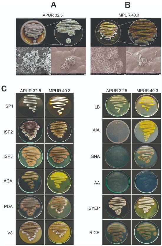
Figure 1.
Morphological and micromorphological characteristics of Streptomyces isolates on different culture media. (A) Macroscopic colonial aspect of Streptomyces sp. APUR 32.5 grown on ISP2 medium (pH 7, 40 °C, 7 days) and its corresponding micromorphology (spiraled spore chains with cubic, wrinkled spores). (B) Macroscopic colonial aspect of Streptomyces murinus MPUR 40.3 grown on ISP2 medium (pH 7, 30 °C, 7 days) and its corresponding micromorphology (well-developed vegetative and aerial mycelia forming closed spiral spore chains with cubic, smooth spores). (C) Comparative overview of the morphological characteristics of Streptomyces sp. APUR 32.5 and Streptomyces murinus MPUR 40.3 isolates cultivated on twelve different culture media after 7 days of incubation at 28 °C, highlighting variations in mycelial coloration and diffusible pigment production.
For the Streptomyces sp. APUR 32.5 isolate, optimal growth (+++) was recorded on ISP2, ISP3, SYEP, V8, and rice agar media. Moderate growth (++) occurred on ISP1, ACA, and PDA, while limited growth (+) was noted on LB, SNA, AA, and AIA. Colony mycelial coloration predominantly ranged from white to brown. Notably, a black diffusible pigment was produced specifically on SYEP medium. Microscopic analysis of APUR 32.5 revealed spiraled spore chains, with the spores exhibiting a cubic shape, a wrinkled surface, and an approximate size of 0.13 μm (Figure 1A,B).
The S. murinus MPUR 40.3 isolate showed optimal growth (+++) on ISP2, ISP3, V8, SYEP, SNA, and rice agar. Moderate growth (++) was observed on ISP1 and PDA, and limited growth (+) was observed on ACA, LB, AIA, and AA. A distinct characteristic of MPUR 40.3 was the prominent production of a yellow diffusible pigment in eight of the twelve tested media (ISP1, ISP2, ACA, PDA, LB, AIA, V8, and SYEP). The aerial mycelium displayed varied colorations, including brown, yellow, lilac, and white tones, depending on the culture medium. Microscopic evaluation of MPUR 40.3 demonstrated well-developed vegetative and aerial mycelia forming closed spiral spore chains, with spores that were cubic in shape and had a smooth surface, and measured approximately 0.15 μm (Figure 1A,B).
3.1.2. Molecular Identification and Phylogenetic Analysis
Molecular identification of the isolates was performed through multilocus phylogenetic analysis based on the concatenated alignment of five housekeeping genes (atpD, gyrB, recA, rpoB, and trpB) from 40 Streptomyces species that are closely related to APUR 32.5 and MPUR 40.3. The final alignment comprised 2529 characters, including gaps, distributed among the genes atpD (496 bp), gyrB (417 bp), recA (504 bp), rpoB (541 bp), and trpB (541 bp).
The most suitable evolutionary models to explain the data according to the AIC criteria were GTR + F + I + G4 for the atpD, recA, and trpB genes, GTR + F + G4 for the gyrB gene, and K3Pu + F+I + G4 for rpoB. The phylogenetic tree obtained by maximum likelihood estimation with 1000 bootstrap replicates revealed that the isolate MPUR 40.3 clustered with high support (99% bootstrap) in the clade including S. murinus DSM 40091T, confirming its identification as belonging to this species (Figure 2).
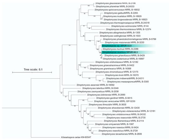
Figure 2.
Phylogenetic tree based on multilocus analysis (atpD, gyrB, recA, rpoB, and trpB) of Streptomyces sp. APUR 32.5 and S. murinus MPUR 40.3 (highlighted in green) and related species. The tree was constructed using the maximum likelihood method with 1000 bootstrap replicates, using Kitasatospora setae KM-6054T as an outgroup. Numbers on the nodes indicate bootstrap support values (%).
Isolate APUR 32.5 formed a distinct branch, yet was closely related to the clade that includes S. murinus, S. griseofuscus, and S. costaricanus (currently considered heterotypic synonyms of S. murinus), suggesting that this isolate represents a putative new species for the genus.
3.2. Antifungal Activity
The results indicated that both Streptomyces strains demonstrated antifungal activity against the seven Colletotrichum species analyzed, with inhibition percentages ranging from 56.3% to 88.6% (Figure 3A,B). Streptomyces sp. APUR 32.5 showed the highest inhibition rates against C. guaranicola (INPA 2939), reaching 86.7 ± 0.2%, Colletotrichum sp. (INPA 2973) with 83.3 ± 0.17%, and C. brevisporum (INPA 2787) with 75.6 ± 0.1%. In turn, S. murinus MPUR 40.3 demonstrated greater efficiency in inhibiting the mycelial growth of C. guaranicola (INPA 2939) with 88.5 ± 0.05%, C. scovillei (INPA 2910) with 85.6 ± 0.43%, and Colletotrichum sp. (INPA 2973) with 85.2 ± 0.3% (Figure 3B). All the tested isolates exhibited inhibition percentages over 50%, confirming the broad spectrum of these strains against the evaluated species.
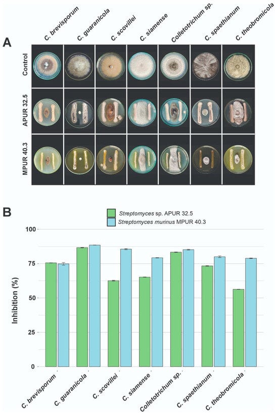
Figure 3.
In vitro antifungal activity of Streptomyces isolates against Colletotrichum species. (A) Representative images of direct antagonism assays on Petri dishes, demonstrating mycelial growth inhibition of seven Colletotrichum species by Streptomyces sp. APUR 32.5 and S. murinus MPUR 40.3 isolates. (B) Bar chart representing the percentage of mycelial growth inhibition (PIC) of the tested phytopathogens after 7 days of cultivation at 28 °C. Bars indicate mean values, and error bars represent the standard deviation of three replicates.
Scanning electron microscopy analyses revealed the differential morphological effects of the Streptomyces strains on the C. scovillei structures. Control cultures containing only the pathogen (Figure 4A–C) displayed intact hyphal networks with a uniform diameter and a smooth surface morphology. In dual cultures with Streptomyces sp. APUR 32.5 (Figure 4D–F), C. scovillei hyphae exhibited morphological alterations that appeared to correlate with proximity to the interaction zone. Hyphae closer to the bacterial colonies showed cell wall disruption and irregular thinning (yellow arrow, Figure 4E), whereas hyphae in more distant regions maintained relatively normal morphology (red arrow, Figure 4E), suggesting a concentration-dependent effect of the diffusible compounds.
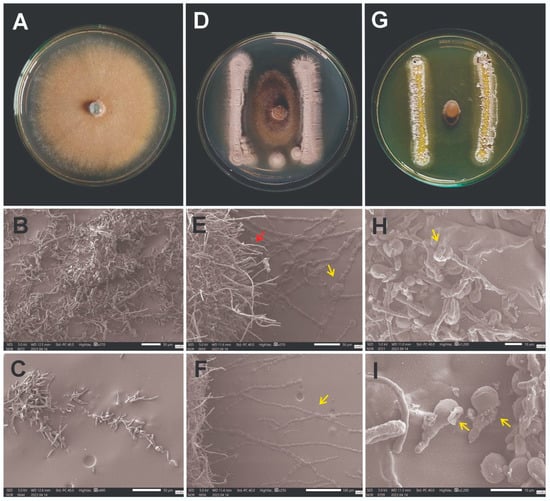
Figure 4.
Images of the interaction between Streptomyces spp. and Colletotrichum scovillei using scanning electron microscopy (SEM). (A–C) Control: pure culture of Colletotrichum scovillei showing normal growth and mycelia with preserved structures, continuous hyphae of uniform diameter and smooth surface. (D–F) Interaction between Streptomyces sp. APUR 32.5 and Colletotrichum scovillei: morphological alterations in phytopathogen mycelia are evident, with irregular hyphal thinning and cell wall disruption (yellow arrow), while some hyphae preserve a morphology that is similar to the control (red arrow). (G–I) Interaction between Streptomyces murinus MPUR 40.3 and Colletotrichum scovillei: cellular lysis of C. scovillei conidia is observed during the germination phase, resulting in structural collapse (yellow arrows) and inhibition of germination. Scale bars are shown in the SEM images.
Co-cultures with S. murinus MPUR 40.3 (Figure 4G–I) exhibited distinct morphological patterns, with C. scovillei conidia showing structural collapse and evident signs of lysis (Figure 4H,I). Although the damage patterns differed from those observed for APUR 32.5, these observations indicate that both isolates inhibit the pathogen through antibiosis. This inference is supported by the absence of direct contact between the antagonist and the fungal structures, together with the presence of diffusible factors causing visible cellular disruption. While the specific metabolites responsible cannot be identified based on SEM images, the results clearly demonstrate that MPUR 40.3 exerts a more pronounced inhibitory effect, preventing the fungal hyphae from approaching the bacterial colony.
3.3. Effect of Streptomyces Isolates on the Control of Colletotrichum Scovillei in Capsicum Chinense Fruits
3.3.1. Evaluation of the Incidence of Disease
After seven days of incubation (at 28 °C), the negative control fruits (treated with sterile water) remained completely healthy, with an intact surface and the absence of any signs of microbial colonization, as confirmed by SEM (Figure 5A–E). In contrast, the positive control fruits (inoculated only with C. scovillei INPA 2010) developed severe symptoms of anthracnose (Figure 5B), characterized by depressed necrotic lesions and abundant sporulation, reaching a disease index of 97.79% (Figure 5F).
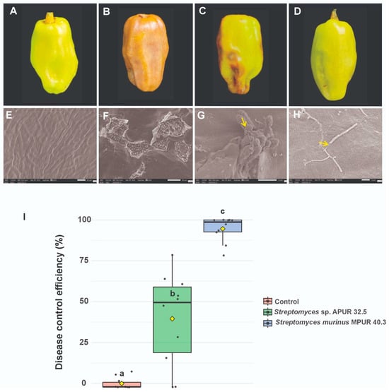
Figure 5.
Evaluation of the interaction between Streptomyces and Colletotrichum scovillei on pepper fruits and ultrastructural analysis of infected tissue surfaces. Macroscopic images of fruits: (A–E) Negative control, showing the intact epicarp of the fruit. (B–F) Positive control, evidencing fungal colonization and fruit surface rupture. (C–G) Fruit treated with Streptomyces sp. APUR 32.5, with reduced lesions and adherence of antagonist spores to pathogen conidia (yellow arrow indicating Streptomyces spores). (D–H) Fruit treated with S. murinus MPUR 40.3, with tissue preservation and structural collapse of C. scovillei hyphae (yellow arrow indicating broken mycelia). (I) Box plot showing the disease control efficiency (%) in different treatments. Means followed by different letters indicate statistically significant differences (p < 0.05).
The application of biocontrol agents resulted in significant protection, albeit with differential efficacy between strains. Fruits treated with S. murinus MPUR 40.3 exhibited high protection, with an infection index of only 4.86%, representing 95% control efficacy relative to the positive control group (Figure 5D–H). Fruits treated with Streptomyces sp. APUR 32.5 showed intermediate protection, with a disease incidence of 49.41% (Figure 5C–G).
Statistical analysis confirmed significant differences (p < 0.05) between all treatments, demonstrating the superior performance of S. murinus MPUR 40.3 as a biocontrol agent (Figure 5I). Importantly, additional controls in which fruits were treated exclusively with Streptomyces strains (without the pathogen) showed no morphological alterations or tissue damage.
3.3.2. Ultrastructural Analysis of the Pathogen-Antagonist Interactions
Scanning electron microscopy enabled the elucidation of the antagonist mechanisms of action and their interaction with the pathogen in host tissues. In the positive control fruits, SEM revealed severe tissue surface damage with extensive fungal colonization and disruption of cellular integrity (Figure 5D). At higher magnifications, pathogen hyphae were observed actively penetrating fruit tissues (Figure 5C, yellow arrow), characterizing the typical infectious process of anthracnose.
In fruits treated with Streptomyces sp. APUR 32.5, the lesions on the plant tissue surfaces appeared less intense and less extensive compared with the positive control, although fruit damage was still observable. The micrographs evidenced a direct interaction between the antagonist and the pathogen, with Streptomyces sp. APUR 32.5 spores adhered to the conidia (Figure 5E, yellow arrow indicating conidium with Streptomyces spores on its surface), confirming the physical contact between the microorganisms. However, some conidia showed morphological alterations with apparent cell wall integrity compromise (Figure 5F, indicated by the yellow arrow), suggesting that the Streptomyces strain may also possess compounds capable of damaging the phytopathogen cell wall.
Treatment with S. murinus MPUR 40.3 resulted in significantly superior protection, with the fruit tissue surface predominantly preserved. Although the macroscopic analysis revealed minimal symptoms, SEM detected small, localized lesions, probably resulting from the initial 24-h pathogen exposure period before application of the antagonist. In the interaction areas, morphological alterations in C. scovillei hyphae were observed, characterized by structural disruption and mycelial disintegration (Figure 5H, indicated by a yellow arrow), demonstrating the potent direct antifungal activity of S. murinus MPUR 40.3.
These ultrastructural results corroborate the data obtained in the in vitro assays (Section 3.2), in which S. murinus MPUR 40.3 also demonstrated greater antagonistic activity against C. scovillei, and suggest that the biocontrol mechanisms involve both direct interaction and the possible production of antifungal compounds that compromise the pathogen’s structural integrity.
3.4. Plant Growth Promotion
The evaluation of the capacity for plant growth promotion by Streptomyces isolates was conducted through the analysis of multiple biometric parameters in Capsicum chinense plants cultivated under controlled conditions for 45 days after bacterial inoculation.
The results demonstrated that inoculation with Streptomyces sp. APUR 32.5 and S. murinus MPUR 40.3 promoted significant alterations in plant root system development, whereas shoot parameters showed no statistically significant differences (Figure 6). Root dry weight analysis revealed substantial increases in treatments with both bacterial isolates compared with the uninoculated control.
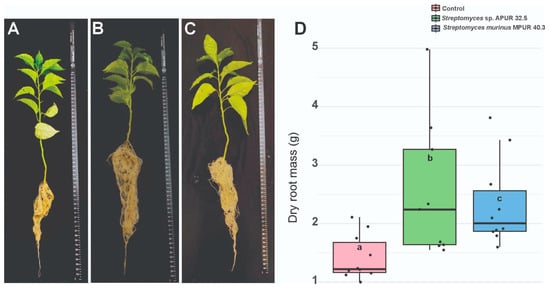
Figure 6.
Effect of Streptomyces isolates on Capsicum chinense root system development after 45 days of cultivation. (A) Root system of control plants (uninoculated); (B) Root system of plants inoculated with Streptomyces sp. APUR 32.5; (C) Root system of plants inoculated with Streptomyces murinus MPUR 40.3; (D) Mean root dry weight (g) per treatment. Bars represent mean ± standard deviation (n = 10). Different letters above the bars indicate statistically significant differences between treatments by Tukey’s test (p < 0.05).
Plants treated with Streptomyces sp. APUR 32.5 showed the greatest increment in root dry weight, with a 79.58% increase relative to the control (p < 0.05), reaching an average of 0.87 ± 0.12 g per plant, while the control averaged 0.48 ± 0.09 g (Figure 6D). Treatment with S. murinus MPUR 40.3 also resulted in a significant increase, with a 64.08% increment in root dry weight (0.79 ± 0.11 g) compared to the control.
Morphological analysis of the root system (Figure 6A–C) evidenced not only an increase in biomass but also alterations in root architecture, with greater branching and lateral root development in the plants treated with Streptomyces, particularly with strain APUR 32.5. These modifications in the root architecture may contribute to improved water and nutrient uptake efficiency, representing an additional benefit beyond the increase in biomass.
Although no statistically significant differences were detected in plant height, stem diameter, or shoot dry weight, the substantial root development observed in the plants treated with Streptomyces suggests the suitability for application of these isolates as growth-promoting inoculants in C. chinense, especially under conditions where root development represents a limiting factor for productivity.
3.5. Enzymatic Assays
3.5.1. Hydrolytic Enzymatic Activities
Both isolates demonstrated the capacity to produce diverse hydrolytic enzymes, with significant variations in enzymatic indices (EI) as a function of temperature and pH conditions (Figure 7A-C). Streptomyces sp. APUR 32.5 exhibited notable amylolytic (EI = 16.0 ± 3.46) and lipolytic (EI = 10.0 ± 2.36) activities, particularly at pH 7.0 and 28 °C (Figure 7A). In contrast, S. murinus MPUR 40.3 displayed superior amylolytic activity (EI = 19.0 ± 0.00) under the same conditions, in addition to moderate cellulolytic (EI = 5.9 ± 1.18) and lipolytic (EI = 5.8 ± 0.70) activities (Figure 7B).
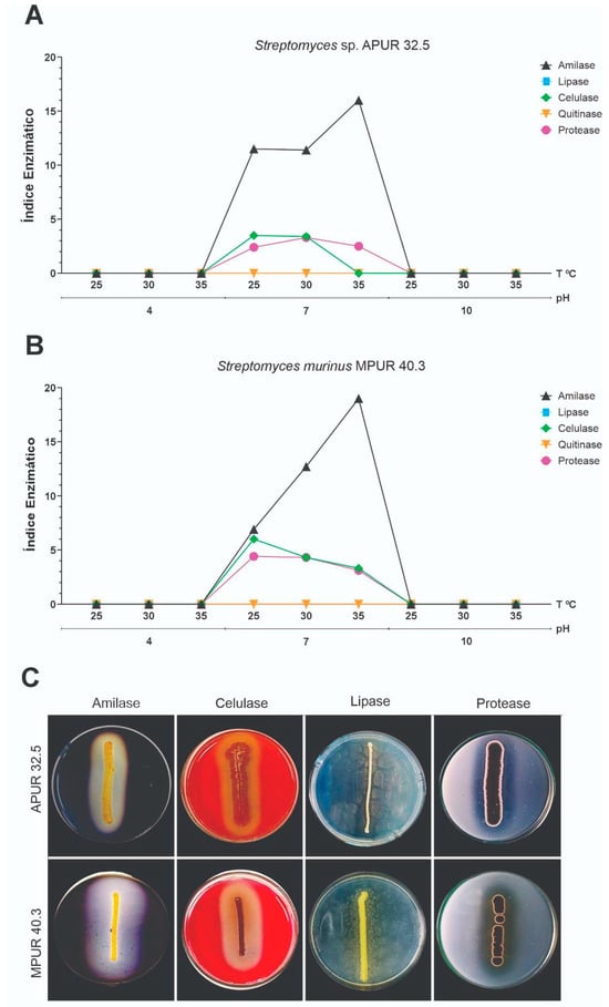
Figure 7.
Hydrolytic enzymatic activities of Streptomyces isolates under different temperature and pH conditions. (A) Enzymatic indices (EI) of Streptomyces sp. APUR 32.5 for amylase, cellulase, and lipase under different temperature combinations. (B) Enzymatic indices of Streptomyces murinus MPUR 40.3 under the same conditions. (C) Representative visualization of enzymatic degradation halos.
Comparative analysis of enzymatic activities under different conditions revealed that both isolates maintained significant hydrolytic activity across a broad pH range (5.0–9.0) and temperature range (25–37 °C), with optimal activity under neutral to slightly alkaline conditions (pH 7.0–8.0) and mesophilic temperatures (28–30 °C). This versatility suggests adaptability to different rhizosphere microenvironments and application under diverse edaphoclimatic conditions.
3.5.2. Siderophore Production and Phosphate Solubilization
The capacity to mobilize essential nutrients was evaluated through qualitative assays for the siderophore production and solubilization of different phosphate sources. Both isolates, Streptomyces sp. APUR 32.5 and S. murinus MPUR 40.3 demonstrated positive results for siderophore production, as evidenced by the characteristic halo formation on CAS (chrome azurol S) medium.
Regarding phosphate solubilization, both isolates showed the capacity to mobilize phosphorus from diverse inorganic sources, including aluminum phosphate (AlPO4), tricalcium phosphate [Ca3(PO4)2], and ferric phosphate (FePO4). This versatility in solubilizing different forms of insoluble phosphate is particularly relevant, considering that phosphorus frequently represents a limiting factor for plant growth in tropical soils.
The combined capacity to produce siderophores and solubilize phosphates constitutes an important mechanism for plant growth promotion, contributing to the increased availability of essential nutrients in the rhizosphere. In addition, siderophore production may act as an indirect biocontrol mechanism through which phytopathogens compete for iron.
3.6. pH and Temperature Tolerance
The capacity to adapt to different environmental conditions is a crucial parameter for evaluating the application of microorganisms as biocontrol agents and plant growth promoters in the field. In this context, the characterization of the tolerance of Streptomyces isolates to different temperatures and pH values was performed, aiming to determine their ecophysiological versatility. Streptomyces sp. APUR 32.5 and S. murinus MPUR 40.3 isolates were evaluated for growth on ISP2 medium under different temperature (25, 30, 35, 40, 45, and 50 °C) and pH (6.0, 7.0, and 8.0) combinations.
Streptomyces sp. APUR 32.5 proved to be a moderately thermotolerant mesophile, with optimal growth (+++) at 35 °C across all tested pH ranges. At lower temperatures (25–30 °C), it showed moderate growth (++) regardless of pH. At 40 °C, it exhibited optimal growth (+++) at pH 6.0, reducing to moderate (++) under neutral to alkaline conditions (pH 7.0–8.0). At an elevated temperature (45 °C), growth was reduced (+) at all pH values, and at 50 °C, no growth was observed, indicating its upper thermal tolerance limit.
S. murinus MPUR 40.3 presented a slightly distinct growth profile, with a preference for more alkaline conditions at lower temperatures. At 25 °C, it demonstrated optimal growth (+++) at pH 8.0 and moderate growth (++) at pH 6.0–7.0. At temperatures of 30–35 °C, it exhibited optimal growth (+++) at pH 7.0–8.0 and moderate growth (++) at pH 6.0. At 40 °C, growth was moderate (++) under all pH conditions. Similarly to the isolate APUR 32.5, it showed reduced growth (+) at 45 °C and absence of growth at 50 °C.
Both isolates demonstrated the capacity to grow across a broad temperature range (25–45 °C) and pH range (6.0–8.0), with distinct preferences reflecting specific ecophysiological adaptations. Streptomyces sp. APUR 32.5 showed greater tolerance to elevated temperatures under slightly acidic conditions (pH 6.0), while S. murinus MPUR 40.3 demonstrated better adaptation to alkaline conditions (pH 8.0) at lower temperatures.
This ecophysiological versatility suggests the application of these isolates under different edaphoclimatic conditions, particularly in tropical and subtropical regions where soil temperatures can vary significantly. Additionally, tolerance to different pH values broadens their application in soils with diverse chemical characteristics.
4. Discussion
The Streptomyces strains investigated in this study show phylogenetic proximity to the clade that includes S. murinus, which is the currently accepted name for this species, while S. griseofuscus and S. costaricanus are considered heterotypic synonyms of S. murinus according to recent taxonomic revisions [35]. Strain MPUR 40.3 was identified as S. murinus, whereas Streptomyces sp. APUR 32.5 represents a distinct sister group, a new putative species within the genus. Complementary phylogenomic analyses (Table S7), including digital DNA-DNA hybridization (dDDH) values below the 70% cutoff for species delineation, further support the phylogenetic results and confirm the distinct taxonomic position of APUR 32.5.
S. murinus was originally described by Frommer in 1959 and is known for its ability to produce the antifungal metabolite pentamycin [36,37], first isolated in 1958 by Umezawa and collaborators. This compound exhibits activity against various pathogenic fungi, including Candida species and dermatophytes. The strain previously classified as S. costaricanus, described in 1995, is recognized for its antibiotic and anti-nematode properties, and is known to produce avermectin [38], a compound also produced by Streptomyces avermitilis [39]. The taxonomic reclassification of these species as S. murinus does not diminish the importance of their bioactive metabolites but rather underscores the complexity of Streptomyces taxonomy and the need for polyphasic approaches for accurate identification.
In the present study, the strains Streptomyces sp. APUR 32.5 and S. murinus MPUR 40.3 demonstrated strong in vitro antagonistic activity against seven distinct Colletotrichum species. Among the species tested, the best performance was observed against C. guaranicola, with inhibition of mycelial growth of over 80%, surpassing previously reported results, in which Streptomyces griseocarneus R132 reduced the mycelial growth of C. guaranicola by 70% [34]. Similarly, inhibition rates of up to 66% were reported against C. siamense using Streptomyces species [40]; whereas, in our study, the APUR 32.5 and MPUR 40.3 strains inhibited the growth of C. siamense by 65% and 79%, respectively. This inhibitory effect may be associated with the production of secondary metabolites, as Streptomyces is well-known for its remarkable ability to synthesize a wide array of bioactive compounds [41,42,43].
In addition to the direct action of microorganisms, Streptomyces extracts have also shown high efficacy against phytopathogens. S. murinus NARZ reduced the mycelial growth of C. scovillei by up to 65%, indicating the presence of secondary metabolites with significant antifungal activity [44]. Similarly, both the extract and the direct action of Streptomyces sp. NEAU-Y11 resulted in significant inhibition of the mycelial growth of Colletotrichum orbiculare [45], reinforcing the potential of this genus as a source of bioactive molecules with antifungal properties. Moreover, metabolites produced by Streptomyces amritsarensis V31 exhibited relevant antifungal activity in the control of different phytopathogenic fungi [46].
In addition to bioactive metabolites, hydrolytic enzymes also represent a promising alternative for controlling phytopathogens. Enzymes such as amylase, cellulase, lipase, glucanase, chitinase, and protease play an essential role in degrading the cell wall, proteins, and structural components of phytopathogenic fungi, establishing an efficient biocontrol mechanism that indirectly supports plant development [47,48]. In the present study, the tested Streptomyces strains demonstrated amylolytic, cellulolytic, lipolytic, and proteolytic activity, suggesting the involvement of these enzymes in the degradation of fungal structures and the suppression of phytopathogens.
The enzymatic index (EI) values for amylase reached significant levels, with S. murinus MPUR 40.3 (EI = 19) and Streptomyces sp. APUR 32.5 (EI = 16) standing out. These results indicate a high capacity for hydrolytic enzyme production, which is possibly associated with the biocontrol mechanism of these strains. The role of enzymes in microbial antagonism is supported by studies that demonstrate the importance of α-amylase (AmyS) produced by Bacillus cereus 0–9 in inhibiting Rhizoctonia cerealis. Through gene editing, the authors observed that the growth of the phytopathogen was reduced by 84.7% in the presence of the strain carrying the functional gene; however, deletion of the same gene (amyS) resulted in a significant decrease in biocontrol efficacy, with inhibition reduced to only 43.8% [49].
The postharvest control of anthracnose using microorganisms and their metabolites is increasingly recognized as an effective biocontrol strategy, crucial for reducing chemical inputs and promoting sustainable agriculture [43]. Our study significantly contributes to this understanding by demonstrating the strong postharvest biocontrol activity of Streptomyces sp. APUR 32.5 and S. murinus MPUR 40.3 against C. scovillei in C. chinense fruits (Section 3.3.1). Specifically, S. murinus MPUR 40.3 achieved a 95% reduction in anthracnose incidence, while Streptomyces sp. APUR 32.5 showed a moderate 39.25% reduction. These findings are highly encouraging and align with the growing body of evidence supporting the efficacy of microbial agents for postharvest disease management. For instance, the significant efficacy of MPUR 40.3 (95%) is comparable to, or even surpasses, some previous reports; as volatile organic compounds from Trichoderma agriamazonicum achieved complete inhibition against C. scovillei in C. chinense [50]. Streptomyces tuirus AR26 [51], Streptomyces lactacystinicus ZZ-84 [52], and Streptomyces olivoreticuli ZZ-21 [53] demonstrated growth inhibition of Colletotrichum scovillei both in vitro and in the assay of disease suppression in planta, and are reported as promising biocontrol strains that are capable of significantly reducing postharvest anthracnose caused by C. scovillei. This reinforces that diverse microbial groups, including Streptomyces, are robust alternatives to chemical inputs. Our results thus underscore the promising role of Streptomyces from Amazonian sediments as potent biocontrol agents for anthracnose, contributing to more sustainable agricultural practices.
SEM analyses revealed marked differences in the mechanisms of action of the antagonistic strains against Colletotrichum scovillei, both in vitro and postharvest biocontrol. In vitro treatment with Streptomyces sp. APUR 32.5 resulted in intense degradation of the pathogen’s mycelia, suggesting the production of lytic enzymes that compromise fungal cell wall integrity. In contrast, S. murinus MPUR 40.3 appeared to release substances that acted directly on germinative cells (conidia), promoting their destruction and consequently preventing germination and establishment of infection.
Postharvest assays showed that, despite the moderate reduction in disease severity provided by strain APUR 32.5, SEM revealed visibly damaged and shriveled C. scovillei spores on the surface of the treated fruits. In contrast, in the treatment with MPUR 40.3, the spores were completely ruptured, corroborating the greater efficacy of this strain. The use of SEM as a tool to visualize interactions between antagonistic microorganisms and pathogens has proven fundamental for elucidating biocontrol mechanisms, as shown in previous studies that revealed structural alterations in different pathogen–antagonist systems [54,55].
The Streptomyces strains tested in this study demonstrated multifunctional activity, acting not only in the biocontrol of phytopathogens but also in the promotion of plant growth. Both isolates were able to produce siderophores and solubilize different forms of phosphate (calcium, iron, and aluminum), in addition to synthesizing hydrolytic enzymes that, in addition to their protective effect against phytopathogens, also contribute to plant development. These effects were evidenced in the growth-promotion assays, particularly through the significant stimulation of C. chinense root system development.
The mechanisms of phosphate solubilization and siderophore production play a fundamental role in promoting plant growth by increasing the availability of essential nutrients in the rhizosphere. The solubilization of inorganic phosphates converts insoluble forms of phosphorus into forms that can be readily absorbed by plants, while siderophores sequester iron under conditions of low availability, whereas siderophore production increases iron uptake by plants and limits iron availability to phytopathogens. These mechanisms highlight the multifunctional role of Streptomyces, with promising applications in both biocontrol and biofertilization, as demonstrated in recent studies [56,57,58].
Taken together, the results obtained in this study reinforce the effectiveness of Streptomyces in controlling phytopathogenic fungi and promoting growth in C. chinense crops, aligning with several recent studies that demonstrate their ability as promising biocontrol agents for sustainable agriculture. The characterization of new strains with both antifungal and growth-promoting activity, particularly those isolated from underexplored environments such as the Amazon, represents a significant contribution to the development of biological alternatives to conventional agrochemicals.
5. Conclusions
This study provides novel insights by thoroughly characterizing the multifunctional potential of two Streptomyces strains, including a putative new species (APUR 32.5) and S. murinus (MPUR 40.3), originally isolated from the highly biodiverse Amazonian sediments. Our findings strongly support the hypothesis that these underexplored ecosystems are rich sources of microorganisms that possess unique and potent biotechnological attributes, which may be applicable in sustainable agricultural solutions. These Amazonian isolates demonstrated robust, broad-spectrum inhibitory capacity against all the tested Colletotrichum species in vitro. More critically, they showed significant efficacy in reducing the incidence of postharvest anthracnose in Capsicum chinense fruits, with S. murinus MPUR 40.3 achieving a remarkable 95% reduction and Streptomyces sp. APUR 32.5 achieving a notable 39.25% reduction. Mechanistic insights from scanning electron microscopy revealed distinct antifungal actions, with MPUR 40.3 primarily inducing conidial lysis during early infection and APUR 32.5 causing hyphal degradation. In addition to biocontrol, these isolates exhibited plant growth-promoting traits, including the production of hydrolytic enzymes and siderophores, as well as phosphate solubilization. Their demonstrated ecophysiological versatility (tolerance to broad ranges of temperature and pH) further enhances their applicability. Collectively, these multifunctional Amazonian Streptomyces isolates represent promising, sustainable candidates for integrated phytopathogen management and enhanced crop productivity in agriculture. Future studies employing metabolomic approaches will aim to elucidate the molecular mechanisms associated with the antifungal activity of these strains. In addition, their efficacy under field conditions and their potential for the development of commercial formulations as bioinoculants and biocontrol agents will be evaluated.
Supplementary Materials
The following supporting information can be downloaded at: https://www.mdpi.com/article/10.3390/microorganisms13122713/s1, Table S1. Composition of the culture media used in this study for the morphological characterization of Streptomyces strains and antagonism analyses. Table S2. Sequences of the primers used for partial amplification of the genes atpD, gyrB, recA, rpoB, and trpB, with their respective annealing temperatures and expected PCR product sizes. Table S3. Sequence data and GenBank accession numbers for Streptomyces strains used in this study. Table S4. Phytopathogens of the genus Colletotrichum used in antagonism assays with Streptomyces sp. APUR 32.5 and Streptomyces murinus MPUR 40.3. Table S5. Composition of the selective culture media used to evaluate the solubilization capacity of different phosphate sources and the production of siderophores by Streptomyces isolates. Table S6. Composition of the culture media used in the enzymatic activity assays and the methods for detecting hydrolysis halos. Table S7. Percentage of digital DNA-DNA hybridization (dDDH) values calculated using formula d4 among genomes of strains MPUR 40.3 and APUR 32.5 compared with phylogenetically closest Streptomyces species using the Type Strain Genome Server (TYGS).
Author Contributions
Conceptualization, I.J.S.d.S. and G.F.d.S.; methodology, I.J.S.d.S., T.F.S., T.M.d.S. and B.M.G.; validation, G.F.d.S., T.F.S., R.E.d.L.P., A.W.M., H.H.F.K. and R.E.H.; formal analysis, A.W.M.; investigation, I.J.S.d.S.; data curation, I.J.S.d.S. and G.F.d.S.; writing—original draft preparation, I.J.S.d.S.; writing—review and editing, I.J.S.d.S. and G.F.d.S.; visualization, I.J.S.d.S. and T.F.S.; supervision, G.F.d.S.; project administration, G.F.d.S. All authors have read and agreed to the published version of the manuscript.
Funding
This work was funded by Coordenação de Aperfeiçoamento de Pessoal de Nível Superior (CAPES-grant number 88881.200469/2018–01 Procad AmazonMicro and Grant No 88887.510218–2020–00 CAPES-Amazônia-Legal) and CAPES—Finance code 001. We also would like to thank Conselho Nacional de Desenvolvimento Científico e Tecnológico (CNPq) for the productivity scholarship grant provided to G.F.d.S (grant number 309680/2025-5). The authors, H.H.F.K., G.F.S., and R.E.H., also acknowledge Fundação de Amparo à Pesquisa do Estado do Amazonas (FAPEAM) for the funding via the projects PROSPAM (call 008/2021), AMAZONAS ESTRATÉGICO (call 004/2018), ÁREAS PRIORITÁRIAS (call 010/2021), and POSGRAD 2022/2023 (call 005/2022).
Institutional Review Board Statement
Not applicable.
Informed Consent Statement
Not applicable.
Data Availability Statement
The original contributions presented in this study are included in the article/Supplementary Materials. Further inquiries can be directed to the corresponding author.
Acknowledgments
The authors thank FAPEAM (Fundação de Amparo à Pesquisa do Estado do Amazonas) for financial support, INPA (National Institute for Amazonian Research), CMABio—UEA (Multiservice Center for Biomedical Phenomena Analysis of the Amazonas State University), and UFAM (Federal University of Amazonas) for their partnerships, and all the technical staff of the Amazonian Applied Microbiology and Biotechnology Laboratory (AmazonMicroBiotech)—Embrapa Western Amazon.
Conflicts of Interest
The authors declare no conflict of interest.
References
- Tudi, M.; Ruan, H.D.; Wang, L.; Lyu, J.; Sadler, R.; Connell, D.; Chu, C.; Phung, D. Agriculture development, pesticide application and its impact on the environment. Int. J. Environ. Res. Public Health 2021, 18, 1112. [Google Scholar] [CrossRef]
- Shahbazi, F.; Golshani, A.; Fattahi, M.; Armin, M. Losses in agricultural produce: A review of causes and solutions, with a specific focus on grain crops. J. Stored Prod. Res. 2025, 111, 102547. [Google Scholar] [CrossRef]
- Nath, B.; Chen, G.; O’sullivan, C.M.; Zare, D. Research and technologies to reduce grain postharvest losses: A review. Foods 2024, 13, 1875. [Google Scholar] [CrossRef]
- Sawicka, B.; Egbuna, C. Pests of agricultural crops and control measures. In Natural Remedies for Pest, Disease and Weed Control; Academic Press: Cambridge, MA, USA, 2020; pp. 1–16. [Google Scholar]
- Zakaria, L. Diversity of Colletotrichum species associated with anthracnose disease in tropical fruit crops—A review. Agriculture 2021, 11, 297. [Google Scholar] [CrossRef]
- Salotti, I.; Baroncelli, R.; Sarrocco, S.; Battilani, P. Development of a model for Colletotrichum diseases with calibration for phylogenetic clades on different host plants. Front. Plant Sci. 2023, 14, 1069092. [Google Scholar] [CrossRef]
- Thanh, L.T.H.; Minh, P.T.; Trang, P.T.; Duong, V.T. First report of Colletotrichum siamense and Colletotrichum endophyticum associated with anthracnose on avocado (Persea americana Mill.) in Vietnam. J. Plant Pathol. 2025, 107, 633–647. [Google Scholar] [CrossRef]
- Torres-Calzada, C.; Tapia-Tussell, R.; Higuera-Ciapara, I.; Pérez-Brito, D. Sensitivity of Colletotrichum truncatum to four fungicides and characterization of thiabendazole-resistant isolates. Plant Dis. 2015, 99, 1590–1595. [Google Scholar] [CrossRef]
- Dowling, M.; Peres, N.A.; Villani, S.M.; Schnabel, G. Managing Colletotrichum on fruit crops: A “complex” challenge. Plant Dis. 2020, 104, 2301–2316. [Google Scholar] [CrossRef]
- Ren, L.; Li, G.Q.; Han, Y.C.; Jiang, D.H.; Huang, H.C. Characterisation of sensitivity of Colletotrichum gloeosporioides and Colletotrichum capsici, causing pepper anthracnose, to picoxystrobin. J. Plant Dis. Prot. 2020, 127, 657–666. [Google Scholar] [CrossRef]
- Karim, M.M.; Wu, C.; Zhang, J.; Li, X.; Liu, P.; Sun, G. Fungicide resistance in Colletotrichum fructicola and Colletotrichum siamense causing peach anthracnose in China. Pestic. Biochem. Physiol. 2024, 203, 106006. [Google Scholar] [CrossRef]
- Giacomin, R.M.; Rodrigues, R.; Gonçalves-Vidigal, M.C.; Silva, C.L. Inheritance of anthracnose resistance (Colletotrichum scovillei) in ripe and unripe Capsicum annuum fruits. J. Phytopathol. 2020, 168, 184–192. [Google Scholar] [CrossRef]
- Caires, N.P.; Félix, C.R.; Tessmann, D.J.; Cardoso, J.E.; Maffia, L.A.; Mizubuti, E.S.G. First report of anthracnose on pepper fruit caused by Colletotrichum scovillei in Brazil. Plant Dis. 2014, 98, 1437. [Google Scholar] [CrossRef]
- Giacomin, R.M.; Colletta, G.D.; Gonçalves-Vidigal, M.C.; Rodrigues, R. Post-harvest quality and sensory evaluation of mini sweet peppers. Horticulturae 2021, 7, 287. [Google Scholar] [CrossRef]
- Shi, N.; Zhang, H.; Liu, P.; Sun, G. Resistance risk and resistance-related point mutations in cytochrome b of florylpicoxamid in Colletotrichum scovillei. Pestic. Biochem. Physiol. 2023, 196, 105617. [Google Scholar] [CrossRef]
- Law, C.X.; Lim, L.; Yap, S.K.; Chee, H.Y. A review on anthracnose disease caused by Colletotrichum spp. in fruits and advances in control strategies. Int. J. Food Microbiol. 2025, 394, 111397. [Google Scholar] [CrossRef]
- Chatterjee, K.; Singh, R.; Yadav, A.N. Application of Streptomycetes in medicine. In Bioeconomy of Streptomyces; CRC Press: Boca Raton, FL, USA, 2025; pp. 119–141. [Google Scholar]
- Patel, S.; Gupta, R.; Singh, H.B. Diversity of secondary metabolites from marine Streptomyces with potential anti-tubercular activity: A review. Arch. Microbiol. 2025, 207, 64. [Google Scholar] [CrossRef]
- Pacios-Michelena, S.; Ríos-Castro, E.; González, M.; Maldonado-Mendoza, I.E. Application of Streptomyces antimicrobial compounds for the control of phytopathogens. Front. Sustain. Food Syst. 2021, 5, 696518. [Google Scholar] [CrossRef]
- Chouyia, F.E.; Ventorino, V.; Pepe, O. Diversity, mechanisms and beneficial features of phosphate-solubilizing Streptomyces in sustainable agriculture: A review. Front. Plant Sci. 2022, 13, 1035358. [Google Scholar] [CrossRef]
- Beroigui, O.; Errachidi, F. Streptomyces at the heart of several sectors to support practical and sustainable applications: A review. Prog. Microbes Mol. Biol. 2023, 6, a0000345. [Google Scholar] [CrossRef]
- Wang, M.; Zhang, Y.; Chen, J.; Li, Q.; Liu, G. Streptomyces strains and their metabolites for biocontrol of phytopathogens in agriculture. J. Agric. Food Chem. 2024, 72, 2077–2088. [Google Scholar] [CrossRef]
- Kumar, M.; Singh, V.; Sharma, R. Potential applications of extracellular enzymes from Streptomyces spp. in various industries. Arch. Microbiol. 2020, 202, 1597–1615. [Google Scholar] [CrossRef]
- Khushboo; Singh, S.; Mishra, P. Biotechnological and industrial applications of Streptomyces metabolites. Biofuels Bioprod. Biorefin. 2022, 16, 244–264. [Google Scholar] [CrossRef]
- Pereira, J.O.; Lima, A.L.; Silva, G.C.; Araújo, J.M. Overview on biodiversity, chemistry, and biotechnological potential of microorganisms from the Brazilian Amazon. In Diversity and Benefits of Microorganisms from the Tropics; Moreira, F.M.S., Huising, E.J., Bignell, D.E., Eds.; Springer International Publishing: Cham, Switzerland, 2017; pp. 71–103. [Google Scholar]
- de Oliveira, A.C.F.M.; Santos, E.M.; Silva, C.F.; da Silva e Silva, L.; de Oliveira Veras, A.A.; das Graças, D.A.; Silva, A.; Baraúna, R.A.; Trivella, D.B.B.; Schneider, M.P.C. A metabologenomics approach reveals the unexplored biosynthetic potential of bacteria isolated from an Amazon Conservation Unit. Microbiol. Spectr. 2024, 13, e00996-24. [Google Scholar] [CrossRef]
- Jose, P.A.; Jha, B. Intertidal marine sediment harbours actinobacteria with promising bioactive and biosynthetic potential. Sci. Rep. 2017, 7, 10041. [Google Scholar] [CrossRef]
- Shirling, E.B.; Gottlieb, D. Methods for characterization of Streptomyces species. Int. J. Syst. Evol. Microbiol. 1966, 16, 313–340. [Google Scholar] [CrossRef]
- Doyle, J.J.; Doyle, J.L. A rapid total DNA preparation procedure for fresh plant tissue. Focus 1990, 12, 13–15. [Google Scholar]
- Labeda, D.P.; Dunlap, C.A.; Rong, X.; Huang, Y.; Doroghazi, J.R.; Ju, K.-S.; Metcalf, W.W. Phylogenetic relationships in the family Streptomycetaceae using multi-locus sequence analysis. Antonie Van Leeuwenhoek 2017, 110, 563–583. [Google Scholar] [CrossRef]
- Thampi, A.; Bhai, R.S. Rhizosphere actinobacteria for combating Phytophthora capsici and Sclerotium rolfsii, the major soil borne pathogens of black pepper (Piper nigrum L.). Biol. Control 2017, 109, 1–13. [Google Scholar] [CrossRef]
- Falguera, J.V.T.; Stratton, K.J.; Bush, M.J.; Jani, C.; Findlay, K.C.; Schlimpert, S.; Nodwell, J.R. DNA damage-induced block of sporulation in Streptomyces venezuelae involves downregulation of ssgB. Microbiology 2022, 168, 001198. [Google Scholar] [CrossRef]
- Ratul, N.; Sharma, G.D.; Madhumita, B. Phosphate solubilization, siderophore production and extracellular enzyme production activities of endophytic fungi isolated from tea (Camellia sinensis) bushes of Assam, India. Res. J. Biotechnol. 2023, 18, 49–57. [Google Scholar] [CrossRef]
- Liotti, R.G.; da Silva Figueiredo, M.I.; Soares, M.A. Streptomyces griseocarneus R132 controls phytopathogens and promotes growth of pepper (Capsicum annuum). Biol. Control 2019, 138, 104065. [Google Scholar] [CrossRef]
- Komaki, H. Reclassification of Streptomyces costaricanus and Streptomyces phaeogriseichromatogenes as later heterotypic synonyms of Streptomyces murinus. Int. J. Syst. Evol. Microbiol. 2021, 71, 004638. [Google Scholar] [CrossRef]
- Gren, T.; Pahl, A.; Nieselt, K.; Heide, L.; Luzhetskyy, A. Characterization and engineering of Streptomyces griseofuscus DSM 40191 as a potential host for heterologous expression of biosynthetic gene clusters. Sci. Rep. 2021, 11, 18301. [Google Scholar] [CrossRef]
- Das, V.; Thomas, L.; Mathew, J.; Joseph, B. Exploration of natural product repository by combined genomics and metabolomics profiling of mangrove-derived Streptomyces murinus THV12 strain. Fermentation 2023, 9, 576. [Google Scholar] [CrossRef]
- Esnard, J.; Potter, T.L.; Zuckerman, B.M. Streptomyces costaricanus sp. nov., isolated from nematode-suppressive soil. Int. J. Syst. Evol. Microbiol. 1995, 45, 775–779. [Google Scholar] [CrossRef]
- Cerna-Chávez, E.; López-Méndez, B.; Rivera-Morales, J.; Martínez, A. Potential of Streptomyces avermitilis: A review on avermectin production and its biocidal effect. Metabolites 2024, 14, 374. [Google Scholar] [CrossRef]
- Evangelista-Martínez, Z.; Torres, M.; Hernández, L.; Reyes, A. Potential of Streptomyces sp. strain AGS-58 in controlling anthracnose-causing Colletotrichum siamense from post-harvest mango fruits. J. Plant Pathol. 2022, 104, 553–563. [Google Scholar] [CrossRef]
- Alam, K.; Tian, J.; Wu, J.; Zhang, L.; Zhang, C. Streptomyces: The biofactory of secondary metabolites. Front. Microbiol. 2022, 13, 968053. [Google Scholar] [CrossRef]
- Ciofini, A.; Di Mattia, T.; Rossi, M.; Santini, C. Management of post-harvest anthracnose: Current approaches and future perspectives. Plants 2022, 11, 1856. [Google Scholar] [CrossRef]
- Nguyen, H.T.; Pham, L.D.; Tran, Q.N.; Le, T.A. Biological control of Streptomyces murinus against Colletotrichum causing anthracnose disease on tomato fruits. J. Pure Appl. Microbiol. 2025, 19, 542–557. [Google Scholar] [CrossRef]
- Song, W.; Yu, X.; Yu, X.; Zhang, H.; Zhang, K.; Guo, L.; Wang, J.-D.; Tian, D.-L.; Yu, Q.; Wang, X.; et al. Antifungal Activity and Potential Mechanisms of Two Bafilomycin Analogues Isolated from Streptomyces sp. NEAU-Y11 against Colletotrichum orbiculare. J. Agric. Food Chem. 2025, 73, 11814–11828. [Google Scholar] [CrossRef]
- Shahid, M.; Singh, B.N.; Verma, S.; Choudhary, P.; Das, S.; Chakdar, H.; Murugan, K.; Goswami, S.K.; Saxena, A.K. Bioactive antifungal metabolites produced by Streptomyces amritsarensis V31 help to control diverse phytopathogenic fungi. Braz. J. Microbiol. 2021, 52, 1687–1699. [Google Scholar] [CrossRef]
- Mishra, P.; Sharma, A.; Singh, V. Microbial enzymes in biocontrol of phytopathogens. In Microbial Enzymes: Roles and Applications in Industries; Academic Press: Cambridge, MA, USA, 2020; pp. 259–285. [Google Scholar]
- Riseh, R.S.; Akbarzadeh, A.; Mousavi, S.M.; Hassanzadeh, S. Unveiling the role of hydrolytic enzymes from soil biocontrol bacteria in sustainable phytopathogen management. Front. Biosci. 2024, 29, 105–125. [Google Scholar] [CrossRef]
- Daunoras, J.; Kačergius, A.; Gudiukaitė, R. Role of soil microbiota enzymes in soil health and activity changes depending on climate change and the type of soil ecosystem. Biology 2024, 13, 85. [Google Scholar] [CrossRef]
- Huang, Q.; Li, Y.; Wang, X.; Chen, H.; Zhou, L. Production of extracellular amylase contributes to the colonization of Bacillus cereus 0–9 in wheat roots. BMC Microbiol. 2022, 22, 205. [Google Scholar] [CrossRef]
- Sousa, T.F.; Oliveira, R.; Lima, J.C.; Gomes, L.F.; Costa, P.R. Trichoderma agriamazonicum sp. nov. (Hypocreaceae), a new ally in the control of phytopathogens. Microbiol. Res. 2023, 275, 127469. [Google Scholar] [CrossRef]
- Renuka, R.; Sathiyabama, M.; Kumar, P. Exploring the potentiality of native actinobacteria to combat the chilli fruit rot pathogens under post-harvest pathosystem. Life 2023, 13, 426. [Google Scholar] [CrossRef]
- Zhong, J.; Bai, X.Y.; Zhang, Z.; Li, X.G.; Zhu, J.Z. Biocontrol potential of Streptomyces lactacystinicus producing volatile organic compounds against postharvest anthracnose of chili pepper. Postharvest Biol. Technol. 2025, 230, 113758. [Google Scholar] [CrossRef]
- Zhong, J.; Bai, X.Y.; Li, X.G.; Zhu, J.Z. Streptomyces olivoreticuli ZZ-21 act as a potential biocontrol strain against pepper anthracnose caused by Colletotrichum scovillei. Int. J. Food Microbiol. 2025, 441, 111319. [Google Scholar] [CrossRef]
- Luan, P.; Li, H.; Zhang, J.; Chen, R.; Wang, S. Biocontrol potential and action mechanism of Bacillus amyloliquefaciens DB2 on Bipolaris sorokiniana. Front. Microbiol. 2023, 14, 1149363. [Google Scholar] [CrossRef]
- Li, X.; Wang, H.; Zhao, J.; Tang, Y. Biocontrol potential of volatile organic compounds produced by Streptomyces corchorusii CG-G2 to strawberry anthracnose caused by Colletotrichum gloeosporioides. Food Chem. 2024, 437, 137938. [Google Scholar] [CrossRef]
- Kawicha, P.; Somwang, P.; Runguphan, W.; Supapvanich, S. Evaluation of soil Streptomyces spp. for the biological control of Fusarium wilt disease and growth promotion in tomato and banana. Plant Pathol. J. 2023, 39, 108–119. [Google Scholar] [CrossRef] [PubMed]
- Al-Quwaie, D.A. The role of Streptomyces species in controlling plant diseases: A comprehensive review. Australas. Plant Pathol. 2024, 53, 1–14. [Google Scholar] [CrossRef]
- Nazari, M.T.; Fakhari, M.; Hemmati, R. Using Streptomyces spp. as plant growth promoters and biocontrol agents. Rhizosphere 2023, 27, 100741. [Google Scholar] [CrossRef]
Disclaimer/Publisher’s Note: The statements, opinions and data contained in all publications are solely those of the individual author(s) and contributor(s) and not of MDPI and/or the editor(s). MDPI and/or the editor(s) disclaim responsibility for any injury to people or property resulting from any ideas, methods, instructions or products referred to in the content. |
© 2025 by the authors. Licensee MDPI, Basel, Switzerland. This article is an open access article distributed under the terms and conditions of the Creative Commons Attribution (CC BY) license (https://creativecommons.org/licenses/by/4.0/).