Article

Functional Analysis of Two Affinity cAMP Phosphodiesterases in the Nematode-Trapping Fungus Arthrobotrys oligospora

1
State Key Laboratory for Conservation and Utilization of Bio-Resources, Key Laboratory for Microbial Resources of the Ministry of Education, School of Life Sciences, Yunnan University, Kunming 650091, China
2
Yunnan Center for Disease Control and Prevention, Kunming 650022, China
*
Author to whom correspondence should be addressed.
These authors contributed equally to this work.
Pathogens 2022, 11(4), 405; https://doi.org/10.3390/pathogens11040405
Submission received: 3 March 2022 / Revised: 22 March 2022 / Accepted: 25 March 2022 / Published: 26 March 2022
(This article belongs to the Special Issue Microbe-Nematode Interactions)

Abstract

:
Phosphodiesterases are essential regulators of cyclic nucleotide signaling with diverse physiological functions. Two phosphodiesterases, PdeH and PdeL, have been identified from yeast and filamentous fungi. Here, the orthologs of PdeH and PdeL were characterized in a typical nematode-trapping fungus Arthrobotrys oligospora by gene disruption and phenotypic comparison. Deletion of AopdeH caused serious defects in mycelial growth, conidiation, stress response, trap formation, and nematicidal efficiency compared to the wild-type strain. In contrast, these phenotypes have no significant difference in the absence of AopdeL. In addition, deletion of AopdeH and AopdeL resulted in a remarkable increase in cAMP level during vegetative growth and trap formation, and the number of autophagosomes was decreased in ΔAopdeH and ΔAopdeL mutants, whereas their volumes considerably increased. Moreover, metabolomic analyses revealed that many metabolites were downregulated in ΔAopdeH mutant compared to their expression in the wild-type strain. Our results indicate that AoPdeH plays a crucial role in mycelial growth, conidiation, stress response, secondary metabolism, and trap formation. In contrast, AoPdeL only plays a minor role in hyphal and conidial morphology, autophagy, and trap formation in A. oligospora. This work expands the roles of phosphodiesterases and deepens the understanding of the regulation of trap formation in nematode-trapping fungi.

1. Introduction

Fungi can sense the changes in various physical and chemical stimuli in the environment and regulate the expression of intracellular related downstream genes to respond to the extracellular stimuli; this biological process involves many signal transduction pathways, in which heterotrimeric G protein (G protein) signaling is the most conserved signal transduction mechanism in eukaryotes [1,2]. In most eukaryotes, G protein signaling is involved in the transduction of intracellular downstream signaling components by using cyclic adenosine monophosphate (cAMP) as a second messenger to mediate extracellular stimuli [3]. The components of the cAMP signaling cascade are highly conserved signaling modules in various fungi that regulate a series of essential cellular processes in growth, development, and morphogenesis [4,5]. For example, cAMP regulates nutrient induction, pseudohyphal differentiation, cell cycle progression, and stress signaling in yeasts [6,7]; cAMP regulates morphogenesis, cell polarity, and asexual development in Neurospora crassa [8] and Aspergillus fumigatus [9]; and cAMP also controls the dimorphic transition in addition to virulence in Ustilago maydis [10]. The balance of intracellular cAMP levels depends on biosynthesis by adenylyl cyclases and hydrolysis by cAMP phosphodiesterases (PDEs) [11]. The adenylate cyclases convert ATP to cAMP, and the cAMP can bind to the regulatory subunit of protein kinase A (PKA) to release catalytic subunits of PKA, which activate downstream transcription factors and other effectors involved in various biological reactions [12,13]. In contrast, the PDEs can hydrolyze cAMP and regulate the total intensity of the signal cascade by inactivation of cAMP [14].
In fungi, PDEs were first discovered in Saccharomyces cerevisiae, containing low-affinity PDE (Pde1/PdeL) and high-affinity PDE (Pde2/PdeH) [15,16]. Pde1 regulates cAMP levels induced by glucose stimulation or intracellular acidification [17,18]. Pde2, in addition to protecting yeast cells from extracellular cAMP, also regulates the basic or steady-state levels of cAMP [7,19]. In addition, the function of PDEs has also been reported in several filamentous fungi. In Magnaporthe oryzae, loss of pdeH leads to an increase in intracellular cAMP accumulation during nutrient and infectious growth, increased conidial production, and highly reduced pathogenicity. In contrast, there was no significant phenotypic difference between the ΔpdeL mutant and the WT [20]. Similarly, loss of bcpde2 caused severe vegetative hyphal growth defects, decreased conidia production, and affected spore germination and virulence in Botrytis cinerea, but the bcpde1-deficient strain showed a wild-type (WT) phenotype [13]. Therefore, PDEs play an indispensable role in asexual and pathogenic development in fungi.
Nematode-trapping (NT) fungi are a special group broadly distributed in terrestrial and aquatic ecosystems in the world [21]. NT fungi can produce specific trapping devices (traps) such as adhesive networks, adhesive knobs, and constricting rings to capture nematodes and extract nutrients from their nematode prey [22,23]. Arthrobotrys oligospora is a representative species of NT fungi, which can develop adhesive networks for nematode predation [24]. Recently, G protein and related signaling pathways have been proved to be involved in the mycelial development and trap formation of A. oligospora, such as receptors of G protein signaling [25,26] and mitogen-activated protein kinase [27,28]. However, the function of PDEs is unknown in A. oligospora and other NT fungi. In this study, the role of two orthologous PDEs (AoPdeH and AoPdeL) in the growth, development, and pathogenicity was identified by gene disruption and multi-phenotypic changes between the A. oligospora and mutant strains. In addition, the role of AoPdeH in secondary metabolism was investigated by the metabolomic approach.

2. Results

2.1. Properties and Conserved Domains of PDEs in A. oligospora

Two orthologous PDEs, AoPdeH and AoPdeL, were retrieved from the fungus A. oligospora. AoPdeH contains 766 amino acid residues with a molecular mass of 85.38 kDa and an isoelectric point of 6.37; AoPdeL contains 992 amino acid residues with a molecular mass of 112.21 kDa and an isoelectric point of 7.17. Both AoPdeH and AoPdeL contain multiple conserved domains, the AoPdeH contains a 3′5′-cyclic nucleotide phosphodiesterase superfamily domain (IPR036971), a phosphodiesterase domain (IPR003607), a 3′5′-cyclic nucleotide phosphodiesterase catalytic domain (IPR002073), and 3ʹ5ʹ-cyclic nucleotide phosphodiesterase conserved site (IPR023174); and AoPdeL contains a ribonuclease Z/hydroxyacyl glutathione hydrolase-like (IPR036866) and a cAMP phosphodiesterase domain (IPR000396). The AoPdeH shares a high degree of similarity with orthologs from NT fungi Duddingtonia flagrans (89.9%) and Dactylellina haptotyla (89.8), and it shares 31.5–34.8% similarity with orthologous proteins of other filamentous fungi (31.5–34.8%) (Supplementary Table S1). Similarly, AoPdeL shares 93.3% and 72.7% sequence similarity with homologous protein from D. flagrans and D. haptotyla, respectively, and the sequence identity between AoPdeL and the orthologs of other filamentous fungi varied from 30.9–43.2% (Supplementary Table S1). Phylogenetic analysis showed that PdeH or PdeL orthologues from different fungi clustered in two clades, and orthologs from NT fungi were distributed in a single branch (clade II) (Supplementary Figure S1).

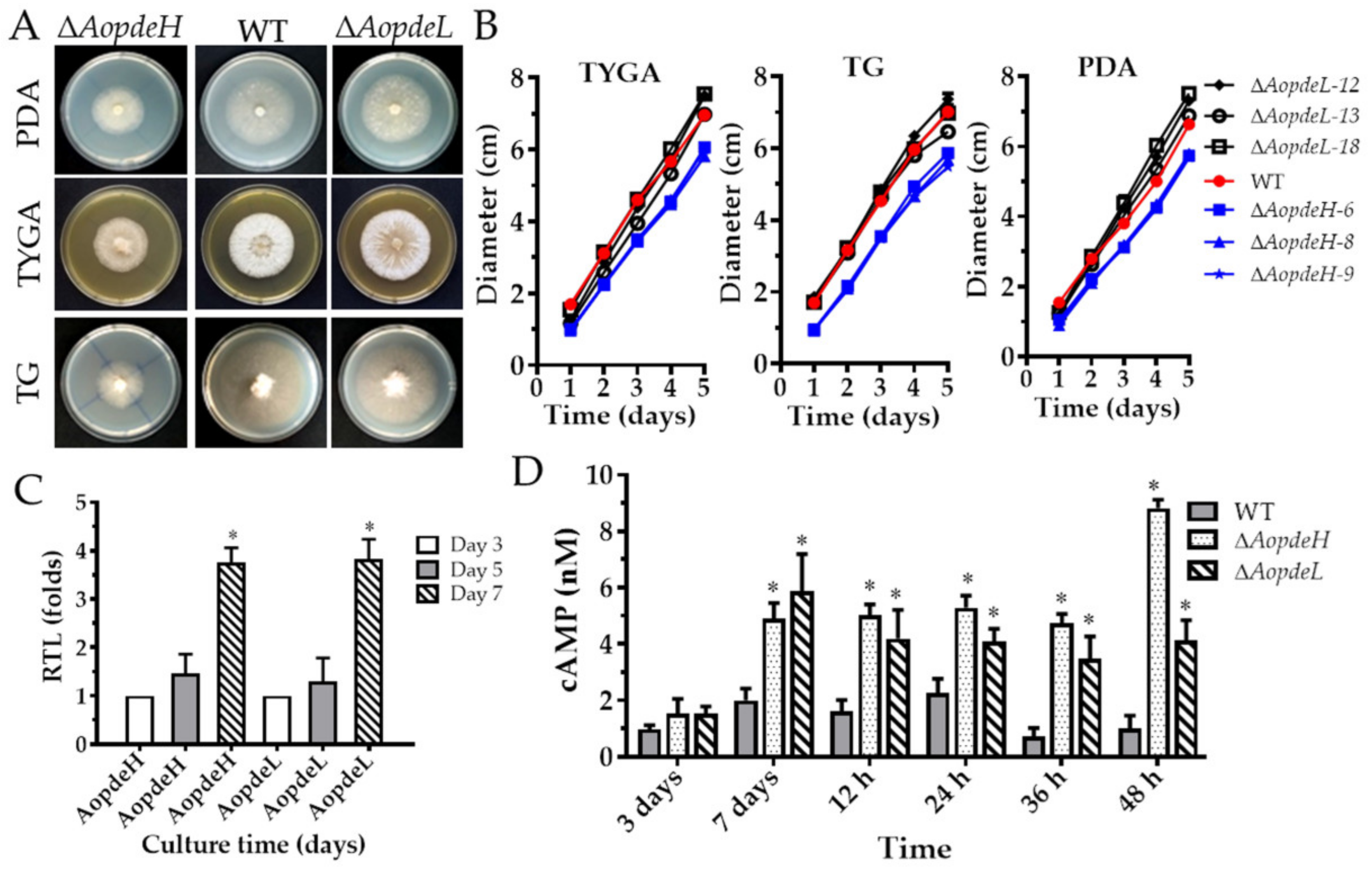
2.2. AoPdeH and AoPdeL Regulate Mycelial Growth and Morphology

Three independent knockout strains for AopdeHAopdeH) and AopdeLAopdeL) were respectively confirmed (Supplementary Figure S2) and used for phenotypic analysis. The growth rate and colony morphology of the WT and mutant strains were compared on different media. As a result, it was found that the ΔAopdeH mutants showed growth defects on the three media at 28 °C for five days post-induction (dpi), showing a significant defect in radial growth and reduction in aerial hyphae growth compared to the WT strain (Figure 1A,B). However, the mycelial growth of the ΔAopdeL mutant was similar to the WT strain. The number of hyphal septa had no significant difference between the WT and mutants, whereas partial hyphal cells of the ΔAopdeH and ΔAopdeL mutants became swollen (Supplementary Figure S3). Moreover, the transcriptional levels of AopdeH and AopdeL in the WT strain were determined, and their expression increased with the extension of culture times; the expression of AopdeH and AopdeL were increased by 3.8-fold at 7 dpi compared to that at 3 dpi (Figure 1C).

2.3. AoPdeH and AoPdeL Regulate Intracellular cAMP Levels

The cAMP content in the mycelium of the WT and mutant strains was measured at different developmental stages. The results showed that the cAMP was in a dynamic state in the WT strain. The cAMP content increased gradually at the vegetative growth and early (12 h post-induction, hpi) and middle stage (24 hpi) of trap formation. In contrast, the cAMP content decreased in the stage of nematode predation (36 and 48 hpi). Compared to the WT strain, the cAMP content significantly increased (p < 0.05) in the mutants at the mycelial mature stage (seven days) and at stages of trap formation and nematode predation (Figure 1D). For example, in the mycelial mature stage (seven days), the mycelial cAMP content of the ΔAopdeH and ΔAopdeL mutants were 2.5-fold and 2.9-fold higher than the WT strain, respectively; In the stage of nematode predation (36 hpi), the mycelial cAMP content of the ΔAopdeH and ΔAopdeL mutants was 6.8-fold and 5.0-fold higher than the WT strain, respectively. These results showed that the absence of AopdeH and AopdeL resulted in an accumulation of intracellular cAMP levels.

2.4. AoPdeH and AoPdeL Affect Autophagy and Cellular Ultrastructure

The autophagic process in the WT and mutants was observed by staining with monodansylcadaverine (MDC) dye; many autophagosomes could be seen in the WT, which distribute in hyphal cells in a punctate pattern. In contrast, the number of autophagosomes was decreased in the hyphal cells of the ΔAopdeH and ΔAopdeL mutants, whereas the volume of autophagosomes in mutants became remarkably enlarged (Figure 2A). In addition, the cellular ultrastructures of the WT and mutants were observed using transmission electron microscopy (TEM; Hitachi, Tokyo, Japan). The vacuole was observed in the hyphal cells of the WT strain, and woronin body could be seen near the septum; similar to the WT, vacuole and woronin body were observed in the hyphal cells of the ΔAopdeL mutant, as well autophagosome-like structure could also be observed. In contrast, less woronin body was observed in the hyphal cells of the ΔAopdeH mutant, and more autophagosome-like structures were observed in the ΔAopdeH mutant (Figure 2B).

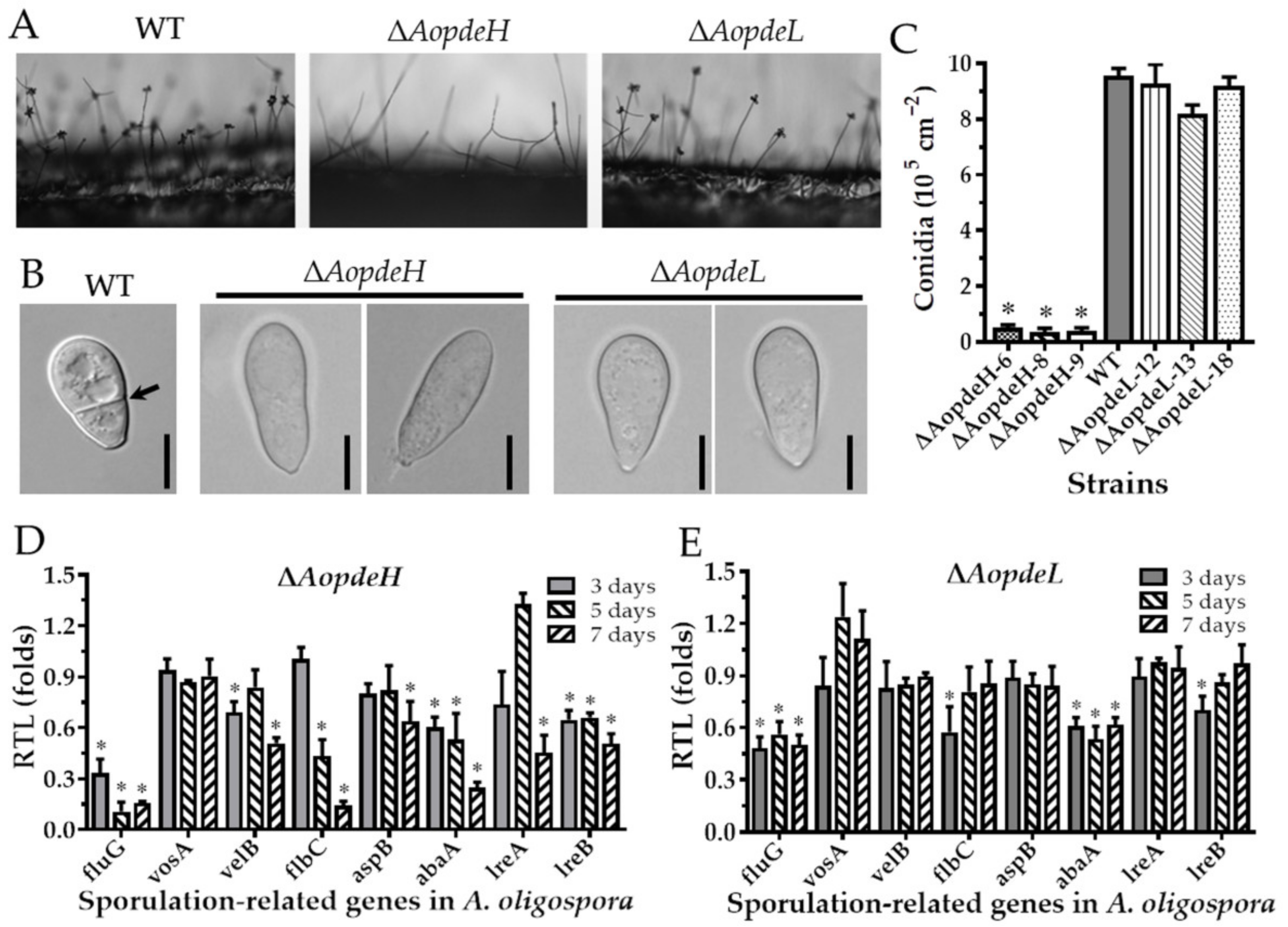
2.5. AoPdeH and AoPdeL Play Different Roles in Conidiation

To explore the regulatory role of PDEs in the asexual development of A. oligospora, we compared the asexual sporulation of the WT and mutants. Firstly, it was found by side-shooting that there was no significant difference between the conidia-forming method of the ΔAopdeL mutant strain and the WT (Figure 3A). Each conidiophore had a round of spores clustered by 4–5 spores. In contrast, the number of conidiophores of the ΔAopdeH mutant was significantly less than that of the WT, and there was almost no spore formation on the conidiophore (Figure 3A). In addition, the conidial morphology of the ΔAopdeH and ΔAopdeL mutants became slender, and their septa disappeared (Figure 3B). Further analysis showed that the conidia yield of the ΔAopdeH mutant was significantly reduced (only 4.2% of the WT), whereas the conidia yield of the ΔAoPdeL mutant was slightly less than the WT, and the difference was not significant (Figure 3C).
To further investigate the role of PDEs to conidiation, eight sporulation-related genes such as abaA, aspB, flbC, fluG, velB, vosA, lreA, and lreB were selected, and their transcriptional levels were determined during the early (three days), middle (five days), and later stages of sporulation. Compared with the WT, the transcription of abaA and fluG was significantly downregulated (p < 0.05) in the ΔAopdeH and ΔAopdeL mutants at different stages of sporulation. Additionally, the transcription of aspB, velB, vosA, lreA, and lreB was significantly downregulated (p < 0.05) in the ΔAopdeH mutant at the later stage of sporulation (Figure 3D), whereas the transcription of these genes had no change in the ΔAopdeL mutant at the same stage (Figure 3E).

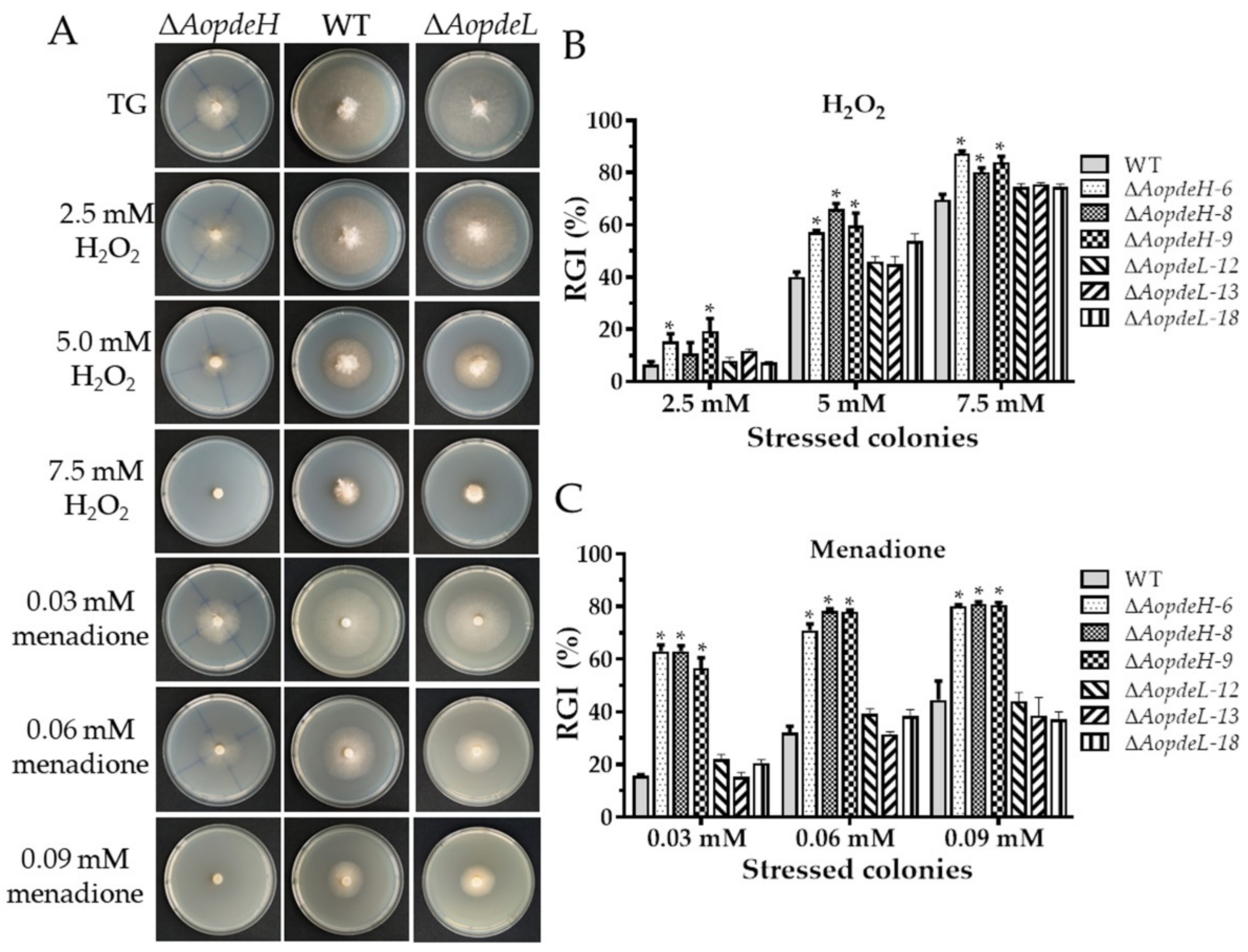
2.6. AoPdeH and AoPdeL Play Different Roles in Stress Response

The WT and mutant stress response was compared against cell wall stress agents, oxidative and osmotic stresses to investigate the effects of PDEs on environmental stresses. The radial mycelial growth of the ΔAopdeH mutants was considerably inhibited by these stressors (Figure 4, Figures S4 and S5). The relative growth inhibition (RGI) value was calculated considering the influence of the growth defects of the mutant strain. Compared with the WT, The AopdeH mutants had a higher RGI value at H2O2 (2.5, 5.0, and 7.5 mM), menadione (0.03, 0.06, and 0.09 mM), sorbitol (0.25, 0.5, and 0.75 M), NaCl (0.2 M), SDS (0.35 and 0.52 mM), and Congo red (0.05 mM). In contrast, the AopdeL-deficient strain showed a WT phenotype to oxidants (H2O2 and menadione) (Figure 4), and it had a higher RGI value at 0.1 M NaCl (Supplementary Figure S4). Nevertheless, the radial growth of the ΔAopdeL mutants was promoted at 0.08–0.12 mg/mL Congo red (Supplementary Figure S5).

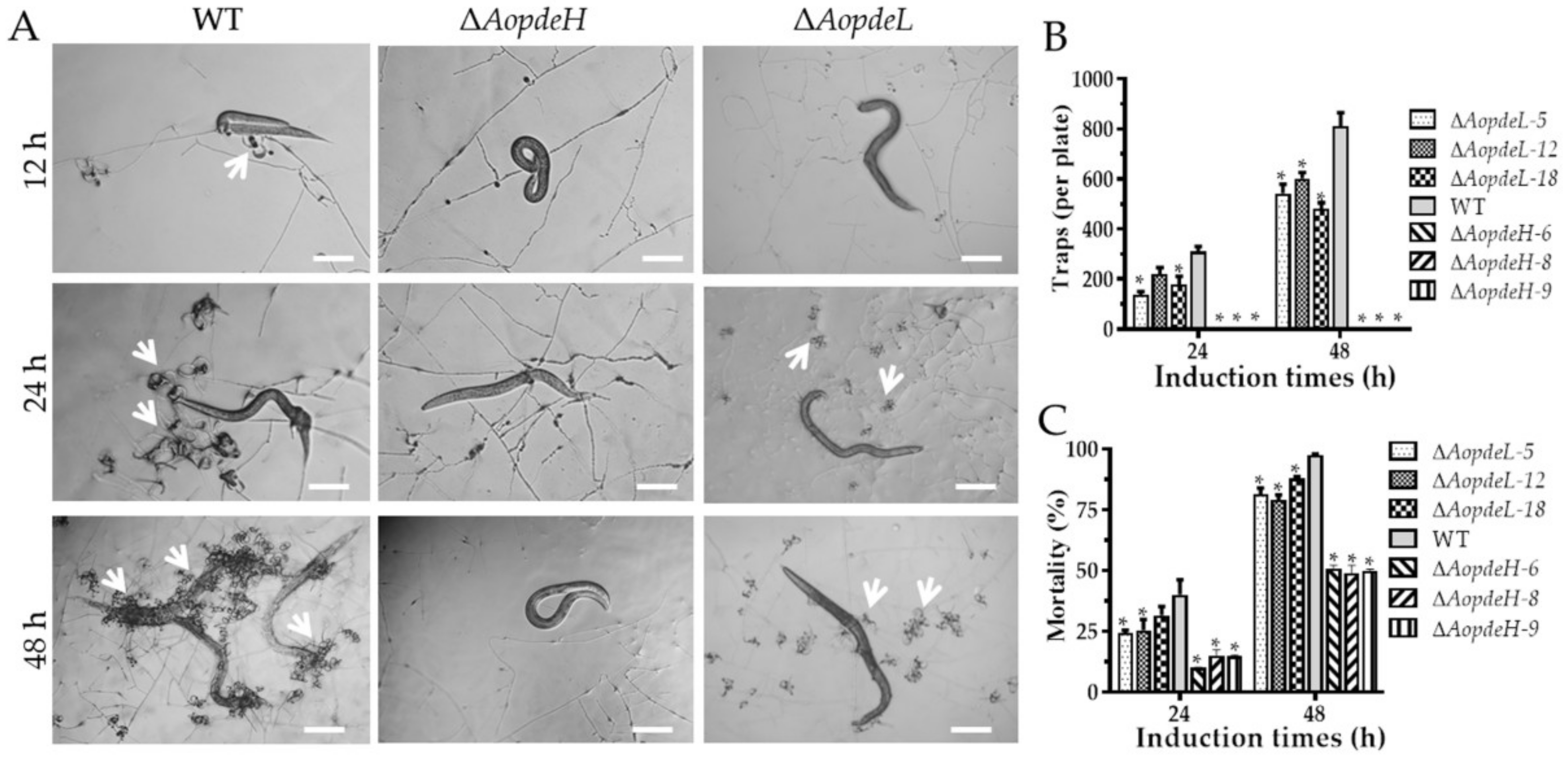
2.7. AoPdeH and AoPdeL Regulate Trap Formation and Nematicidal Activity

Trap formation was evaluated after the addition of nematodes. The WT strain produced immature traps containing one or two hyphal loops at 12 hpi, mature traps were observed, and nematodes were captured at 24 hpi. Then the majority of the nematodes were captured at 36 hpi and digested by the WT strain at 48 hpi (Figure 5A). However, the absence of AopdeH and AopdeL resulted in a remarkable reduction in trap formation. The ΔAopdeH mutant could not produce traps, and the number of traps produced by the ΔAopdeL mutant was considerably reduced compared with the WT (Figure 5A,B). For example, the WT strain produced approximately 309 and 811 traps per plate at 24 and 48 hpi, respectively, while the ΔAopdeL mutant produced 178 and 542 traps per plate at the same time points (Figure 5B). Correspondingly, the nematicidal efficiency of the ΔAopdeH mutant and ΔAopdeL mutant was remarkably decreased. At 24 hpi, the nematode mortality rates of WT, ΔAopdeL, and ΔAopdeH mutant strains were 40.1, 26.7, and 15.8%, respectively, while at 48 hpi, the nematode mortality rates of them were 97.6, 81.3, and 43%, respectively (Figure 5C).

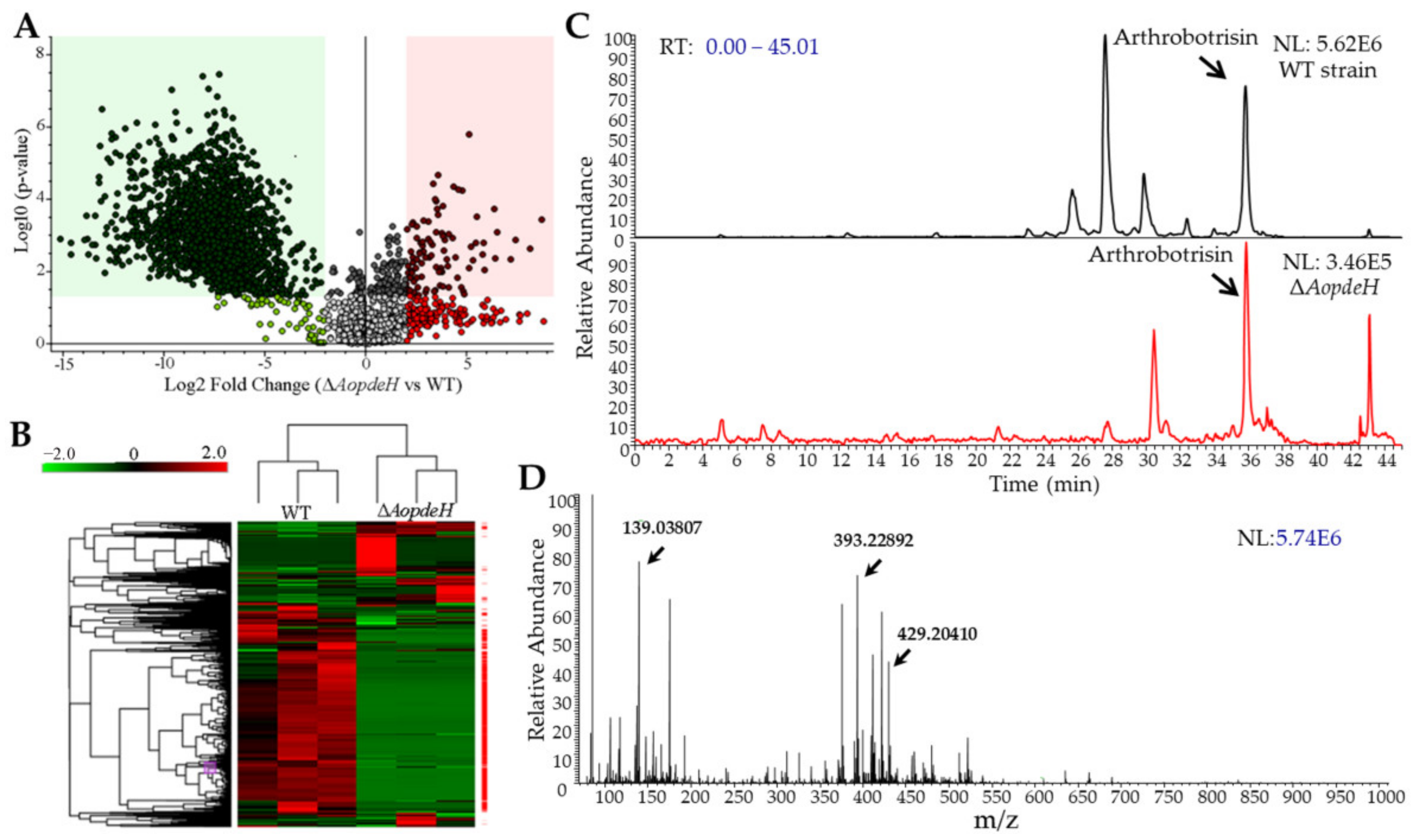
2.8. AoPdeH Regulates Secondary Metabolism

The hyphae and supernatants of the WT and ΔAopdeH mutant strains were quantified, and the hyphal wet biomass of the WT (2.43 g) was 2.02-fold higher than that of the ΔAopdeH mutant (1.20 g). Based on the biomass levels of hyphae, 200 and 405 mL of supernatants were collected from the WT and ΔAopdeH mutant strains, respectively. The supernatants were extracted with ethyl acetate and vacuum-evaporated to obtain a crude extract. Then, the metabolites in the crude extracts were analyzed using liquid chromatography-mass spectrometry (LC-MS). According to the chromatogram, the WT strain produces plentiful metabolites, contrasting to the peaks of many compounds not detected or markedly decreased in the ΔAopdeH mutants (Supplementary Figure S6). Volcano plot analysis showed substantially more downregulated compounds in the ΔAopdeH mutant than in the WT strain; there were 99 upregulated and 1855 downregulated compounds in the ΔAopdeH mutant compared with the WT (Figure 6A). Correspondingly, more downregulated metabolic pathways were found in the ΔAopdeH mutant (Figure 6B). The differential metabolic pathways were mainly enriched in degradation of the aromatic compounds toluene, naphthalene, mandelate, and L-tyrosine, biosynthesis of aromatic amino acid, 4-hydroxybenzoate, rosmarinic acid, novobiocin, and chorismate metabolism (Supplementary Table S2). In addition, arthrobotrisins, a kind of specific sesquiterpenyl epoxy-cyclohexenoid (SEC) metabolites found in A. oligospora and other NT fungi [29,30], were detected in the ΔAopdeH mutant and WT strain (diagnostic fragments ion at m/z 139, 393, and 429) (Figure 6C,D), and the relative peak area of arthrobotrisins in ΔAopdeH mutant (MA: 5319713) is remarkably less than that in the WT strain (MA: 76693347) (Supplementary Figure S7).

3. Discussion

Previous studies have shown that PDEs can regulate intracellular cAMP levels in yeasts and filamentous fungi, thus playing a crucial regulatory role in nutrient sensing, asexual reproduction, and pathogenic development in yeasts and filamentous fungi [5,7,13,17,20]. In this study, we investigated the significance of PDE-mediated regulation of intracellular cAMP in the asexual and pathogenic development of A. oligospora. Our results showed that two PDEs AoPdeH and AoPdeL play a different role in the asexual growth, development, and pathogenicity of A. oligospora.
In S. cerevisiae and C. albicans, deletion of pde2 resulted in increased accumulation of intracellular cAMP levels [7,31]. Similar results were observed in several filamentous fungi. In M. oryzae, the ΔpdeL mutant accumulated 1.5-fold higher levels of cAMP than the WT, while the ΔpdeH mutant and the ΔpdeLpdeH double mutant accumulated ~3-fold and ~4.5-fold higher levels of cAMP than the WT, respectively [11]. In B. cinerea, intracellular cAMP levels of the bcpde1 deletion strains were similar to those of the WT, while Δbcpde2 and ΔΔccpde1/2 mutants showed slightly decreased cAMP levels [13]. Similarly, the A. flavus ΔpdeH strain accumulated 1.8-fold higher cAMP levels than the WT and 1.6-fold higher than the ΔpdeL [5]. In this study, the absence of AopdeH and AopdeL caused the increased accumulation of intracellular cAMP levels during the hyphal stage, stages of trap formation, and nematode predation. Recently, resistance to inhibitors of cholinesterase 8 (Ric8), a negative regulator of G protein signaling, its deletion resulted in a significant reduction in cAMP level in A. oligospora [26]. In contrast, the deletion of genes encoding for regulators of G protein signaling (RGSs) caused increased intracellular cAMP levels in A. oligospora [25]. These findings suggested that PDEs govern G protein signaling by regulating cAMP levels in fungi.
As expected, the deletion of AopdeH caused significant growth defects. The radial growth of ΔAopdeH mutants became slow, and aerial hyphae were abnormally thin compared with the WT; however, there was no significant difference in the growth rate and colony morphology of the ΔAopdeL mutants compared to the WT strain. Similar growth defects caused by disruption of PDEs were found in several filamentous fungi such as M. oryzae [20], B. cinerea [13], and A. flavus [5]. In M. oryzae, the ΔpdeH colony was flat due to reduced aerial hyphal growth and displayed enhanced pigmentation and marginally slower radial growth, whereas the ΔpdeL was similar to the WT strain and showed no apparent defects in aerial or radial growth and colony morphology [20]. These findings showed that PdeH plays a crucial role in aerial or radial growth and colony morphology, but PdeL only plays a minor role in mycelial growth.
Compared with the WT strain, the conidiophores and conidia yield of the ΔAopdeH mutants were significantly reduced, and the conidial morphology was also deformed, becoming narrow and without a septum. In contrast, the conidiophores and conidia yield of ΔAopdeL mutants were not significantly different from that of WT. However, the ΔAopdeL spore morphology is similar to that of the ΔAopdeH mutants. Similarly, in N. crassa, the pde2 deletion mutant does not produce any conidia [32]. In B. cinerea, deletion of bcpde2 causes reduced spores (20%) compared to the WT strain, and the conidia morphology becomes narrow [13]. In contrast, in M. oryzae, the conidia yield of the ΔpdeH mutant is increased 2–3 folds. The number of conidiophores increased 2-fold, whereas the ΔpdeL mutant had no difference in conidia production or conidiophores from WT [20], but both ΔpdeL and ΔpdeH mutants produced elongated and thinner conidia [11]. In addition, the transcription of several sporulation-related genes such as abaA, fluG, and lreB was significantly downregulated (p < 0.05) in the ΔAopdeH mutant, which is consistent with the suppression of conidia yield. The AbaA and FluG are the key regulators of conidiation in several filamentous fungi, such as Aspergillus nidulans and A. fumigatus [33], and Beauveria bassiana [34,35]. Therefore, PdeH is indispensable for conidiation in A. oligospora and other fungi, and PdeL plays a specific role in conidial morphology.
Our previous studies revealed that G protein signaling regulates multi-stress responses in A. oligospora [26,36]. In this study, the absence of the AopdeH caused significant defects in tolerance to osmotic salts, oxidants, and cell wall stress agents; loss of AopdeL only altered the sensitivity to NaCl and Congo red. The results are similar to S. cerevisiae, deletion of the pde2 causes the cells to be sensitive to freezing and thawing, and the oxidative stress response of the pde2 deletion mutant can be induced by H2O2 or paraquat, but the absence of pde1 does not affect cell stress resistance [7]. Similarly, the Candida albicans pde2 deletion strains exhibited substantial growth defects in tolerance to ionic stress (NaCl and KCl) and high temperature, but not to sorbitol. However, pde1 deletion did not affect the tolerance to these stresses [31]. In contrast, in M. oryzae, there are no phenotypic changes between the ΔpdeH mutant and the WT strain in response to stress agents, such as ionic stress, oxidative stress, osmotic stress, or cell wall-disturbing agents [11]. These findings showed that PDEs involve regulating stress signaling, and the function of PDEs is varied in different fungi, of which, PdeH likely plays a predominant role in regulating adverse stresses.
Previous studies have been revealed that PDEs are required for full virulence of C. albicans [31] and several pathogenic fungi [11,20]. In C. albicans, the adhesion ability of the Δpde2 mutant was approximately 10-fold lower than that of the WT, and deletion of pde2 caused a drastic reduction in virulence and double deletion of pde1 and pde2 completely eliminated virulence [31]. In M. oryzae, loss of pdeH significantly accelerated appressorium formation on non-inductive surfaces. However, ΔpdeH conidia failed to infect the host efficiently and cause typical blast lesions owing to its compromised ability to form proper infection hyphae and further advance its growth and spread in the host tissue [11,20]. Similarly, deletion of bcpde2 resulted in severely affected full virulence of B. cinerea, whereas the bcpde1 deletion strain displayed a WT-like phenotype [13]. In this study, deletion of AopdeH abolished the trap formation and caused a remarkable reduction in predation efficiency, and deletion of AopdeL also hindered the trap formation and predation efficiency. These findings suggested that PDEs, especially PdeH, play a more prominent role than PdeL in infectious structure development and pathogenicity.
Like other filamentous fungi, NT fungi can produce numerous metabolites during vegetative growth and predator-prey interaction [30,37,38]. Recently, G protein signaling has been revealed to regulate the secondary metabolism in A. oligospora, such as the regulator of G-protein [26] and small GTPases [39]. In this study, the absence of AopdeH caused a remarkable alteration in secondary metabolism; 99 compounds were upregulated and 1855 downregulated in the ΔAopdeH mutant compared to the WT. In particular, the specific SEC metabolites arthrobotrisins were identified, the relative content of which decreased by 93.1% in the ΔAopdeH mutant. SEC metabolites can regulate the conidiation and morphological switch of NT fungi [30,38,40]. Similarly, the content of arthrobotrisins was downregulated, respectively, by 9.57- and 9.95-fold in the ΔAoras2 and ΔAorheb mutants as compared with that in the WT strain [39]. Similarly, in A. flavus, deletion of both pdeH and pdeL caused a significant increase in aflatoxin production compared to the WT strain [5]. Therefore, PDEs play a vital role in secondary metabolism in NT fungi and other fungi.
PDEs are also involved in autophagy; loss of AopdeH and AopdeL caused a reduction in the number of autophagosomes and increased the volume of autophagosomes. Recently, several autophagy-related genes such as atg1, atg4, atg5, and atg8 have been revealed to regulate conidiation and trap the formation of A. oligospora [41,42]. However, the regulatory mechanism of PDEs to the autophagic process needs to be studied further. In summary, our works revealed the important role of PDEs in the mycelial growth, autophagy, conidiation, stress response, trap formation, and secondary metabolism in A. oligospora, and AoPdeH plays a more prominent role than AoPdeL in regulating the growth, development, and pathogenicity through modulation of intracellular cAMP levels. Our results expanded the understanding of PDEs in NT fungi, which provides a basis for exploring the regulation mechanism underlying trap development and environmental adaptation of NT fungi.

4. Materials and Methods

4.1. Fungal Strains, Culture Conditions, and Media

The WT strain of A. oligospora (ATCC24927) and mutants generated in this study were routinely maintained as previously described [43]. Uracil-deficient S. cerevisiae FY834 was used as the host to construct the knockout vector, which was cultured in YPD (10 g/L yeast extract, 20 g/L peptone, and 20 g/L dextrose) [44]. Escherichia coli strain DH5α (Takara, Shiga, Japan) was used to store the plasmid pCSN44 containing the hygromycin resistance gene (hph) fragment and the plasmid pRS426 used to construct the knockout vector [45].
Potato dextrose agar (PDA), tryptone-glucose (TG), and tryptone yeast extract glucose agar (TYGA) media were prepared as previously described [43] and used to analyze mycelial growth. A liquid TG medium was used to prepare the mycelium for protoplast production or DNA extraction. TB3 medium (3 g/L hydrolyzed casein, 200 g/L sucrose, 3 g/L tryptone, and 0.75% agar powder) supplemented with 200 μg/mL hygromycin for protoplast regeneration and screening of positive transformants. Conidia were induced in the CMY medium (20 g/L maizena, 20 g/L agar, and 5 g/L yeast extract). Oatmeal water medium was used for culturing the nematode Caenorhabditis elegans, which was used for bioassay.

4.2. Sequence and Phylogenetic Analysis of PDEs

The orthologous AoPdeH (AOL_s00083g160) and AoPdeL (AOL_s00097g378) were identified in A. oligospora by comparing the protein sequences of homologous PDEs in the model fungi S. cerevisiae, A. nidulans, and N. crassa. The conserved domains of PDEs were predicted by InterProScan 4.8 (http://www.ebi.ac.uk/Tools/pfa/iprscan/, accessed on 19 October 2019), and the theoretical isoelectric point (pI) and molecular weight of PDEs were calculated using the pI/MW tool (http://web.expasy.org/compute_pi/, accessed on 19 October 2019). Orthologous PDEs from different fungi were downloaded from the GenBank, and a phylogenetic tree was constructed using the Mega software package (version 7.0) [46].

4.3. Vector Construction and Targeted Gene Deletion

AopdeH and AopdeL were disrupted by homologous recombination as described previously [44,47]. The hygromycin fragment hph was used as a selection marker conferring hygromycin B resistance on transformants. The homologous regions of the target gene and hph cassette were amplified by PCR using the primer pairs (Supplementary Table S3). The pRS426 plasmid was digested with EcoRI and XhoI, and the amplified fragments were co-transformed into S. cerevisiae by electroporation. The correct plasmid was screened on the SC-Ura plates and transformed into E. coli DH5α for expansion culture. The gene fragment for AopdeH and AopdeL disruption was amplified with primer pairs 160-5F/160-3R and 378-5F/378-3R (Supplementary Table S3) using the corresponding knockout vector plasmid as a template. Finally, the knockout construct was transformed into A. oligospora protoplasts by chemical transformation as previously described [47,48]. The hygromycin-resistant transformants were screened in TB3 plates and further confirmed by PCR amplification using primer pairs (Supplementary Table S3), respectively, and the transformants were further confirmed by Southern blot analysis as previously described [49].

4.4. Analysis of Mycelial Growth and Colonial Morphology

After synchronizing the WT and mutant strains, their growth rate and colonial morphology were compared on PDA, TYGA, and TG plates at 28 °C. The colonial diameters of the WT and mutant strains were determined every 24 h and photographed on the fifth day to record their colony morphology. The fungal cell wall and hyphal septum were dyed by Calcofluor white (Sigma-Aldrich, St. Louis, MO, USA) staining, as previously described [28], to observe hyphal morphology.

4.5. Analysis of Conidiation and Autophagy

The same size of the mycelial blocks was individually inoculated on the CMY medium and incubated at 28 °C for 14 days to evaluate the sporulation ability of the fungal strains. The conidiophore was observed using the side-shot approach described previously [50]. Conidia were harvested in 40 mL sterile water, followed by filtration through six layers of lens tissues to remove mycelial debris, then the conidial production was counted with a hemocytometer as previously described [25].
The WT and mutants were incubated on PDA plates with sterile coverslips at 28 °C for five days to detect autophagy. The mycelia were treated with 30–50 µL of 100 mg/mL MDC staining solution at 37 °C in the dark for 30 min. Then the images were observed by fluorescence microscopy (Leica, Mannheim, Germany).

4.6. Quantification of Intracellular cAMP

Quantification of intracellular levels of cAMP was carried out as previously described [51]. The WT and mutant strains were incubated on TYGA plates at 28 °C, and mycelial samples were harvested at 3 and 7 dpi, respectively. In addition, the strains were incubated on TYGA plates for seven days, then about 300 nematodes were added to the plates, and mycelia were harvested at 12, 24, 36, and 48 hpi. The mycelia samples were treated with 1 M HCl for 30 min, then frozen in liquid nitrogen. Intracellular cAMP was extracted, and cAMP levels were quantified using the Direct cAMP ELISA kit (Enzo Life Sciences, Farmingdale, NY, USA).

4.7. Analysis of Stress Tolerance

Several chemical stressors, such as cell wall stress agents (SDS and Congo red), oxidants (menadione and H2O2), and osmotic agents (NaCl and sorbitol), were used to evaluate the stress response of the WT and mutants as previously described [25,27]. The strains were cultured at 28 °C for five days, the diameter of the colony was measured and photographed to record the colony morphology. The RGI value of the mutant versus WT strain was calculated as previously described [25,27].

4.8. Trap Formation and Bioassay

Approximately 3 × 10⁴ conidia of WT, ΔAopdeH, and ΔAopdeL mutant strains were inoculated in water agar plates, after culturing at 28 °C for five days, and about 300 nematodes were added to the plates to analyze the trap formation and nematode predation efficiency, respectively. The number of traps and captured nematodes were calculated at 24 and 48 hpi, respectively.

4.9. Reverse Transcription-Quantitative PCR (RT-qPCR)

The fungal strains were cultured in TYGA plates at 28 °C, and mycelial samples were collected at 3, 5, and 7 dpi. Total RNA of mycelia was isolated and used to reverse-transcribe the cDNA. The cDNA from each strain was used as a template for RT-qPCR, and the A. oligospora β-tubulin gene was used as an internal standard (Supplementary Table S4). The transcriptional levels of AopdeH, AopdeL, and sporulation-related genes at different stages were determined by RT-qPCR. The relative transcript levels of each gene were calculated using the 2−ΔΔCt method [52].

4.10. Comparison of Metabolites between WT and ΔAopdeH Mutant Strains

The WT and ΔAopdeH mutant strains were cultured for seven days in PD broth at 28 °C and 180 rpm. The hyphae of fermentation broth of the WT and ΔAopdeH mutant strains were harvested and qualified. The fermentation broth was treated, and the metabolites were analyzed with LC-MS as previously described [39]. Three replicates were maintained for the analysis. Untargeted metabolomics analysis was performed using the Compound Discoverer 3.0 software (Thermo Fisher Scientific, Waltham, MA, USA).

4.11. Statistical Analyses

Data were expressed as standard deviations (SDs) of the means from three biological replicates, and subjected to one-way analysis of variance, followed by Tukey’s honestly significant difference (HSD) test. p < 0.05 indicates a significant difference.

Supplementary Materials

The following supporting information can be downloaded at: https://www.mdpi.com/article/10.3390/pathogens11040405/s1, Figure S1: Phylogenetic tree analysis based on the amino acid sequences of PdeH and PdeL homologous proteins from different fungi; Figure S2: Deletion of AopdeH and AopdeL in A. oligospora; Figure S3: Comparison of the hyphal septum between WT and mutants; Figure S4: Comparison of osmotic stress responses between WT and mutants; Figure S5: Comparison of cell wall stress agent responses between WT and mutants; Figure S6: Comparison of high-performance liquid chromatography profiles of the WT and ΔAopdeH mutant strains; Figure S7: Comparison of the relative peak area of arthrobotrisins between WT and ΔAopdeH mutant strains; Table S1: The sequence similarity between AoPdeH/AoPdeL and orthologs from different fungi; Table S2: Comparison of the differential compounds-metabolic pathways between the WT and ΔAopdeH mutant strains; Table S3: List of primers used for gene disruption in this study; Table S4: List of RT-PCR primers used in this study.

Author Contributions

N.M.: Investigation, Formal analysis, Writing—original draft. K.-X.J.: Formal analysis, Writing—original draft. N.B. and D.-N.L.: Data analysis and discussion. K.-Q.Z.: Conceptualization, Funding acquisition. J.-K.Y.: Conceptualization, Funding acquisition, Writing—review & editing. All authors have read and agreed to the published version of the manuscript.

Funding

This research was funded by the National Natural Science Foundation of China (nos. 31960556 and U1402265), and the Applied Basic Research Foundation of Yunnan Province (202001BB050004).

Institutional Review Board Statement

Not applicable.

Informed Consent Statement

Not applicable.

Data Availability Statement

Not applicable.

Acknowledgments

We would be grateful to Microbial Library of the Germplasm Bank of Wild Species from Southwest China for preserving and providing experimental strains, and Guo Yingqi (Kunming Institute of Zoology, Chinese Academy of Sciences) for her help of taking and analyzing TEM images.

Conflicts of Interest

The authors declare no conflict of interest.

References

  1. Wang, Y.; Geng, Z.; Jiang, D.; Long, F.; Zhao, Y.; Su, H.; Zhang, K.Q.; Yang, J. Characterizations and functions of regulator of G protein signaling (RGS) in fungi. Appl. Microbiol. Biotechnol. 2013, 97, 7977–7987. [Google Scholar] [CrossRef] [PubMed]
  2. Park, H.S.; Kim, M.J.; Yu, J.H.; Shin, K.S. Heterotrimeric G-protein signalers and RGSs in Aspergillus fumigatus. Pathogens 2020, 9, 902. [Google Scholar] [CrossRef] [PubMed]
  3. Daniel, P.B.; Walker, W.H.; Habener, J.F. Cyclic AMP signaling and gene regulation. Annu. Rev. Nutr. 1998, 18, 353–383. [Google Scholar] [CrossRef] [PubMed]
  4. D’Souza, C.A.; Heitman, J. Conserved cAMP signaling cascades regulate fungal development and virulence. FEMS Microbiol. Rev. 2001, 25, 349–364. [Google Scholar] [CrossRef] [PubMed]
  5. Yang, K.; Liu, Y.; Liang, L.; Li, Z.; Qin, Q.; Nie, X.; Wang, S. The high-affinity phosphodiesterase PdeH regulates development and aflatoxin biosynthesis in Aspergillus flavus. Fungal Genet. Biol. 2017, 101, 7–19. [Google Scholar] [CrossRef] [PubMed]
  6. Pan, X.; Heitman, J. Cyclic AMP-dependent protein kinase regulates pseudohyphal differentiation in Saccharomyces cerevisiae. Mol. Cell. Biol. 1999, 19, 4874–4887. [Google Scholar] [CrossRef] [PubMed] [Green Version]
  7. Park, J.I.; Grant, C.M.; Dawes, I.W. The high-affinity cAMP phosphodiesterase of Saccharomyces cerevisiae is the major determinant of cAMP levels in stationary phase: Involvement of different branches of the Ras-cyclic AMP pathway in stress responses. Biochem. Biophys. Res. Commun. 2005, 327, 311–319. [Google Scholar] [CrossRef]
  8. Banno, S.; Ochiai, N.; Noguchi, R.; Kimura, M.; Yamaguchi, I.; Kanzaki, S.; Murayama, T.; Fujimura, M. A catalytic subunit of cyclic AMP-dependent protein kinase, PKAC-1, regulates asexual differentiation in Neurospora crassa. Genes Genet. Syst. 2005, 80, 25–34. [Google Scholar] [CrossRef] [Green Version]
  9. Lafon, A.; Han, K.H.; Seo, J.A.; Yu, J.H.; d’Enfert, C. G-protein and cAMP-mediated signaling in aspergilli: A genomic perspective. Fungal Genet. Biol. 2006, 43, 490–502. [Google Scholar] [CrossRef]
  10. Dürrenberger, F.; Wong, K.; Kronstad, J.W. Identification of a cAMP-dependent protein kinase catalytic subunit required for virulence and morphogenesis in Ustilago maydis. Proc. Natl. Acad. Sci. USA 1998, 95, 5684–5689. [Google Scholar] [CrossRef] [Green Version]
  11. Zhang, H.; Liu, K.; Zhang, X.; Tang, W.; Wang, J.; Guo, M.; Zhao, Q.; Zheng, X.; Wang, P.; Zhang, Z. Two phosphodiesterase genes, PDEL and PDEH, regulate development and pathogenicity by modulating intracellular cyclic AMP levels in Magnaporthe oryzae. PLoS ONE 2011, 6, e17241. [Google Scholar] [CrossRef] [Green Version]
  12. Lengeler, K.B.; Davidson, R.C.; D’souza, C.; Harashima, T.; Shen, W.C.; Wang, P.; Pan, X.; Waugh, M.; Heitman, J. Signal transduction cascades regulating fungal development and virulence. Microbiol. Mol. Biol. Rev. 2000, 64, 746–785. [Google Scholar] [CrossRef] [Green Version]
  13. Harren, K.; Brandhoff, B.; Knödler, M.; Tudzynski, B. The high-affinity phosphodiesterase BcPde2 has impact on growth, differentiation and virulence of the phytopathogenic ascomycete Botrytis cinerea. PLoS ONE 2013, 8, e78525. [Google Scholar] [CrossRef] [Green Version]
  14. Houslay, M.D.; Baillie, G.S.; Maurice, D.H. cAMP-Specific phosphodiesterase-4 enzymes in the cardiovascular system: A molecular toolbox for generating compartmentalized cAMP signaling. Circ. Res. 2007, 100, 950–966. [Google Scholar] [CrossRef]
  15. Uno, I.; Matsumoto, K.; Ishikawa, T. Characterization of a cyclic nucleotide phosphodiesterase-deficient mutant in yeast. J. Biol. Chem. 1983, 258, 3539–3542. [Google Scholar] [CrossRef]
  16. Londesborough, J.; Suoranta, K. The zinc-containing high Km cyclic nucleotide phosphodiesterase of bakers’ yeast. J. Biol. Chem. 1983, 258, 2966–2972. [Google Scholar] [CrossRef]
  17. Ma, P.; Wera, S.; Van Dijck, P.; Thevelein, J.M. The PDE1-encoded low-affinity phosphodiesterase in the yeast Saccharomyces cerevisiae has a specific function in controlling agonist-induced cAMP signaling. Mol. Biol. Cell 1999, 10, 91–104. [Google Scholar] [CrossRef] [Green Version]
  18. Wera, S.; Ma, P.; Thevelein, J.M. Glucose exerts opposite effects on mRNA versus protein and activity levels of Pde1, the low-affinity cAMP phosphodiesterase from budding yeast, Saccharomyces cerevisiae. FEBS Lett. 1997, 420, 147–150. [Google Scholar] [CrossRef] [Green Version]
  19. Wilson, R.B.; Renault, G.; Jacquet, M.; Tatchell, K. The pde2 gene of Saccharomyces cerevisiae is allelic to rca1 and encodes a phosphodiesterase which protects the cell from extracellular cAMP. FEBS Lett. 1993, 325, 191–195. [Google Scholar] [CrossRef] [Green Version]
  20. Ramanujam, R.; Naqvi, N.I. PdeH, a high-affinity cAMP phosphodiesterase, is a key regulator of asexual and pathogenic differentiation in Magnaporthe oryzae. PLoS Pathog. 2010, 6, e1000897. [Google Scholar] [CrossRef] [Green Version]
  21. Ji, X.; Li, H.; Zhang, W.; Wang, J.; Liang, L.; Zou, C.; Yu, Z.; Liu, S.; Zhang, K.Q. The lifestyle transition of Arthrobotrys oligospora is mediated by microRNA-like RNAs. Sci. China Life Sci. 2020, 63, 543–551. [Google Scholar] [CrossRef] [Green Version]
  22. Nordbring-Hertz, B.; Jansson, H.B.; Tunlid, A. Nematophagous fungi. In Els; John Wiley & Sons, Ltd.: Chichester, UK, 2011. [Google Scholar]
  23. Su, H.; Zhao, Y.; Zhou, J.; Feng, H.; Jiang, D.; Zhang, K.Q.; Yang, J. Trapping devices of nematode-trapping fungi: Formation, evolution, and genomic perspectives. Biol. Rev. 2017, 92, 357–368. [Google Scholar] [CrossRef]
  24. Yang, J.; Wang, L.; Ji, X.; Feng, Y.; Li, X.; Zou, C.; Xu, J.; Ren, Y.; Mi, Q.; Wu, J.; et al. Genomic and proteomic analyses of the fungus Arthrobotrys oligospora provide insights into nematode-trap formation. PLoS Pathog. 2011, 7, e1002179. [Google Scholar] [CrossRef] [Green Version]
  25. Ma, N.; Zhao, Y.; Wang, Y.; Yang, L.; Li, D.; Yang, J.; Jiang, K.; Zhang, K.Q.; Yang, J. Functional analysis of seven regulators of G protein signaling (RGSs) in the nematode-trapping fungus Arthrobotrys oligospora. Virulence 2021, 12, 1825–1840. [Google Scholar] [CrossRef]
  26. Bai, N.; Zhang, G.; Wang, W.; Feng, H.; Yang, X.; Zheng, Y.; Yang, L.; Xie, M.; Zhang, K.Q.; Yang, J. Ric8 acts as a regulator of G-protein signalling required for nematode-trapping lifecycle of Arthrobotrys oligospora. Environ. Microbiol. 2021, in press. [Google Scholar] [CrossRef]
  27. Zhen, Z.; Xing, X.; Xie, M.; Yang, L.; Yang, X.; Zheng, Y.; Chen, Y.; Ma, N.; Li, Q.; Zhang, K.Q.; et al. MAP kinase Slt2 orthologs play similar roles in conidiation, trap formation, and pathogenicity in two nematode-trapping fungi. Fungal Genet. Biol. 2018, 116, 42–50. [Google Scholar] [CrossRef]
  28. Xie, M.; Yang, J.; Jiang, K.; Bai, N.; Zhu, M.; Zhu, Y.; Zhang, K.Q.; Yang, J. AoBck1 and AoMkk1 are necessary to maintain cell wall integrity, vegetative growth, conidiation, stress resistance, and pathogenicity in the nematode-trapping fungus Arthrobotrys oligospora. Front. Microbiol. 2021, 12, 649582. [Google Scholar] [CrossRef]
  29. Wei, L.X.; Zhang, H.X.; Tan, J.L.; Chu, Y.S.; Li, N.; Xue, H.X.; Wang, Y.L.; Niu, X.M.; Zhang, Y.; Zhang, K.Q. Arthrobotrisins A-C, oligosporons from the nematode-trapping fungus Arthrobotrys oligospora. J. Nat. Prod. 2011, 74, 1526–1530. [Google Scholar] [CrossRef]
  30. He, Z.Q.; Tan, J.L.; Li, N.; Zhang, H.X.; Chen, Y.H.; Wang, L.J.; Zhang, K.Q.; Niu, X.M. Sesquiterpenyl epoxy-cyclohexenoids and their signaling functions in nematode-trapping fungus Arthrobotrys oligospora. J. Agric. Food Chem. 2019, 67, 13061–13072. [Google Scholar] [CrossRef]
  31. Wilson, D.; Tutulan-Cunita, A.; Jung, W.; Hauser, N.C.; Hernandez, R.; Williamson, T.; Piekarska, K.; Rupp, S.; Young, T.; Stateva, L. Deletion of the high-affinity cAMP phosphodiesterase encoded by PDE2 affects stress responses and virulence in Candida Albicans. Mol. Microbiol. 2007, 65, 841–856. [Google Scholar] [CrossRef]
  32. Greenwald, C.J.; Kasuga, T.; Glass, N.L.; Shaw, B.D.; Ebbole, D.J.; Wilkinson, H.H. Temporal and spatial regulation of gene expression during asexual development of Neurospora crassa. Genetics 2010, 186, 1217–1230. [Google Scholar] [CrossRef] [PubMed] [Green Version]
  33. Park, H.S.; Yu, J.H. Genetic control of asexual sporulation in filamentous fungi. Curr. Opin. Microbiol. 2012, 15, 669–677. [Google Scholar] [CrossRef] [PubMed]
  34. Guo, C.T.; Peng, H.; Tong, S.M.; Ying, S.H.; Feng, M.G. Distinctive role of fluG in the adaptation of Beauveria bassiana to insect-pathogenic lifecycle and environmental stresses. Environ. Microbiol. 2021, 23, 5184–5199. [Google Scholar] [CrossRef] [PubMed]
  35. Zhang, A.X.; Mouhoumed, A.Z.; Tong, S.M.; Ying, S.H.; Feng, M.G. BrlA and AbaA govern virulence-required dimorphic switch, conidiation, and pathogenicity in a fungal insect pathogen. mSystems 2019, 4, e00140-19. [Google Scholar] [CrossRef] [Green Version]
  36. Xie, M.; Wang, Y.; Tang, L.; Yang, L.; Zhou, D.; Li, Q.; Niu, X.; Zhang, K.Q.; Yang, J. AoStuA, an APSES transcription factor, regulates the conidiation, trap formation, stress resistance and pathogenicity of the nematode-trapping fungus Arthrobotrys oligospora. Environ. Microbiol. 2019, 21, 4648–4661. [Google Scholar] [CrossRef]
  37. Kuo, T.H.; Yang, C.T.; Chang, H.Y.; Hsueh, Y.P.; Hsu, C.C. Nematode-trapping fungi produce diverse metabolites during predator-prey interaction. Metabolites 2020, 10, 117. [Google Scholar] [CrossRef] [Green Version]
  38. Zhang, H.X.; Tan, J.L.; Wei, L.X.; Wang, Y.L.; Zhang, C.P.; Wu, D.K.; Zhu, C.Y.; Zhang, Y.; Zhang, K.Q.; Niu, X.M. Morphology regulatory metabolites from Arthrobotrys oligospora. J. Nat. Prod. 2012, 75, 1419–1423. [Google Scholar] [CrossRef]
  39. Yang, L.; Li, X.; Xie, M.; Bai, N.; Yang, J.; Jiang, K.; Zhang, K.Q.; Yang, J. Pleiotropic roles of Ras GTPases in the nematode-trapping fungus Arthrobotrys oligospora identified through multi-omics analyses. iScience 2021, 24, 102820. [Google Scholar] [CrossRef]
  40. Yu, X.; Hu, X.; Pop, M.; Wernet, N.; Kirschhöfer, F.; Brenner-Weiß, G.; Keller, J.; Bunzel, M.; Fischer, R. Fatal attraction of Caenorhabditis elegans to predatory fungi through 6-methyl-salicylic acid. Nat. Commun. 2021, 12, 5462. [Google Scholar] [CrossRef]
  41. Zhou, D.; Zhu, Y.; Bai, N.; Yang, L.; Xie, M.; Yang, J.; Zhu, M.; Zhang, K.Q.; Yang, J. AoATG5 plays pleiotropic roles in vegetative growth, cell nucleus development, conidiation, and virulence in the nematode-trapping fungus Arthrobotrys oligospora. Sci. China Life Sci. 2021, 65, 412–425. [Google Scholar] [CrossRef]
  42. Zhou, D.; Zhu, Y.; Bai, N.; Xie, M.; Zhang, K.-Q.; Yang, J. Aolatg1 and Aolatg13 regulate autophagy and play different roles in conidiation, trap formation, and pathogenicity in the nematode-trapping fungus Arthrobotrys oligospora. Front. Cell. Infect. Microbiol. 2022, 11, 824407. [Google Scholar] [CrossRef]
  43. Yang, X.; Ma, N.; Yang, L.; Zheng, Y.; Zhen, Z.; Li, Q.; Xie, M.; Li, J.; Zhang, K.Q.; Yang, J. Two Rab GTPases play different roles in conidiation, trap formation, stress resistance, and virulence in the nematode-trapping fungus Arthrobotrys oligospora. Appl. Microbiol. Biotechnol. 2018, 102, 4601–4613. [Google Scholar] [CrossRef]
  44. Park, G.; Colot, H.V.; Collopy, P.D.; Krystofova, S.; Crew, C.; Ringelberg, C.; Litvinkova, L.; Altamirano, L.; Li, L.; Curilla, S.; et al. High-throughput production of gene replacement mutants in Neurospora crassa. Methods Mol. Biol. 2011, 722, 179–189. [Google Scholar]
  45. Christianson, T.W.; Sikorski, R.S.; Dante, M.; Shero, J.H.; Hieter, P. Multifunctional yeast high-copy-number shuttle vectors. Gene 1992, 110, 119–122. [Google Scholar] [CrossRef]
  46. Kumar, S.; Stecher, G.; Tamura, K. MEGA7: Molecular evolutionary genetics analysis version 7.0 for Bigger datasets. Mol. Biol. Evol. 2016, 33, 1870–1874. [Google Scholar] [CrossRef] [Green Version]
  47. Tunlid, A.; Ahman, J.; Oliver, R.P. Transformation of the nematode-trapping fungus Arthrobotrys oligospora. FEMS Microbiol. Lett. 1999, 173, 111–116. [Google Scholar] [CrossRef]
  48. Yang, L.; Li, X.; Bai, N.; Yang, X.; Zhang, K.Q.; Yang, J. Transcriptomic analysis reveals that Rho GTPases regulate trap development and lifestyle transition of the nematode-trapping fungus Arthrobotrys oligospora. Microbiol. Spectr. 2022, 10, e0175921. [Google Scholar] [CrossRef]
  49. Ma, Y.; Yang, X.; Xie, M.; Zhang, G.; Yang, L.; Bai, N.; Zhao, Y.; Li, D.; Zhang, K.Q.; Yang, J. The Arf-GAP AoGlo3 regulates conidiation, endocytosis, and pathogenicity in the nematode-trapping fungus Arthrobotrys oligospora. Fungal Genet. Biol. 2020, 138, 103352. [Google Scholar] [CrossRef]
  50. Yao, G.; Zhang, F.; Nie, X.; Wang, X.; Yuan, J.; Zhuang, Z.; Wang, S. Essential APSES transcription factors for mycotoxin synthesis, fungal development, and pathogenicity in Aspergillus Flavus. Front. Microbiol. 2017, 8, 2277. [Google Scholar] [CrossRef]
  51. Liu, H.; Suresh, A.; Willard, F.S.; Siderovski, D.P.; Lu, S.; Naqvi, N.I. Rgs1 regulates multiple Galpha subunits in Magnaporthe pathogenesis, asexual growth and thigmotropism. EMBO J. 2007, 26, 690–700. [Google Scholar] [CrossRef] [Green Version]
  52. Livak, K.J.; Schmittgen, T.D. Analysis of relative gene expression data using real-time quantitative PCR and the 2−ΔΔCT method. Methods 2001, 25, 402–408. [Google Scholar] [CrossRef]
Figure 1. Comparison of mycelial growth, transcript of PDEs genes, and cAMP levels between WT and mutants. (A) Colonial morphologies of WT and mutants incubated on different media at 28 °C for five days. (B) Colonial diameters of WT and mutants incubated on different media. (C) Relative transcription levels (RTLs) of AopdeH and AopdeL in WT strain at different culture times. An asterisk indicates a significant difference between the RTL of AopdeH and AopdeL on the seventh day and the third day (Tukey’s HSD, p < 0.05). (D) Comparison of cAMP levels in the WT and mutant at different culture times and trap induction. An asterisk indicates a significant difference between ΔAopdeH and ΔAopdeL mutants and WT strain (Tukey’s HSD, p < 0.05).
Pathogens 11 00405 g001
Figure 2. Comparison of autophagy and cellular ultrastructure between WT and mutants. (A) Comparison of autophagosomes in the hyphal cells of the WT and mutants. White arrows: autophagosomes. Bar = 10 μm. (B) Cellular ultrastructure of the WT and mutants. V, vacuole; S, hyphal septum; W, woronin body; A, autophagic vacuole.
Pathogens 11 00405 g002
Figure 3. Comparison of conidiation, spore morphology, and transcript of sporulation-related genes between WT and mutants. (A) Conidiophores of the WT and mutants. (B) Spore morphology of the WT and mutants. (C) Conidia yields of the WT and mutants. (D,E) Relative transcription levels (RTLs) of sporulation-related genes in ΔAopdeH and ΔAopdeL mutant compared to WT strain at different culture times. An asterisk (CE) indicates a significant difference between mutant and WT strains (Tukey’s HSD, p < 0.05).
Pathogens 11 00405 g003
Figure 4. Comparison of oxidative stress responses between WT and mutants. (A) Colonial morphology of fungal strains under oxidative stresses. (B,C) Relative growth inhibition (RGI) of fungal colonies after being grown for five days at 28 °C on TG plates supplemented with different concentrations of H2O2 and menadione, respectively. An asterisk indicates a significant difference between mutant and the WT strain (Tukey’s HSD, p < 0.05).
Pathogens 11 00405 g004
Figure 5. Comparison of trap formation and nematode predation efficiency between WT and mutants. (A) Trap formation and nematode predation at different time points. White arrows: Traps. Bar = 100 μm. (B) Comparison of traps produced by WT and mutants at 24 and 48 h. (C) Comparison of nematodes captured by WT and mutants at 24 and 48 h. An asterisk (B,C) indicates a significant difference between mutant and the WT strain (Tukey’s HSD, p < 0.05).
Pathogens 11 00405 g005
Figure 6. Comparison of differential expressed metabolites between WT and ΔAopdeH mutant strains. (A) Volcano plot of differential metabolites between ΔAopdeH mutant and WT strain. (B) Heatmap of differential metabolic pathways between ΔAopdeH mutant and WT strain determined via KEGG enrichment. (C) Comparison of ion chromatograms of arthrobotrisin between WT and mutant strains. Arrow, the peak of arthrobotrisin. (D) Mass spectrogram of arthrobotrisins in the WT strain. Arrow, diagnostic fragments ion at m/z 139, 393, and 429.
Pathogens 11 00405 g006
Publisher’s Note: MDPI stays neutral with regard to jurisdictional claims in published maps and institutional affiliations.

