Article

Nanostructured Coatings (Ti,Zr)N as a Barrier to Hydrogen Diffusion into Ti0.16Pd (wt.%) Alloy

1
Labortory of Shape Memory Alloys, Institute of Strength Physics and Materials Science of the Siberian Branch of the Russian Academy of Science, 634055 Tomsk, Russia
2
Laboratory of Vaccum Plasma Coating, Physical Technical Institute NASB, 220141 Minsk, Belarus
*
Author to whom correspondence should be addressed.
Metals 2021, 11(9), 1332; https://doi.org/10.3390/met11091332
Submission received: 28 June 2021 / Revised: 13 August 2021 / Accepted: 18 August 2021 / Published: 24 August 2021

Abstract

:
The results of a study of structure, phase, and chemical compositions of nanostructured (Ti,Zr)N coatings formed by vacuum arc deposition on Ti0.16Pd (wt.%) alloy substrates are reported. The coating composition was varied depending on the quasi-binary system δ—TiN—δ—ZrN. The coatings were formed in two modes: without (mode 1) and with (mode 2) rotation of the substrates in a plasma flow. It was shown that irrespective of the deposition regime, the coatings have a single-phase nanograined (grain size ≤ 20 nm) structure of δ-nitrides TiN, (Ti,Zr)N, and ZrN. It is found out that the coatings deposited in accordance with modes 1 and 2 significantly differ in their microstructure. It is demonstrated that in the case of electrolytic hydrogenation in a physiological saline solution (0.9% NaCl), the barrier properties of the coatings deposited via mode 2 are substantially better than those deposited via mode 1 (irrespective of the chemical coating compositions). In the coatings with a regular columnar structure (mode 1), there is a high concentration of hydrogen homogeneously distributed over the coating thickness. In the coatings formed via mode 2 (without columnar microstructure), a high concentration of hydrogen was observed in the subsurface area only. It is found out that there is no hydrogen diffusion into the substrate of these coating both immediately after hydrogenation and after storing for 430 h at room temperature. It was shown that the highest barrier properties were exhibited by the (Ti,Zr)N coatings with the least correlation of spatial distribution of nanograins and Zr/Ti ≤ 1. The hydrogen absorption in the coating based on zirconium nitride increases by a factor of 2.

1. Introduction

High corrosion resistance, biocompatibility, and high specific strength ensure titanium and its alloys in different engineering applications and as materials for medicine. In a large group of titanium alloys, the Ti0.2Pd (0.16–0.20 wt.%) alloy is noted for its high corrosion resistance in acid and alkaline media [1,2], which predetermines its industrial production (Grade 7, 4200-in Russia) and application in the chemical industry [3]. Nakagawa et al. [4,5] have demonstrated that even in the solutions containing fluorine ions, the corrosion resistance of Ti0.2Pd is noticeably higher than that of other titanium alloys. It is thought to be a promising medical material [5,6]. On the other hand, the use of Ti0.2Pd as a material for medical implants is inhibited by its comparatively low mechanical properties. In particular, the yield stress of industrial coarse-grained specimens of Ti0.2Pd alloy is 370 MPa. At the same time, we showed earlier [7] that the formation of submicrocrystalline (SMC) structure in the Ti0.2Pd alloy with the grain-subgrain sizes of ~0.25 µm by the method of equal channel angular pressing improves its mechanical properties (yield stress increases to 570 MPa) and corrosion resistance [8]. The formation of SMC and ultrafine-grained (UFG) structures is also a promising trend reducing the tendency of titanium alloys towards developing so-called hydrogen embrittlement in the practical use of these materials. Titanium-based alloys (including Ti0.2Pd) in prolonged contact with hydrogen-containing media can absorb many hydrogen atoms [9,10,11,12]. This gives rise to the formation of hydride phases, a decrease in plasticity, the tendency to cracking and fracture. The authors [8,13,14] demonstrated that a grain size reduction down to 1 µm and smaller favorably affects the stability of titanium alloys towards the development of hydrogen embrittlement. In the alloys with microcrystalline, SMC, and UFG structures, an increase in the grain-boundary density (the effective centers of trapping of diffusing hydrogen atoms) results in the formation of microcrystalline or nanosized globular hybrid phases, which do not effectively concentrate the stresses initiating cracking and fracture.
Within the recent two decades, there has been intensive research into the influence of thin surface films and coatings formed on metals and alloys on hydrogen diffusion and, hence, on reducing the negative effect of hydrogen on these materials. Such coatings with thicknesses less than 1. 5 µm, made of AlN, BN, Al2O3, TiC, TiN, TiN+TiC formed on steels (SS316L, HR-2), Ti-6Al-4V alloy, palladium, tantalum, and other materials, are capable of reducing hydrogen (or its isotopes – deuterium and tritium) absorption by 2–6 orders of magnitude [15,16,17,18]. Obrosov et al. [19] demonstrated that hydrogen diffusion in Cr2N+CrN coatings is insignificant, and no penetration of hydrogen into the substrate is observed. In the case of titanium alloys of medical applications, to be used as barriers inhibiting hydrogen diffusion, the most efficient coatings are titanium nitride (TiN) and titanium dioxide (TiO2), which exhibit high biocompatibility with physiological media. At the same time, the mechanical properties of the latter films are much lower than those of TiN. In particular, when used in modular body hip implants made of titanium alloys, the surface oxide film fractures due to functioning-induced friction, which accelerates hydrogen absorption [9]. Therefore TiN-based coatings are more preferable protective barrier films, especially given low hydrogen solubility in TiN [20]. The results of a study of hydrogen permeability through different-type coatings on titanium alloys and other materials allowed identifying several common factors affecting the efficiency of these coatings. It has been found out that the most efficient barriers to hydrogen penetration are ultrafine-grained coatings without columnar structure [21,22]. Hydrogen penetration in these coatings decreases by two orders of magnitude as the grain size decreases from 100 to 20 nm [21,22]. The results reported by Tamura et al. [22] showed that nanostructured titanium nitride is more efficient compared to nanostructured TiC and Al2O3. The hardening and wear-resistant coatings on steels and VT1-0 titanium alloy are made of nanostructured (Ti,Zr)N [23,24,25,26,27]. The (Ti,Zr)N coatings possess higher microhardness and wear resistance than titanium nitride.
On the other hand, Lin et al. and Latushkina et al. [26,27] showed that the corrosion resistance of the VT1-0 titanium alloy and a (Ti,Zr)N-coated Ti0.2Pd alloy is higher than that of the initial VT1-0 and Ti0.2Pd alloys. Lin et al. [26] demonstrated that an increase in the content of Zr up to 26 at.% in the (Ti,Zr)N coatings formed on steel 304 increases its corrosion resistance compared to that of TiN coatings. Unfortunately, no studies of (Ti,Zr)N as barrier coatings preventing hydrogen penetration into titanium alloys (including Ti0.2Pd alloy) are available at present.
The purpose of this work is to study peculiar features of hydrogen absorption during electrolytic hydrogenation in a physiological saline solution (0.9% NaCl) of Ti0.2Pd alloy with nanocrystalline coatings (Ti,Zr)N at different Zr to Ti ratios in terms of the TiN—ZrN quasi binary system.

2. Materials and Methods
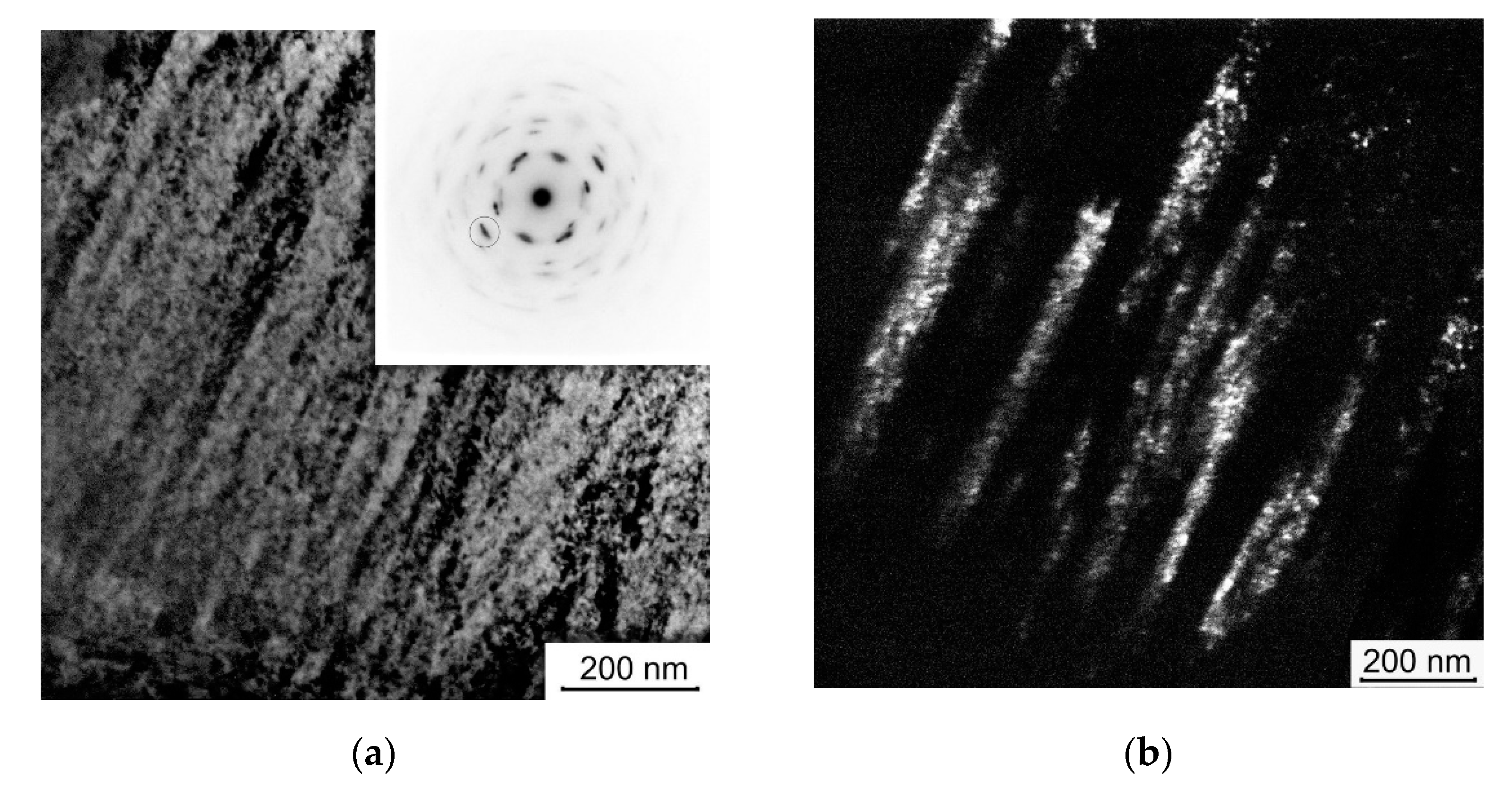
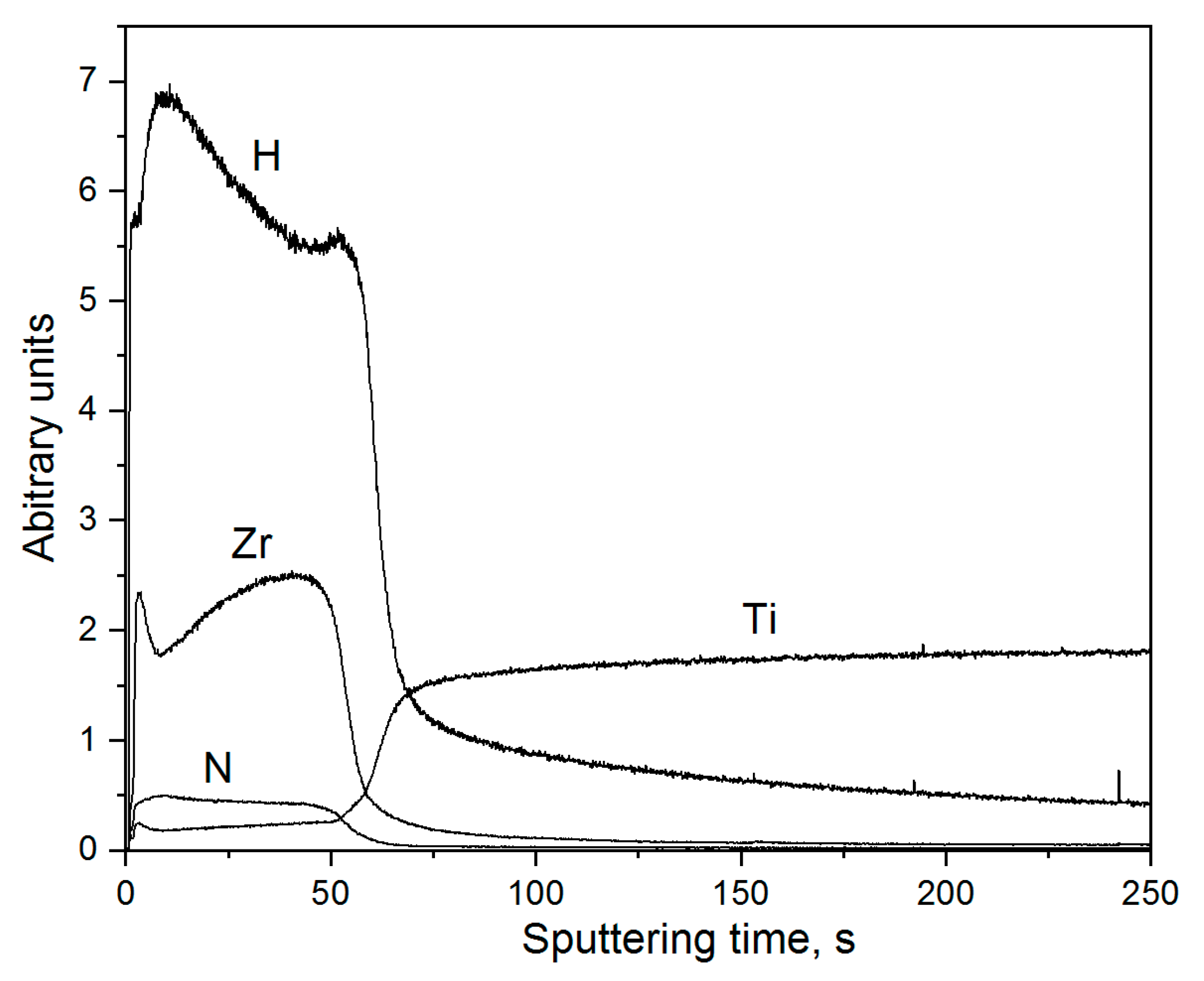
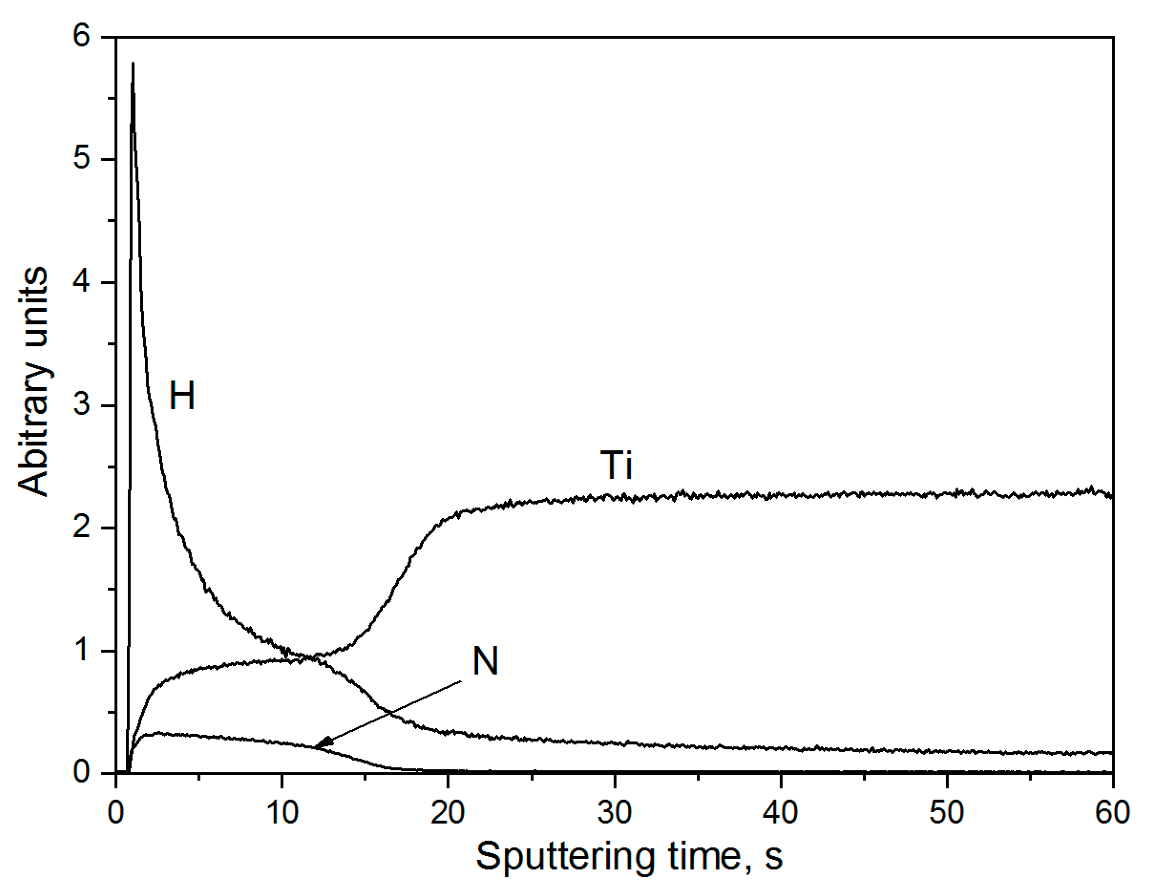
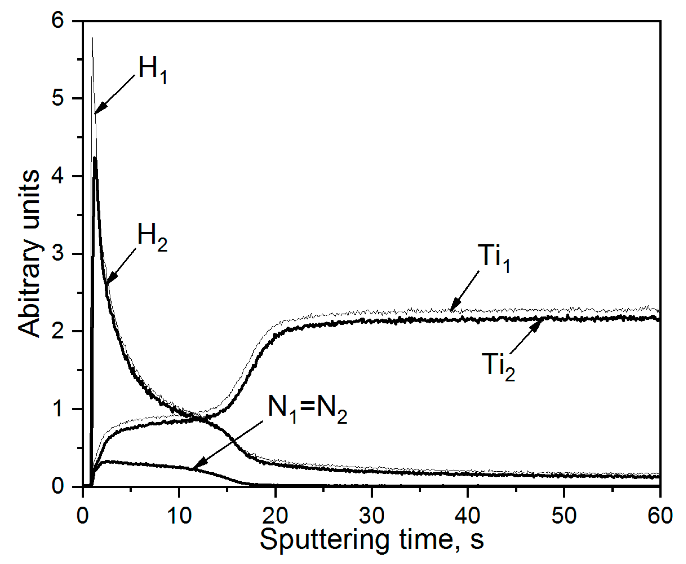
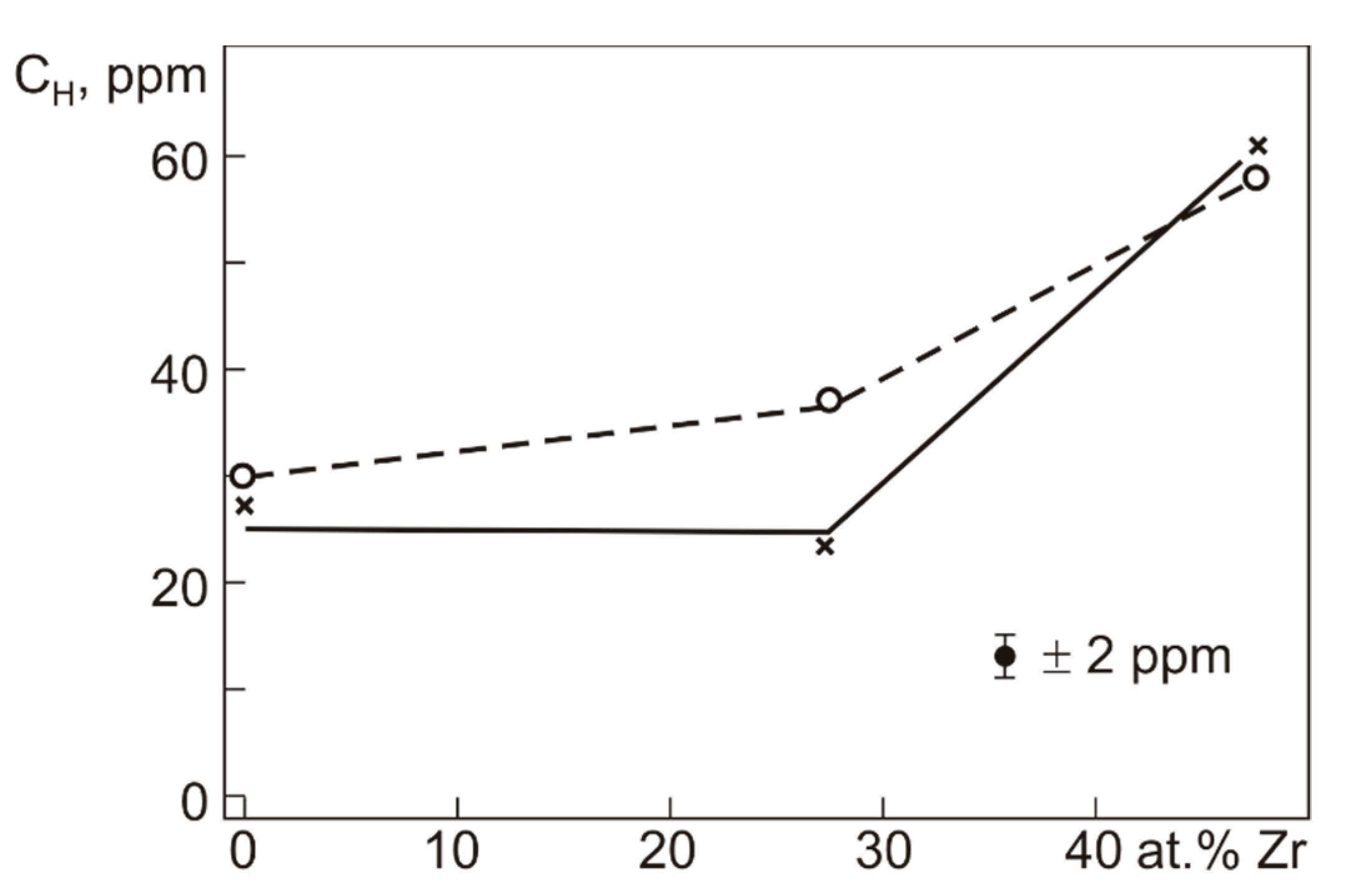
The (Ti,Zr)N coatings were formed on the Ti0.16Pd (wt.%) substrates by the method of vacuum arc deposition in a URMZ. 279.048 modernized installation (Kaliningrad mechanical engineering plant, Kaliningrad, Russia) at a low pressure (2 × 10⁻² Pa) of high-purity nitrogen. The setup includes a Y-shaped filter of simultaneously evaporated materials of the titanium and zirconium cathodes ruling out the presence of the microdroplet fraction and ensuring the transport of the ion component of the plasma flow only to the substrate surfaces. The chemical compositions of the coatings were changed by varying the ratio of the arc discharge currents of the titanium and zirconium cathodes. The preparation of the specimen surfaces in the working chamber involved their preliminary irradiation by argon ions (pressure 0.04 Pa) for 5 min. Then a sublayer of titanium was deposited onto the specimen surfaces for 5 min (without the supply of nitrogen at a titanium cathode arc current of 50 A, a bias voltage of –70 V), after which (Ti,Zr)N coatings were deposited at the technological parameters listed in Table 1. The substrates were made of Ti0.16Pd alloy with microcrystalline structure (average grain size of 10 μm) and hardened specimens with submicrocrystalline structure formed by equal-channel angular pressing (average grain-subgrain size of 0.25 μm). In what follows, these specimens were denoted by M and SMC symbols, respectively. Their basic phase composition was made up of the hexagonal α-phase of titanium (irrespective of the type of their microstructure). The detailed phase composition of the specimens with M and SMC structures is presented elsewhere [7]. The coatings were deposited simultaneously on the specimens with M and SMC structures at all technological process parameters. For testing their structure, phase, and chemical compositions, the specimens were shaped as plates measuring 10 mm × 20 mm × 1 mm. The specimens for testing the average hydrogen concentration after electrolytic hydrogenation were shaped as bars (the quadratic cross-section of 1 mm × 1 mm and the length of 20 mm). All faces of each specimen (both plates and bars with the quadratic cross-sections) were prepared by SiC abrasive paper. The diamond paste made the final fine mechanical polishing.
The tests were performed on the specimens with the coatings deposited via two different regimens. In mode 1, the coatings were deposited on immobile specimens placed perpendicular to the plasma flow axis. In mode 2, the coatings were deposited onto the substrates placed on a table rotating with respect to the vertical axis of the working chamber. The specimens were placed along a circle with a radius of 80 mm from the axis of rotation. The distance along the circle between specimens was 50 mm. The rotation speed of the table was 1.5 rpm. The rotation was continuous for the duration of coating deposition. Consequently, coatings were deposited on all faces parallel to the axis of rotation (including the narrow faces of plate specimens). Under the conditions of a periodical variation of the specimen’s positions in the plasma flow, the coatings with the same microstructure on all specimen faces oriented along the rotation axis were formed. It was found out that irrespective of the substrate structure (M or SMC), deposition regimes (modes 1 or 2) and chemical compositions, the coatings had a single-phase nanocrystalline structure (δ—TiN, δ—(Ti,Zr)N or δ—ZrN). However, and this was of uttermost importance, the coatings deposited via modes 1 and 2 differed in their microstructure, which accounts for their cardinal difference in the barrier properties with respect to hydrogen diffusion into the substrates during electrolytic hydrogenations.
The specimens were hydrogenated in a physiological saline solution (0.9% NaCl) at a current density of 20 A/m2 within 3 h. The structure and phase composition of the coatings were examined by the methods of X-ray diffraction analysis in a DRON-7 diffractometer (Bourevestnik JSC, Saint-Petersburg, Russia) and transmission electron microscopy (TEM) in a JEOL-2100 electron microscope (JEOL Ltd., Tokyo, Japan). The grinding made the thin foils for TEM studies, mechanical polishing to a thickness of 0.1 mm and ion thinning on a JEOL EM 09100IS ion slicer (JEOL Ltd., Tokyo, Japan). The chemical compositions of the coatings were determined by a JEOL JXA 8500-F electron probe microanalyzer (JEOL Ltd., Tokyo, Japan). The distribution of chemical elements in the coated hydrogenated specimens was performed using a GD-OES Profiler 2 (Jobin Yvon Emission Horiba Group, Longjumeau Cedex, France). The average hydrogen content in the coated hydrogenated specimens was determined using a LECO RHEN 602 gas analyzer (LECO, Saint Joseph, MI, USA).

3. Results

The diffraction analysis results demonstrated that the coatings had a single-phase structure irrespective of the substrate types (M or SMC) and deposition regimes (modes 1 or 2). The diffraction patterns contained only the reflections of the fcc-structure of the NaCl type (in addition to the α-phase reflections of the titanium substrate) characteristic for the quasi binary δ—TiN—δ—ZrN system. The lattice parameters of the δ-phase increase with the content of zirconium (Table 2) and are close to their well-known values [28].
TEM microstructural studies have demonstrated that all coatings (independent of the deposition mode) have a nanocrystalline structure. Figure 1 presents TEM images of a δ-(Ti,Zr)N coating deposited via mode 1. The ratio Zr/Ti = 1.34, Table 2. The coating has a highly regular columnar microstructure. The mesofragments in the cross-section had sizes within 50–60 nm. These mesofragments consist of nanograins measuring 6–12 nm.
The distribution of Ti, Zr, nitrogen, and hydrogen immediately after hydrogenation of the TZ1SMC specimen with a coating deposited via mode 1 is given in Figure 2. It is clear from Figure 1 that the maximum content of absorbed hydrogen was localized in the initial zone of the coating (~0.4 μm from the surface). The content of hydrogen decreases but slightly (by ~20%) towards the beginning of the coating–substrate transition zone (Ti content increases and Zr content decreases) and sharply decreases at the lower boundary of this zone. A gradual decrease in the hydrogen content was observed in the substrate. Since the average hydrogen content in the initial Ti0.16Pd alloy was 10 ppm, the estimated hydrogen content at the interface with the substrate does not exceed ~30 ppm. Therefore, on the whole, the δ—(Ti,Zr)N coating, formed by deposition on the immobile specimen, presented a sufficiently efficient barrier to hydrogen penetration during electrolytic hydrogenation. However, the columnar structure of this coating allowed hydrogen to penetrate through all its thickness and partially to the Ti0.16Pd alloy substrate.
The microstructure of the coatings, deposited via mode 2 on the substrates rotating in the plasma flow, markedly differed from that of the coatings deposited via mode 1. Using mode 2, the coatings made of δ—TiN, δ—(Ti,Zr)N with the ratio Zr/Ti = 1.28 (close to the ratio of these elements in the above-discussed coating) and a coating formed of nearly pure δ—ZrN (Zr/Ti = 10.15) on the substrates with microcrystalline and SMC structures were formed, as shown in Table 1 and Table 2. Typical bright- and dark-field microstructural images from these coatings are presented in Figure 3 and Figure 4. It is to be noted that no substantial microstructural differences in the coatings deposited on the substrates with the initial and SMC structures were observed. Moreover, the coatings with different Zr/Ti ratios, which were deposited via mode 2, had (to a first approximation) qualitatively similar microstructures. A continuous variation of the crystallite growth conditions on the rotating substrates dictates the formation of coatings with less pronounced regularity of their microstructure. The mesoscale microstructural fragments, consisting of the grains of ≤20 nm in size (Figure 3 and Figure 4), are substantially different in shape from the lengthy regular mesofragments in the coatings deposited on immobile specimens (Figure 1). The longitudinal size of these fragments is generally smaller than the coating thickness. Moreover, these fragments can be misoriented with respect to each other and normal to the substrate surface at angles up to ~30°. On the whole, it is practically impossible to single out the regular, lengthy structures of a columnar type in the coatings deposited via mode 2. The lowest correlation degree of the spatial distribution of nanograins was observed in a thin surface zone of the coating and the substrate-coating transition zone.
The microstructure of the coatings deposited via mode 2 substantially changes the hydrogen absorption and its distribution during electrolytic hydrogenation in a physiological saline solution. Figure 5 presents Ti, Zr, nitrogen, and hydrogen distribution profiles in the TZ2SMC specimen with a coating deposited via mode 2. The composition of this δ—(Ti,Zr)N coating with the ratio Zr/Ti = 1.28 was close to that of the coating on the TZ1SMC specimen (Zr/Ti = 1.35) deposited via mode 1. It is clear from Figure 5 that hydrogen in the TZ2SMC specimen is mainly concentrated in the surface zone of the coating, with a width of ~0.2 µm. The content of hydrogen as we moved from the surface decreased sharply, and at the coating–substrate interface (~0.84 µm from the surface), the concentration of hydrogen was nearly equal to its content in the initial alloy (10 ppm). A similar pattern was demonstrated by the hydrogen distributions in the TZ2M specimens and δ-ZrN-based specimens (TZ3M and TZ3SMC with a hydrogen content of ~4.7 at.%). For the δ—TiN specimens (TNM and TNSMC), the distribution of hydrogen is on the whole close to that given in Figure 6. Note that there is no substantial penetration of hydrogen into the substrate of the hydrogenated specimens. The similarity of hydrogen distribution profiles in nanocrystalline coatings deposited via mode 2, irrespective of the Zr/Ti ratio in their composition, was due to the peculiarities of microstructure in these coatings with a low degree of spatial correlation of nanograin distribution (suppression of formation of a regular columnar microstructure in the course of deposition). In general, this decreases the effective diffusion coefficient of hydrogen in these coatings, ensures its high content in the surface area of the coatings, and prevents its penetration into the substrate. Thus, the barrier properties of the coatings deposited via mode 2 were much better than those of the coatings having regular columnar microstructure, which were deposited on immobile specimens (mode 1).
Figure 7 presents the thickness of the coatings, deposited via Mode 2, as a function of Zr content, which was obtained from a combined analysis of the TEM results and the element distribution profiles. From Figure 7, it can be estimated that the coating deposition rate from δ-TiN is noticeably lower (~8 nm/s) than that of Zr-enriched coatings (~11 nm/s). However, the differences in the coating deposition rate and thickness do not significantly affect their barrier properties.
Hydrogen absorbed during electrolytic hydrogenation was retained in the coating composition during their long-term (430 h) storage at room temperature in air environment. At the same time, there was a marked diffusion-induced hydrogen redistribution in the volume of the coating with Zr/Ti = 1/28 (TZ2SMC) and TZ2M) and in the coatings from practically pure δ—ZrN (TZ3M and TZ3SMC). From a comparison of Figure 5 and Figure 8, it is clear that while holding the hydrogenated TZ2SMC specimens at room temperature, the maximum hydrogen content in the thin surface zone decreases while the hydrogen content in the adjacent zone increases. However, there was no hydrogen penetration into the substrate. More pronounced hydrogen redistribution occurs in the hydrogenated TZ2M specimens during storage at room temperature in the air environment, Figure 9. After hydrogenation of these specimens, the distribution of hydrogen in them is qualitatively similar to that presented in Figure 5. After 430 h of holding at room temperature, the hydrogen content in the surface zone also decreases. Note, however, that there was more intensive hydrogen accumulation in the coating–substrate transition zone than in the central coating area. Another hydrogen distribution maximum was formed. As this goes on, the level of hydrogen content in the substrate does not increase. The least significant hydrogen redistribution is observed in the hydrogenated TNM and TNSMC with δ-TiN coatings during their long-term storage, as shown in Figure 10. To sum up, in the course of storage after hydrogenation, the barrier properties of the coatings deposited via Mode 2 are retained for a long time.
Considering that practically all hydrogen in the coated specimens is concentrated in the coating zones with qualitatively similar microstructures and hydrogen distributions, such parameter as the average hydrogen content in the hydrogenated specimens acquires great importance. Since the coating thickness ratio to the specimen size (1 mm × 1 mm) is small and practically the same for all of them, the measured average hydrogen concentration (CH) characterizes the effect of the coating composition on hydrogen absorption during electrolytic hydrogenation. The coatings were deposited via mode 2 simultaneously with the specimens used for TEM structural examination and determination of hydrogen distribution in the coatings. The graph of the dependence of CH on the coating composition is presented in Figure 11. The δ—TiN coatings absorbed the least content of hydrogen (27–30 ppm). As the content of Zr in the coating composition was increased to 27.2 at.% (TZ2M and TZSMC specimens with (Zr/Ti = 1.28), the amount of absorbed hydrogen slightly differed from CH in the δ—TiN coatings. It increased to 37 ppm in the coatings on SMC substrates and 23 ppm on the substrates with the initial microcrystalline structure. This difference was very like due to minor fluctuations in the composition during deposition. In the coatings formed from practically pure δ—ZrN (~4.7 at.% Ti, ratio Ti/Zr = 10.15), the hydrogen absorption during electrolytic hydrogenation was maximal, and CH = 60 ppm. Hence, the δ—(Ti,Zr)N coatings with Zr/Ti ≤ 1 are more preferable as protective coatings with high barrier properties ruling out hydrogen diffusion into the substrate made of the Ti0.16Pd alloy.

4. Discussion

It should be primarily noted that the microstructures of the (Ti,Zr)N coatings formed in this study are qualitatively consistent with the well-known classification of the microstructures of coatings deposited by vacuum arc processes. In particular, in Thornton’s structure-zone model [29], depending on the argon pressure and the temperature ratio T/Tm (T – substrate temperature, Tm – coating material melting temperature), three main types of coating microstructures can form on the substrate surface: coarse-grained, fine-grained and columnar structures. The formation of columnar structure corresponds to Zone 2 (0.3 < T/Tm < 0.5) [29]. This microstructure in (Ti,Zr)N coatings with a highly regular distribution of columnar mesofragments and nanograined structure inside them, oriented strictly along the normal to the substrate surface, is formed on the surface of immobile specimens (mode 1). The length of these mesofragments practically coincides with the coating thickness. It should be noted that the mesofragment boundaries apparently have a high density of free volumes. This ensures the penetration of many hydrogen atoms into the entire coating thickness during hydrogenation in a physiological saline solution. In coating deposition via mode 2, the working chamber atmosphere and the deposition regime remain the same as in mode 1. Inside the mesofragments of the coatings, there is still a nanograined structure. However, the continuous variation in the facet orientation with respect to the plasma flow axis gives rise to substantial changes in the spatial distributions of mesofragments and nanograins. The length of mesofragments becomes much smaller than the coating thickness. The mesofragments are misoriented by the angles of 20°–30° relative to the normal substrate surface. These circumstances considerably complicate the diffusion paths for hydrogen penetration into the coatings. Note that the highest hydrogen content within the period of electrolytic hydrogenation is observed in the thin coating layer near its outer surface. In contrast, in the central zones and near the interface with the substrate, its content is much smaller, as shown in Figure 5 and Figure 6. The microstructural difference between the coatings deposited on immobile and rotating substrates was also observed by Boon et al. [30]. The results obtained in our work demonstrate that a change in geometry, with the other deposition parameters being constant, exerts a noticeable influence both on the coating microstructure and the hydrogen absorption by these coatings.
The results of our work are consistent with the common patterns of the influence of microstructure of the coatings, deposited on the substrates made of different materials, on hydrogen absorption during hydrogenation. For instance, qualitatively similar results were obtained by Tamura et al. [21,22]. They showed that Al2O3, TiC, and TiN nanostructured coatings with columnar microstructure exhibited much higher permeability for hydrogen penetration than similar coatings without a pronounced columnar (fine-grained structures in References [21,22]). Note that the barrier properties of the coatings increase by ~2 orders of magnitude as the nanograin size is decreased from 100 nm to 20 nm.
To sum up, it should be underlined that the barrier properties towards hydrogen absorption are controlled not only by their microstructure and grain size, but they also depend on the crystallographic orientation of the grains (coating texture), crystal lattice defects, and residual microstresses in them, which are beyond the scope of this study. The formation of (Ti,Zr)N coatings with the highest barrier properties requires that such studies be performed in the future.

5. Conclusions

Similar single-phase nanostructure coatings with a chemical composition close to the quasi-binary TiN—ZrN system have been formed on the Ti0.16Pd alloy substrates with microcrystalline and SMC structures by the method of vacuum arc deposition from separated plasma flows. Dependence of the δ—(Ti,Zr)N coating microstructure on the deposition modes has been established. The coatings deposited via mode 1 (on immobile substrates oriented perpendicular to the plasma flow) have a highly regular columnar microstructure oriented perpendicular to the substrate surface. The coatings deposited via mode 2 (with rotation of the table with the substrates flow around the vertical axis of the working chamber) have a much less pronounced correlation of the spatial distribution of nanograins.
It has been found out that the microstructural differences between the coatings deposited via modes 1 and 2 determine the peculiarities of hydrogen absorption and its distribution in the specimens as a result of electrolytic hydrogenation in a physiological saline solution (0.9% NaCl). In the case of a highly regular columnar structure of the coatings deposited via mode 1, the high hydrogen content is observed throughout the entire coating cross-section and a sharp decrease in its concentration in the coating-substrate transition zone. It is noteworthy that hydrogen is partially diffused into the substrate. In the coating with a low regularity of the spatial distribution of the microstructure elements, which were deposited via mode 2, the highest content of absorbed hydrogen is observed in a thin surface zone (~0.2 µm). In contrast, the hydrogen content sharply drops on approach to the coating-substrate interface. There is no hydrogen diffusion from the coating to the substrate immediately after hydrogenation and after long-term (430 h) storage of the hydrogenated specimens at room temperature in the air environment.
It has been shown that the content of hydrogen absorbed during electrolytic hydrogenation of the specimens coated via mode 2 depends on the ratio between zirconium and titanium in δ—(Ti,Zr)N. The hydrogen absorption in the coatings based on zirconium nitride increases by a factor of ~2.

Author Contributions

Conceptualization, A.L., V.G., S.L. and V.K., investigations, A.B., V.G., S.L., N.G., D.Z. and V.T.; writing—original draft preparation, V.G.; writing—review and editing, V.G., A.L. and D.Z.; project administration, A.L. and V.G.; funding acquisition, A.L. All authors have read and agreed to the published version of the manuscript.

Funding

The work was perfomed accoding to the government research assignement for ISPMS SB RAS, project FWRW-2021-0004 and was funded by Russian Foundation for Basic Research, grant number 18-58-00036.

Institutional Review Board Statement

Not applicable.

Informed Consent Statement

Not applicable.

Data Availability Statement

Not applicable.

Conflicts of Interest

The authors declare no conflict of interest.

References

  1. Zwicker, U. Titan and Titanlegiarungen; Springer: Berlin/Heidelberg, Germany, 1974. [Google Scholar]
  2. Tomashev, N.D. Titanium and Its Corrosion-Resistant Alloys; Metallurgiya: Moscow, Soviet Union, 1985. (In Russian) [Google Scholar]
  3. Fokin, M.N.; Ruskol, Y.S.; Mosolov, A.V. Titanium and Its Alloys in Chemical Industry; Khimiya: Leningrad, Soviet Union, 1978. (In Russian) [Google Scholar]
  4. Nakagawa, M.; Matsua, S.; Udoh, K. Corrosion behavior of pure titanium and titanium alloys in fluoride-containing solution. Dent. Mater. J. 2001, 20, 305–314. [Google Scholar] [CrossRef] [PubMed]
  5. Nakagawa, M.; Matono, Y.; Matsuya, S.; Udoh, K. The effect of Pt and Pd alloying additions on the corrosion behavior of titanium in fluoride-containing environments. Biomaterials 2005, 26, 2239–2246. [Google Scholar] [CrossRef] [PubMed]
  6. Wang, Z.B.; Hu, H.X.; Zheng, Y.G.; Ke, W.; Qiao, Y.X. Comparison of the corrosion behavior of pure titanium and its alloys in fluoride-containing sulfuric acid. Corros. Sci. 2016, 103, 50–65. [Google Scholar] [CrossRef]
  7. Grishkov, V.; Kopylov, V.; Lotkov, A.; Latushkina, S.; Baturin, A.; Girsova, N.; Timkin, V.; Zhapova, D. Effect of warm equal channel angular pressing on the structure and mechanical properties of Ti0.16Pd0.14Fe (wt.%) alloy. Rev. Adv. Mater. Sci. 2019, 58, 22–31. [Google Scholar] [CrossRef]
  8. Liu, B.; Zhow, Q.; Qu, R.-F.; Chang, W.-T. Effect of microstructure on corrosion resistance of CP-Ti and Ti-0.2Pd. Chin. J. Nonferrous Met. 2015, 25, 959–966. [Google Scholar]
  9. Rodrigues, D.C.; Urban, R.M.; Jacobs, J.J.; Gilbert, J.L. In vivo severe corrosion and hydrogen embrittlement of retrieved modular body titanium alloy hip-implants. J. Biomed Mater. Res. B Appl. Biomater. 2009, 88, 206–219. [Google Scholar] [CrossRef] [Green Version]
  10. Ervin Tal-Gutelmacher and Dan Eliezer. Hydrogen-assisted degradation of titanium based alloys. Mater. Trans. 2004, 45, 1594–1600. [Google Scholar] [CrossRef] [Green Version]
  11. Yokoyama, K.; Ichikawa, T.; Murakami, H.; Miyamoto, Y.; Asaoka, K. Fracture mechanisms of retrieved titanium screw thread in dental implant. Biomaterials 2002, 23, 2459–2465. [Google Scholar] [CrossRef]
  12. Greene, C.A.; Henry, A.J.; Brossia, C.S.; Ahn, T.V. Evaluation of the passible susceptibility of titanium Grade 7 to hydrogen embrittlement in geologic repository environment. Mat. Res. Soc. Symp. Proc. 2001, 663, 1–9. [Google Scholar]
  13. Murzinova, M.A.; Salishchev, G.A. Effect of decrease of hydride-induced embrittlement in nanocrystalline titanium. Adv. Eng. Mater. 2010, 12, 765–768. [Google Scholar] [CrossRef]
  14. Kolobov, Y.R.; Torganchuk, V.I.; Fokin, V.N.; Tarasov, B.P. Structural features of the hydride phase formation in nanostructured titanium. IOP Conf. Ser. Mater. Sci. Eng. 2015, 81, 012053. [Google Scholar] [CrossRef] [Green Version]
  15. Tamura, M.; Noma, M.; Yamashita, M. Characteristic change of hydrogen permeation in stainless steel plate by BN coating. Surf. Coat. Technol. 2014, 260, 148–154. [Google Scholar] [CrossRef]
  16. Yilbas, B.S.; Coban, A.; Kahraman, R.; Khaled, M.M. Hydrogen embrittlement of Ti-6Al-4V alloy with surface modification dy TiN coating. Int. J. Hydrogen Energy 1998, 23, 483–489. [Google Scholar] [CrossRef]
  17. Shan, C.; Wu, A.; Li, Y.; Zhao, Z.; Chen, Q.; Huang, Q.; Shi, S. The behavior of diffusion and permeation of tritium through 316L stainless steel with coating of TiC and TiC+TiN. J. Nucl. Mater. 1992, 191, 221–225. [Google Scholar] [CrossRef]
  18. Van Hove, R.P.; Sierevelt, I.N.; Van Royen, B.J.; Nolte, P.A. Titanium-nitride coating of orthopaedic implants: A review of literature. Biomed. Tes. Int. 2015, 2015, 485975. [Google Scholar] [CrossRef] [PubMed] [Green Version]
  19. Obrosov, A.; Sutygina, A.; Volinsky, A.; Manakhov, A.; Weiβ, S.; Kashkarov, E. Effect of Hydrogen exposure on mechanical and tribological behavior of CrxN coating depsited at different pressures on IN718. Materials 2017, 10, 563. [Google Scholar] [CrossRef] [PubMed] [Green Version]
  20. Em, V.T. Structure and Phase Transformations of Interstitial Alloys of Transition Metals of the IV-V Groups. Ph.D. Thesis, Sciences of the Nuclear Physics Institute of the Uzbek SSR AS, Tashkent, Soviet Union, 1988. (In Russian). [Google Scholar]
  21. Tamura, M. Hydrogen permeation characteristics of TiN coated stainless steel. J. Mater. Sci. Eng. A 2015, 5, 204–208. [Google Scholar] [CrossRef] [Green Version]
  22. Tamura, M.; Eguchi, T. Nanostructured thin films for hydrogen-permeation barrier. J. Vac. Sci. Technol. A 2015, 33, 041503-1–041503-6. [Google Scholar] [CrossRef] [Green Version]
  23. Wang, D.-Y.; Chang, C.-L.; Hsu, C.-H.; Lin, H.-N. Sinthesis of (Ti,Zr)N hard coatings by unbalanced magnetron sputtering. Surf. Coat. Technol. 2000, 130, 64–68. [Google Scholar] [CrossRef]
  24. Uglov, V.V.; Anishchik, A.M.; Khodasevich, V.V.; Prikhodko, Z.L.; Zlotski, S.V.; Abadias, G.; Dub, S.N. Structural characterization and mechanical properties of Ti-Zr-N coatings depsited by vacuum arc. Surf. Coat. Technol. 2004, 180–181, 519–525. [Google Scholar] [CrossRef]
  25. Ramana, J.V.; Kumar, S.; David, C.; Raju, V.S. Structure, Composition and microhardness of (Ti,Zr)N and (Ti,Al)N coatings prepared by DS magnetron sputtering. Mater. Lett. 2004, 58, 2553–2558. [Google Scholar] [CrossRef]
  26. Lin, Y.-W.; Huang, J.-H.; Yu, G.-P. Microstructure and corrosion resistance of nanocrystalline TiZrN films jn AISI 304 stainless substrate. J. Vac. Sci. Technol. A 2010, 28, 774–778. [Google Scholar] [CrossRef]
  27. Latushkina, S.D.; Lotkov, A.I.; Kopylov, V.I.; Posylkina, O.I.; Shkrobot, V.A. Formation of protective vacuum-plasma TiZrN coating on titanium alloys after equal channel angular pressing. In Proceedings of the 13th International Conference Interraction of Radiation with Solids, Minsk, Belarus, 30 September–3 October 2019. [Google Scholar]
  28. Knotek, O.; Barimani, A. On spinodal decomposition in magnetron-sputtered (Ti,Zr) nitride and carbide thin films. Thin Solid Films 1989, 174, 51–56. [Google Scholar] [CrossRef]
  29. Tornton, J.A. High rate thick film growth. Ann. Res. Mater. Sci. 1977, 7, 239–260. [Google Scholar] [CrossRef]
  30. Boone, D.H.; Strangman, T.E.; Wilson, L.W. Some effects of structure and composition on properties of electron beam vapor deposited coatings for gas turbine superalloys. J. Vac. Sci. Technol. 1974, 11, 641–646. [Google Scholar] [CrossRef]
Figure 1. Bright-field electron microscopy images (a) of the coating microstructure of the TZ1SMC specimens with a microdiffraction pattern from the central coating area(inset); dark-field images (b), (200)-type reflection) from the cubic structure of the δ—(Ti,Zr)N—encircled with an ellipse.
Metals 11 01332 g001
Figure 2. Distribution of Ti, Zr, nitrogen, and hydrogen atoms in the TZ1SMC specimens (coating formation time—90 min) after electrolytic hydrogenation in a physiological saline solution.
Metals 11 01332 g002
Figure 3. Bright-field (a) microstructural image (TEM) of the (Ti,Zr)N coating from the TZ2SMC specimen; microdiffraction pattern (b) from the central area of the coating (reflections of quasi-rings of the following types are indicated: 1—(111), 2—(200), 3—(220), 4—(222)); dark-field images (c,d) of the coating microstructure (reflections of the (111) and (220) type are indicated in Figure 3b by arrows).
Metals 11 01332 g003
Figure 4. Bright-field (a) image (TEM) of the (Ti,Zr)N coating microstructure from the TZ3M specimen; microdiffraction pattern (b) from the central coating area (reflections of the following types are indicated: 1—(111), 2—(200), 3—(220), 4—(311), 5—(400)); dark-field images (c,d) of the coating microstructure (reflections of the (200) and (111) types are indicated in Figure 4b by arrows).
Metals 11 01332 g004
Figure 5. Element distribution profiles in the TZ2SMC specimens of the Ti0.16Pd alloy with a δ—(Ti,Zr)N coating (ratio Zr/Ti = 1.28) immediately after cathode hydrogenation in a physiological saline solution.
Metals 11 01332 g005
Figure 6. Element distribution profiles in the TNSMC specimens of the Ti0.16Pd with a δ—TiN coating immediately after cathode hydrogenation in a physiological saline solution.
Metals 11 01332 g006
Figure 7. Thickness of the coatings formed on the Ti0.16Pd alloy versus the content of Zr in (Ti,Zr)N (●—from element distribution profiles; ○—from TEM data).
Metals 11 01332 g007
Figure 8. Element distribution profiles in the TZ2SMC specimens of the Ti0.16Pd with a δ—(Ti,Zr)N coating (ratio Zr/Ti = 1.28) after holding (430 h) at room temperature in air environment.
Metals 11 01332 g008
Figure 9. Element distribution profiles in the TZ2M specimens of the Ti0.16Pd with a δ—(Ti,Zr)N coating (ratio Zr/Ti = 1.28) after holding (430 h) at room temperature in air environment.
Metals 11 01332 g009
Figure 10. Element distribution profiles in the TNSMC specimens with a δ—TiN coating immediately after cathode hydrogenation in a physiological saline solution (H1, Ti1, N1) and after holding (430 h) at room temperature in air environment (H2, Ti2, N2).
Metals 11 01332 g010
Figure 11. Average hydrogen concentration in the coated Ti0.16Pd alloy specimens immediately after hydrogenation versus the content of Zr in the coating (Table 2). Substrate: Ti0.16Pd with microcrystalline (o) and SMC (×) structure.
Metals 11 01332 g011
Table 1. Technological parameters of coating deposition processes.
SpecimensSubstrate StructureCathode Arc Currents, ADeposition Time, minMode
TiZr
TZ1SMCSMC505090Immobile specimens perpendicular to the beam
TZ2Mmicrocrystalline808075Rotation of tables with specimens
TZ2SMCSMC
TZ3Mmicrocrystalline6010075Rotation of tables with specimens
TZ3SMCSMC
TNMmicrocrystalline60-75Rotation of tables with specimens
TNSMCSMC
Note: Common parameters for all deposition processes: bias voltage –80V, nitrogen pressure in the chamber—2 × 10⁻² Pa. Note: TZ—(Ti,Zr)N coatings; TN—TiN coatings.
Table 2. Elemental composition of coatings and lattice parameters of δ—(Ti,Zr)N.
SpecimensTi, at.%Zr, at.%N, at.%Zr/Tia, Å
TZ1SMC23.331.245.61.344.496
TZ2M21.227.251.51.284.479
TZ2SMC
TZ3M4.747.747.710.15 (~ZrN)4.575
TZ3SMC
TNM50.2-49.80.00 (TiN)4.245
TNSMC
Publisher’s Note: MDPI stays neutral with regard to jurisdictional claims in published maps and institutional affiliations.

