Article

Apoptosis and Relevant Genes Are Engaged in the Response of Apis mellifera Larvae to Ascosphaera apis Invasion

1
College of Bee Science and Biomedicine, Fujian Agriculture and Forestry University, Fuzhou 350002, China
2
National & Local United Engineering Laboratory of Natural Biotoxin, Fuzhou 350002, China
3
Apitherapy Research Institute of Fujian Province, Fuzhou 350002, China
*
Authors to whom correspondence should be addressed.
These authors contributed equally to this work.
Insects 2025, 16(9), 925; https://doi.org/10.3390/insects16090925
Submission received: 24 July 2025 / Revised: 30 August 2025 / Accepted: 1 September 2025 / Published: 2 September 2025
(This article belongs to the Section Social Insects and Apiculture)

Simple Summary

Honey bee larvae are threatened by a deadly fungus called chalkbrood, caused by Ascosphaera apis (A. apis). This disease kills honey bee larvae and hurts beekeeping. We wanted to understand if and how a natural process in the bee larvae’s gut, called apoptosis, helps defend against this fungus or makes things worse. We infected larvae with the fungus and looked at what happened to key cell death signals. We found that the fungus tricks the larvae into turning on too much cell death in their gut. Next, we tested two chemicals: one that stops cell death and one that forces it. Blocking cell death helped infected larvae survive much better and weakened the fungus. Forcing more cell death made survival worse and strengthened the fungus. Our results show that the fungus manipulates the larvae’s own cell death process against them to cause disease. Controlling this harmful cell death could be a new way to protect honey bees from this chalkbrood, helping save these important honey-producing insects and the crops they pollinate.

Abstract

Apoptosis is a genetically controlled process vital for homeostasis. This study examined the apoptotic response in the gut of Apis mellifera (A. mellifera) larvae to infection by Ascosphaera apis (A. apis) and its impact on host resistance and pathogen virulence. Here, Worker larvae of A. mellifera were inoculated with purified A. apis spores. We then quantified the expression of key apoptosis-related genes (AmCaspase-3, AmBax, and AmBcl-2) in the host gut and detected apoptotic cells via TUNEL assay. To functionally assess the role of apoptosis, larvae were treated with either the apoptosis inhibitor Z-VAD-FMK or the activator PAC-1, after which host survival, expression of apoptosis-associated genes, and the fungal virulence factor gene Ste11-like were analyzed. Our results showed that infection with A. apis significantly upregulated the expression of AmCaspase-3 and AmBax (p < 0.05) at 1–3 days post-inoculation (dpi), while the expression of AmBcl-2 was significantly reduced at 1 and 3 dpi (p < 0.05). Consistent with this, TUNEL assays revealed a markedly stronger green fluorescence signal in the guts of inoculated larvae at 3 dpi compared to uninfected controls, with clear co-localization of TUNEL and nuclear signals, confirming increased apoptosis. Pharmacological inhibition of apoptosis significantly enhanced the survival rate of A. apis-infected larvae, whereas apoptosis activation decreased larval survival. Accordingly, inhibiting apoptosis significantly suppressed the expression of AmCaspase-3 and AmBax (p < 0.001) and upregulated AmBcl-2 (p < 0.001). Conversely, apoptosis activation upregulated AmCaspase-3 (p > 0.05) and AmBax (p < 0.001), while significantly down-regulating AmBcl-2. Furthermore, apoptosis inhibition significantly down-regulated the fungal virulence gene Ste11-like, while its activation had the opposite effect. In summary, A. apis infection induces apoptosis in the larval gut by activating AmCaspase-3 and AmBax and suppressing AmBcl-2. Inhibiting this apoptotic response enhanced host survival by modulating the expression of host apoptosis-related genes and the fungal Ste11-like virulence factor. These findings provide new insights into the host response to A. apis and suggest a potential strategy for controlling chalkbrood disease.

1. Introduction

Honeybees are critically important pollinators in global ecosystems, essential for maintaining biodiversity and supporting agricultural productivity. Among bee species, the Western honeybee (Apis mellifera, A. mellifera) is highly suitable for beekeeping due to its superior biological characteristics, and is therefore the most widely used species in commercial apiculture worldwide. It is valued for its contributions to ecological stability, scientific research, and economic development [1,2].
Ascosphaera apis (A. apis) is a filamentous fungus that infects honey bee larvae, causing chalkbrood disease. This lethal infection imposes substantial economic losses on the apicultural industry due to severe declines in colony population and productivity [3]. After spores are ingested by the larvae, they germinate within the gut lumen [4]. The mycelia then penetrate the peritrophic matrix and the gut wall, eventually breaching the body wall and proliferating across the integument. This process ultimately leads to larval death and the formation of a characteristic white, grey, or black mummified cadaver [5].
Apoptosis is a genetically programmed and highly conserved form of cell death that plays vital roles in development, tissue homeostasis, and host defense against pathogens in multicellular organisms [6]. This process is mediated by caspase-mediated protease cascades, which trigger characteristic biochemical and morphological changes such as cell shrinkage, chromatin condensation [7], DNA fragmentation, and the formation of apoptotic bodies [8]. These bodies are efficiently phagocytosed by neighboring cells or professional phagocytes, thereby avoiding inflammatory responses. In invertebrates, including insects, apoptosis serves not only as a key mechanism governing development but also as an essential component of the innate immune response against microbial infections.
A growing body of evidence indicates that filamentous fungal infections in insect hosts often dysregulate apoptotic pathways [9,10]. Infected hosts may initiate apoptosis in compromised or adjacent cells to restrict pathogen spread and nutrient acquisition [11]. Conversely, successful entomopathogenic fungi frequently employ molecular strategies to suppress, evade, or exploit host apoptosis, thereby promoting their own colonization, proliferation, and dissemination [12]. Such apoptotic modulation has been observed in multiple insect-pathogen systems, including the silkworm (Bombyx mori) [13], wax moth (Galleria mellonella) [14], cotton bollworm (Helicoverpa armigera) [15], and fruit fly (Drosophila melanogaster) [16], highlighting the complex role of apoptosis regulation in host–pathogen interactions.
Although apoptotic regulatory mechanisms have been elucidated in several model insects, the molecular basis of apoptosis in A. mellifera—particularly in response to A. apis infection—remains unexplored. Current research on honeybee apoptosis has primarily focused on infections by microsporidian pathogens (Nosema spp.), which suppress midgut Caspase-3 expression in midgut [17]. In contrast, A. apis, as an obligate tissue-invasive fungus, likely interacts with host apoptotic pathways in distinct and uncharacterized ways. Therefore, elucidating how A. apis influences apoptotic signaling in honeybee larvae will not advance our understanding of chalkbrood pathogenesis but may also uncover strategies used by the pathogen to evade host immune defenses.
This study investigated the apoptotic response in the gut of A. mellifera larvae following A. apis infection. Using pharmacological modulation of apoptosis—via inhibitors and activators—we assessed larval survival and the expression of key apoptosis-related genes. Our work aims to clarify the role of apoptosis as an innate immune defense mechanism during fungal infection in honeybee larvae. Furthermore, these results may inform novel pest management strategies that target apoptotic pathways and expand the understanding of agricultural insect immunology.

2. Materials and Methods

2.1. Bee Larvae and Fungal Spores

The A. m. ligustica worker larvae were derived from three colonies kept in the apiary of College of Bee Science and Biomedicine, Fujian Agriculture and Forestry University, Fuzhou city, China. Honeybee colonies exhibited no clinical symptoms of chalkbrood disease, and PCR assays returned negative results for A. apis.
According to the established technical procedures [18], preserved A. apis spores were inoculated onto Potato Dextrose Agar (PDA) solid medium for activation culture. Following incubation at 33 °C for 10 days in a biochemical incubator, black spore masses were observed covering the medium surface. Purification of A. apis spores was carried out as follows: In a laminar flow hood, surface hyphae were removed from sporangia by scraping. The sporangia were then transferred to tubes containing sterile water and vortexed. After centrifugation, the pellets were washed three times with sterile water. Purified spores, as confirmed by microscopy, were frozen in liquid nitrogen and stored at −80 °C. Spore suspensions were adjusted to working concentrations of 1.57 × 10⁸ mL−1 for infection or 1.57 × 10⁵ mL−1 for validation assays. A. apis was deposited in the China General Microbiological Culture Collection Center (CGMCC; Accession No. 40895).

2.2. Preparation of Gut Samples

Selected strong colonies provided a brood comb containing eggs and larvae, which was transferred to the laboratory. Purified spores were inoculated into 3-day-old larvae, divided into an inoculated treatment group (n = 48) and a non-inoculated control group (n = 48). Larvae were reared under laboratory conditions until they reached 4–6 days old. At 1–3 days post-inoculation (dpi), larval guts were extracted following a previously described method [19]. Specifically, midgut dissection was performed under sterile conditions in a laminar flow hood using sterilized microscissors and fine forceps.

2.3. TUNEL Assay

Midgut tissues from freshly collected 6-day-old A. apis-infected and uninfected larvae were immediately fixed in 4% paraformaldehyde for 24 h at 4 °C. The samples were then dehydrated through a graded ethanol series, cleared in xylene, and embedded in paraffin. Serial sections of 4–6 μm thickness were obtained using a microtome (Leica, Shanghai, China) and mounted on poly-L-lysine-coated slides. After deparaffinization and rehydration, sections were treated with 20 μg/mL proteinase K at 37 °C for 15 min to expose DNA fragmentation sites. Following three washes with PBS, sections were incubated with TUNEL reaction mixture (containing fluorescein-labeled dUTP and terminal deoxynucleotidyl transferase, TdT) in a humidified dark chamber at 37 °C for 1 h. Negative controls omitted the TdT enzyme. Nuclei were counterstained with DAPI for 5 min. Slides were mounted with anti-fade medium (ProLong™ Gold) and immediately examined under a fluorescence microscope (Nikon, Shanghai, China). All procedures were protected from light to maintain fluorescent signal stability.

2.4. Inhibition and Activation of Apoptosis

Three-day-old A. m. ligustica previously inoculated with A. apis were orally administered the apoptosis inhibitor Z-VAD-FMK or activator PAC-1 (5 μL/larva) at concentrations of 10 μM, 50 μM, or 100 μM. Larvae were divided into four groups per compound (three treatment groups + one control group, n = 20 larvae/group). Controls received 0.1% DMSO. Larvae were subsequently maintained on an artificial diet. Survival was monitored every 24 h, and survival curves were generated. The concentration demonstrating the most significant effect was selected as optimal. Optimal concentrations of activators and inhibitors were selected and administered to 3-day-old worker larvae via dietary supplementation. Midguts were dissected from 6-day-old larvae (n = 3 per treatment group) for further analysis.

2.5. Quantitative Real-Time PCR (qRT-PCR)

Total RNA was extracted from gut samples (n = 3 biological replicates) using the SteadyPure Quick RNA Extraction Kit (Accurate Biology, Hunan, China). cDNA was synthesized from total RNA using the Reverse Transcription Kit (Vazyme, Nanjing, China) and stored at −20 °C. Using qPCR, we quantified the expression of AmCaspase-3, AmBax, and AmBcl-2 genes, with GAPDH as the reference gene. Additionally, the relative expression of virulence factor genes was analyzed via qPCR using A. apis Actin as the reference gene [20]. Midguts from worker bee larvae treated with 0.1% DMSO were used as the control group.
The qPCR reaction mixture (20 μL total volume) contained 10 μL SYBR Green Mix (Yeasen, Shanghai, China), 1 μL each of forward and reverse primers (2.5 μmol/L), 1 μL of cDNA template, and 7 μL of DEPC-treated water. The primer sequences used are listed in Table 1. The thermal cycling protocol was as follows: initial denaturation at 95 °C for 3 min; 45 cycles (or 50 cycles for A. apis virulence factor detection) of denaturation at 95 °C for 15 s, annealing at 55 °C for 30 s, and extension at 72 °C for 15 s. Each sample was analyzed with three biological replicates, each comprising three technical replicates. Gene expression was quantified using the 2−ΔΔCT method, and statistical significance was determined by one-way ANOVA in GraphPad Prism 8, with results presented graphically.

3. Results

3.1. A. apis Infection Induces the Apoptosis of the A. mellifera Worker Larval Gut Cells

Compared to uninoculated larval guts, the expression level of AmCaspase-3 showed significant upregulation (p < 0.01) in A. apis-inoculated larval guts at 1 to 3 dpi (Figure 1A). Similarly, AmBax expression levels were significantly elevated (p < 0.01) at 1–3 dpi (Figure 1B). Conversely, the significant downregulation of AmBcl-2 (p < 0.05) was observed at 4 dpi and 6 dpi (Figure 1C).
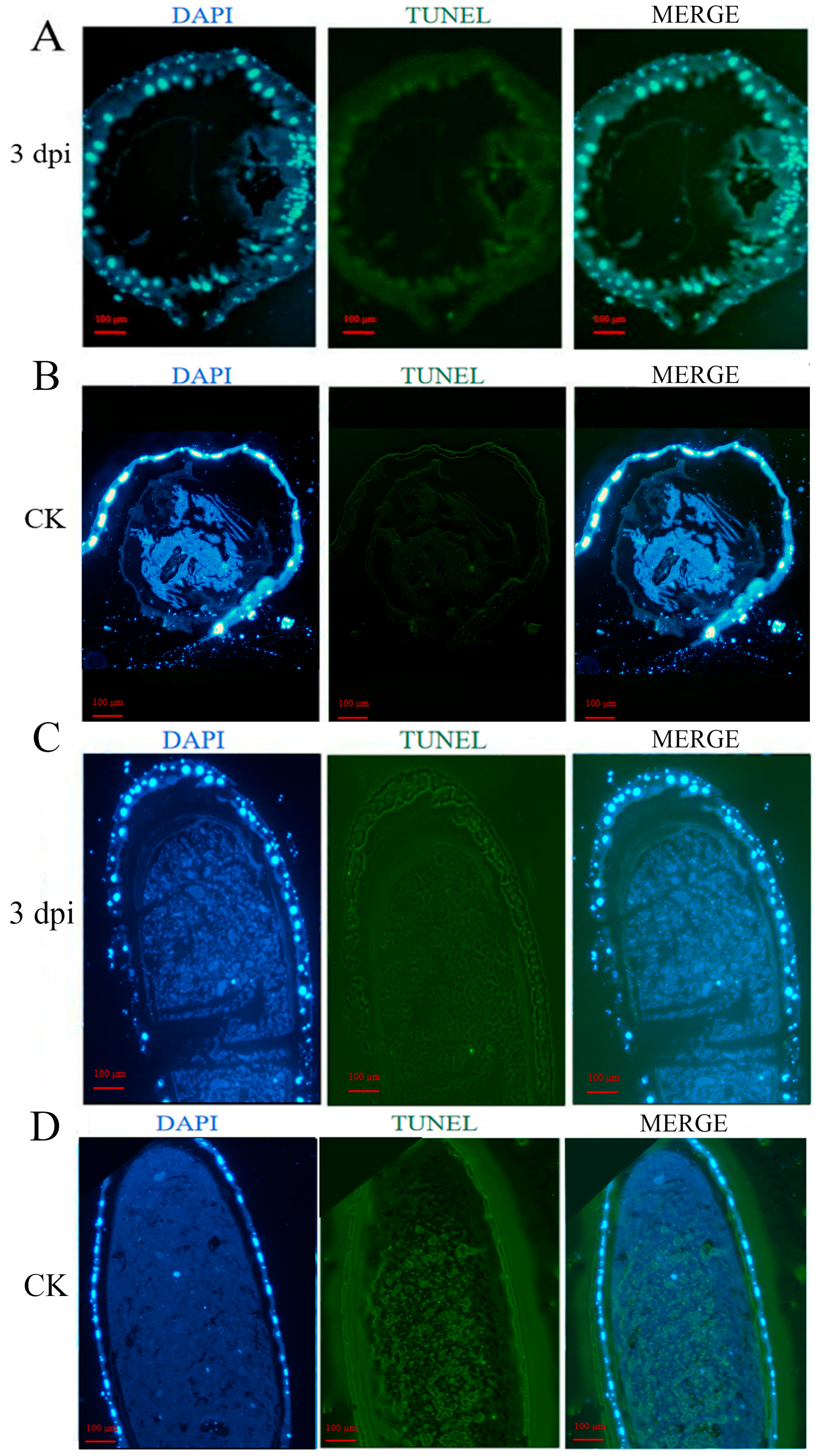
TUNEL staining revealed minimal green fluorescence in uninfected larval guts, whereas strong fluorescent signals were observed in A. apis-infected samples. Merged images confirmed co-localization of the TUNEL signal (green) and cell nuclei (blue) in infected larval guts (Figure 2), indicating that A. apis infection induced significant apoptosis.

3.2. A. apis Affects the Survival of Worker Larvae Through the Apoptosis Process

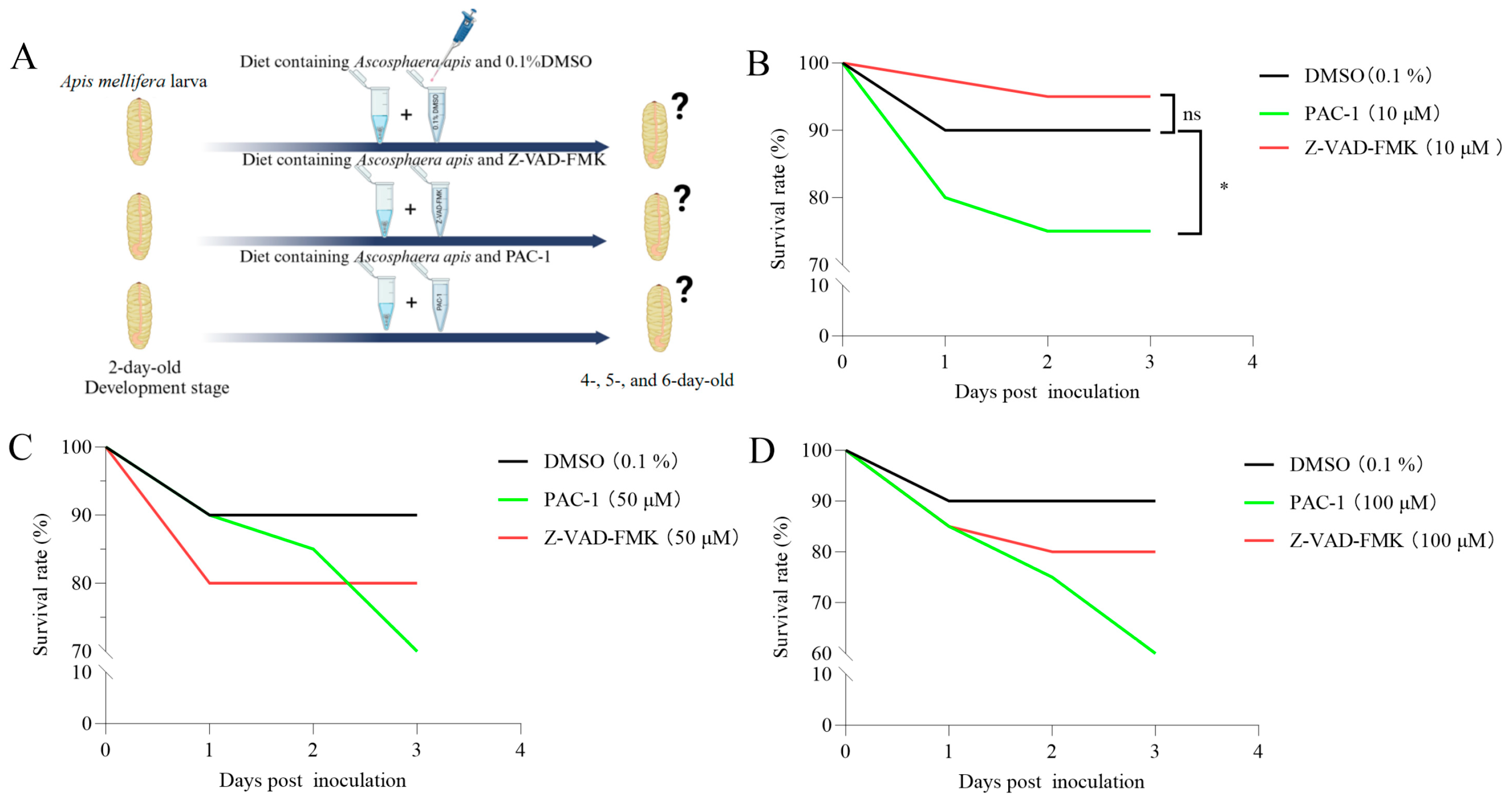
To assess the impact of A. apis-induced apoptosis on worker larval survival, infected larvae were treated with the apoptosis inhibitor Z-VAD-FMK or the activator PAC-1 (Figure 3A). Treatment with the apoptosis inhibitor (10 μM) significantly improved the survival of infected larvae, whereas the apoptosis activator (10 μM) reduced survival. At higher concentrations (50 μM and 100 μM), both compounds led to a decrease in survival; however, inhibitor-treated larvae consistently exhibited higher survival rates than those receiving the activator (Figure 3C,D).

3.3. Inhibition and Activation of Apoptosis Affected the Expression of Apoptosis-Relevant Genes in the Infected Larval Guts and A. apis Virulence Factor Ste11-like Gene

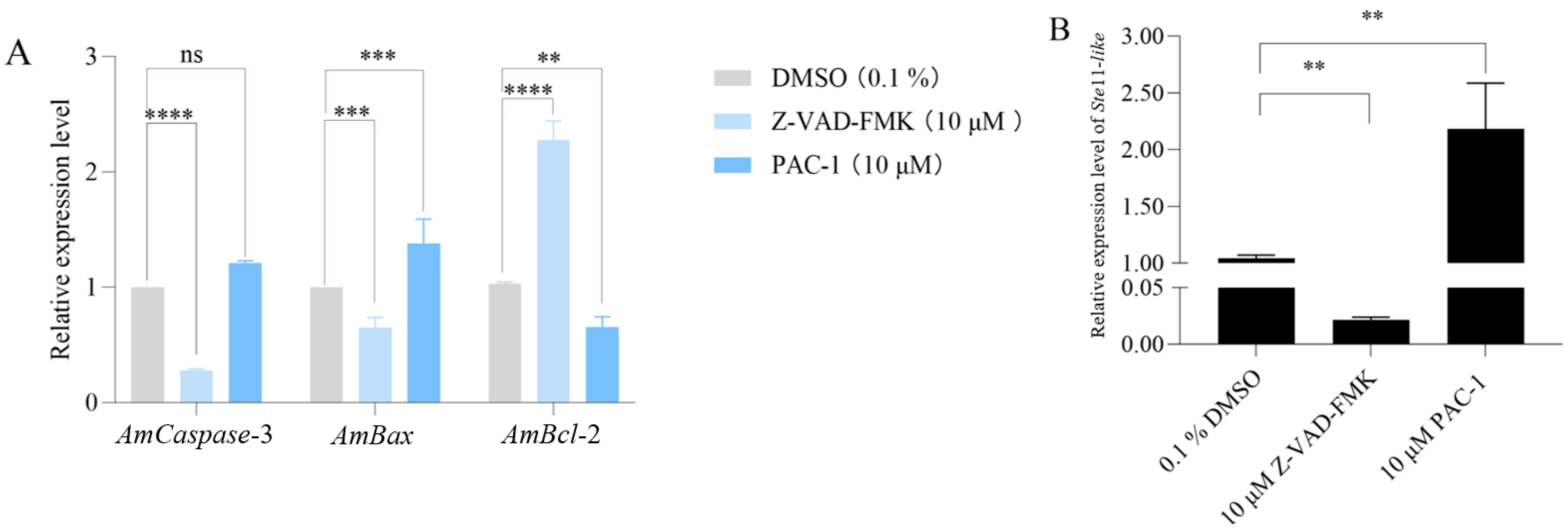
RT-qPCR analysis revealed that inhibition of apoptosis in A.Apis-infected larvae significantly downregulated AmCaspase-3 (p < 0.001), while apoptosis activation markedly increased its expression. Similarly, AmBax expression decreased significantly upon apoptosis inhibition (p < 0.001) and increased upon activation (p < 0.001). In contrast, AmBcl-2 expression was significantly upregulated when apoptosis was inhibited (p < 0.001) and downregulated following apoptosis activation (p < 0.01) (Figure 4A). Additionally, inhibition of host apoptosis resulted in significantly reduced expression of the A. apis Ste11-like gene (p < 0.01), whereas apoptosis activation induced its significant upregulation (p < 0.01) (Figure 4B).

4. Discussion

The pro-apoptotic gene Bax acts as a molecular “switch” that triggers the host’s apoptotic defense response [21]. In contrast, the anti-apoptotic Bcl-2 serves as a “brake,” indicating fungal hijacking of host anti-apoptosis mechanisms for immune evasion [22]. The apoptosis executor Caspase-3 directly confirms completion of the apoptotic program [23]. Correlating these three biomarkers with disease phenotypes and molecular mechanisms may identify therapeutic targets for chalkbrood disease and establish a foundation for developing apoptosis-targeted control strategies (e.g., RNAi or specific inhibitors) [24]. Following the A. apis inoculation, the expression levels of AmCaspase-3 and AmBax in honeybee larval midguts were significantly upregulated at 1–3 dpi (Figure 1A,B), whereas the AmBcl-2 exhibited marked downregulation at 1 dpi and 3 dpi (Figure 1C). This coordinated dysregulation likely facilitates apoptosis, potentially benefiting fungal pathogenesis. As an anti-apoptotic regulator, the downregulation of AmBcl-2, a key suppressor of mitochondrial apoptosis, implies a weakening of host intrinsic mechanisms aimed at maintaining cellular integrity [25]. Concurrently, the upregulation of AmCaspase-3 and AmBax drives irreversible commitment to apoptosis. This process may be mediated by A. apis, potentially through fungal effectors targeting host apoptotic pathways. This mechanism aligns with strategies observed in human fungal pathogens, where virulence factors activate host caspase family proteins to promote cell death and dissemination [26]. Furthermore, sustained and overwhelming activation of executioner caspases like Caspase-3 by virulence factors during infection progression would ultimately lead to irreversible apoptosis.
TUNEL assays revealed intensive DNA fragmentation signals in the larval gut at 3 dpi with A. apis, whereas only sporadic signals were observed in the control gut (Figure 2C). The results are consistent with gene expression profiles, indicating that the A. apis infection induced significant apoptosis of the challenged larval gut. Notably, DAPI staining was suggestive of intact nuclear morphology, confirming that apoptosis occurred via programmed cell death rather than necrosis. This aligns with the “apoptosis induction for nutrient acquisition” strategy commonly employed by fungal pathogens [27]. Candida albicans secretes toxins to trigger epithelial apoptosis and disrupt host immune barriers [28]. Similarly, A. apis was likely to activate the host Caspase-3 pathway to promote intestinal epithelial apoptosis, potentially hyphal invasion through compromised tissue integrity.
Treatment with the inhibitor Z-VAD-FMK increased larval survival rates, whereas the activator PAC-1 significantly decreased survival, offering direct evidence for the dual role of apoptosis in the pathogenesis of A. apis (Figure 3B). On one hand, moderate apoptosis may serve as a host defense mechanism to eliminate infected cells [29]. On the other hand, excessive apoptosis may lead to loss of intestinal barrier function, accelerating pathogen dissemination [30]. A similar phenomenon was also observed in the immune responses of insects to bacterial infection. Following infection with Bacillus bombysepticus, the silkworm (B. mori) triggered comprehensive immune responses encompassing cellular immune (e.g., phagocytosis) and humoral immune (e.g., antimicrobial peptide expression). While a moderate immune response can clear the pathogen, overactivation may induce systemic inflammation or tissue damage [31]. In the present study, Z-VAD-FMK, by inhibiting Caspase-3 activity and reducing apoptosis levels, likely delayed intestinal epithelial damage, thereby providing the host with additional time to clear the pathogen.
Upon the inhibition of apoptosis, the expression of the A. apis virulence factor gene Ste11-like was significantly reduced. Conversely, activation of apoptosis led to its marked upregulation (p < 0.01) (Figure 4B). These results suggested that the apoptotic state of host cells may feedback-regulate pathogen virulence gene expression through an unknown signaling pathway. The Ste11 protein in fungi typically participates in the MAPK signaling pathway, regulating hyphal development and stress responses [32]. It is hypothesized that A. apis may sense host cell apoptosis-associated metabolic alterations (e.g., fluctuations in ATP levels or reactive oxygen species bursts), subsequently upregulating Ste11-like to enhance its fitness. A similar strategy of adapting to host-derived signals is documented in studies of human pathogens, e.g., the infection with Bacillus anthracis causes anthrax in humans and animals [33,34]. During the infection process of host macrophages, B. anthracis sensed and rapidly adapted to the intracellular environment and modulated its metabolic pathways, including energy metabolism and cofactor biosynthesis, to enhance its intracellular survival [35]. Mycobacterium tuberculosis subverted the pyroptosis by hijacking the host ubiquitin system to remodel membrane lipid homeostasis [36]. This ability of pathogens to sense and adapt to host-derived cues, akin to B. anthracis and M. tuberculosis in macrophages, underscores the sophistication of the feedback regulation we observed between larval apoptosis and A. apis virulence.

5. Conclusions

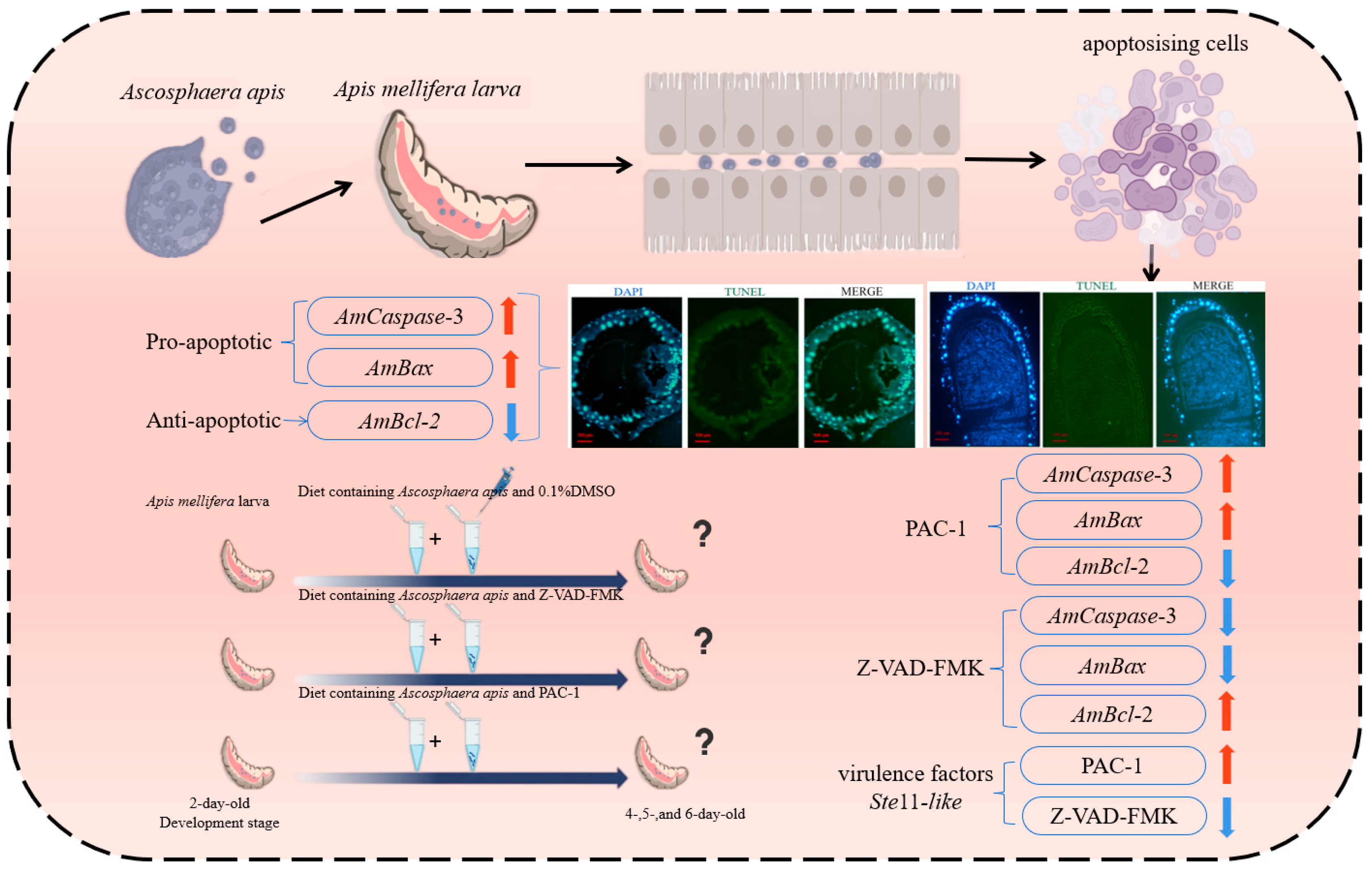
A. apis employs multi-level modulation of the host apoptosis pathway (Caspase-3/Bcl-2/Bax axis) to adapt to the infection microenvironment, while demonstrating a dynamic interplay between host apoptotic status and pathogen virulence factor gene expression. This work lays the groundwork for the theoretical basis of targeted control strategies focusing on the apoptosis-virulence interplay node (Figure 5).

Author Contributions

Conceptualization, R.G. and J.Q.; methodology, T.Z. and J.L.; software, T.Z., J.L., J.Y., X.F., S.M., X.G., M.D., X.L. and P.Z.; validation, T.Z., J.L., J.Y., X.F., S.M., X.G., M.D., X.L. and P.Z.; formal analysis, T.Z., J.L., J.Y., X.F., S.M., X.G., M.D., X.L. and P.Z.; data curation, T.Z., J.L., J.Y., X.F., S.M., X.G., M.D., X.L. and P.Z.; writing—original draft preparation, T.Z. and J.L.; visualization, J.Y., X.F., S.M., X.G., M.D., X.L. and P.Z.; supervision, Q.T., D.C., R.G. and J.Q.; project administration, R.G. and J.Q.; funding acquisition, R.G. All authors have read and agreed to the published version of the manuscript.

Funding

This work was financially supported by the National Natural Science Foundation of China (32372943, 32172792), the Earmarked fund for China Agriculture Research System (CARS-44-KXJ7), the Natural Science Foundation of Fujian Province (2025J01616, 2023J01447), the Master Supervisor Team Fund of Fujian Agriculture and Forestry University (Rui Guo), the Special Fund for Science and Technology Innovation of Fujian Agriculture and Forestry University (KFb22060XA), and the Undergraduate Innovation and Entrepreneurship Training Program of Fujian Province (202510389033, S2025103890106).

Data Availability Statement

All the data are contained within the article.

Acknowledgments

We thank all editors and reviewers for their constructive comments and recommendations.

Conflicts of Interest

The authors declare no conflicts of interest.

References

  1. Aronstein, K.A.; Murray, K.D. Chalkbrood disease in honey bees. J. Invertebr. Pathol. 2010, 103, S20–S29. [Google Scholar] [CrossRef]
  2. Aizen, M.A.; Garibaldi, L.A.; Cunningha, S.A.; Klein, A.M. How much does agriculture depend on pollinators lessons from long-term trends in crop production. Ann. Bot. 2009, 103, 1579–1588. [Google Scholar] [CrossRef] [PubMed]
  3. von Knoblauch, T.; Jensen, A.B.; Mülling, C.K.W.; Aupperle-Lellbach, H.; Genersch, E. Chalkbrood disease caused by Ascosphaera apis in honey bees (Apis mellifera)-morphological and histological changes in infected larvae. Vet. Sci. 2024, 11, 415. [Google Scholar] [CrossRef]
  4. Alvarado, I.; Phui, A.; Elekonich, M.M.; Abel-Santos, E. Requirements for in vitro germination of Paenibacillus larvae spores. J. Bacteriol. 2013, 195, 1005–1011. [Google Scholar] [CrossRef] [PubMed]
  5. Garcia-Gonzalez, E.; Genersch, E. Honey bee larval peritrophic matrix degradation during infection with Paenibacillus larvae, the aetiological agent of American foulbrood of honey bees, is a key step in pathogenesis. Environ. Microbiol. 2013, 15, 2894–2901. [Google Scholar] [CrossRef]
  6. Genersch, E. Honey bee pathology: Current threats to honey bees and beekeeping. Appl. Microbiol. Biotechnol. 2010, 87, 87–97. [Google Scholar] [CrossRef]
  7. Liang, Q.; Chen, D.F. Apicultural Protection, 2nd ed.; China Agriculture Press: Beijing, China, 2009; pp. 83–85. [Google Scholar]
  8. Jensen, A.B.; Palmer, K.A.; Boomsma, J.J.; Pedersen, B.V. Varying degrees of Apis mellifera ligustica introgression in protected populations of the black honeybee, Apis mellifera mellifera, in northwest Europe. Mol. Ecol. 2005, 14, 93–106. [Google Scholar] [CrossRef]
  9. Vertyporokh, L.; Hułas-Stasiak, M.; Wojda, I. Host-pathogen interaction after infection of Galleria mellonella with the filamentous fungus Beauveria bassiana. Insect Sci. 2020, 27, 1079–1089. [Google Scholar] [CrossRef]
  10. Li, B.; Song, S.; Wei, X.; Tang, G.; Wang, C. Activation of microlipophagy during early infection of insect hosts by Metarhizium robertsii. Autophagy 2022, 18, 608–623. [Google Scholar] [CrossRef]
  11. Nainu, F.; Shiratsuchi, A.; Nakanishi, Y. Induction of Apoptosis and subsequent phagocytosis of virus-infected cells as an antiviral mechanism. Front. Immunol. 2017, 8, 1220. [Google Scholar] [CrossRef]
  12. Tu, C.; Zhang, Y.; Zhu, P.; Sun, L.; Xu, P.; Wang, T.; Luo, J.; Yu, J.; Xu, L. Enhanced toxicity of entomopathogenic fungi Beauveria bassiana with bacteria expressing immune suppressive dsRNA in a leaf beetle. Pestic. Biochem. Physiol. 2023, 193, 105431. [Google Scholar] [CrossRef]
  13. Zheng, Y.; Meng, H.; Jiang, X.; Huang, S. Bombyx mori UFL1 facilitates BmNPV proliferation by regulating host cell apoptosis through PERK. Arch. Insect Biochem. Physiol. 2024, 116, e22127. [Google Scholar] [CrossRef]
  14. Wrońska, A.K.; Kaczmarek, A.; Kazek, M.; Boguś, M.I. Infection of Galleria mellonella (Lepidoptera) larvae with the entomopathogenic fungus Conidiobolus coronatus (Entomophthorales) induces apoptosis of hemocytes and affects the concentration of eicosanoids in the hemolymph. Front. Physiol. 2022, 12, 774086. [Google Scholar] [CrossRef] [PubMed]
  15. Yu, H.; Xiao, H.Y.; Li, N.; Yang, C.J.; Huang, G.H. An ascovirus utilizes different types of host larval regulated cell death mechanisms to produce and release vesicles. J. Virol. 2023, 97, e0156622. [Google Scholar] [CrossRef]
  16. Zhou, L. P53 and Apoptosis in the Drosophila model. Adv. Exp. Med. Biol. 2019, 1167, 105–112. [Google Scholar]
  17. Higes, M.; Juarranz, Á.; Dias-Almeida, J.; Lucena, S.; Botías, C.; Meana, A.; García-Palencia, P.; Martín-Hernández, R. Apoptosis in the pathogenesis of Nosema ceranae (Microsporidia: Nosematidae) in honey bees (Apis mellifera). Environ. Microbiol. Rep. 2013, 5, 530–536. [Google Scholar] [CrossRef] [PubMed]
  18. Xu, X.J.; Guo, R.; Luo, Q.; Xiong, C.L.; Liang, Q.; Zhang, C.L.; Zheng, Y.Z.; Zhang, Z.N.; Huang, Z.J.; Zhang, L.; et al. De novo transcriptome assembly for Apis cerana cerana larval gut and identification of SSR molecular markers. Sci. Agric. Sin. 2017, 50, 1157–1166. [Google Scholar]
  19. Guo, R.; Du, Y.; Xiong, C.L.; Zheng, Y.Z.; Fu, Z.M.; Xu, G.J.; Wang, H.P.; Chen, H.Z.; Geng, S.H.; Zhou, D.D.; et al. Differentially expressed MicroRNA and their regulation networks during the developmental process of Apis mellifera ligustica larval gut. Sci. Agric. Sin. 2018, 51, 4197–4209. [Google Scholar]
  20. Getachew, A.; Abejew, T.A.; Wu, J.; Xu, J.; Yu, H.; Tan, J.; Wu, P.; Tu, Y.; Kang, W.; Wang, Z.; et al. Transcriptome profiling reveals insertional mutagenesis suppressed the expression of candidate pathogenicity genes in honeybee fungal pathogen, Ascosphaera apis. Sci. Rep. 2020, 10, 7532. [Google Scholar] [CrossRef]
  21. Peña-Blanco, A.; García-Sáez, A.J. Bax, Bak and beyond—Mitochondrial performance in apoptosis. FEBS J. 2018, 285, 416–431. [Google Scholar] [CrossRef]
  22. Gross, A.; McDonnell, J.M.; Korsmeyer, S.J. BCL-2 family members and the mitochondria in apoptosis. Genes Dev. 1999, 13, 1899–1911. [Google Scholar] [CrossRef]
  23. McComb, S.; Chan, P.K.; Guinot, A.; Hartmannsdottir, H.; Jenni, S.; Dobay, M.P.; Bourquin, J.P.; Bornhauser, B.C. Efficient apoptosis requires feedback amplification of upstream apoptotic signals by effector caspase-3 or -7. Sci. Adv. 2019, 5, eaau9433. [Google Scholar] [CrossRef]
  24. Stroschein-Stevenson, S.L.; Foley, E.; O’Farrell, P.H.; Johnson, A.D. Phagocytosis of Candida albicans by RNAi- treated Drosophila S2 cells. Methods Mol. Biol. 2009, 470, 347–358. [Google Scholar]
  25. Rui, Y.X.; Xie, H.X.; Li, D.; Ma, X.Y.; Geng, F.N.; Liu, R. Mesaconine inhibits apoptosis through the BAX/BCL-2/Caspase 3 signaling pathway to improve renal fibrosis in rats with chronic renal failure. Chin. Med. Pharmaco. Clin. 2024, 40, 42–48. [Google Scholar]
  26. Yin, S.; Lu, B.Y.; Luo, S. Value of combined diagnosis using serum Caspase-1 and MMP-1 for postoperative pelvic infection in ovarian cyst patients and analysis of pathogenic characteristics. Anhui Med. J. 2025, 46, 483–486. [Google Scholar]
  27. Lamkanfi, M.; Dixit, V.M. Manipulation of host cell death pathways during microbial infections. Cell Host Microbe 2010, 8, 44–54. [Google Scholar] [CrossRef] [PubMed]
  28. Poulain, D. Candida albicans, plasticity and pathogenesis. Crit. Rev. Microbiol. 2015, 41, 208–217. [Google Scholar] [CrossRef] [PubMed]
  29. Orzalli, M.H.; Kagan, J.C. Apoptosis and necroptosis as host defense strategies to prevent viral infection. Trends Cell Biol. 2017, 27, 800–809. [Google Scholar] [CrossRef]
  30. Priyamvada, S.; Jayawardena, D.; Bhalala, J.; Kumar, A.; Anbazhagan, A.N.; Alrefai, W.A.; Borthakur, A.; Dudeja, P.K. Cryptosporidium parvum infection induces autophagy in intestinal epithelial cells. Cell Microbiol. 2021, 23, e13298. [Google Scholar] [CrossRef]
  31. Huang, L.; Cheng, T.; Xu, P.; Cheng, D.; Fang, T.; Xia, Q. A genome-wide survey for host response of silkworm, Bombyx mori during pathogen Bacillus bombyseptieus infection. PLoS ONE 2009, 4, e8098. [Google Scholar] [CrossRef]
  32. Monge, R.A.; Román, E.; Nombela, C.; Pla, J. The MAP kinase signal transduction network in Candida albicans. Microbiology 2006, 152, 905–912. [Google Scholar] [CrossRef]
  33. LaForce, F.M. Anthrax. Clin. Infect. Dis. 1994, 19, 1009–1014. [Google Scholar] [CrossRef]
  34. Dutz, W.; Kohout, E. Anthrax. Annu. Rev. Pathol. 1971, 6, 209–248. [Google Scholar] [CrossRef]
  35. Bergman, N.H.; Anderson, E.C.; Swenson, E.E.; Janes, B.K.; Fisher, N.; Niemeyer, M.M.; Miyoshi, A.D.; Hanna, P.C. Transcriptional profiling of Bacillus anthracis during infection of host macrophages. Infect. Immun. 2007, 75, 3434–3444. [Google Scholar] [CrossRef]
  36. Chai, Q.; Yu, S.; Zhong, Y.; Lu, Z.; Qiu, C.; Yu, Y.; Zhang, X.; Zhang, Y.; Lei, Z.; Qiang, L.; et al. A bacterial phospholipid phosphatase inhibits host pyroptosis by hijacking ubiquitin. Science 2022, 378, eabq0132. [Google Scholar] [CrossRef]
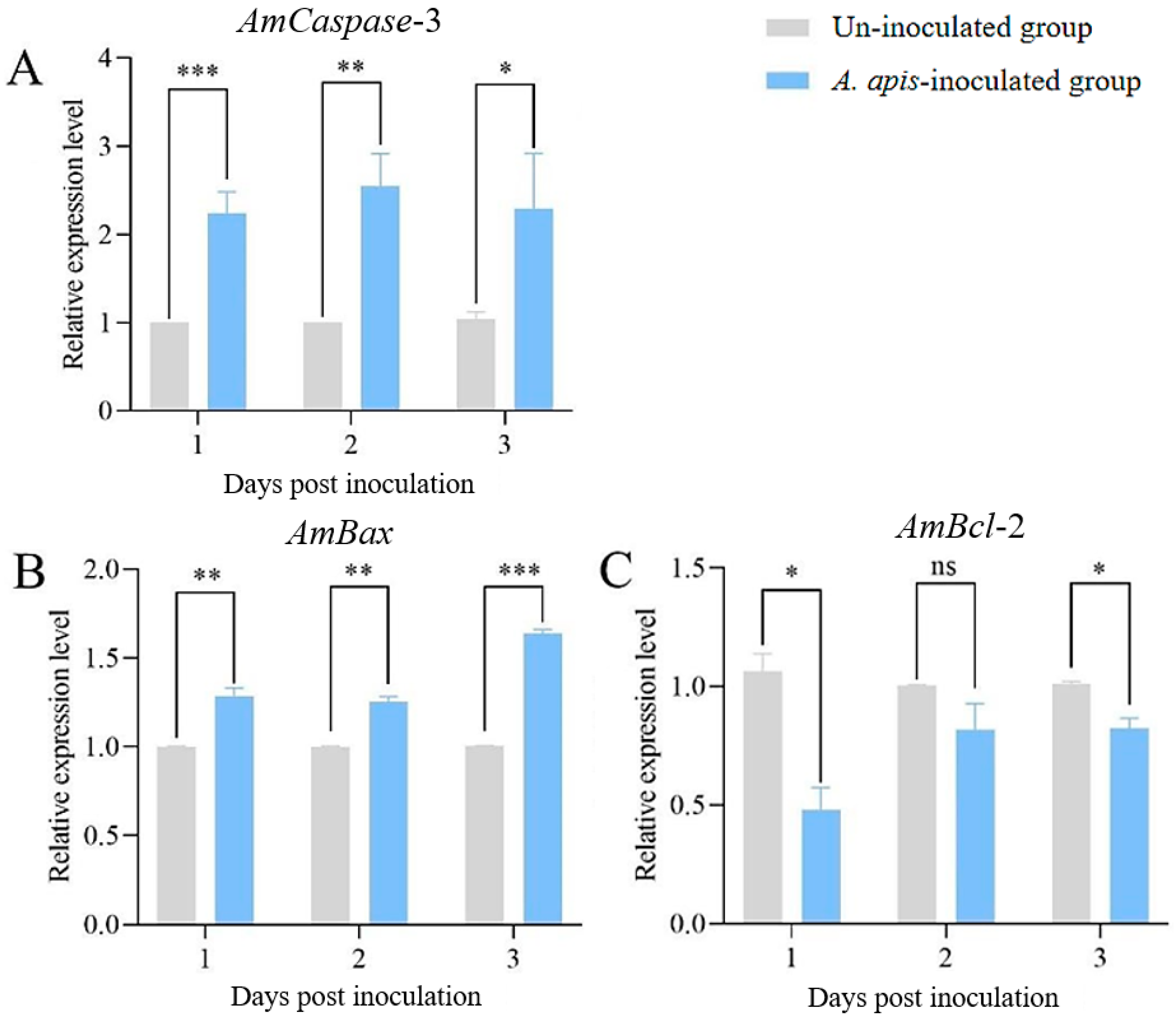
Figure 1. The relative expression levels of AmCaspase-3 (A), AmBax (B), and AmBcl-2 (C) in the guts of worker larvae inoculated with A. apis. Data are presented as mean ± SEM. Multiple t tests, Holm–Sidak method, *, p < 0.05; **, p < 0.01; ***, p < 0.001; ns, not significant.
Insects 16 00925 g001
Figure 2. TUNEL assay of apoptosis in the gut. Cross-section of A. apis-infected larval gut (A), Cross-section of uninfected control larva (B), Longitudinal section of A. apis-infected larval gut (C), Longitudinal section of uninfected control larva (D).
Insects 16 00925 g002
Figure 3. The survival rate of worker larvae fed apoptosis inhibitors and activators. (A) Schematic diagram of the feeding method with apoptosis inhibitors and activators. (BD) Larval survival rates following administration of 10 μM (B), 50 μM (C), or 100 μM (D) activators/inhibitors. log-rank test, *, p < 0.05; ns, not significant.
Insects 16 00925 g003
Figure 4. Relative expression levels of AmCaspase-3, AmBax, and AmBcl-2 following the inhibition and activation of apoptosis in the gut of worker larvae infected by A. apis (A), and the virulence factor Ste11-like in A. apis (B). Data are presented as mean ± SEM. Multiple t tests, Holm–Sidak method, **, p < 0.01; ***, p < 0.001, ****, p < 0.0001; ns, not significant.
Insects 16 00925 g004
Figure 5. Schematic diagram depicting the apoptotic status of host gut cells, expression changes in apoptosis-related genes, and expression patterns of fungal virulence factors in the gut of A. apis-infected honeybee larvae during pathogenesis. The upward red arrow indicates a upregulated expression of the gene; the downward blue arrow indicates a downregulated expression of the gene.
Insects 16 00925 g005
Table 1. Primer information.
GeneSequence (5′-3′)Purpose
GAPDHF: CACCTTCTGCAAAATTATGGCG
R: ACCTTTGCCAAGTCTAACTGTTAA
Reference gene for RT-qPCR
Caspase-3F: ACCTGATCACTCGTTCCACT
R: AGCAAGATGGAAAACGTGTGT
RT-qPCR
Bcl-2F: GAATATGTAGCCCGCATCTTTT
R: CTTTGTTGATTAGACTTGCCGA
BaxF: CTGCTGCAGGAGTTTACATTCA
R: AAGTAAAGGACCCAGGCCAA
Actin apisF: CATGATTGGTATGGGTCAG
R: CGTTGAAGGTCTCGAAGAC
Reference gene for detection of virulence factors in A. apis
Ste11-likeF: GGGAAGATTGCCAGGCC
R: CACTTGTAGTCCGGATG
Detection of virulence factors in A. apis
Disclaimer/Publisher’s Note: The statements, opinions and data contained in all publications are solely those of the individual author(s) and contributor(s) and not of MDPI and/or the editor(s). MDPI and/or the editor(s) disclaim responsibility for any injury to people or property resulting from any ideas, methods, instructions or products referred to in the content.