
Subcapsular Biloma following Endoscopic Retrograde Cholangiopancreatography and Endoscopic Biliary Sphincterotomy: A Case Report with a Mini Review of Literature

 , , , ,
  ,
, 
Abstract
1. Introduction
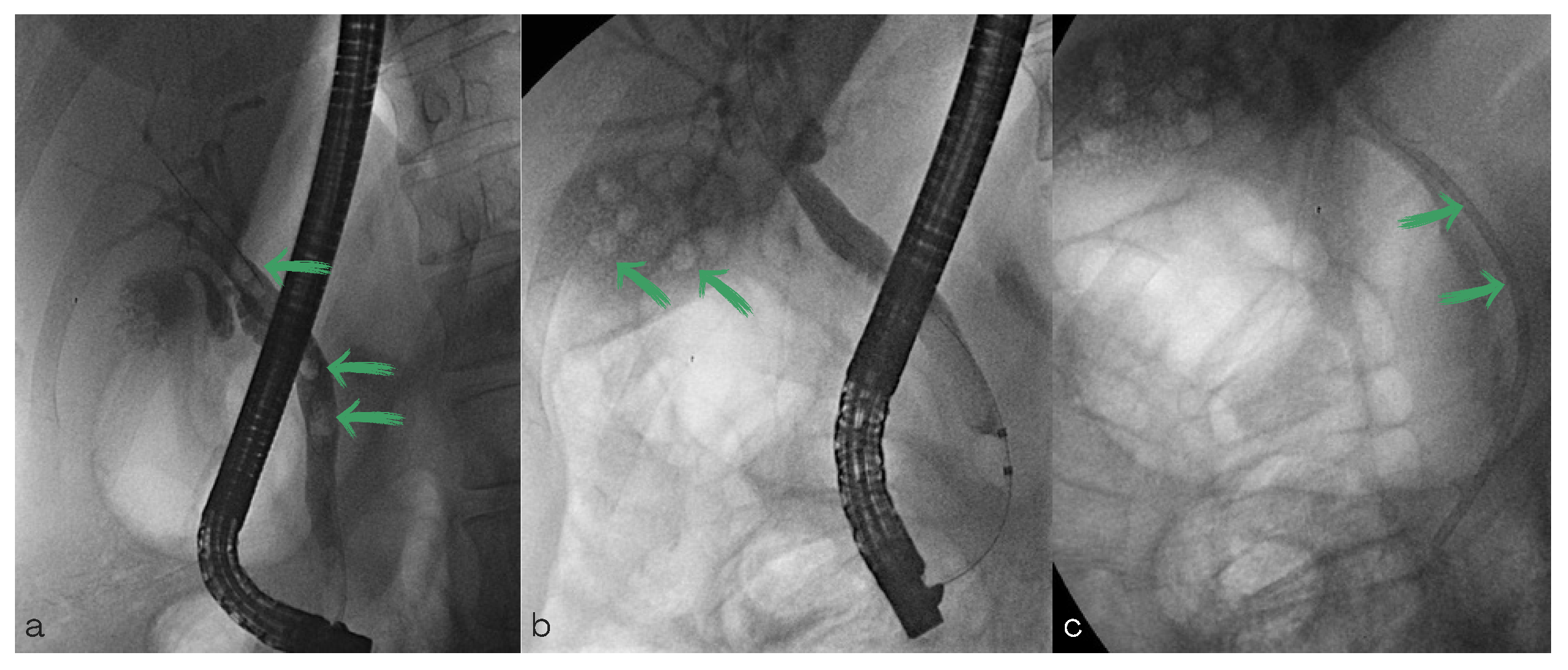
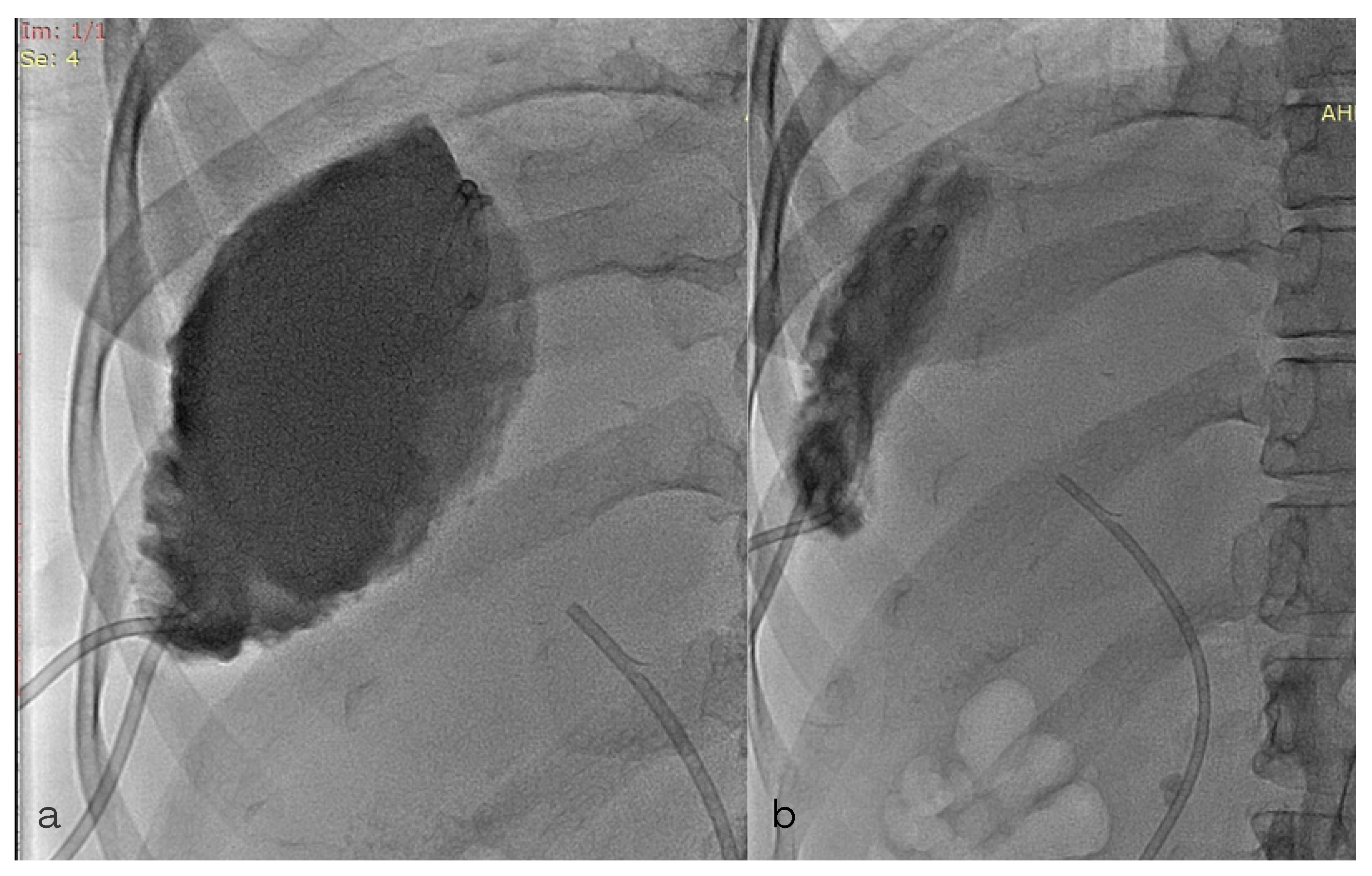
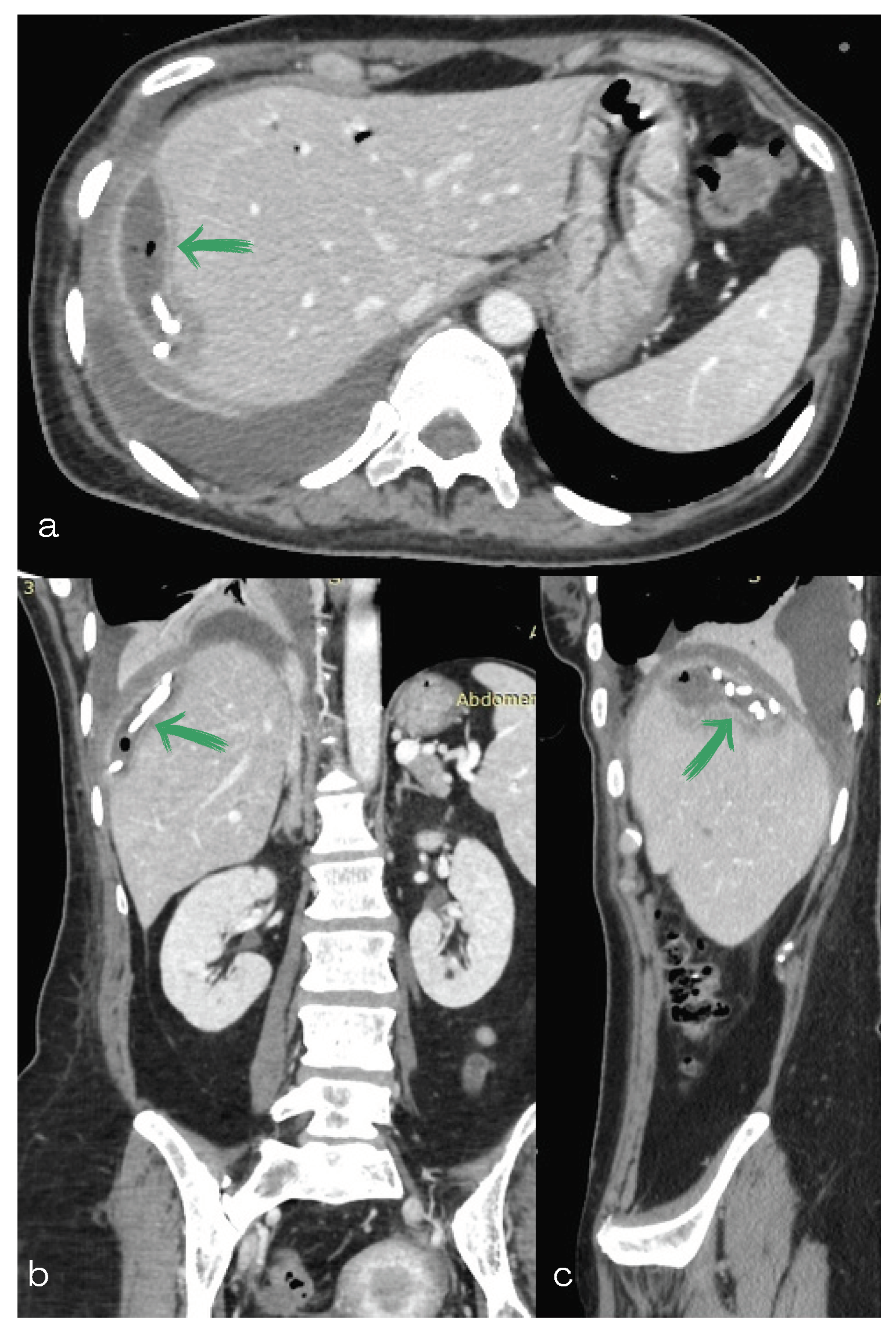
2. Case Report
3. Discussion
Author Contributions
Funding
Institutional Review Board Statement
Informed Consent Statement
Data Availability Statement
Conflicts of Interest
References
- Gould, L.; Patel, A. Ultrasound detection of extrahepatic encapsulated bile: “Biloma”. Am. J. Roentgenol. 1979, 132, 1014–1015. [Google Scholar] [CrossRef] [PubMed]
- Copelan, A.; Bahoura, L.; Tardy, F.; Kirsch, M.; Sokhandon, F.; Kapoor, B. Etiology, Diagnosis, and Management of Bilomas: A Current Update. Tech. Vasc. Interv. Radiol. 2015, 18, 236–243. [Google Scholar] [CrossRef] [PubMed]
- Faisaluddin, M.; Bansal, R.; Iftikhar, P.M.; Arastu, A.H. A Rare Case Report of Biloma After Cholecystectomy. Cureus 2019, 11, e5459. [Google Scholar] [CrossRef] [PubMed]
- Balfour, J.; Ewing, A. Hepatic Biloma. In StatPearls [Internet]; StatPearls Publishing: Treasure Island, FL, USA, 2022. [Google Scholar]
- Yadav, A.; Condati, N.K.; Mukund, A. Percutaneous Transhepatic Biliary Interventions. J. Clin. Interv. Radiol. ISVIR 2018, 2, 27–37. [Google Scholar] [CrossRef]
- Yousaf, M.N.; D’Souza, R.G.; Chaudhary, F.; Ehsan, H.; Sittambalam, C. Biloma: A Rare Manifestation of Spontaneous Bile Leak. Cureus 2020, 12, e8116. [Google Scholar] [CrossRef]
- Meseeha, M.; Attia, M. Endoscopic Retrograde Cholangiopancreatography; StatPearls Publishing: Treasure Island, FL, USA, 2022. [Google Scholar]
- Kwon, C.; Song, S.H.; Hahm, K.B.; Ko, K.H. Unusual complications related to endoscopic retrograde cholangiopancreatography and its endoscopic treatment. Clin. Endosc. 2013, 46, 3. [Google Scholar] [CrossRef]
- Dupas, J.L.; Mancheron, H.; Sevenet, F.; Delamarre, J.; Delcenserie, R.; Capron, J. Hepatic subcapsular biloma. An unusual complication of endoscopic retrogade cholangiopancreatography. Gastroenterology 1988, 94, 1225–1227. [Google Scholar] [CrossRef]
- Alkhateeb, H.M.; Aljanabi, T.J.; Al-azzawi, K.H.; Alkarboly, T.A. Huge biloma after endoscopic retrogade cholangiopancreatography and endoscopic biliary sphincterotomy. Int. J. Surg. Case Rep. 2015, 16, 7–11. [Google Scholar] [CrossRef]
- Jafari, A. A rare case of hepatic subcapsular biloma after laparoscopic cholecystectomy and subsequent endoscopic retrograde cholangiopancreatography. Caspian J. Intern. Med. 2018, 9, 198–200. [Google Scholar]
- Szary, N.M.; Al-Kawas, F.H. Complications of endoscopic retrograde cholangiopancreatography: How to avoid and manage them. Gastroenterol. Hepatol. 2013, 9, 496–504. [Google Scholar]
- Harshna, V.V.; Ronald, S.A. Imaging and Intervention of Biliary Leaks and Bilomas. Dig. Dis. Interv. 2017, 1, 014–021. [Google Scholar]
- Weerakkody, Y.; Jones, J.B. Reference Article. Available online: https://radiopaedia.org/ (accessed on 1 September 2022).
- Snyder, E.; Banks, K.P. Hepatobiliary Scintigraphy. In StatPearls [Internet]; StatPearls Publishing: Treasure Island, FL, USA, 2022. [Google Scholar]
- Fatima, J.; Baron, T.H.; Topazian, M.D.; Houghton, S.G.; Iqbal, C.W.; Ott, B.J.; Farley, D.R.; Farnell, M.B.; Sarr, M.G. Pancreaticobiliary and Duodenal Perforations After Periampullary Endoscopic Procedures: Diagnosis and Management. Arch Surg. 2007, 142, 448–455. [Google Scholar] [CrossRef]


Disclaimer/Publisher’s Note: The statements, opinions and data contained in all publications are solely those of the individual author(s) and contributor(s) and not of MDPI and/or the editor(s). MDPI and/or the editor(s) disclaim responsibility for any injury to people or property resulting from any ideas, methods, instructions or products referred to in the content. |
© 2023 by the authors. Licensee MDPI, Basel, Switzerland. This article is an open access article distributed under the terms and conditions of the Creative Commons Attribution (CC BY) license (https://creativecommons.org/licenses/by/4.0/).
Share and Cite
Pentara, N.V.; Ioannidis, A.; Tzikos, G.; Kougias, L.; Karlafti, E.; Chorti, A.; Tsalkatidou, D.; Michalopoulos, A.; Paramythiotis, D. Subcapsular Biloma following Endoscopic Retrograde Cholangiopancreatography and Endoscopic Biliary Sphincterotomy: A Case Report with a Mini Review of Literature. Diagnostics 2023, 13, 831. https://doi.org/10.3390/diagnostics13050831
Pentara NV, Ioannidis A, Tzikos G, Kougias L, Karlafti E, Chorti A, Tsalkatidou D, Michalopoulos A, Paramythiotis D. Subcapsular Biloma following Endoscopic Retrograde Cholangiopancreatography and Endoscopic Biliary Sphincterotomy: A Case Report with a Mini Review of Literature. Diagnostics. 2023; 13(5):831. https://doi.org/10.3390/diagnostics13050831
Chicago/Turabian StylePentara, Natalia Valeria, Aristidis Ioannidis, Georgios Tzikos, Leonidas Kougias, Eleni Karlafti, Angeliki Chorti, Despoina Tsalkatidou, Antonios Michalopoulos, and Daniel Paramythiotis. 2023. "Subcapsular Biloma following Endoscopic Retrograde Cholangiopancreatography and Endoscopic Biliary Sphincterotomy: A Case Report with a Mini Review of Literature" Diagnostics 13, no. 5: 831. https://doi.org/10.3390/diagnostics13050831
APA StylePentara, N. V., Ioannidis, A., Tzikos, G., Kougias, L., Karlafti, E., Chorti, A., Tsalkatidou, D., Michalopoulos, A., & Paramythiotis, D. (2023). Subcapsular Biloma following Endoscopic Retrograde Cholangiopancreatography and Endoscopic Biliary Sphincterotomy: A Case Report with a Mini Review of Literature. Diagnostics, 13(5), 831. https://doi.org/10.3390/diagnostics13050831