
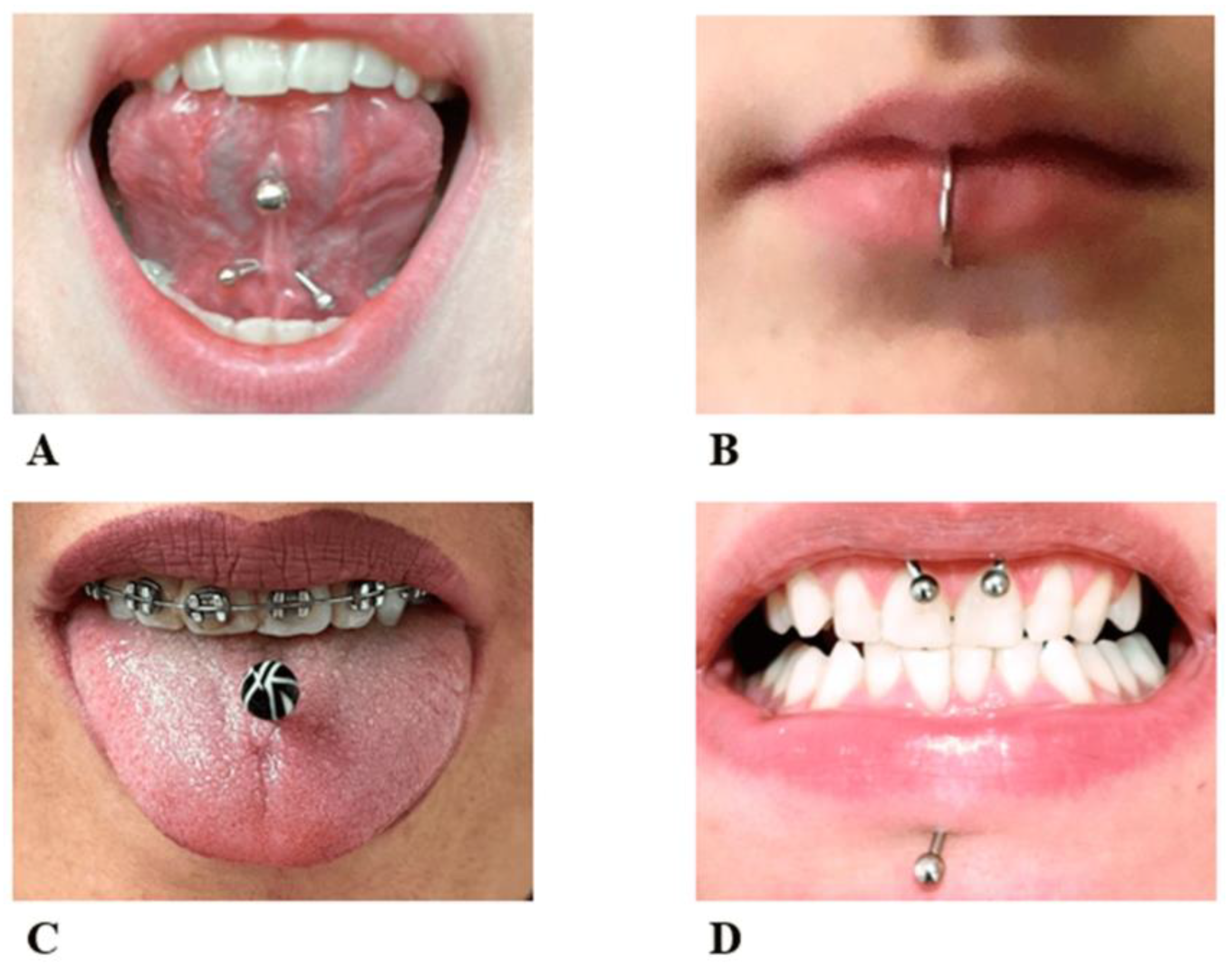
Oral Complications Associated with the Piercing of Oral and Perioral Tissues and the Corresponding Degree of Awareness among Public and Professionals: A Systematic Review

 ,
,  ,
,  and
and
Abstract
:1. Introduction
2. Materials and Methods
2.1. Search Strategy
2.2. Inclusion and Exclusion Criteria
2.3. Screening
2.4. Data Extraction
2.5. Assessment of Quality
3. Results
3.1. Study Design
3.2. Study Characteristics
3.3. Quality Assessment
3.4. Studied Outcomes
3.4.1. Public/Professional Awareness
3.4.2. Periodontal/Peri-Implant Complications
3.4.3. Microbiological Analyses
3.4.4. General and Hard/Soft Tissue Complications
3.4.5. Frequency of Complications
4. Discussion
Biases and Potential Confounders
5. Conclusions
Supplementary Materials
Author Contributions
Funding
Institutional Review Board Statement
Informed Consent Statement
Data Availability Statement
Conflicts of Interest
References
- Pejcic, A.; Kojovic, D.; Mirkovic, D. Oral Piercing and Its Complications in Two Serbian Youths A Case Report and Review of the Literature. W. Indian Med. J. 2012, 61, 838–843. [Google Scholar] [PubMed]
- Ziebolz, D.; Hildebrand, A.; Proff, P.; Rinke, S.; Hornecker, E.; Mausberg, R.F. Long-term effects of tongue piercing—A case control study. Clin. Oral Investig. 2012, 16, 231–237. [Google Scholar] [CrossRef] [PubMed]
- Abbasi, K.; Tavakolizadeh, S.; Hadi, A.; Hosseini, M.; Soufdoost, R.S.; Heboyan, A.; Alam, M.; Fani-Hanifeh, S. The wound healing effect of collagen/adipose-derived stem cells (ADSCs) hydrogel: In vivo study. Vet. Med. Sci. 2023, 9, 282–289. [Google Scholar] [CrossRef] [PubMed]
- Covello, F.; Salerno, C.; Giovannini, V.; Corridore, D.; Ottolenghi, L.; Vozza, I. Piercing and oral health: A study on the knowledge of risks and complications. Int. J. Environ. Res. Public Health 2020, 17, 613. [Google Scholar] [CrossRef] [PubMed]
- Muldoon, K.A. Body piercing in adolescents. J. Pediatr. Health Care 1997, 11, 298–301. [Google Scholar] [CrossRef]
- Marya, A.; Rokaya, D.; Heboyan, A.; Fernandes, G.V.O. Biomolecular and Biochemical Aspects of the Oral Cavity. Molecules 2022, 27, 8676. [Google Scholar] [CrossRef]
- Greif, J.; Hewitt, W.; Armstrong, M.L. Tattooing and body piercing: Body art practices among college students. Clin. Nurs. Res. 1999, 8, 368–385. [Google Scholar] [CrossRef]
- Mayers, L.B.; Judelson, D.A.; Moriarty, B.W.; Rundell, K.W. Prevalence of Body Art (Body Piercing and Tattooing) in University Undergraduates and Incidence of Medical Complications. Mayo Clin. Proc. 2002, 77, 29–34. [Google Scholar] [CrossRef]
- Plessas, A.; Pepelassi, E. Dental and periodontal complications of lip and tongue piercing: Prevalence and influencing factors. Aust. Dent. J. 2012, 57, 71–78. [Google Scholar] [CrossRef]
- Ziebolz, D.; Söder, F.; Hartl, J.F.; Kottmann, T.; Rinke, S.; Schmalz, G. Comprehensive assessment of dental behaviour and oral status in patients with tongue piercing—Results of a cross-sectional study. Clin. Oral Investig. 2020, 24, 971–977. [Google Scholar] [CrossRef]
- Samoilenko, A.; Orishchenko, V.Y.; Strelchenia, T.; Strelchenia, O. Oral piercing is a new challenge in the dentistry. Medicni Perspekt. 2019, 24, 133–144. [Google Scholar] [CrossRef]
- Hennequin-Hoenderdos, N.; Slot, D.; Van der Weijden, G. The prevalence of oral and peri-oral piercings in young adults: A systematic review. Int. J. Dent. Hyg. 2012, 10, 223–228. [Google Scholar] [CrossRef]
- Hennequin-Hoenderdos, N.; Slot, D.; Van der Weijden, G. Complications of oral and peri-oral piercings: A summary of case reports. Int. J. Dent. Hyg. 2011, 9, 101–109. [Google Scholar] [CrossRef] [PubMed]
- Sindoni, A.; Valeriani, F.; Protano, C.; Liguori, G.; Spica, V.R.; Vitali, M.; Gallè, F. Health risks for body pierced community: A systematic review. Public Health 2022, 205, 202–215. [Google Scholar] [CrossRef] [PubMed]
- Malcangi, G.; Patano, A.; Palmieri, G.; Riccaldo, L.; Pezzolla, C.; Mancini, A.; Inchingolo, A.D.; Di Venere, D.; Piras, F.; Inchingolo, F.; et al. Oral Piercing: A Pretty Risk-A Scoping Review of Local and Systemic Complications of This Current Widespread Fashion. Int. J. Env. Res. Public Health 2023, 20, 5744. [Google Scholar] [CrossRef]
- Passos, P.F.; Pintor, A.V.B.; Marañón-Vásquez, G.A.; Campos, T.; Abrahão, A.C.; Ferreira, D.; Maia, L.C.; Primo, L.G.; Visconti, M.A. Oral manifestations arising from oral piercings: A systematic review and meta-analyses. Oral Surg. Oral Med. Oral Pathol. Oral Radiol. 2022, 134, 327–341. [Google Scholar] [CrossRef]
- Srimaneepong, V.; Heboyan, A.; Zafar, M.S.; Khurshid, Z.; Marya, A.; Fernandes, G.V.O.; Rokaya, D. Fixed Prosthetic Restorations and Periodontal Health: A Narrative Review. J. Funct. Biomater. 2022, 13, 15. [Google Scholar] [CrossRef]
- Page, M.J.; McKenzie, J.E.; Bossuyt, P.M.; Boutron, I.; Hoffmann, T.C.; Mulrow, C.D.; Shamseer, L.; Tetzlaff, J.M.; Akl, E.A.; Brennan, S.E.; et al. The PRISMA 2020 statement: An updated guideline for reporting systematic reviews. BMJ 2021, 372, n71. [Google Scholar] [CrossRef]
- Higgins, J.P. Cochrane Handbook for Systematic Reviews of Interventions, Version 5.0.1; The Cochrane Collaboration: London, UK, 2008; Available online: http://www.cochrane-handbook.org (accessed on 20 June 2023).
- Moga, C.; Guo, B.; Schopflocher, D.; Harstall, C. Development of a Quality Appraisal Tool for Case Series Studies Using a Modified DELPHI Technique; Institute of Health Economics: Edmonton, AB, Canada, 2012. [Google Scholar]
- Stang, A. Critical evaluation of the Newcastle-Ottawa scale for the assessment of the quality of nonrandomized studies in meta-analyses. Eur. J. Epidemiol. 2010, 25, 603–605. [Google Scholar] [CrossRef]
- Ibraheem, W.I.; S Preethanath, R.; Devang Divakar, D.; Al-Askar, M.; Al-Kheraif, A.A. Effect of tongue piercing on periodontal and peri-implant health: A cross-sectional case-control study in adults. Int. J. Dent. Hyg. 2022, 20, 282–290. [Google Scholar] [CrossRef]
- Junco, P.; Barrios, R.; Ruiz, M.J.; Bravo, M. Educational intervention about oral piercing knowledge among dental students and adolescents at schools. Int. Dent. J. 2017, 67, 294–298. [Google Scholar] [CrossRef] [PubMed]
- King, E.; Brewer, E.; Brown, P. Oral piercings and their complications–how confident are we as a profession? Br. Dent. J. 2018, 224, 887–895. [Google Scholar] [CrossRef] [PubMed]
- Mejersjö, C.; Ovesson, D.; Mossberg, B. Oral parafunctions, piercing and signs and symptoms of temporomandibular disorders in high school students. Acta Odontol. Scand. 2016, 74, 279–284. [Google Scholar] [CrossRef] [PubMed]
- Schmidt, J.C.; Calderaro, S.; Weiger, R.; Walter, C. On the association between oral piercings and periodontal conditions—A case series. Int. J. Dent. Hyg. 2019, 17, 318–326. [Google Scholar] [CrossRef] [PubMed]
- Vozza, I.; Fusco, F.; Bove, E.; Ripari, F.; Corridore, D.; Ottolenghi, L. Awareness of risks related to oral piercing in Italian piercers. Pilot study in Lazio Region. Ann.Stomatol. 2014, 5, 128. [Google Scholar]
- Ziebolz, D.; Söder, F.; Hartl, J.F.; Kottmann, T.; Rinke, S.; Merle, C.L.; Schmalz, G. Prevalence of periodontal pathogenic bacteria at different oral sites of patients with tongue piercing—Results of a cross sectional study. Diagn. Microbiol. Infect. Dis. 2019, 95, 114888. [Google Scholar] [CrossRef]
- Ziebolz, D.; Hornecker, E.; Mausberg, R. Microbiological findings at tongue piercing sites–implications to oral health. Int. J. Dent. Hyg. 2009, 7, 256–262. [Google Scholar] [CrossRef]
- Zadik, Y.; Burnstein, S.; Derazne, E.; Sandler, V.; Ianculovici, C.; Halperin, T. Colonization of Candida: Prevalence among tongue-pierced and non-pierced immunocompetent adults. Oral Dis. 2010, 16, 172–175. [Google Scholar] [CrossRef]
- Vilchez-Perez, M.A.; Angeles Fuster-Torres, M.; Figueiredo, R.; Valmaseda-Castellón, E.; Gay-Escoda, C. Periodontal health and lateral lower lip piercings: A split-mouth cross-sectional study. J. Clin. Periodontol. 2009, 36, 558–563. [Google Scholar] [CrossRef]
- Vieira, E.P.; Ribeiro, A.L.R.; João de Jesus, V.P.; Alves Jr, S.d.M. Oral piercings: Immediate and late complications. J. Oral Maxillofac. Surg. 2011, 69, 3032–3037. [Google Scholar] [CrossRef]
- Ventä, I.; Lakoma, A.; Haahtela, S.; Peltola, J.; Ylipaavalniemi, P.; Turtola, L. Oral piercings among first-year university students. Oral Surg. Oral Med. Oral Pathol. Oral Radiol. Endodontol. 2005, 99, 546–549. [Google Scholar] [CrossRef] [PubMed]
- Saquet, P.N.; Saleh, S.B.; da Costa MARCHIORI, J.; Pozzobon, R. Perfil dos usuários depiercingoral e implicações decorrentes de seu uso. RGO 2009, 57, 41–45. [Google Scholar]
- Pearose, M.M.; Perinpanayagam, M.K.; ChinKit-Wells, M.D. Trends in oral piercing in Buffalo, New York, high schools. N. Y. State Dent. J. 2006, 72, 30. [Google Scholar] [PubMed]
- Oberholzer, T.G.; George, R. Awareness of complications of oral piercing in a group of adolescents and young South African adults. Oral Surg. Oral Med. Oral Pathol. Oral Radiol. Endodontol. 2010, 110, 744–747. [Google Scholar] [CrossRef] [PubMed]
- Lorenzini, G.; Magnani, M.; Di Vece, L.; Viviano, M.; Bertelli, E. Oral piercing: Risks and complications. Dent. Cadmos 2008, 76, 55. [Google Scholar]
- López-Jornet, P.; Navarro-Guardiola, C.; Camacho-Alonso, F.; Vicente-Ortega, V.; Yánez-Gascon, J. Oral and facial piercings: A case series and review of the literature. Int. J. Dermatol. 2006, 45, 805–809. [Google Scholar] [CrossRef]
- López-Jornet, P.; Camacho-Alonso, F. Oral and dental complications of intra-oral piercing. J. Adolesc. Health 2006, 39, 767–769. [Google Scholar] [CrossRef]
- Kapferer, I.; Beier, U.S.; Persson, R.G. Tongue piercing: The effect of material on microbiological findings. J. Adolesc. Health 2011, 49, 76–83. [Google Scholar] [CrossRef]
- Kapferer, I.; Beier, U.S. Lateral lower lip piercing--Prevalence of associated oral complications: A split-mouth cross-sectional study. Quintessence Int. 2012, 43, 747–752. [Google Scholar]
- Inchingolo, F.; Tatullo, M.; Abenavoli, F.M.; Marrelli, M.; Inchingolo, A.D.; Palladino, A.; Inchingolo, A.M.; Dipalma, G. Oral piercing and oral diseases: A short time retrospective study. Int. J. Med. Sci. 2011, 8, 649. [Google Scholar] [CrossRef]
- Hickey, B.; Schoch, E.; Bigeard, L.; Musset, A. Complications following oral piercing. A study among 201 young adults in Strasbourg, France. Community Dent. Health 2010, 27, 35. [Google Scholar]
- Giuca, M.; Pasini, M.; Nastasio, S.; D’Ercole, S.; Tripodi, D. Dental and periodontal complications of labial and tongue piercing. J. Biol. Regul. Homeost. Agents 2012, 26, 553–560. [Google Scholar]
- Gill, J.B.; Karp, J.M.; Kopycka-Kedzierawski, D.T. Oral piercing injuries treated in United States emergency departments, 2002–2008. Pediatr. Dent. 2012, 34, 56–60. [Google Scholar] [PubMed]
- Firoozmand, L.M.; Paschotto, D.R.; Almeida, J.D. Oral piercing complications among teenage students. Oral Health Prev. Dent. 2009, 7, 77. [Google Scholar] [PubMed]
- Campbell, A.; Moore, A.; Williams, E.; Stephens, J.; Tatakis, D.N. Tongue piercing: Impact of time and barbell stem length on lingual gingival recession and tooth chipping. J. Periodontol. 2002, 73, 289–297. [Google Scholar] [CrossRef] [PubMed]
- Pires, I.L.O.; Cota, L.O.M.; Oliveira, A.C.B.; Costa, J.E.; Costa, F.O. Association between periodontal condition and use of tongue piercing: A case–control study. J. Clin. Periodontol. 2010, 37, 712–718. [Google Scholar] [CrossRef] [PubMed]
- Kieser, J.; Thomson, W.; Koopu, P.; Quick, A. Oral piercing and oral trauma in a New Zealand sample. Dent. Traumatol. 2005, 21, 254–257. [Google Scholar] [CrossRef] [PubMed]
- Levin, L.; Zadik, Y.; Becker, T. Oral and dental complications of intra-oral piercing. Dent. Traumatol. 2005, 21, 341–343. [Google Scholar] [CrossRef] [PubMed]
- De Moor, R.; De Witte, A.; Delmé, K.; De Bruyne, M.; Hommez, G.; Goyvaerts, D. Dental and oral complications of lip and tongue piercings. Br. Dent. J. 2005, 199, 506–509. [Google Scholar] [CrossRef]
- De Moor, R.; De Witte, A.; De Bruyne, M. Tongue piercing and associated oral and dental complications. Dent. Traumatol. Case Rep. 2000, 16, 232–237. [Google Scholar] [CrossRef]
- Escudero-Castaño, N.; Perea-García, M.A.; Campo-Trapero, J.; Cano, S.; Bascones-Martínez, A. Oral and perioral piercing complications. Open Dent. J. 2008, 2, 133–136. [Google Scholar] [CrossRef] [PubMed]
- Miranda-Rius, J.; Brunet-Llobet, L.; Lahor-Soler, E. The Periodontium as a Potential Cause of Orofacial Pain: A Comprehensive Review. Open Dent. J. 2018, 12, 520–528. [Google Scholar] [CrossRef] [PubMed]
- Alarcón-Sánchez, M.A.; Heboyan, A.; Fernandes, G.V.O.; Castro-Alarcón, N.; Romero-Castro, N.S. Potential Impact of Prosthetic Biomaterials on the Periodontium: A Comprehensive Review. Molecules 2023, 28, 1075. [Google Scholar] [CrossRef]
- Hennequin-Hoenderdos, N.L.; Slot, D.; Van der Weijden, G. The incidence of complications associated with lip and/or tongue piercings: A systematic review. Int. J. Dent. Hyg. 2016, 14, 62–73. [Google Scholar] [CrossRef] [PubMed]
- Des Jarlais, D.C.; Lyles, C.; Crepaz, N.; Group, T. Improving the reporting quality of nonrandomized evaluations of behavioral and public health interventions: The TREND statement. Am. J. Public Health 2004, 94, 361–366. [Google Scholar] [CrossRef]
- Dundas, K.; Johnson, N.A.; Paras, L.; Hancock, S.; Barker, D.; Chiu, S.; James, E.L. Does Increasing the Experiential Component Improve Efficacy of the “This Is Public Health” Photo Essay Task? A Nonrandomized Trial. Pedagog. Health Promot. 2019, 5, 178–189. [Google Scholar] [CrossRef]
- Karobari, M.I.; Siddharthan, S.; Adil, A.H.; Khan, M.M.; Venugopal, A.; Rokaya, D.; Heboyan, A.; Marya, C.M.; Marya, A. Modifiable and Non-modifiable Risk Factors Affecting Oral and Periodontal Health and Quality of Life in South Asia. Open Dent. J. 2022, 16, e187421062209270. [Google Scholar] [CrossRef]
- Mosaddad, S.A.; Hussain, A.; Tebyaniyan, H. Green Alternatives as Antimicrobial Agents in Mitigating Periodontal Diseases: A Narrative Review. Microorganisms 2023, 11, 1269. [Google Scholar] [CrossRef]
- Stroup, D.F.; Berlin, J.A.; Morton, S.C.; Olkin, I.; Williamson, G.D.; Rennie, D.; Moher, D.; Becker, B.J.; Sipe, T.A.; Thacker, S.B. Meta-analysis of observational studies in epidemiology: A proposal for reporting. Jama 2000, 283, 2008–2012. [Google Scholar] [CrossRef]
- Dissemination, C. Systematic Reviews: CRD’s Guidance for Undertaking Reviews in Healthcare; CRD, University of York: York, UK, 2009. [Google Scholar]
- Reeves, B.C.; Deeks, J.J.; Higgins, J.P.; Wells, G.A. Cochrane Handbook for Systematic Reviews of Interventions; Cochrane Book Series; Cochrane: Chichester, UK, 2008; pp. 389–432. [Google Scholar]
- Kapferer, I.; Benesch, T.; Gregoric, N.; Ulm, C.; Hienz, S. Lip piercing: Prevalence of associated gingival recession and contributing factors. A cross-sectional study. J. Periodontal Res. 2007, 42, 177–183. [Google Scholar] [CrossRef]
- Dougherty, S.L.; Tervort-Bingham, K. Asessment of the Alveolar Bone Surrounding the Mandibular Anterior Teeth of Individuals Wearing a Tongue Stud. Am. Dent. Hyg. Assoc. 2005, 79, 8. [Google Scholar]


| Study | Design (Male/Female Ratio) | Population | No. of Cases (Age Mean ± SD) | Piercing | Evaluation | Outcome | Conclusion |
|---|---|---|---|---|---|---|---|
| Covello et al. (2020) | Case-series (NA) | Adolescents with oral piercing | 387 (NA) | Oral | They asked 387 people with oral piercings to complete a questionnaire anonymously. Additionally, 70 people were examined regarding dental health and gingiva recession. | The results of the analysis of the questionnaires showed that 46.8% of people did not know about the dangers of piercing, 70.6% of people said that they were not aware of the gingiva problems that may arise, and 60.4% of the people said that they did not know about the dangers of piercing to cause dental issues. Among the people under examination, 52.8% had poor oral health conditions, 42% had symptoms of generative gingivitis, 20% had 3 to 4 mm cavities, and 22% had tooth fracture(s) due to piercing. | There is not enough awareness about the complications caused by piercing, and the correct methods of maintaining oral piercings and oral piercings can be considered a potential risk to oral and dental health. Therefore, periodic examinations by specialists and dentists can effectively prevent and reduce the complications caused by oral piercing. |
| Ibraheem et al. (2022) | Case (15 (31.25%)/33 (68.75%)) control 19 (38.77%)/30 (61.23%) | Adolescents with oral piercing | 48 (38.2 ± 0.5)/49 (37.5 ± 0.2) | Tongue | They divided people into two experimental (48) and control (49) groups, including people with and without tongue piercing. The required information was collected via a questionnaire. | These people were examined regarding oral and dental health indicators such as entire mouth plaque and around the implant, gingival index, clinical attachment loss, and bone loss. Their results showed that in the experimental group, plaque index around the implant, gingival index, probing depth, and crestal bone loss were significantly higher in the anterior mandible. | Tongue piercing can increase the probability of periodontal diseases around the implant, especially in the anterior mandibular part. |
| Junco et al. (2017) | Case (25 (37.9%)/41 (62.1%)) Control (11 (45.8)/13 (54.2)) | dental students | 66 (21.6 ± 1.7)/28 (23.0 ± 1.4) | Oral | They designed a training program for 66 dental students, during which dental students’ knowledge about oral piercing was evaluated before, immediately after, and 12 months after the training program by answering a questionnaire. | The study’s findings showed a statistically significant difference between the groups of dental students before and after the educational intervention. | The oral piercing educational intervention had a favorable effect on dental students, especially among those more engaged in the learning process. |
| King et al. (2018) | Case-series (NA) | dental students | 53 (NA) | They collected this information from 200 dentists via a questionnaire. Only fifty-three dentists answered the questions. | Of this number, 24.5% were very aware of the side effects of piercing. However, most dentists (73.6%) stated that they obtained information empirically, and the recommendations provided varied significantly. | Dentists do not know enough about the risks and preventive recommendations for patients with piercings. | |
| Mejersjo et al. (2016) | Case-series (71 (57.25%)/53 (47.75%)) | High school students | 124 (18.1 ± 2.9) | Oral | Students in their study answered questionnaire questions about different functions and symptoms of temporomandibular disorders. Of this number, 116 students underwent clinical examination of the temporomandibular system and tooth wear estimation. | They included 124 high schools. 14% of students had oral piercings. Female students had more headaches, more severe symptoms, more medical consultations, and used more painkillers. They also had more oral piercings than boys. Oral piercing was associated with symptoms of headache, muscle sensitivity, daily nail-biting, and tooth wear. | There is a relationship between gingival health, nail-biting, and mouth piercing and symptoms of temporomandibular disorders. |
| Schmidt et al. (2019) | Case-series (4 (22.2%)/14 (77.8%)) | Adolescents with oral piercing | 18 (28.3 ± 7.7) | Tongue, lip, tongue, and lip | Their study included eighteen patients with tongue and lip piercings. This number had 14 tongue holes and seven lip holes. | In patients with tongue piercings, the percentage of bleeding sites on probing, probing pocket depth ≥6 mm, clinical attachment loss ≥6 mm, and gingival recession ≥2 mm increased in teeth compared to teeth unaffected by piercing. In patients with lip piercing, the periodontal findings in the teeth close to the piercing were not significantly different from those unaffected. | Tongue piercing could negatively affect the periodontal conditions of the teeth close to the piercing. |
| Vozza et al. (2014) | Case-series (NA) | Adolescents with oral piercing | 30 (NA) | Oral | They asked 30 people with piercings to answer a 20-question questionnaire. | 66.6% of people answered the questions. Only 20% of the people had enough information about the anatomy of the oral cavity, none of them knew about the anatomy of the tongue and gingiva, and only 10% said that a dental visit was necessary. Additionally, 40% of the respondents stated the need to take care of piercings. | The participant’s awareness of the potential risks of oral piercing is deficient. |
| Ziebolz et al. (2019) | Cross-sectional (Case-control) (20%/80%) | Adolescents with oral piercing | 50 NA/NA (28.3 ± 7.1/28.2 ± 7.1) | Tongue | Fifty participants were placed in each group. They took samples from the piercing surface, periodontal pocket, and tongue and examined for the presence of 11 potential periodontal pathogenic bacteria. | Most of the investigated bacteria were identified in the periodontal pocket of the piercing group compared to the control group, and a significant relationship was observed between the piercing surface and the periodontal pocket. | The tongue-piercing surface should be considered an important ecological site and reservoir for periodontal pathogens. |
| Ziebolz et al. (2020) | Case-control | Adolescents with oral piercing | 50/50 | Tongue | They put 50 participants with tongue piercings and 50 without piercings into two experimental and control groups, respectively. The dental examination included missing- and filled-teeth-index and non-carious tooth defects. The periodontal examination had periodontal probing depth, bleeding on probing, and recession. The factors related to piercing and oral health-related quality of life were evaluated using questionnaires. | People with tongue piercings suffered worse from missing- and filled-teeth-index, periodontal probing depth, bleeding on probing, and recession. In addition, a higher prevalence of tooth enamel cracks and dent-shaped scratches was observed in the piercing group, and most of the participants had tongue piercings, worse verbal behavior, insufficient cleaning of piercings, and in 80% of cases, mass formation on the surface of the piercing, as well as oral health-related quality of life. | Patients with tongue piercing lack good dental and periodontal health, so paying more attention to patients who use tongue piercing in dentistry is necessary. |
| Ziebolz et al. (2009) | Case-series (7 (58.33%)/5 (41.64%)) | Adolescents with tongue piercings | 12 (24.0 ± 2.8) | Tongue | Participants filled out a particular questionnaire detailing their piercing details, including the material used, the length of time the device was in place, their personal oral and piercing cleanliness practices, and whether or not they smoked. The DNA of the 11 periodontopathogenic bacteria was also analyzed by polymerase chain reaction (PCR) from microbiological samples obtained from the surface of the piercing jewelry next to the tongue hole. | The duration of their tongue piercings ranged from 2 years to 8 years. According to the microbiological examination, all instances had an elevated or significantly elevated concentration of periodontopathogenic bacteria. It became clear that the transition from bacteria with a moderate periodontopathogenic potential to microorganisms with a high periodontopathogenic potential was more prominent the longer a piercing had been in situ. | In the absence of good oral hygiene, prolonged use of tongue jewelry might lead to the colonization of periodontopathogenic bacteria at the piercing site. It is essential to educate both prospective and present piercers about the potential for adverse consequences, including risks to oral health, and on the requirement of frequently cleaning piercing jewelry with a CHX solution or another suitable disinfectant. |
| Ziebolz et al. (2012) | Case (46 (100%)/0 (0%))-control (46 (100%)/0 (0%)) | Adolescents with tongue piercings | 46 (22.2)/46 (22.1) | Tongue | Those with TP from the German Federal Armed Forces (group TP) and a control group with similar demographics (group C) volunteered for the research. It was recorded how long TP was left in place, where it was found, and what it was made of. During their dental checkups, they checked for caries, calculus, plaque, gingival disease, enamel fissures (EF), enamel fractures (EC), and recessions (R). | Enamel fissures, enamel fractures, and lingual recessions are more common in those who have tongue piercings. However, compared to the control group, the EF, EC, and R number was more significant in the tongue-piercing group. There was a statistically significant (p < 0.001) gap between the two groups. | This case–control research, within its scope, has shown that tongue piercing has negative long-term consequences. Enamel fissures, enamel fractures, and gingival recessions (particularly in the lingual area of mandibular incisors) were shown to be much more common in those who had tongue piercings. |
| Zadik et al. (2010) | Case (NA)-control (NA) | Adolescents with tongue piercings | 115 (20.4 ± 1.6)/86 (20.7 ± 1.7) | Tongue | Swabs were taken from the anterior lingual mucosa of young people who had just pierced their tongues. Additionally, patients with non-intra-oral face piercings served as a control group. Colonization by Candida was studied by light microscopy. Chromagar Candida plates were used to re-cultivate the specimens that tested positive. | Tongue-pierced people were more likely to be colonized with Candida than facially pierced people. Candida albicans were found in every single colony. Current tongue ornament wearers and non-wearers did not vary significantly from one another. Multivariate analysis revealed that tongue piercing and smoking more than ten cigarettes daily were the only significant positive influencing variables on colonization. | There was no correlation between the presence or absence of an ornament in a tongue piercing and an increased risk of Candida albicans colonization. |
| Vilchez-Perez et al. (2009) | Cross-sectional (11 (22%)/39 (78%)) | Adolescents with lip piercings | 50 (21.3 ± 4.4) | Lateral lower lip | Patients had their piercing and non-piercing sides examined for periodontal disease, dental health, and mucosal health. | Women made up the vast majority (78%) of piercing enthusiasts. Gingival recession was more common, and the quantity of keratinized and connected gingiva was less on the piercing side. Most people lost their canine and initial bicuspid teeth. The piercing side had double the rate of tooth fractures and cracks compared to the non-pierced side. Seven individuals had abnormalities in their mucosal lining. | Piercing the lower lip on the side increases gingival recession and decreases the quantity of keratinized, connected gingiva. These accessories are also linked to broken or cracked teeth. |
| Vieira et al. (2011) | Case-series (13 (33.3%)/26 (66.7%)) | Adolescent with oral piercings | 39 (NA) | Oral | Complications from oral piercings were examined in 42 instances, including 39 young people who were now or formerly pierced in the mouth. | Twenty-nine patients had immediate problems, the most common of which were prolonged bleeding (69%) and discomfort (52.3%). There were two reported occurrences of syncope. In 97.6% of instances, patients reported late problems such as discomfort or edema at the insertion site of the piercing. In 33.3% and 31% of cases, respectively, problems involving the surrounding tissues, such as dental discomfort and tongue lacerations, were reported. | Anyone considering getting an oral piercing should be informed of the risks involved, most of which are limited to the immediate area. Those interested in being pierced should only do so under trained specialists’ care and maintain regular dental checkups to ensure early diagnosis of any potential complications. |
| Ventä et al. (2005) | Case-control (49 (21%)/185 (79%) | First-year university students with oral piercing | 234 (20.6 ± 0.6) | Oral | Using the Beck Depression Inventory and other questionnaires, they measured the number of teeth with decay, gaps, and restorations (DMF index). | 3.4% of the population has an oral piercing. There were no significant differences between the groups in any of the DMF indicators—students who had piercings tended to drool more often than their peers who did not. The study group had much higher rates of cigarette and illegal drug use, as well as depression, than the control group. | Oral piercings need special attention to aftercare because of the potential for oral complications. |
| Saquet et al. (2009) | Case-series (NA) | Adolescents with oral piercings | 51 (NA) | Tongue or lip | Fifty-one people who have had oral piercings were surveyed. | Most respondents who got piercings in their mouths said they did so to show their individuality. Over half of those who did so reported experiencing oral and/or general changes due to piercings. | Most piercings are bad for the person getting them; thus, dentists should advise their patients who have oral piercings to be prepared for any complications and provide them the attention they need to avoid serious health issues. |
| Pearose et al. (2006) | Case-series (NA) | High school adolescents obtaining an oral piercing | 508 (NA) | Oral | Five high schools were polled by questionnaire. | Only 49 out of 508 respondents (10%) reported having an oral piercing. Swelling, soreness, numbness, taste loss, bleeding, and pus were some of the side effects of the piercing. Little to no care was given to oral piercings. Injuries to the mouth, especially the teeth, were prevalent. | Researchers found that although the number of adolescents undergoing oral piercings was low, those who did so without parental permission often had infection symptoms. |
| Oberholzer et al. (2010) | Case-series (55 (22%)/195 (78%)) | South African adults with intraoral piercings | 250 (19.6 ± 5.3) | Intraoral | They were given a questionnaire to fill up with their knowledge of oral piercing risks. Then, two qualified dentists checked them out to see if there were any problems with their teeth from the piercing. | Most responders (59.4%) said they had no idea oral piercing may cause difficulties. In the recent year, 24% of respondents had an intraoral piercing, while another 17.2% got one between the ages of 5 and 7 years ago. | The dentistry community should address the widespread lack of knowledge about the risks of mouth piercing. |
| Lorenzini et al. (2008) | Cross-sectional (NA) | People having oral and perioral piercing | 44 (NA) | Oral and perioral | Any person was given a complete physical examination and had their medical and dental histories recorded. | Oral and perioral piercings were associated with at least one problem in 96% of the sample, either immediately after the procedure or later on. Mucosal atrophy, difficulties eating or speaking, gingival recessions, tooth wear, enamel chipping or cracking, dentinal hypersensitivity, and excessive salivation were the most common adverse effects. | The observed problems are consistent with those described in the published literature. The authors noticed a correlation between the number of times people had mouth piercings and how long those piercings lasted. |
| López-Jornet et al. (2006) | Case-series (27 (18.7%)/43 (81.3)) | Healthy individuals with oral and facial piercings | 70 (17.08 ± 2.61) | Oral and facial | A standardized methodology was implemented to evaluate potential side effects (such as inflammation, discomfort, or tooth changes). | Pain was the most common reported problem, occurring in 60% of patients, followed by inflammation (34.3%), bleeding (24%), tooth fractures or fissures (20%), and gingival damage (26.7%). | Pain, swelling, and dental issues are all things you might expect after getting your tongue pierced. |
| López-Jornet et al. (2006) | Case-series (29 (29.9%)/68 (70.1%)) | Individuals with intra-oral piercings | 98 (20.06 ± 4.75) | Intraoral | The potential side effects were evaluated by dental examination. | In 23 patients (23.5%), recessions of the gingiva were seen, most often around the mandibular incisors. Thirteen instances (13.3%) had tooth structural damage. | Therefore, it is important to warn patients about the potential dangers of intra-oral piercings to their teeth and gingiva. |
| Kapferer et al. (2011) | Case-series (12 (15%)/68 (85%)) | Individuals with intra-Tongue piercings | 85 (22.74 ± 4.47) | Tongue | Bacterial samples were taken from tongue piercings constructed of various materials and analyzed using checkerboard deoxyribonucleic acid-deoxyribonucleic acid hybridization to determine the microbial differences between the materials. | There were 61 lingual recessions reported by 28.8% of participants, and 5% had at least one chipped tooth. Samples taken from studs and piercing channels seldom included periodontitis-related bacteria, except for Aggregatibacter actinomycetemcomitans (Y4), Fusobacterium nucleatum species, and Parvimonas micra. Compared to polytetrafluoroethylene and polypropylene perforations, a sample from stainless steel revealed considerably more significant levels of 67 of the 80 bacterial species studied. | There is little chance of contracting an oral infection after getting your tongue pierced, as shown by the low bacterial numbers in the piercing channels. Steel studs were more conducive to biofilm formation than polytetrafluoroethylene or polypropylene studs, indicating that the latter may be more resistant to bacterial colonization. If the piercing channel is contaminated, the presence of Staphylococci on the steel or titanium stud may indicate a higher risk for complications. |
| Kapferer et al. (2012) | Cross-sectional (NA) | Nondental setting population with lateral lower lip piercing | 47 (NA) | Lateral lower lip | Full-mouth plaque and bleeding indexes, probing depth, recession, clinical attachment level, periodontal biotype, assessment of hard tissues, occlusal damage, stud features, and mucosal inspection and palpation were all part of the comprehensive clinical examination. | Four of the experimental teeth and one of the control teeth showed signs of mid-buccal recession. Most people lost their canines and front teeth. One of the test teeth chipped, while none of the control teeth did. Teeth in the experimental group had substantially more plaque than teeth in the control group. | Plaque buildup on teeth next to piercings is much more significant in those who have had their lower lips pierced laterally. Some people have tooth chipping or buccal recession after a lateral lower lip piercing. |
| Inchingolo et al. (2011) | Retrospective (74 (69%)/34 (31%)) | Individuals who had an oral piercing performed by a healthcare professional | 108 (NA) | Oral | Clinical examinations were performed on all of the patients to detect the occurrence of any late problems. | No severe problems occurred among the 108 individuals. All patients claimed that they had followed the piercers’ recommendations, yet 96% of them still had postoperative local issues, such as bleeding within 12 h of the piercing (90%), perilesional edema for 3.5 days after the piercing procedure (80%), and persistent mucosal atrophy (70%). | There are fewer risks and fewer infection opportunities with a medical practitioner doing the piercing. |
| Hickey et al. (2010) | Case-series (55 (27.36%)/146 (72.64%)) | Individuals with oral piercing | 201 (22.7) | Oral | They were asked to complete a questionnaire on what they knew about the dangers of oral piercings. | Only about a quarter of piercings go well, with the complication rate varying widely depending on how close the piercing was to the mouth. 8.5% of the group with knowledge of difficulties had a gingival recession, while 6.9% experienced chipped teeth. The recession was linked to titanium, stainless steel, and Teflon in 52.9%, 23.5%, and 9% of this population, whereas chipped teeth occurred in 35.7%, 42.5%, and 14.3%. | Complications happened a lot. More research, including dental checkups and public education, is required. |
| Giuca et al. (2012) | Case (11 (44%)/14 (56%))-control (11 (44%)/14 (56%)) | Patients with a minimum of one labial or tongue piercing | 25 (23.4 ± 3.6)/25 (NA) | Labial and tongue | Each patient was asked to complete a questionnaire and undergo a physical examination. | Labial and lingual piercing individuals were likelier to have irregular tooth wear and chipped teeth. In addition, those who’d had their tongues or lips pierced had a more significant gingival recession than those who had not. No discernible changes were found when comparing the two groups for clinical attachment loss and pocket depth. Thirteen people who had piercings for more than four years had a significantly higher incidence of tooth and periodontal problems, suggesting a correlation between piercing length and dental malformations. | Tongue piercings are more common than lip piercings, and so are dental abnormalities. Tongue and lip piercings both cause gingival recession. Dental abnormalities, gingival recession, attachment loss, and probing depth of teeth next to pierced locations are more common, and the longer the period, the more severe tongue and lip piercings are worn. Gingival recession rates are related to the morphology of ornaments. |
| Gill et al. (2012) | Retrospective 6794 (27.78%)/ 17665 (72.22%) | Oral piercing injuries collected from 2002 to 2008 | 24459 (NA) | Oral | They analyzed by injury type, anatomic site, and mechanism of injury according to age, gender, and race. | Lip and tongue wounds accounted for 46%, while tooth injuries accounted for 10% of all reported cases. The leading causes of damage were infections (42%), followed by soft tissue puncture wounds (29%). Overgrown mucosa around oral piercings is the leading cause of emergency department visits (39%). The need for hospitalization was very uncommon. | Teenagers and young adults account for most oral piercing injuries seen in American hospital emergency rooms. Data collected nationally suggests that emergency room dentists should be prepared to treat hard and soft tissue issues resulting from oral piercings. |
| Firoozmand et al. (2009) | Case-series (506 (55.58%)/421 (45.45%)) | Teenage students with oral piercing | 927 (16.14 ± 1.03) | Oral | Clinical examinations were performed on all subjects, and a questionnaire was sent to gather information on demographics, including gender, piercing site, mouth problems or modifications, and frequency of cleaning. | Only 3.6% of the children evaluated had oral piercings, with 69.70% attending public schools and 30.30% attending private ones. A slight majority of men (54.55%) outnumbered girls (45.45%). Most people (66.6%) had their tongues pierced. In 74.3% of the instances, piercing led to the difficulties and modifications expected from its usage. | Oral piercing was only seen in 3.6% of high school students in the group investigated, and it was linked to some minor health issues in those who had it done. |
| Campbell et al. (2002) | Case-series (31 (60%)/21 (30%)) | Adults with tongue piercings | 52 (22 ± 5) | Tongue | Patients were examined for gingival recession on the lingual aspect of the 12 anterior teeth and tooth chipping anywhere in the mouth. | No one had lingual recession or tooth chipping within the group of people whose tongues were pierced between 0 and 2 years ago. Half of the participants who wore lengthy barbells for 2+ years had lingual regression on their mandibular central incisors. After four years or more of having their tongues pierced, 47% of patients had chipped molars or premolars. | The front mandibular teeth and the posterior molars are more likely to have lingual recession after a tongue piercing. These side effects are more common in those who use a tongue barbell often. Recession and chipping seem to affect barbells of varying stem lengths. Efforts to end tongue piercing should focus on young adults, who comprise most of those who acquire them. |
| Pires et al. (2010) | Case (27 (45%)/33 (55.5%)) control (43 (35.8%)/77 (64.2%)) | Individuals with tongue piercings | 60 (18.9 ± 3.9)/120 (17.78 ± 3.8) | Tongue | Periodontal characteristics and tooth fractures were among the factors considered in the clinical review of patients’ oral health records. | It was shown that gingival recession was more common and severe in the case group than in the control group. Tongue piercings were associated with an increased risk of gingival recession in the anterior lingual mandibular area by a factor of 11 compared to those without piercings. In the front lingual mandibular area, piercing usage, older age, male gender, and bleeding on probing were all linked with gingival recession. | There was a significant correlation between tongue piercings and gingival recession in the anterior lingual mandibular area. |
| Kieser et al. (2005) | Case-series (3 (7%)/40 (93%)) | Adult dental patients with tongue and lip piercings | 43 (21 ± 5) | Tongue and lip | A questionnaire was given to patients after they had undergone an intraoral examination. | 76.7% of participants had their tongues pierced, 34.9% had their lips pierced, and 11.6% had both. Only 4 of the piercings had been performed by a medical professional—problems arising after a piercing were noted by 34.9% of patients. Gingival recession was present in at least one labial piercing site in eighty percent of those with them and almost one-third of those with tongue piercings. The chances of developing lingual recession increased by 1.17 for every year beyond the age of 14; this trend was statistically significant. While age did predict the incidence of labial recession and the number of afflicted sites, it did not predict the number of lingual sites with recession. No correlations between piercings and either abnormal tooth wear or damage were found. | These results indicate a possible link between oral piercings and localized gingival recession, suggesting that patients should be warned of the potential risks to their periodontal health as part of the informed consent process. |
| Levin et al. (2005) | Case-series (210 (54%)/179 (46%)) | Young adults with oral piercing | 389 (20.08 ± 1.1) | Oral | Patients were given a questionnaire on oral piercing, complication awareness, and piercing-related complication incidence before their dental checkups. | Lingual piercing was the most prevalent form of oral piercing, with 79 individuals (20.3%) reporting they had one. Forty-one (51.9%) and thirty-six (45.7%) individuals experienced swelling and bleeding after piercing. Of the whole sample, 225 people (57.8%) had no idea that having an intra-oral piercing may be harmful. On closer inspection, 15 teeth were broken in 11 (or 13.9%) of the pierces. Twenty-one patients (or 26.6%) were found to have gingival recessions, most often around the mandibular incisors. | As the number of patients with pierced intra- and perioral locations rises, dentists should be prepared to advise their patients on the risks and benefits of this body modification. |
| De Moor et al. (2000) | Cross-sectional Study (NA) | Patients with tongue-piercing | 15 (NA) | Body of the tongue, anterior to the lingual frenum | Individuals were evaluated based on clinical and radiological criteria | Teeth chipping was reported as the most prevalent dental issue. In addition, there were two broken teeth and four broken cusps. Selective dental abrasion was reported once—the majority of patients presented with trauma to the anterior lingual gingiva. Only 2 of the 15 polled people reported experiencing saliva flow. Patients did not report any difficulties with eating, chewing, or swallowing. A single incident of galvanic current generation due to the appliance was reported. | Based on the collected data, it was concluded that the dentistry community should play a more significant role in informing patients about tongue and oral piercing risks. |
| De Moor et al. (2005) | Case-series (13 (26%)/37 (74%)) | Patients with oral piercing | 50 (NA) | Oral and perioral | Individuals were evaluated based on clinical and radiological criteria | Chipped teeth were the most often reported dental issue, and they were most prevalent after getting a tongue piercing. Lip piercing with studs was associated with gingival regression. Oedema, bleeding, and infection were some of the postoperative problems. | More power should be provided to dentists and oral and maxillofacial surgeons to counsel patients who have or are considering oral and facial piercings. |
| Studies | Selection | Comparability | Outcome | Score |
|---|---|---|---|---|
| Ibraheem et al. (2022) | ★★★★ | ★ | ★★ | 7 |
| Schmidt et al. (2019) | ★★★★ | ★★ | ★★ | 8 |
| Ziebolz et al. (2019) | ★★★★ | ★ | ★★ | 7 |
| Ziebolz et al. (2020) | ★★★★ | ★★ | ★★ | 8 |
| Ziebolz et al. (2012) | ★★ | ★★ | ★★ | 6 |
| Zadik et al. (2010) | ★★ | ★ | ★★ | 5 |
| Ventä et al. (2005) | ★★ | ★★ | ★★ | 6 |
| Giuca et al. (2012) | ★★ | ★★ | ★★ | 6 |
| Pires et al. (2010) | ★★★ | ★★ | ★★ | 7 |
| Studies | Selection | Comparability | Outcome | Score |
|---|---|---|---|---|
| Inchingolo et al. (2011) | ★★★ | ★ | ★★ | 6 |
| Gill et al. (2012) | ★★★★ | ★★ | ★★ | 8 |
| Studies | Selection | Comparability | Outcome | Score |
|---|---|---|---|---|
| Vilchez-Perez et al. (2009) | ★★ | ★ | ★★ | 5 |
| Lorenzini et al. (2008) | ★★★ | ★★ | ★★★ | 8 |
| Kapferer et al. (2012) | ★★★★ | ★★ | ★★ | 8 |
| De Moor et al. (2000) | ★★ | ★ | ★ | 4 |
| Study Objective | Study Design | Study Population | Intervention and Co-Intervention | Outcome Measure | Statistical Analysis | Results and Conclusions | Estimated Risk of Bias | ||||||||||||||
|---|---|---|---|---|---|---|---|---|---|---|---|---|---|---|---|---|---|---|---|---|---|
| QN | 1 | 2 | 3 | 4 | 5 | 6 | 7 | 8 | 9 | 10 | 11 | 12 | 13 | 14 | 15 | 16 | 17 | 18 | 19 | 20 | |
| Study | |||||||||||||||||||||
| Covello et al. (2020) | + | + | + | + | + | + | + | NA | NA | + | NA | + | NA | + | + | NA | + | + | + | + | low |
| King et al. (2018) | + | + | ? | + | + | + | + | NA | NA | + | NA | + | NA | + | + | NA | + | + | + | + | low |
| Mejersjo et al. (2016) | + | + | + | + | + | + | + | NA | NA | + | NA | + | NA | + | + | NA | + | + | + | + | low |
| Schmidt et al. (2019) | + | + | + | + | + | + | + | NA | NA | + | NA | + | NA | + | + | NA | + | + | + | + | low |
| Vozza et al. (2014) | + | + | - | + | + | + | + | NA | NA | + | NA | + | NA | + | + | NA | + | + | + | + | low |
| Ziebolz et al. (2009) | + | + | - | + | + | + | + | NA | NA | + | NA | + | NA | + | + | NA | + | + | + | - | low |
| Vieira et al. (2011) | + | + | + | + | - | + | + | NA | NA | + | NA | + | NA | + | + | NA | + | + | + | - | low |
| Saquet et al. (2009) | + | + | + | + | ? | + | + | NA | NA | + | NA | + | NA | + | + | NA | ? | + | + | + | low |
| Pearose et al. (2006) | + | + | + | + | + | + | + | NA | NA | + | NA | + | NA | + | + | NA | + | + | + | + | low |
| Oberholzer et al. (2010) | + | + | + | + | + | - | + | NA | NA | + | NA | + | NA | + | + | NA | + | + | + | + | low |
| López-Jornet et al. (2006) | + | + | - | + | + | + | + | NA | NA | + | NA | + | NA | + | + | NA | + | + | + | + | low |
| López-Jornet et al. (2006) (2) | + | + | - | + | + | + | + | NA | NA | + | NA | + | NA | + | + | NA | + | + | + | - | low |
| Kapferer et al. (2011) | + | + | + | + | ? | + | + | NA | NA | + | NA | + | NA | + | + | NA | + | + | + | - | low |
| Hickey et al. (2010) | + | + | + | + | + | + | + | NA | NA | + | NA | + | NA | + | + | NA | + | + | + | + | low |
| Firoozmand et al. (2009) | + | + | - | + | + | - | + | NA | NA | + | NA | + | NA | - | - | NA | - | - | + | ? | high |
| Campbell et al. (2002) | + | + | + | - | - | + | + | NA | NA | + | NA | + | NA | - | + | NA | - | + | + | + | moderate |
| Kieser et al. (2005) | + | + | - | - | + | - | + | NA | NA | + | NA | ? | NA | + | + | NA | - | + | + | - | moderate |
| Levin et al. (2005) | + | + | + | + | + | - | + | NA | NA | + | NA | + | NA | + | + | NA | - | + | ? | - | moderate |
| De Moor et al. (2005) | + | + | + | + | - | + | + | NA | NA | ? | NA | ? | NA | - | + | NA | + | + | + | - | moderate |
Disclaimer/Publisher’s Note: The statements, opinions and data contained in all publications are solely those of the individual author(s) and contributor(s) and not of MDPI and/or the editor(s). MDPI and/or the editor(s) disclaim responsibility for any injury to people or property resulting from any ideas, methods, instructions or products referred to in the content. |
© 2023 by the authors. Licensee MDPI, Basel, Switzerland. This article is an open access article distributed under the terms and conditions of the Creative Commons Attribution (CC BY) license (https://creativecommons.org/licenses/by/4.0/).
Share and Cite
Mosaddad, S.A.; Talebi, S.; Hemmat, M.; Karimi, M.; Jahangirnia, A.; Alam, M.; Abbasi, K.; Yazadaniyan, M.; Hussain, A.; Tebyaniyan, H.; et al. Oral Complications Associated with the Piercing of Oral and Perioral Tissues and the Corresponding Degree of Awareness among Public and Professionals: A Systematic Review. Diagnostics 2023, 13, 3371. https://doi.org/10.3390/diagnostics13213371
Mosaddad SA, Talebi S, Hemmat M, Karimi M, Jahangirnia A, Alam M, Abbasi K, Yazadaniyan M, Hussain A, Tebyaniyan H, et al. Oral Complications Associated with the Piercing of Oral and Perioral Tissues and the Corresponding Degree of Awareness among Public and Professionals: A Systematic Review. Diagnostics. 2023; 13(21):3371. https://doi.org/10.3390/diagnostics13213371
Chicago/Turabian StyleMosaddad, Seyed Ali, Sahar Talebi, Maryam Hemmat, Mohammadreza Karimi, Alireza Jahangirnia, Mostafa Alam, Kamyar Abbasi, Mohsn Yazadaniyan, Ahmed Hussain, Hamid Tebyaniyan, and et al. 2023. "Oral Complications Associated with the Piercing of Oral and Perioral Tissues and the Corresponding Degree of Awareness among Public and Professionals: A Systematic Review" Diagnostics 13, no. 21: 3371. https://doi.org/10.3390/diagnostics13213371
APA StyleMosaddad, S. A., Talebi, S., Hemmat, M., Karimi, M., Jahangirnia, A., Alam, M., Abbasi, K., Yazadaniyan, M., Hussain, A., Tebyaniyan, H., & Abdollahi Namanloo, R. (2023). Oral Complications Associated with the Piercing of Oral and Perioral Tissues and the Corresponding Degree of Awareness among Public and Professionals: A Systematic Review. Diagnostics, 13(21), 3371. https://doi.org/10.3390/diagnostics13213371