
Comparison of Bleb Morphology following PRESERFLO® MicroShunt and Trabeculectomy Using Anterior Segment OCT


Abstract
:1. Introduction
2. Materials and Methods
2.1. Surgical Technique
TET with Application of MMC
2.2. Bleb Examination Using AS-OCT
3. Results
4. Discussion
Author Contributions
Funding
Institutional Review Board Statement
Informed Consent Statement
Data Availability Statement
Conflicts of Interest
References
- Rulli, E.; Biagioli, E.; Riva, I.; Gambirasio, G.; De Simone, I.; Floriani, I.; Quaranta, L. Efficacy and safety of trabeculectomy vs nonpenetrating surgical procedures: A systematic review and meta-analysis. JAMA Ophthalmol. 2013, 131, 1573–1582. [Google Scholar] [CrossRef]
- Picht, G.; Grehn, F. Development of the filtering bleb after trabeculectomy. Classification, histopathology, wound healing process. Ophthalmologe 1998, 95, W380–W387. [Google Scholar] [CrossRef]
- Cantor, L.B.; Mantravadi, A.; WuDunn, D.; Swamynathan, K.; Cortes, A. Morphologic classification of filtering blebs after glaucoma filtration surgery: The Indiana Bleb Appearance Grading Scale. J. Glaucoma 2003, 12, 266–271. [Google Scholar] [CrossRef]
- Khaw, P.T.; Chang, L.; Wong, T.T.; Mead, A.; Daniels, J.T.; Cordeiro, M.F. Modulation of wound healing after glaucoma surgery. Curr. Opin. Ophthalmol. 2001, 12, 143–148. [Google Scholar] [CrossRef]
- Wells, A.P.; Crowston, J.G.; Marks, J.; Kirwan, J.F.; Smith, G.; Clarke, J.C.; Shah, R.; Vieira, J.; Bunce, C.; Murdoch, I.; et al. A pilot study of a system for grading of drainage blebs after glaucoma surgery. J. Glaucoma 2004, 13, 454–460. [Google Scholar] [CrossRef]
- Savini, G.; Zanini, M.; Barboni, P. Filtering blebs imaging by optical coherence tomography. Clin. Exp. Ophthalmol. 2005, 33, 483–489. [Google Scholar] [CrossRef]
- Leung, C.K.; Yick, D.W.; Kwong, Y.Y.; Li, F.C.; Leung, D.Y.; Mohamed, S.; Tham, C.C.; Chung-chai, C.; Lam, D.S. Analysis of bleb morphology after trabeculectomy with Visante anterior segment optical coherence tomography. Br. J. Ophthalmol. 2007, 91, 340–344. [Google Scholar] [CrossRef]
- Singh, M.; Chew, P.T.; Friedman, D.S.; Nolan, W.P.; See, J.L.; Smith, S.D.; Zheng, C.; Foster, P.J.; Aung, T. Imaging of trabeculectomy blebs using anterior segment optical coherence tomography. Ophthalmology 2007, 114, 47–53. [Google Scholar] [CrossRef]
- Khamar, M.B.; Soni, S.R.; Mehta, S.V.; Srivastava, S.; Vasavada, V.A. Morphology of functioning trabeculectomy blebs using anterior segment optical coherence tomography. Indian J. Ophthalmol. 2014, 62, 711–714. [Google Scholar] [CrossRef]
- Howlett, J.; Vahdani, K.; Rossiter, J. Bulbar Conjunctival and Tenon’s Layer Thickness Measurement using Optical Coherence Tomography. J. Curr. Glaucoma Pract. 2014, 8, 63–66. [Google Scholar] [CrossRef]
- Tan, J.C.K.; Muntasser, H.; Choudhary, A.; Batterbury, M.; Vallabh, N.A. Swept-Source Anterior Segment Optical Coherence Tomography Imaging and Quantification of Bleb Parameters in Glaucoma Filtration Surgery. Bioengineering 2023, 10, 1186. [Google Scholar] [CrossRef]
- Solus, J.F.; Jampel, H.D.; Tracey, P.A.; Gilbert, D.L.; Loyd, T.L.; Jefferys, J.L.; Quigley, H.A. Comparison of limbus-based and fornix-based trabeculectomy: Success, bleb-related complications, and bleb morphology. Ophthalmology 2012, 119, 703–711. [Google Scholar] [CrossRef]
- Gambini, G.; Carla, M.M.; Giannuzzi, F.; Boselli, F.; Grieco, G.; Caporossi, T.; De Vico, U.; Savastano, A.; Baldascino, A.; Rizzo, C.; et al. Anterior Segment-Optical Coherence Tomography Bleb Morphology Comparison in Minimally Invasive Glaucoma Surgery: XEN Gel Stent vs. PreserFlo MicroShunt. Diagnostics 2022, 12, 1250. [Google Scholar] [CrossRef]
- Hasan, S.M.; Theilig, T.; Papadimitriou, M.; Meller, D. A Comparative Analysis of Morphology and Dimensions of Functional Blebs following PRESERFLO-Microshunt and XEN-Gel-Stent, a Study Using Anterior Segment OCT. Diagnostics 2023, 13, 2318. [Google Scholar] [CrossRef]
- Beckers, H.J.M.; Aptel, F.; Webers, C.A.B.; Bluwol, E.; Martinez-de-la-Casa, J.M.; Garcia-Feijoo, J.; Lachkar, Y.; Mendez-Hernandez, C.D.; Riss, I.; Shao, H.; et al. Safety and Effectiveness of the PRESERFLO(R) MicroShunt in Primary Open-Angle Glaucoma: Results from a 2-Year Multicenter Study. Ophthalmol. Glaucoma 2022, 5, 195–209. [Google Scholar] [CrossRef]
- Ibarz Barbera, M.; Hernandez-Verdejo, J.L.; Bragard, J.; Morales-Fernandez, L.; Rodriguez-Carrillo, L.; Martinez Galdon, F.; Tana, P.; Teus, M.A. Bleb geometry and morphology after Preserflo Microshunt surgery: Risk factors for surgical failure. PLoS ONE 2023, 18, e0286884. [Google Scholar] [CrossRef]
- Ibarz Barbera, M.; Morales Fernandez, L.; Tana Rivero, P.; Gomez de Liano, R.; Teus, M.A. Anterior-segment optical coherence tomography of filtering blebs in the early postoperative period of ab externo SIBS microshunt implantation with mitomycin C: Morphological analysis and correlation with intraocular pressure reduction. Acta Ophthalmol. 2022, 100, e192–e203. [Google Scholar] [CrossRef]
- Lenzhofer, M.; Strohmaier, C.; Hohensinn, M.; Hitzl, W.; Sperl, P.; Gerner, M.; Steiner, V.; Moussa, S.; Krall, E.; Reitsamer, H.A. Longitudinal bleb morphology in anterior segment OCT after minimally invasive transscleral ab interno Glaucoma Gel Microstent implantation. Acta Ophthalmol. 2019, 97, e231–e237. [Google Scholar] [CrossRef]
- Hasan, S.M.; Theilig, T.; Tarhan, M.; Papadimitriou, M.; Unterlauft, J.D.; Meller, D. Novel Tomographical Bleb Classification Following ab-interno Implantation of Gel-Stent Using Anterior Segment Optical Coherence Tomography. J. Glaucoma 2022, 32, 117–126. [Google Scholar] [CrossRef]
- Oh, L.J.; Wong, E.; Lam, J.; Clement, C.I. Comparison of bleb morphology between trabeculectomy and deep sclerectomy using a clinical grading scale and anterior segment optical coherence tomography. Clin. Exp. Ophthalmol. 2017, 45, 701–707. [Google Scholar] [CrossRef]
- Baker, N.D.; Barnebey, H.S.; Moster, M.R.; Stiles, M.C.; Vold, S.D.; Khatana, A.K.; Flowers, B.E.; Grover, D.S.; Strouthidis, N.G.; Panarelli, J.F.; et al. Ab-Externo MicroShunt versus Trabeculectomy in Primary Open-Angle Glaucoma: One-Year Results from a 2-Year Randomized, Multicenter Study. Ophthalmology 2021, 128, 1710–1721. [Google Scholar] [CrossRef]
- Batlle, J.F.; Corona, A.; Albuquerque, R. Long-term Results of the PRESERFLO MicroShunt in Patients with Primary Open-angle Glaucoma from a Single-center Nonrandomized Study. J. Glaucoma 2021, 30, 281–286. [Google Scholar] [CrossRef]
- Hasan, S.M.; Theilig, T.; Tarhan, M.; Papadimitriou, M.; Meller, D. Bleb morphology using optical coherence tomography: After primary implantation of XEN gel stent and open conjunctival revision. Die Ophthalmol. 2022, 120, 529–537. [Google Scholar] [CrossRef]
- Waibel, S.; Spoerl, E.; Furashova, O.; Pillunat, L.E.; Pillunat, K.R. Bleb Morphology after Mitomycin-C Augmented Trabeculectomy: Comparison between Clinical Evaluation and Anterior Segment Optical Coherence Tomography. J. Glaucoma 2019, 28, 447–451. [Google Scholar] [CrossRef]
- Tominaga, A.; Miki, A.; Yamazaki, Y.; Matsushita, K.; Otori, Y. The assessment of the filtering bleb function with anterior segment optical coherence tomography. J. Glaucoma 2010, 19, 551–555. [Google Scholar] [CrossRef]
- Hamanaka, T.; Omata, T.; Sekimoto, S.; Sugiyama, T.; Fujikoshi, Y. Bleb analysis by using anterior segment optical coherence tomography in two different methods of trabeculectomy. Investig. Ophthalmol. Vis. Sci. 2013, 54, 6536–6541. [Google Scholar] [CrossRef]
- Kojima, S.; Inoue, T.; Nakashima, K.; Fukushima, A.; Tanihara, H. Filtering blebs using 3-dimensional anterior-segment optical coherence tomography: A prospective investigation. JAMA Ophthalmol. 2015, 133, 148–156. [Google Scholar] [CrossRef]


| Parameter | Abbreviation | Description | Remarks |
|---|---|---|---|
|
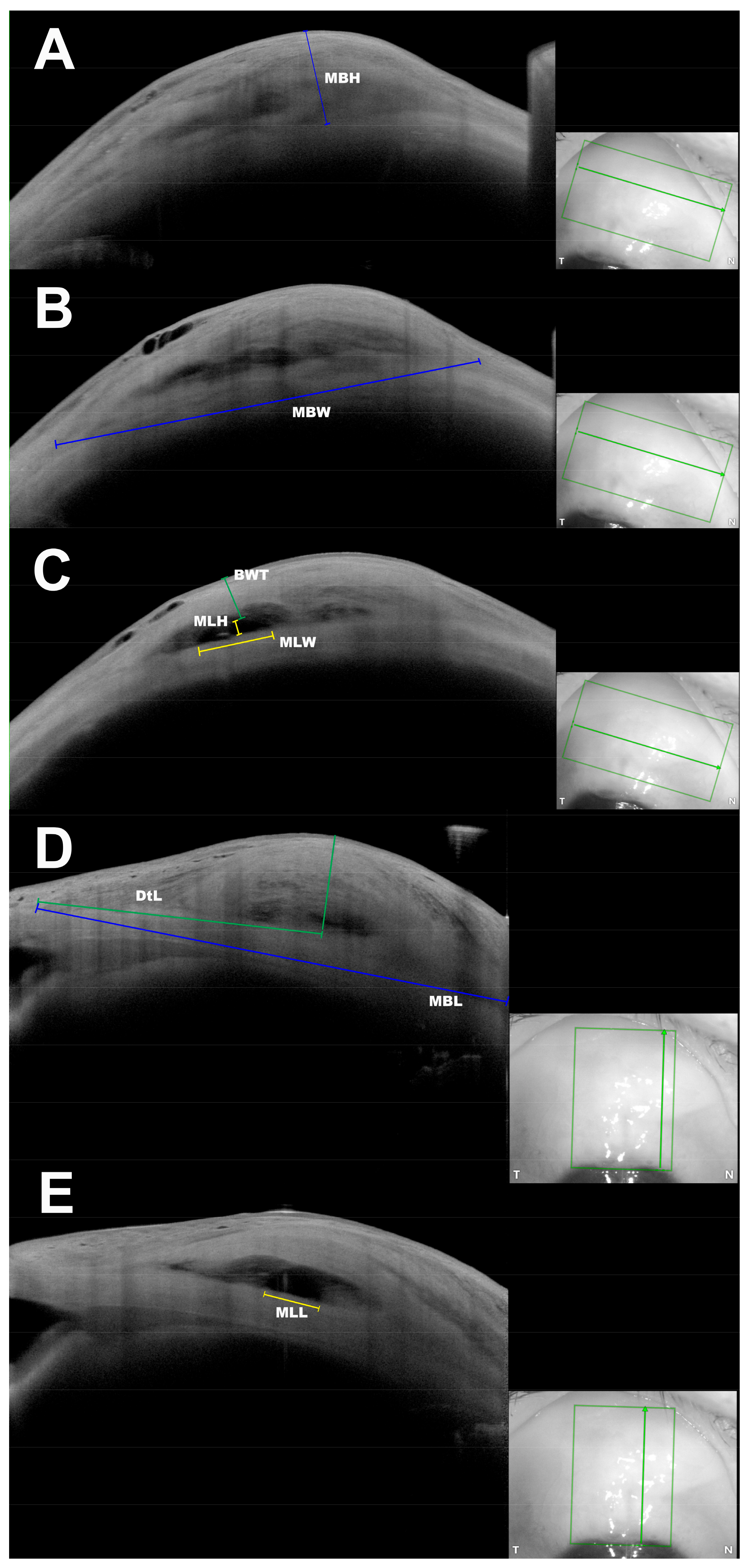
Maximum Bleb Height Figure 1A | MBH | The maximum height of the bleb seen in the tangential scans, measured as the maximum perpendicular distance from the sclera to the first reflex at the conjunctiva. | |
|
Maximum Bleb Width Figure 1B | MBW | The maximum width of the bleb seen in tangential scans, measured as a direct line between two points: begin of changes in tenon thickness nasally to end of tenon changes temporally. | If the whole width of the bleb could not be captured in a single image, the maximum visible width was measured. |
|
Maximal Bleb Length Figure 1D | MBL | The maximum posterior extension of the bleb seen in radial scans, measured as a direct line between two points: from the first changes in tenon thickness anteriorly to the last visible tenon change posteriorly. If the bleb extended over the cornea, measurement was started at the level of the scleral spur. | If the whole length of the bleb could not be captured in a single image, the maximum visible length was measured. |
|
Maximum Lake Height Figure 1C | MLH | The maximum height of the episcleral lake (ES1 according to JBGS) in the tangential scans, measured as maximum perpendicular distance from the inferior to the superior edge of the episcleral lake. | MLH was measurable only in blebs showing the pattern ES1. |
|
Maximum Lake Width Figure 1C | MLW | The maximum width of episcleral lake seen in tangential scans, measured as a direct line between two points: begin of episcleral lake nasally to its end temporally. | MLW was measurable only in blebs showing the ES1-Pattern. |
|
Maximal Lake Length Figure 1E | MLL | The maximum posterior extension of the episcleral lake seen in a radial scan, measured as a linear distance between two points: begin of the episcleral lake anteriorly to its end posteriorly. | If the whole length of the episcleral lake could not be captured in a single image, the maximum visible length was measured. |
|
Bleb Wall Thickness Figure 1C | BWT | Minimal thickness of the bleb wall at the scan with the MLH, measured as the minimal perpendicular distance between the end of the episcleral lake and the first reflex at the conjunctiva. | BWT was measurable only in blebs showing the ES1-Pattern. |
|
Distance to Limbus Figure 1D | DtL | The linear distance between two points: point of corneal surface corresponding to the scleral spur and point of scleral surface corresponding to the highest point of the bleb in radial scans. |
| Parameter | PRESERFLO-Group | Trabeculectomy-Group | p Value |
|---|---|---|---|
| Age (years) | 68.1 ± 12.9 | 70.3 ± 9.0 | 0.38 |
| Sex: Male (%) | 25 (43.8%) | 14 (38.9%) | 0.64 |
| Preoperative IOP (mmHg) | 25.8 ± 10.6 | 26.2 ± 8.7 | 0.82 |
| Preoperative NoM | 2.9 ± 1.3 | 2.8 ± 1.2 | 0.67 |
Type of Glaucoma: n (%)
|
|
| 0.22 |
| TaS (days) | 213.6 ± 171 | 270.6 ± 215 | 0.16 |
| Postoperative IOP (mmHg) | 11.5 ± 3.3 | 10.0 ± 3.4 | 0.036 |
| IOP reduction (mmHg) | 14.2 ± 10.3 | 16.2 ± 7.9 | 0.33 |
| IOP reduction (%) | 50.2 ± 17.7 | 59.7 ± 15.8 | 0.01 |
| Morphological Pattern | PRESERFLO-Group | Trabeculectomy-Group | p Value |
|---|---|---|---|
| C0 | 5 (8.8) | 2 (5.5) | 0.5 |
| C1 | 2 (3.5) | 8 (22.2) | 0.007 |
| C2 | 46 (80.7) | 26 (72.2) | 0.086 |
| T0 | 0 (0) | 0 (0) | - |
| T1 | 16 (28.0%) | 5 (13.8) | 0.084 |
| T2 | 32 (56.1) | 20 (55.5) | 0.727 |
| T3 | 6 (10.5) | 11 (30.5) | 0.021 |
| ES1 | 50 (87.7) | 28 (77.8) | 0.1 |
| Parameter (mm) | PRESERFLO-Group | Trabeculectomy-Group | p Value |
|---|---|---|---|
| MBH | 2.17 ± 0.47 | 2.15 ± 0.43 | 0.83 |
| MBW | 10.67 ± 2.23 | 11.23 ± 2.25 | 0.24 |
| MBL | 9.59 ± 1.57 | 9.9 ± 1.56 | 0.39 |
| MLH | 0.52 ± 0.24 | 0.67 ± 0.3 | 0.017 |
| MLW | 3.69 ± 1.95 | 3.51 ± 2.22 | 0.72 |
| MLL | 4.12 ± 1.54 | 3.23 ± 1.64 | 0.024 |
| BWT | 1.52 ± 0.46 | 1.10 ± 0.37 | 0.00004 |
| DtL | 6.16 ± 1.36 | 4.87 ± 1.34 | 0.00005 |
Disclaimer/Publisher’s Note: The statements, opinions and data contained in all publications are solely those of the individual author(s) and contributor(s) and not of MDPI and/or the editor(s). MDPI and/or the editor(s) disclaim responsibility for any injury to people or property resulting from any ideas, methods, instructions or products referred to in the content. |
© 2023 by the authors. Licensee MDPI, Basel, Switzerland. This article is an open access article distributed under the terms and conditions of the Creative Commons Attribution (CC BY) license (https://creativecommons.org/licenses/by/4.0/).
Share and Cite
Hasan, S.M.; Theilig, T.; Meller, D. Comparison of Bleb Morphology following PRESERFLO® MicroShunt and Trabeculectomy Using Anterior Segment OCT. Diagnostics 2023, 13, 3373. https://doi.org/10.3390/diagnostics13213373
Hasan SM, Theilig T, Meller D. Comparison of Bleb Morphology following PRESERFLO® MicroShunt and Trabeculectomy Using Anterior Segment OCT. Diagnostics. 2023; 13(21):3373. https://doi.org/10.3390/diagnostics13213373
Chicago/Turabian StyleHasan, Somar M., Theresa Theilig, and Daniel Meller. 2023. "Comparison of Bleb Morphology following PRESERFLO® MicroShunt and Trabeculectomy Using Anterior Segment OCT" Diagnostics 13, no. 21: 3373. https://doi.org/10.3390/diagnostics13213373
APA StyleHasan, S. M., Theilig, T., & Meller, D. (2023). Comparison of Bleb Morphology following PRESERFLO® MicroShunt and Trabeculectomy Using Anterior Segment OCT. Diagnostics, 13(21), 3373. https://doi.org/10.3390/diagnostics13213373