A Recurrent Exertional Syncope and Sudden Cardiac Arrest in a Young Athlete with Known Pathogenic p.Arg420Gln Variant in the RYR2 Gene

 ,
, 
Abstract
1. Introduction
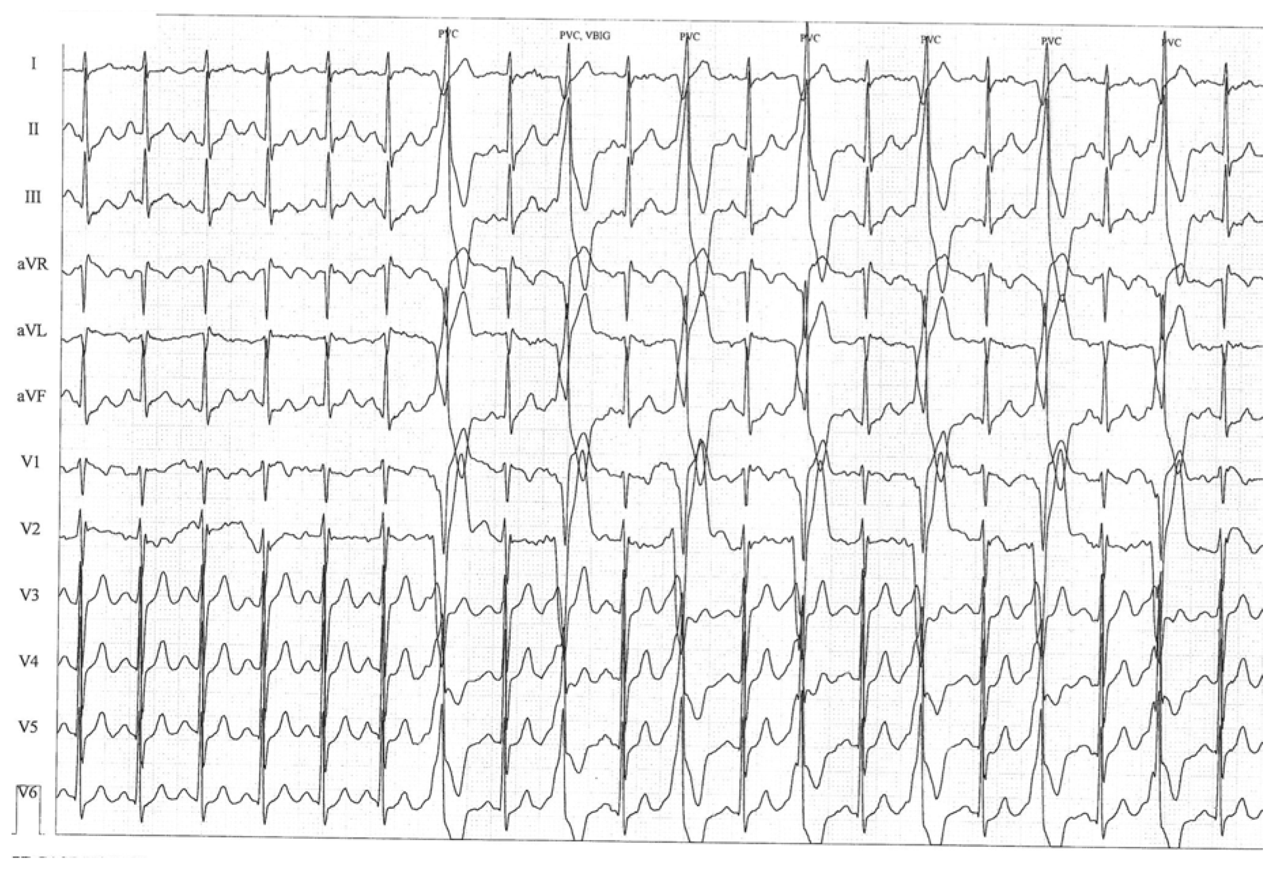
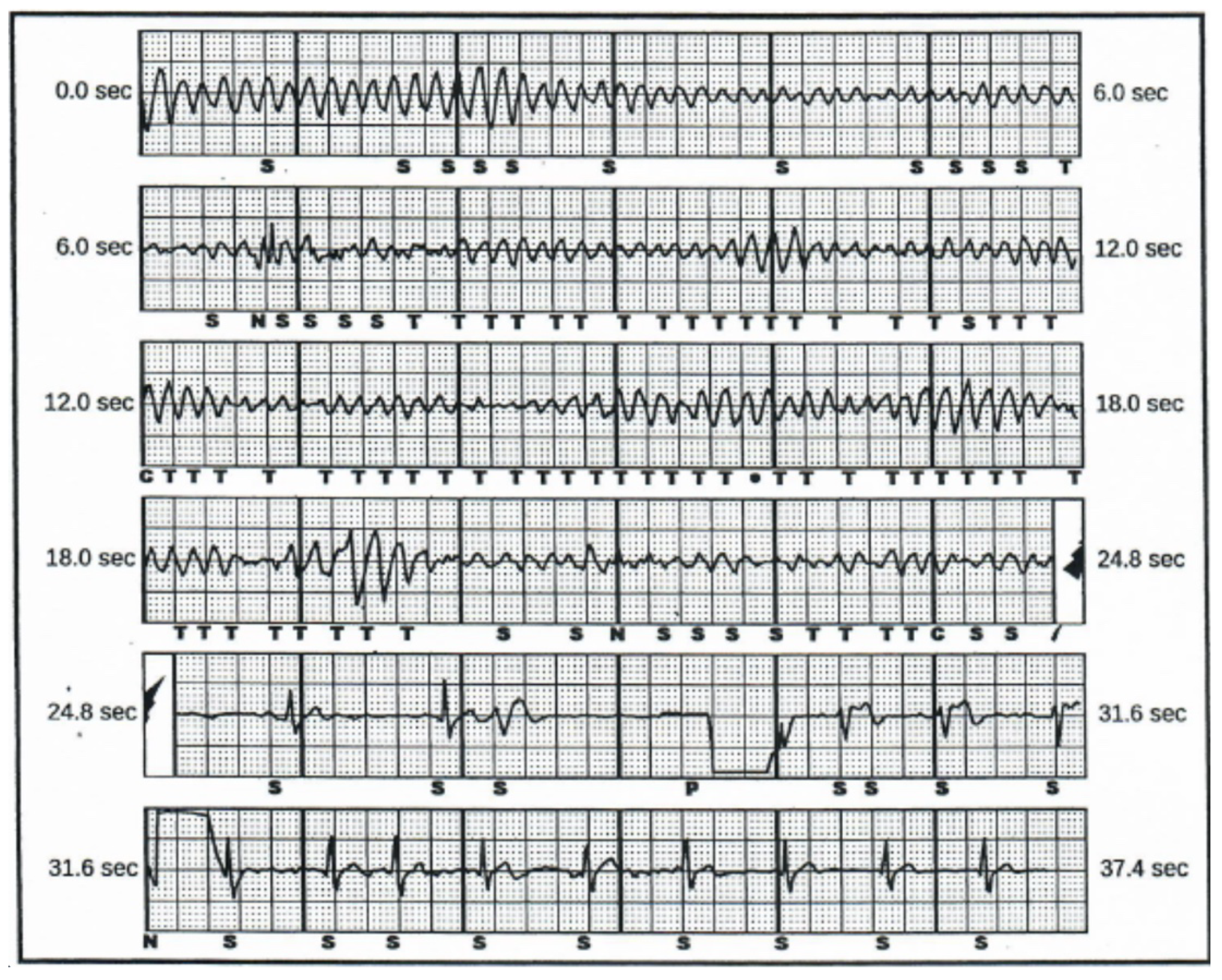
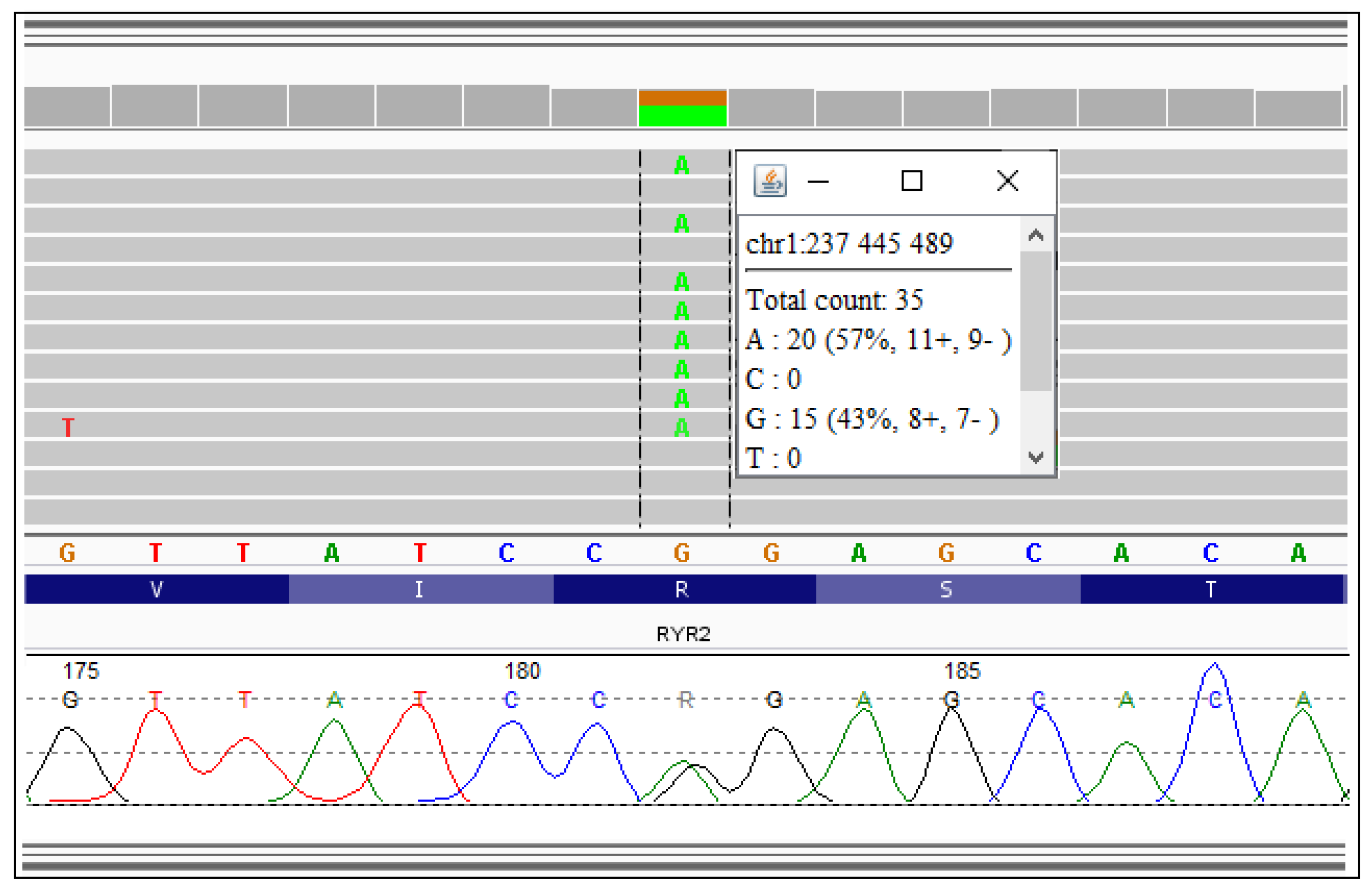
2. Case Description
3. Discussion
3.1. Difficulties in Diagnosis—The Role of Genetics
3.2. Therapeutic Issues
4. Conclusions
Author Contributions
Funding
Conflicts of Interest
References
- Priori, S.G. 2015 ESC Guidelines for the management of patients with ventricular arrhythmias and the prevention of sudden cardiac death: The Task Force for the Management of Patients with Ventricular Arrhythmias and the Prevention of Sudden Cardiac Death of the European Society of Cardiology (ESC). Endorsed by: Association for European Paediatric and Congenital Cardiology (AEPC). Eur. Heart J. 2015, 36, 2793–2867. [Google Scholar] [PubMed]
- Stepien-Wojno, M. Sudden cardiac arrest in patients without overt heart disease: A limited value of next generation sequencing. Pol. Arch. Intern. Med. 2018, 128, 721–730. [Google Scholar] [CrossRef] [PubMed]
- Stępień-Wojno, M. A different background of arrhythmia in siblings with a positive family history of sudden death at young age. Ann. Noninvasive Electrocardiol. 2019, e12707. [Google Scholar] [CrossRef]
- Emery, M.S.; Kovacs, R.J. Sudden Cardiac Death in Athletes. JACC Heart Fail. 2018, 6, 30–40. [Google Scholar] [CrossRef]
- Hayashi, M. Incidence and risk factors of arrhythmic events in catecholaminergic polymorphic ventricular tachycardia. Circulation 2009, 119, 2426–2434. [Google Scholar] [CrossRef]
- Lieve, K.V.; van der Werf, C.; Wilde, A.A. Catecholaminergic Polymorphic Ventricular Tachycardia. Circ. J. 2016, 80, 1285–1291. [Google Scholar] [CrossRef]
- Medeiros-Domingo, A. The RYR2-encoded ryanodine receptor/calcium release channel in patients diagnosed previously with either catecholaminergic polymorphic ventricular tachycardia or genotype negative, exercise-induced long QT syndrome: A comprehensive open reading frame mutational analysis. J. Am. Coll. Cardiol. 2009, 54, 2065–2074. [Google Scholar]
- Van der Werf, C. Flecainide therapy reduces exercise-induced ventricular arrhythmias in patients with catecholaminergic polymorphic ventricular tachycardia. J. Am. Coll. Cardiol. 2011, 57, 2244–2254. [Google Scholar] [CrossRef]
- Ohno, S.; Hasegawa, K.; Horie, M. Gender Differences in the Inheritance Mode of RYR2 Mutations in Catecholaminergic Polymorphic Ventricular Tachycardia Patients. PLoS ONE 2015, 10, e0131517. [Google Scholar] [CrossRef]
- Shigemizu, D. Exome Analyses of Long QT Syndrome Reveal Candidate Pathogenic Mutations in Calmodulin-Interacting Genes. PLoS ONE 2015, 10, e0130329. [Google Scholar] [CrossRef]
- Domingo, D. Non-ventricular, Clinical, and Functional Features of the RyR2(R420Q) Mutation Causing Catecholaminergic Polymorphic Ventricular Tachycardia. Rev. ESP Cardiol. 2015, 68, 398–407. [Google Scholar] [CrossRef] [PubMed]
- Okudaira, N. A knock-in mouse model of N-terminal R420W mutation of cardiac ryanodine receptor exhibits arrhythmogenesis with abnormal calcium dynamics in cardiomyocytes. Biochem. Biophys. Res. Commun. 2014, 452, 665–668. [Google Scholar] [CrossRef] [PubMed]
- Baltogiannis, G.G. CPVT: Arrhythmogenesis, Therapeutic Management, and Future Perspectives. A Brief. Review of the Literature. Front. Cardiovasc. Med. 2019, 6, 92. [Google Scholar] [CrossRef] [PubMed]
- Willis, B.C. Constitutive Intracellular Na+ Excess in Purkinje Cells Promotes Arrhythmogenesis at Lower Levels of Stress Than Ventricular Myocytes From Mice With Catecholaminergic Polymorphic Ventricular Tachycardia. Circulation 2016, 133, 2348–2359. [Google Scholar] [CrossRef]
- Ostby, S.A. Competitive Sports Participation in Patients with Catecholaminergic Polymorphic Ventricular Tachycardia: A Single Center’s Early Experience. JACC Clin. Electrophysiol. 2016, 2, 253–262. [Google Scholar] [CrossRef]
- Postma, A.V. Catecholaminergic polymorphic ventricular tachycardia: RYR2 mutations, bradycardia, and follow up of the patients. J. Med. Genet. 2005, 42, 863–870. [Google Scholar] [CrossRef]
- Sumitomo, N. Association of atrial arrhythmia and sinus node dysfunction in patients with catecholaminergic polymorphic ventricular tachycardia. Circ. J. 2007, 71, 1606–1609. [Google Scholar] [CrossRef]
- Miyake, C.Y. Circadian Variation of Ventricular Arrhythmias in Catecholaminergic Polymorphic Ventricular Tachycardia. JACC Clin. Electrophysiol. 2017, 3, 1308–1317. [Google Scholar] [CrossRef]
- Pflaumer, A. 50 Years of Catecholaminergic Polymorphic Ventricular Tachycardia (CPVT)—Time to Explore the Dark Side of the Moon. Heart Lung Circ. 2020, 29, 520–528. [Google Scholar] [CrossRef]
- Leren, I.S. Nadolol decreases the incidence and severity of ventricular arrhythmias during exercise stress testing compared with β1-selective β-blockers in patients with catecholaminergic polymorphic ventricular tachycardia. Heart Rhythm 2016, 13, 433–440. [Google Scholar] [CrossRef]
- Van der Werf, C. Implantable cardioverter-defibrillators in previously undiagnosed patients with catecholaminergic polymorphic ventricular tachycardia resuscitated from sudden cardiac arrest. Eur. Heart J. 2019, 40, 2953–2961. [Google Scholar] [CrossRef] [PubMed]
- Dusi, V. Cardiac Sympathetic Denervation in Channelopathies. Front. Cardiovasc. Med. 2019, 6, 27. [Google Scholar] [CrossRef] [PubMed]
- Roses-Noguer, F. Outcomes of defibrillator therapy in catecholaminergic polymorphic ventricular tachycardia. Heart Rhythm 2014, 11, 58–66. [Google Scholar] [CrossRef] [PubMed]
- Probst, V. Subcutaneous implantable cardioverter defibrillator indication in prevention of sudden cardiac death in difficult clinical situations: A French expert position paper. Arch. Cardiovasc. Dis. 2020, 113, 359–366. [Google Scholar] [CrossRef]
- Rudic, B. Low Prevalence of Inappropriate Shocks in Patients with Inherited Arrhythmia Syndromes with the Subcutaneous Implantable Defibrillator Single Center Experience and Long-Term Follow-Up. J. Am. Heart Assoc. 2017, 6, e006265. [Google Scholar] [CrossRef]
- Kannankeril, P.J. Atropine-induced sinus tachycardia protects against exercise-induced ventricular arrhythmias in patients with catecholaminergic polymorphic ventricular tachycardia. Europace 2020, 22, 643–648. [Google Scholar] [CrossRef]


© 2020 by the authors. Licensee MDPI, Basel, Switzerland. This article is an open access article distributed under the terms and conditions of the Creative Commons Attribution (CC BY) license (http://creativecommons.org/licenses/by/4.0/).
Share and Cite
Stępień-Wojno, M.; Ponińska, J.; Biernacka, E.K.; Foss-Nieradko, B.; Chwyczko, T.; Syska, P.; Płoski, R.; Bilińska, Z.T. A Recurrent Exertional Syncope and Sudden Cardiac Arrest in a Young Athlete with Known Pathogenic p.Arg420Gln Variant in the RYR2 Gene. Diagnostics 2020, 10, 435. https://doi.org/10.3390/diagnostics10070435
Stępień-Wojno M, Ponińska J, Biernacka EK, Foss-Nieradko B, Chwyczko T, Syska P, Płoski R, Bilińska ZT. A Recurrent Exertional Syncope and Sudden Cardiac Arrest in a Young Athlete with Known Pathogenic p.Arg420Gln Variant in the RYR2 Gene. Diagnostics. 2020; 10(7):435. https://doi.org/10.3390/diagnostics10070435
Chicago/Turabian StyleStępień-Wojno, Małgorzata, Joanna Ponińska, Elżbieta K. Biernacka, Bogna Foss-Nieradko, Tomasz Chwyczko, Paweł Syska, Rafał Płoski, and Zofia T. Bilińska. 2020. "A Recurrent Exertional Syncope and Sudden Cardiac Arrest in a Young Athlete with Known Pathogenic p.Arg420Gln Variant in the RYR2 Gene" Diagnostics 10, no. 7: 435. https://doi.org/10.3390/diagnostics10070435
APA StyleStępień-Wojno, M., Ponińska, J., Biernacka, E. K., Foss-Nieradko, B., Chwyczko, T., Syska, P., Płoski, R., & Bilińska, Z. T. (2020). A Recurrent Exertional Syncope and Sudden Cardiac Arrest in a Young Athlete with Known Pathogenic p.Arg420Gln Variant in the RYR2 Gene. Diagnostics, 10(7), 435. https://doi.org/10.3390/diagnostics10070435