Article

Dynamics of Dopamine and Other Monoamines Content in Rat Brain after Single Low-Dose Carbon Nuclei Irradiation

by
Viktor S. Kokhan
1,*,
Alexey A. Ustyugov
2 and
Vladimir A. Pikalov
3
1
V.P. Serbsky Federal Medical Research Centre for Psychiatry and Narcology, 119034 Moscow, Russia
2
Institute of Physiologically Active Compounds RAS, 142432 Chernogolovka, Russia
3
Institute for High Energy Physics Named by A.A. Logunov of National Research Centre “Kurchatov Institute”, 142281 Protvino, Russia
*
Author to whom correspondence should be addressed.
Life 2022, 12(9), 1306; https://doi.org/10.3390/life12091306
Submission received: 15 July 2022 / Revised: 10 August 2022 / Accepted: 20 August 2022 / Published: 25 August 2022
(This article belongs to the Special Issue Advances in Space Biology)

Abstract

:
Space radiation, presented primarily by high-charge and -energy particles (HZEs), has a substantial impact on the central nervous system (CNS) of astronauts. This impact, surprisingly, has not only negative but also positive effects on CNS functions. Despite the fact that the mechanisms of this effect have not yet been elucidated, several studies indicate a key role for monoaminergic networks underlying these effects. Here, we investigated the effects of acute irradiation with 450 MeV/n carbon (12C) nuclei at a dose of 0.14 Gy on Wistar rats; a state of anxiety was accessed using a light–dark box, spatial memory in a Morris water maze, and the dynamics of monoamine metabolism in several brain morphological structures using HPLC. No behavioral changes were observed. Irradiation led to the immediate suppression of dopamine turnover in the prefrontal cortex, hypothalamus, and striatum, while a decrease in the level of norepinephrine was detected in the amygdala. However, these effects were transient. The deferred effect of dopamine turnover increase was found in the hippocampus. These data underscore the ability of even low-dose 12C irradiation to affect monoaminergic networks. However, this impact is transient and is not accompanied by behavioral alterations.

1. Introduction

Space radiation, presented primarily by high-charge and -energy particles (HZEs), has a substantial impact on the central nervous system (CNS) of astronauts. HZEs are very deeply ionizing highly charged nuclei with an atomic number of more than two. An important HZE parameter is linear energy transfer (LET)—the rate of particle energy loss, measured in keV/µm of water. During interplanetary missions, the vast majority of collisions will be produced by low-LET particles (<15 keV/µm), while the number of collisions and, accordingly, the absorbed dose for high-LET particles will be relatively small [1,2]. 12C nuclei are the most encountered after alpha particles among the nuclei flux in interplanetary space and the most encountered among HZEs [3]. Despite the obvious negative effect of HZEs on CNS [4,5], studies on the latest modern ground-based models that most appropriately reproduce outer space conditions have shown rather unexpected results. Among these findings was the recently discovered phenomenon of the composite effect of radiation. Combined exposure did not cause any cognitive impairments [6,7] that have been shown recently at comparable space-related doses (<1 Sv) under exposure to only one nuclei beam [8,9,10]. Moreover, combined irradiation revealed a pro-cognitive (12C, 0.14 Gy, 450 MeV, 10.3 keV/µm, and 0.4 Gy γ-rays) [11,12] and nootropic (12C 0.18 Gy, 450 MeV, 10.3 keV/μm, and γ-rays 0.24 Gy) [13] effect. The molecular mechanisms of these phenomena remain poorly understood, and the exact pathways that are involved in the composite action of ionizing radiation, including HZEs, remain unknown. At the same time, the growing body of research indicates that monoaminergic neural networks could be involved and serve as a key link in the implementation of the described phenomena [14,15].
One of the first pieces of evidence of functional alterations of the dopaminergic system was the detection of cocaine-induced locomotor response impairment after irradiation with 56Fe (1.2 or 2.4 Gy, 1 GeV, 147 keV/µm) with dose-dependent persistence in time [16]. It has been shown in two independent studies using H+ (150 MeV, 0.4 keV/µm) that irradiation caused a significant decrease (0.5 Gy, including non-sensitive to radiation animals) or increase (1 or 2 Gy) in dopamine (DA) transporter (DAT) levels and also increased (1 Gy) the level of DA receptor D2 in a brain slice (which included the caudate–putamen, nucleus accumbens, ventral tegmental area, and substantia nigra) and in the frontal cortex (only DAT). These observations were made in a cohort of irradiated rats only with behavioral impairments, and this group was designated as “sensitive to radiation” [17,18].
On the other hand, the overexpression of D2 receptors in the striatum after combined exposure to radiation (combined H+, 1.5 Gy, 170 MeV, 0.4 keV/µm, and 3 Gy γ-rays) and antiorthostatic suspension (a simulated model of microgravity) have suggested a neuroreparative effect of irradiation [19]. The analysis of serotonin metabolism has shown that irradiation suppressed serotonin turnover in the prefrontal cortex and hippocampus [15,19,20,21,22,23] but activated serotonin turnover in the hypothalamus [15]. The overexpression and increased protein level of 5-HT2a receptors under combined irradiation (combined H+ 1.5 Gy, 170 MeV, 0.4 keV/µm, and 3 Gy γ-rays or combined 12C, 0.14 Gy, 450 MeV, 10.3 keV/µm, and 0.4 Gy γ-rays) have revealed pro-cognitive effects [19,24].
Despite the generally positive research data for future space missions, there is still a great need for fundamental comprehensive research on the influence of ionizing radiation, particularly HZEs, on biological subjects. A number of positive effects of the combined irradiation with γ-rays and 12C nuclei on the CNS and their connections with monoamine turnover and metabolism have been identified, but the effects of 12C beam irradiation in comparative doses are not yet thoroughly characterized.
To address these knowledge gaps, here, we define how 12C irradiation of the head affects behavior and monoamine metabolism and dynamics in male Wistar rats. The irradiation model used in the study is not relevant to interplanetary flights but is designed to facilitate an understanding of the interaction nature of γ-rays and 12C nuclei when combined.

2. Materials and Methods

2.1. Animals

Male 3-month-old Wistar rats weighing 270–300 g were used in this study. The rats were maintained at the Center for Collective Use of the Institute of Physiologically Active Compounds RAS (Chernogolovka, Russia). Rats were housed in a standard environment (12 h light/dark cycle, 18–23 °C, and 30–70% relative humidity) with food and water ad libitum.
Experimental groups were formed by random assignment of rats to: control animals (C, n = 16) and irradiated rats (R, n = 16). At Day 1 after irradiation, half of the control (C1, n = 8) and irradiated (R1, n = 8) rats were euthanized for the HPLC study. The remaining animals were subjected to a battery of behavioral tests and euthanized on Day 25 after irradiation: control (C25, n = 8) and irradiated (R25, n = 8) groups.
The study was approved by the Ethics Committee of Institute of Physiologically Active Compounds (protocol code 55 on 26 March 2021).

2.2. Carbon (12C) Nuclei Irradiation

The heads of experimental animals (R group) were subjected to a single exposure of 12C nuclei (450 MeV/n, 140 ± 15 mGy, LET 10.3 keV/μm) in a U-70 accelerator (NRC “Kurchatov Institute”—IHEP, Protvino, Russia). Dosimetric monitoring of irradiation was performed using a DKS-AT5350/1 dosimeter (Atomtex, Minsk, Belarus) with a TM30010-1 ionization chamber (PTW-Freiburg, Freiburg, Germany). A collimator from carbon (density 1.8 g/cm3, thickness 50 cm, diameter of beam 65 mm) was used to restrict the 12C beam. The unevenness of the irradiation field was <±2.5% within the sweep radius R = 30 mm. The animals were placed in special poly(methyl methacrylate) cases that restrict movement and were positioned so that only the head and a small area of the neck fell into the 12C beam.
The control animal group (C1 and C25 groups) was transported together with irradiated animals and also placed in cases but was kept separately in another laboratory room and, thus, not exposed to ionizing radiation.

2.3. Dark–Light Box

A dark–light box (OpenScience, Moscow, Russia) for rats was used where the light compartment was evenly lit (60 lx). Each rat was placed in the dark compartment, and movement activity indicators were recorded for 5 min.

2.4. Morris Water Maze

For these experiments, a black pool 180 cm in diameter was used. A black plastic platform (invisible to the animal) 10 cm in diameter was immersed 2 cm under water, always in the same place. The rat was released into the water at the edge of the pool at a chosen point and allowed to swim freely for 60 s. The same starting point was used for all rats within one trial. There were four trials per day, each time changing the starting point. The probe test was performed after the final learning at Day 6 of learning. The platform was removed from the pool, the rat was released into the water’s edge, and swim time to find the platform in the same quadrant as the platform was recorded within 60 s of total swim time.

2.5. Tissue Collection and High-Performance Liquid Chromatography

On Day 1 and Day 25 after irradiation, the appropriate group of rats were euthanized by decapitation, and the following brain morphological structures were isolated on a thermoelectric surface (+2 °C) and frozen in liquid nitrogen: the prefrontal cortex (PFC), hippocampus (HPC), striatum (ST), amygdala (AMY), and hypothalamus (HYP).
Brain tissue homogenization and extraction were carried out in 0.1 M HClO4 solution with the addition of 0.25 nM/mL 3,4-dioxybensilamine as an internal standard. Samples were centrifuged at 10,000× g for 15 min 4 °C. The content of monoamines (NA, 5-HT, DA) and their metabolites 5-hidroxyindoacetic acid (5-HIAA), homovanillic acid (HVA), 3,4-dihydroxyphenylacetic acid (DOPAC), and 3-methoxytyramine (3-MT) was determined in the supernatant using high-performance liquid chromatography (ion-pair chromatography) with electrochemical detection on a System Gold liquid chromatograph (Beckman Coulter, Inc., Brea, CA, USA) equipped with a Rheodyne 7125 injector (Rohnert Park, CA, USA) with a 20 μL sample loop. The samples were separated on a reverse-phase column Nucleodur C18 Gravity, 4.6 × 250 mm, pore diameter of 5 μm (Mashery-Nagel GmbH & Co., KG, Düren, Germany). Mobile phase (0.1 M citrate-phosphate buffer pH 3.0 containing 1.1 mM octanesulfonic acid, 0.1 mM EDTA, and 9% acetonitrile) flow rate of 1 mL/min at a pressure of 200 atm was achieved using a System Gold 125 pump (Beckman Coulter, Inc.). Measurements were performed using an EC3000 electrochemical detector (RECIPE Chemicals + Instruments GmbH, Munich, Germany) equipped with a ClinLab ECD cell, Model Sputnik, with a glassy carbon working electrode (+0.85 V) and a Ag/AgCl reference electrode. Sample registration and chromatogram processing were performed using Multichrom v.1.5 software (Ampersand, Moscow, Russia).

2.6. Data Processing

Data are presented as the mean ± standard deviation (SD). Standard data processing was performed using Statistica 12 software (StatSoft Inc., Tulsa, OK, USA). The Shapiro–Wilk test was used to assess the normality of the data distribution; for p > 0.05, parametric analysis methods were used. Intergroup differences in dark–light box were analyzed using Student’s t-test. Intergroup differences in Morris water maze were analyzed using repeated-measures analysis of variance (ANOVA). HPLC data were processed using two-way ANOVA. Duncan’s posterior test was calculated if necessary.

3. Results

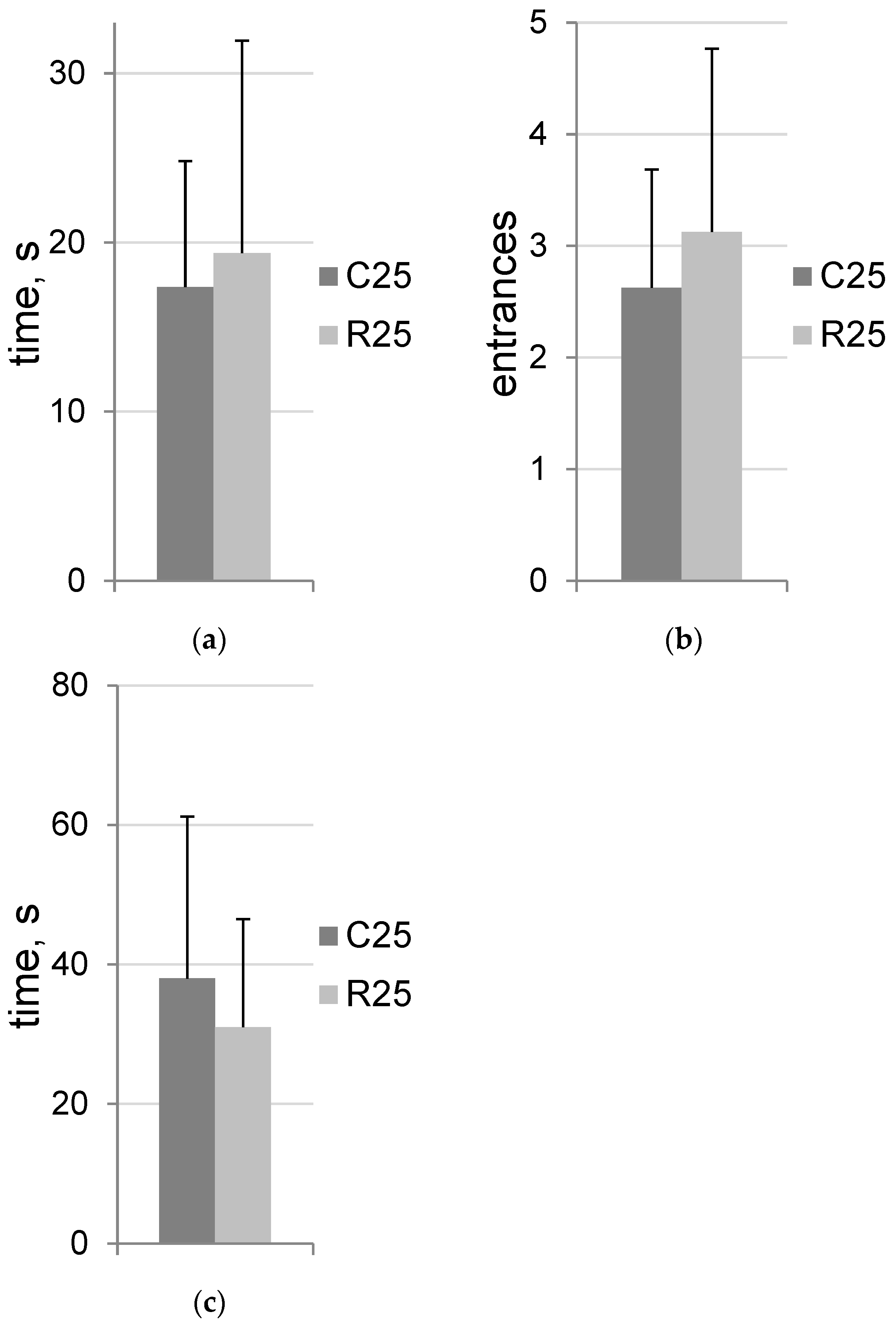
3.1. Irradiation Does Not Change Trait Anxiety

The dark–light box data are presented in Figure 1. No statistically significant differences were detected. All tested groups showed rodent-specific exploratory behavior without signs of increased state anxiety.

3.2. Irradiation Does Not Affect Spatial Training

The Morris water maze data are presented in Figure 2. No statistically significant differences were detected, both in the dynamics of learning (Figure 2a) and during the probe test (Figure 2b). Experimental groups of rats were characterized by good learning dynamics.

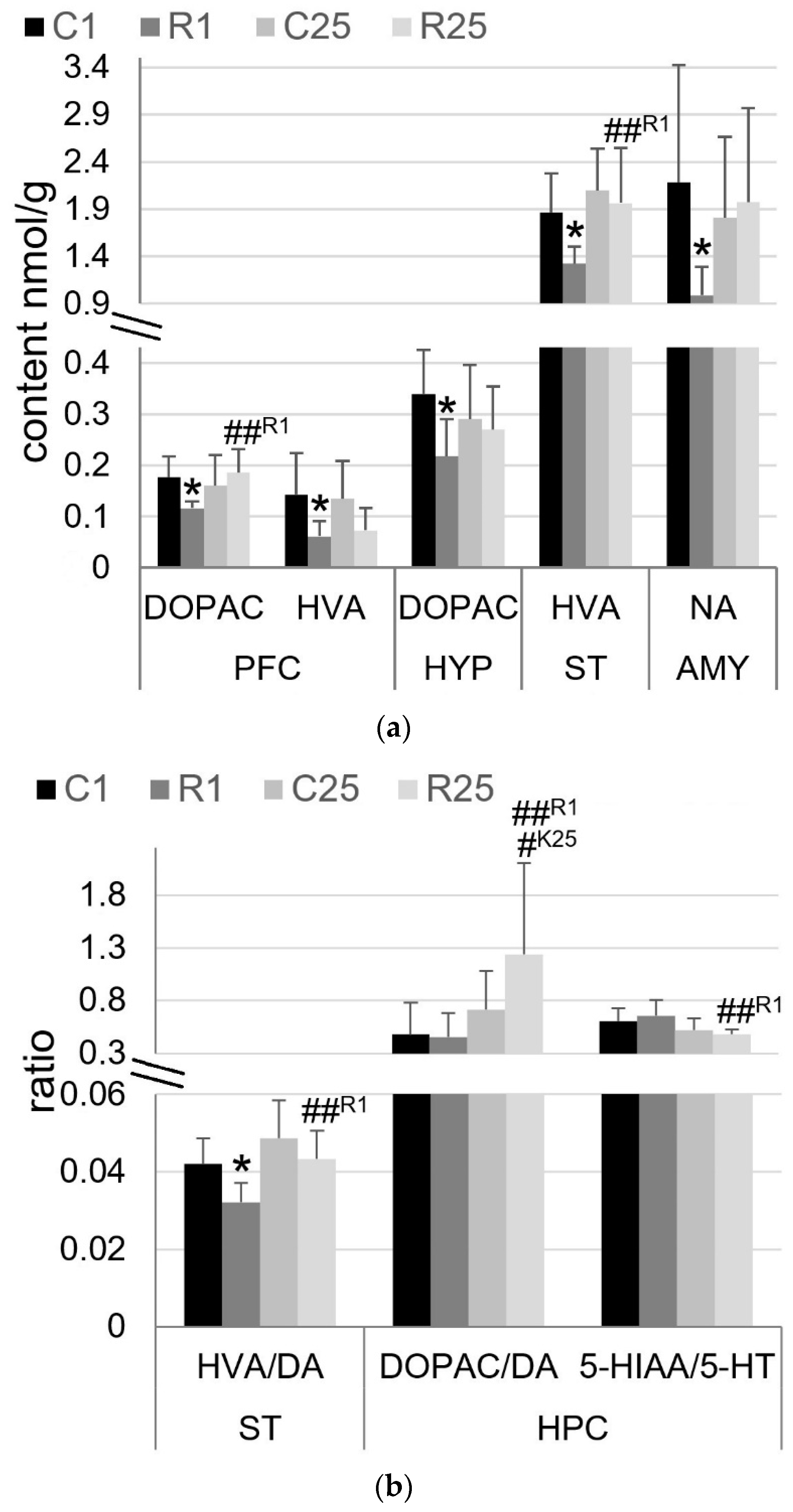
3.3. Irradiation Leads to Immediate and Transient Changes in Monoamine Metabolism, with the Exception of Hippocampus, Where It Appears Later

The following statistically significant differences in monoamines and their metabolites were found in PFC (Figure 3a): DOPAC (F1,28 = 7.8, p = 0.009; radiation × time factor interaction) and HVA (F1,28 = 11.1, p = 0.002; time factor). The irradiated R1 group of rats showed a reduction in the monoamines DOPAC and HVA, at 34% (p = 0.011) and 57% (p = 0.019), respectively, in comparison with control C1 rats. In a separate group of irradiated rats—R25—DOPAC content was 60% (p = 0.005) higher than that in R1-rats.
Statistically significant differences in the following monoamines and their metabolites were found in HPC (Figure 3b): DOPAC/DA ratio (F1,28 = 8.1, p = 0.008; time factor) and 5-HIAA/5-HT ratio (F1,28 = 10.4, p = 0.003; time factor). The DOPAC/DA ratio was greater in the R25 group of irradiated rats: 73% (p = 0.049) compared to the value of control C25-rats and 172% (p = 0.008) compared to the value of R1-rats. In contrast, the 5-HIAA/5-HT ratio was decreased in R25-rats at 26% (p = 0.008) compared to that seen in R1-rats.
Statistically significant differences in the following monoamines and their metabolites were found in ST (Figure 3a,b): HVA (F1,28 = 4.9, p = 0.003, radiation factor; F1,28 = 8.3, p = 0.008, time factor) and HVA/DA ratio (F1,28 = 8.6, p = 0.007, radiation factor; F1,28 = 12, p = 0.002, time factor). In the R1 group of rats, decreased HVA content was found with mean HVA/DA ratios at 29% (p = 0.018) and 24% (p = 0.012) in comparison with C1-rats. In the R25 group of irradiated rats, the HVA content and mean HVA/DA ratio were 48% (p = 0.007) and 35% (p = 0.006) higher than those in R1-rats.
Statistically significant differences in the following monoamine and its metabolites were found in AMY (Figure 3a): NA (F1,28 = 4.3, p = 0.046; radiation × time factors interaction). The NA content in the R1 group was 55% (p = 0.023) compared with the C1 control group.
Statistically significant differences in the following monoamine and its metabolites were found in HYP (Figure 3a): DOPAC (F1,28 = 5.2, p = 0.03; radiation factor). The DOPAC content was lower in the R1 group at 36% (p = 0.016) compared to the value seen in C1 control rats.

4. Discussion

The importance of studying anxiety behavior is based on a number of data showing the relationship between radiation-induced anxiety and changes in the cognitive abilities of experimental animals. Thus, radiation-induced anxiety-associated cognition enhancement [15,25], decline [26,27], and the absence of changes against the background of anxiolytic drugs [28] were shown. The anxiogenic effect of ionizing radiation has been revealed in a number of relevant irradiation scenarios: γ-rays in a dose range of 0.5–2 Gy [8], combined irradiation (0.4 Gy γ-rays and 12C 0.14 Gy, 10.3 keV/μm) [15,25], and chronic exposure to neutrons and γ-rays (252Cf, 1 mGy/day, 0.4 Gy totally, but not ~0.12 or 0.2 Gy) [12].
During behavioral testing, no anxiogenic action of irradiation was detected. The decrease in NE content in AMY demonstrated an overall neurochemical picture indicating the absence of any anxiogenic effect caused by irradiation. Indeed, the reduced efficiency of NE innervation of AMY leads to an anxiolytic effect [29,30], whereas activation, conversely, is accompanied by the enhancement of signs of anxiety-like behavior [31,32]. At the same time, reduced NE content in AMY may be responsible for memory impairment in behavior tests that are based on electric foot shock and other aversive stimuli [33,34,35].
We found no impairment, and more importantly, no enhancement in the spatial learning of irradiated rats. The absence of learning disabilities in the Morris water maze is not surprising since a number of studies have not previously revealed any impairments in this test in close dose/nature of irradiation [36,37,38]. At the same time, the enhancement of spatial learning is detected at the same radiation doses of 12C irradiation under the condition of γ-ray (0.4 Gy) pre-irradiation [15].
Thus, the lack of anxiogenic action and spatial memory changes distinguishes acute 12C irradiation from previously observed data. Relying on combined irradiation data [15,25,28], we believe that the irradiation caused by the γ-ray component in combined irradiation conditions is key to inducing anxiogenic effects and cognition changes.
The analysis of the content of monoamines revealed that acute irradiation led to the immediate suppression of DA turnover in PFC, ST, and HYP. This conclusion is supported by the fact that an increased concentration of metabolites in the tissue ex vivo and/or elevated metabolite/neurotransmitter ratio represents increased neurotransmitter signaling in vivo [39,40]. However, on Day 25 after irradiation, these changes were no longer detected. The suppression of DA turnover in PFC and ST is also characteristic of acute irradiation with high-LET H+ (1 and 2 Gy, spread-out Bragg peak) [41] but not by low-LET H+ (1.5 and 3 Gy, 165 MeV) [20]. Taking into account the results of previous studies, we hypothesize that the suppression of DA turnover is dose-dependent in nature: from the transient in the present study to persistent (it immediately appears and lasts up to 30 but not 90 days after irradiation) when irradiated by 12C (500 MeV) in the absorbed dose 1 Gy, which was shown previously [21,23]. The decreasing content of DOPAC and HVA, first of all in PFC and HYP, may be associated with a decrease in the content or activity of monoamine oxidases but not catechol-O-methyltransferase. This assumption finds its confirmation in the absence of changes in the 3-MT content [42]. On the contrary, large doses of (1-3 Gy) low- and high-LET H+ led to significant changes in the content of 3-MT but not DOPAC [20,41], and in close conditions of irradiation, a change in the expression of catechol-O-methyltransferase was shown [19], which indirectly confirms our assumption. The change in the expression level of monoamine oxidase A is a similar indirect confirmation of our assumption [28]. However, the previous data [28] were obtained in several other conditions of irradiation (combined γ-ray and 12C) and on the background of anxiolytic pharmaceutical therapy; therefore, they must be considered with due caution.
The suppression of DA turnover in PFC, ST, and HYP may indicate a neurotoxic effect as a result of biomolecular damage, oxidative stress, and neuroinflammation caused by irradiation [43,44,45]. On the contrary, the increase in DA turnover in HPC could be considered as pro-cognitive and a sign of neurorecovery [46,47,48]. However, these assumptions are speculative and require future precision studies. Moreover, the observed neurochemical changes did not reach the degree in which CNS functional disorders would be observed in behavioral tests.
It is known that irradiation with γ-rays (fractionated 0.5 Gy × 6 times, 661.7 keV, and 3 Gy) does not affect monoamine turnover [49] or cognition (2 Gy acute or fractions 0.2 Gy × 10 days) [50]. Thus, we hypothesize that pre-irradiation with γ-rays before HZE irradiation produces unique effects that are not based on synergism or simple summation of individual effects. Moreover, γ-ray pre-irradiation, when combined in the irradiation model used, caused protective/recovery effects in the CNS [15,25]. A similar effect is probably observed when irradiation involves ultra-low-LET particle components, such as H+ or 4He, in combination with low-, moderate-, and high-LET nuclei in space-related doses (about 1 Sv) [6,7]. Additionally, the modulation of monoaminergic neural networks under irradiation makes a significant contribution to the exercise of these effects.
It is important to note that the doses used in the presented study are relevant neither to ionizing radiation during deep space missions nor to the therapeutic beam in radiotherapy. The main purpose of this work was to show the cardinal difference between mono-beam HZE irradiation and irradiation using pre-irradiation with ultra-low-LET sources (γ-rays in our previous study). At the same time, in the comparative analysis, we relied on a number of data from combined irradiation (γ-rays and 12C) obtained earlier [15,25,28].

Author Contributions

Conceptualization, V.S.K. and A.A.U.; Data curation, V.S.K.; Formal analysis, V.S.K. and A.A.U.; Funding acquisition, A.A.U.; Investigation, V.S.K. and V.A.P.; Methodology, V.S.K. and V.A.P.; Resources, A.A.U.; Writing—original draft, V.S.K. All authors have read and agreed to the published version of the manuscript.

Funding

The study was funded by the grant agreement No. 075-15-2020-795, contract No. 13.1902.21.0027 of 29 September 2020 unique project ID: RF-190220X0027.

Institutional Review Board Statement

The study was conducted according to the guidelines of the European Communities Council Directive of 24 November 1986 (86/609/EEC) and the Regulations for laboratory practice in the Russian Federation 2003 and approved by the Ethics Committee of Institute of Physiologically Active Compounds (protocol code 55 on 26 March 2021).

Informed Consent Statement

Not applicable.

Data Availability Statement

The data presented in this study are openly available in Mendeley Data, link Kokhan, Viktor (2021), “Dynamics of dopamine and other monoamines content in some rat brain morphological structures after single low-dose carbon nuclei irradiation”, Mendeley Data, V1, doi:10.17632/xn3z368d4t.1.

Conflicts of Interest

The authors declare no conflict of interest.

References

  1. Zeitlin, C.; Hassler, D.M.; Cucinotta, F.A.; Ehresmann, B.; Wimmer-Schweingruber, R.F.; Brinza, D.E.; Kang, S.; Weigle, G.; Bottcher, S.; Bohm, E.; et al. Measurements of energetic particle radiation in transit to Mars on the Mars Science Laboratory. Science 2013, 340, 1080–1084. [Google Scholar] [CrossRef] [PubMed]
  2. Hassler, D.M.; Zeitlin, C.; Wimmer-Schweingruber, R.F.; Ehresmann, B.; Rafkin, S.; Eigenbrode, J.L.; Brinza, D.E.; Weigle, G.; Bottcher, S.; Bohm, E.; et al. Mars’ surface radiation environment measured with the Mars Science Laboratory’s Curiosity rover. Science 2014, 343, 1244797. [Google Scholar] [CrossRef] [PubMed]
  3. Lezniak, J.A.; Webber, W.R. The charge composition and energy spectra of cosmic-ray nuclei from 3000 MeV per nucleon to 50 GeV per nucleon. Astrophys. J. 1978, 223, 676–696. [Google Scholar] [CrossRef]
  4. Cucinotta, F.A.; Alp, M.; Sulzman, F.M.; Wang, M. Space radiation risks to the central nervous system. Life Sci. Space Res. 2014, 2, 54–69. [Google Scholar] [CrossRef]
  5. Cekanaviciute, E.; Rosi, S.; Costes, S.V. Central Nervous System Responses to Simulated Galactic Cosmic Rays. Int. J. Mol. Sci. 2018, 19, 3669. [Google Scholar] [CrossRef]
  6. Raber, J.; Fuentes Anaya, A.; Torres, E.R.S.; Lee, J.; Boutros, S.; Grygoryev, D.; Hammer, A.; Kasschau, K.D.; Sharpton, T.J.; Turker, M.S.; et al. Effects of Six Sequential Charged Particle Beams on Behavioral and Cognitive Performance in B6D2F1 Female and Male Mice. Front. Physiol. 2020, 11, 959. [Google Scholar] [CrossRef]
  7. Raber, J.; Yamazaki, J.; Torres, E.R.S.; Kirchoff, N.; Stagaman, K.; Sharpton, T.; Turker, M.S.; Kronenberg, A. Combined Effects of Three High-Energy Charged Particle Beams Important for Space Flight on Brain, Behavioral and Cognitive Endpoints in B6D2F1 Female and Male Mice. Front. Physiol. 2019, 10, 179. [Google Scholar] [CrossRef]
  8. Rabin, B.M.; Carrihill-Knoll, K.L.; Shukitt-Hale, B. Comparison of the Effectiveness of Exposure to Low-LET Helium Particles ((4)He) and Gamma Rays ((137)Cs) on the Disruption of Cognitive Performance. Radiat. Res. 2015, 184, 266–272. [Google Scholar] [CrossRef]
  9. Carr, H.; Alexander, T.C.; Groves, T.; Kiffer, F.; Wang, J.; Price, E.; Boerma, M.; Allen, A.R. Early effects of (16)O radiation on neuronal morphology and cognition in a murine model. Life Sci. Space Res. 2018, 17, 63–73. [Google Scholar] [CrossRef]
  10. Britten, R.A.; Jewell, J.S.; Duncan, V.D.; Davis, L.K.; Hadley, M.M.; Wyrobek, A.J. Spatial Memory Performance of Socially Mature Wistar Rats is Impaired after Exposure to Low (5 cGy) Doses of 1 GeV/n (48)Ti Particles. Radiat. Res. 2017, 187, 60–65. [Google Scholar] [CrossRef]
  11. Whoolery, C.W.; Yun, S.; Reynolds, R.P.; Lucero, M.J.; Soler, I.; Tran, F.H.; Ito, N.; Redfield, R.L.; Richardson, D.R.; Shih, H.Y.; et al. Multi-domain cognitive assessment of male mice shows space radiation is not harmful to high-level cognition and actually improves pattern separation. Sci. Rep. 2020, 10, 2737. [Google Scholar] [CrossRef] [PubMed]
  12. Perez, R.E.; Younger, S.; Bertheau, E.; Fallgren, C.M.; Weil, M.M.; Raber, J. Effects of chronic exposure to a mixed field of neutrons and photons on behavioral and cognitive performance in mice. Behav. Brain Res. 2020, 379, 112377. [Google Scholar] [CrossRef]
  13. Chicheva, M.M.; Mal’tsev, A.V.; Kokhan, V.S.; Bachurin, S.O. The Effect of Ionizing Radiation on Cognitive Functions in Mouse Models of Alzheimer’s Disease. Dokl. Biol. Sci. 2020, 494, 225–227. [Google Scholar] [CrossRef] [PubMed]
  14. Kokhan, V.S.; Matveeva, M.I.; Mukhametov, A.; Shtemberg, A.S. Risk of defeats in the central nervous system during deep space missions. Neurosci. Biobehav. Rev. 2016, 71, 621–632. [Google Scholar] [CrossRef]
  15. Kokhan, V.S.; Shakhbazian, E.V.; Markova, N.A. Psycho-emotional status but not cognition are changed under the combined effect of ionizing radiations at doses related to deep space missions. Behav. Brain Res. 2019, 362, 311–318. [Google Scholar] [CrossRef]
  16. Rice, O.V.; Grande, A.V.; Dehktyar, N.; Bruneus, M.; Robinson, J.K.; Gatley, S.J. Long-term effects of irradiation with iron-56 particles on the nigrostriatal dopamine system. Radiat. Environ. Biophys. 2009, 48, 215–225. [Google Scholar] [CrossRef] [PubMed]
  17. Davis, C.M.; DeCicco-Skinner, K.L.; Roma, P.G.; Hienz, R.D. Individual differences in attentional deficits and dopaminergic protein levels following exposure to proton radiation. Radiat. Res. 2014, 181, 258–271. [Google Scholar] [CrossRef]
  18. Davis, C.M.; DeCicco-Skinner, K.L.; Hienz, R.D. Deficits in Sustained Attention and Changes in Dopaminergic Protein Levels following Exposure to Proton Radiation Are Related to Basal Dopaminergic Function. PLoS ONE 2015, 10, e0144556. [Google Scholar] [CrossRef]
  19. Kokhan, V.S.; Lebedeva-Georgievskaya, K.B.; Kudrin, V.S.; Bazyan, A.S.; Maltsev, A.V.; Shtemberg, A.S. An investigation of the single and combined effects of hypogravity and ionizing radiation on brain monoamine metabolism and rats’ behavior. Life Sci. Space Res. 2019, 20, 12–19. [Google Scholar] [CrossRef]
  20. Shtemberg, A.S.; Bazian, A.S.; Lebedeva-Georgievskaya, K.B.; Matveeva, M.I.; Kudrin, V.S.; Narkevich, V.B.; Klodt, P.M.; Kokhan, V.S. Effects of exposure to high-energy protons on rat’s behavior and underlying neurochemical mechanisms. Aerosp. Environ. Med. 2013, 47, 54–60. [Google Scholar]
  21. Matveeva, M.I.; Shtemberg, A.S.; Timoshenko, G.N.; Krasavin, E.A.; Narkevich, V.B.; Klodt, P.M.; Kudrin, V.S.; Bazyan, A.S. The effects of irradiation by 12C carbon ions on monoamine exchange in several rat brain structures. Neurochem. J. 2013, 7, 303–307. [Google Scholar] [CrossRef]
  22. Kokhan, V.S.; Matveeva, M.I.; Bazyan, A.S.; Kudrin, V.S.; Mukhametov, A.; Shtemberg, A.S. Combined effects of antiorthostatic suspension and ionizing radiation on the behaviour and neurotransmitters changes in different brain structures of rats. Behav. Brain Res. 2017, 320, 473–483. [Google Scholar] [CrossRef] [PubMed]
  23. Belov, O.V.; Belokopytova, K.V.; Bazyan, A.S.; Kudrin, V.S.; Narkevich, V.B.; Ivanov, A.A.; Severiukhin, Y.S.; Timoshenko, G.N.; Krasavin, E.A. Exposure to (12)C particles alters the normal dynamics of brain monoamine metabolism and behaviour in rats. Phys. Med. 2016, 32, 1088–1094. [Google Scholar] [CrossRef] [PubMed]
  24. Kokhan, V.S. Some aspects of the effect of combined irradiation by gamma-rays and carbon nuclei (12C) on the serotonergic system in rat brain. J. Biomed 2020, 16, 68–72. [Google Scholar] [CrossRef]
  25. Kokhan, V.S.; Anokhin, P.K.; Belov, O.V.; Gulyaev, M.V. Cortical Glutamate/GABA Imbalance after Combined Radiation Exposure: Relevance to Human Deep-Space Missions. Neuroscience 2019, 416, 295–308. [Google Scholar] [CrossRef]
  26. Parihar, V.K.; Allen, B.D.; Caressi, C.; Kwok, S.; Chu, E.; Tran, K.K.; Chmielewski, N.N.; Giedzinski, E.; Acharya, M.M.; Britten, R.A.; et al. Cosmic radiation exposure and persistent cognitive dysfunction. Sci. Rep. 2016, 6, 34774. [Google Scholar] [CrossRef] [PubMed]
  27. Acharya, M.M.; Baulch, J.E.; Klein, P.M.; Baddour, A.A.D.; Apodaca, L.A.; Kramar, E.A.; Alikhani, L.; Garcia, C., Jr.; Angulo, M.C.; Batra, R.S.; et al. New Concerns for Neurocognitive Function during Deep Space Exposures to Chronic, Low Dose-Rate, Neutron Radiation. eNeuro 2019, 6, ENEURO.0094-19. [Google Scholar] [CrossRef]
  28. Kokhan, V.S.; Mariasina, S.; Pikalov, V.A.; Abaimov, D.A.; Somasundaram, S.G.; Kirkland, C.E.; Aliev, G. Neurokinin-1 Receptor Antagonist Reverses Functional CNS Alteration Caused by Combined gamma-rays and Carbon Nuclei Irradiation. CNS Neurol. Disord. Drug Targets 2022, 21, 278–289. [Google Scholar] [CrossRef]
  29. Hurlemann, R.; Walter, H.; Rehme, A.K.; Kukolja, J.; Santoro, S.C.; Schmidt, C.; Schnell, K.; Musshoff, F.; Keysers, C.; Maier, W.; et al. Human amygdala reactivity is diminished by the beta-noradrenergic antagonist propranolol. Psychol. Med. 2010, 40, 1839–1848. [Google Scholar] [CrossRef]
  30. Morilak, D.A.; Barrera, G.; Echevarria, D.J.; Garcia, A.S.; Hernandez, A.; Ma, S.; Petre, C.O. Role of brain norepinephrine in the behavioral response to stress. Prog. Neuro-Psychopharmacol. Biol. Psychiatry 2005, 29, 1214–1224. [Google Scholar] [CrossRef]
  31. McCall, J.G.; Siuda, E.R.; Bhatti, D.L.; Lawson, L.A.; McElligott, Z.A.; Stuber, G.D.; Bruchas, M.R. Locus coeruleus to basolateral amygdala noradrenergic projections promote anxiety-like behavior. Elife 2017, 6, e18247. [Google Scholar] [CrossRef] [PubMed]
  32. Galvez, R.; Mesches, M.H.; McGaugh, J.L. Norepinephrine release in the amygdala in response to footshock stimulation. Neurobiol. Learn. Mem. 1996, 66, 253–257. [Google Scholar] [CrossRef] [PubMed]
  33. Tully, K.; Li, Y.; Tsvetkov, E.; Bolshakov, V.Y. Norepinephrine enables the induction of associative long-term potentiation at thalamo-amygdala synapses. Proc. Natl. Acad. Sci. USA 2007, 104, 14146–14150. [Google Scholar] [CrossRef] [PubMed]
  34. Tully, K.; Bolshakov, V.Y. Emotional enhancement of memory: How norepinephrine enables synaptic plasticity. Mol. Brain 2010, 3, 15. [Google Scholar] [CrossRef] [PubMed]
  35. Gu, Y.; Piper, W.T.; Branigan, L.A.; Vazey, E.M.; Aston-Jones, G.; Lin, L.; LeDoux, J.E.; Sears, R.M. A brainstem-central amygdala circuit underlies defensive responses to learned threats. Mol. Psychiatry 2020, 25, 640–654. [Google Scholar] [CrossRef]
  36. Haley, G.E.; Yeiser, L.; Olsen, R.H.; Davis, M.J.; Johnson, L.A.; Raber, J. Early effects of whole-body (56)Fe irradiation on hippocampal function in C57BL/6J mice. Radiat. Res. 2013, 179, 590–596. [Google Scholar] [CrossRef]
  37. Howe, A.; Kiffer, F.; Alexander, T.C.; Sridharan, V.; Wang, J.; Ntagwabira, F.; Rodriguez, A.; Boerma, M.; Allen, A.R. Long-Term Changes in Cognition and Physiology after Low-Dose (16)O Irradiation. Int. J. Mol. Sci. 2019, 20, 188. [Google Scholar] [CrossRef]
  38. Kiffer, F.; Alexander, T.; Anderson, J.E.; Groves, T.; Wang, J.; Sridharan, V.; Boerma, M.; Allen, A.R. Late Effects of (16)O-Particle Radiation on Female Social and Cognitive Behavior and Hippocampal Physiology. Radiat. Res. 2019, 191, 278–294. [Google Scholar] [CrossRef]
  39. Wang, H.; Wang, Y. Heavier ions with a different linear energy transfer spectrum kill more cells due to similar interference with the Ku-dependent DNA repair pathway. Radiat. Res. 2014, 182, 458–461. [Google Scholar] [CrossRef]
  40. Thorre, K.; Chaouloff, F.; Sarre, S.; Meeusen, R.; Ebinger, G.; Michotte, Y. Differential effects of restraint stress on hippocampal 5-HT metabolism and extracellular levels of 5-HT in streptozotocin-diabetic rats. Brain Res. 1997, 772, 209–216. [Google Scholar] [CrossRef]
  41. Shtemberg, A.S.; Kokhan, V.S.; Matveeva, M.I.; Lebedeva-Georgievskaia, K.V.; Timoshenko, G.N.; Molokanov, A.G.; Krasavin, E.A.; Narkevich, V.B.; Klodt, P.M.; Bazian, A.S. The impact of high-energy protons in Bragg peak on the behavior of rats and exchange of monoamines in some brain structures. Neurochem. J. 2015, 9, 78–85. [Google Scholar] [CrossRef]
  42. Meiser, J.; Weindl, D.; Hiller, K. Complexity of dopamine metabolism. Cell Commun. Signal 2013, 11, 34. [Google Scholar] [CrossRef] [PubMed]
  43. Jackson-Lewis, V.; Przedborski, S.; Kostic, V.; Suber, F.; Fahn, S.; Cadet, J.L. Partial attenuation of chronic fluphenazine-induced changes in regional monoamine metabolism by D-alpha-tocopherol in rat brain. Brain Res. Bull. 1991, 26, 251–258. [Google Scholar] [CrossRef]
  44. Loghin, F.; Chagraoui, A.; Asencio, M.; Comoy, E.; Speisky, H.; Cassels, B.K.; Protais, P. Effects of some antioxidative aporphine derivatives on striatal dopaminergic transmission and on MPTP-induced striatal dopamine depletion in B6CBA mice. Eur. J. Pharm. Sci. 2003, 18, 133–140. [Google Scholar] [CrossRef]
  45. Liu, M.; Bachstetter, A.D.; Cass, W.A.; Lifshitz, J.; Bing, G. Pioglitazone Attenuates Neuroinflammation and Promotes Dopaminergic Neuronal Survival in the Nigrostriatal System of Rats after Diffuse Brain Injury. J. Neurotrauma 2017, 34, 414–422. [Google Scholar] [CrossRef]
  46. Laatikainen, L.M.; Sharp, T.; Bannerman, D.M.; Harrison, P.J.; Tunbridge, E.M. Modulation of hippocampal dopamine metabolism and hippocampal-dependent cognitive function by catechol-O-methyltransferase inhibition. J. Psychopharmacol. 2012, 26, 1561–1568. [Google Scholar] [CrossRef]
  47. Krishna, G.; Beitchman, J.A.; Bromberg, C.E.; Currier Thomas, T. Approaches to Monitor Circuit Disruption after Traumatic Brain Injury: Frontiers in Preclinical Research. Int. J. Mol. Sci. 2020, 21, 588. [Google Scholar] [CrossRef]
  48. Meythaler, J.M.; Brunner, R.C.; Johnson, A.; Novack, T.A. Amantadine to improve neurorecovery in traumatic brain injury-associated diffuse axonal injury: A pilot double-blind randomized trial. J. Head Trauma Rehabil. 2002, 17, 300–313. [Google Scholar] [CrossRef]
  49. Shtemberg, A.S.; Lebedeva-Georgievskaia, K.V.; Matveeva, M.I.; Kudrin, V.S.; Narkevich, V.B.; Klodt, P.M.; Bazian, A.S. Effect of space flight factors simulated in ground-based experiments on the behavior, discriminant learning, and exchange of monoamines in different brain structures of rats. Izv. Akad. Nauk. Seriia Biol./Ross. Akad. Nauk. 2014, 41, 168–175. [Google Scholar] [CrossRef]
  50. Peng, S.; Yang, B.; Duan, M.Y.; Liu, Z.W.; Wang, W.F.; Zhang, X.Z.; Ren, B.X.; Tang, F.R. The Disparity of Impairment of Neurogenesis and Cognition After Acute or Fractionated Radiation Exposure in Adolescent BALB/c Mice. Dose Response 2019, 17, 1559325818822574. [Google Scholar] [CrossRef]
Figure 1. Dark–light box. (a) Latency time of light compartment entrance; (b) number of entrances; (c) total time spent in the light compartment. C25 (n = 8)—control group of rats; R25 (n = 8)—irradiated group of rats on Day 25 after exposure.
Life 12 01306 g001
Figure 2. Morris water maze. (a) Learning dynamics; (b) probe test. C25 (n = 8)—control group of rats; R25 (n = 8)—irradiated group of rats on Day 25 after exposure.
Life 12 01306 g002
Figure 3. Content of monoamines and metabolites. (a) Absolute values of monoamine content, nmol/g; (b) values of metabolite/monoamine ratios. DA—dopamine, 5-HT—serotonin, NA—norepinephrine, 5-HIAA—5-hydroxyindoleacetic acid, DOPAC—3,4-dihydroxyphenylacetic acid, HVA—homovanillic acid, AMY—amygdala, HPC—hippocampus, HYP—hypothalamus, PFC—prefrontal cortex, ST—striatum. C1 (n = 8) and R1 (n = 8)—control and irradiated group of rats on Day 1 after exposure, respectively; C25 (n = 8) and R25 (n = 8)—control and irradiated group of rats on Day 25 after exposure, respectively. Asterisks (*) indicate statistically significant differences between K1 and R1 group of rats (* p < 0.05; post hoc Duncan’s test). Hash (#) indicates statistically significant differences between experimental groups except the K1 group of rats (# p < 0.05; ## p < 0.01; post hoc Duncan’s test).
Life 12 01306 g003
Publisher’s Note: MDPI stays neutral with regard to jurisdictional claims in published maps and institutional affiliations.

