Article

Genome-Wide Analysis of the LBD Gene Family in Melon and Expression Analysis in Response to Wilt Disease Infection

Department of Biology, Luoyang Normal University, Luoyang 471934, China
*
Author to whom correspondence should be addressed.
Genes 2024, 15(4), 442; https://doi.org/10.3390/genes15040442
Submission received: 1 February 2024 / Revised: 25 March 2024 / Accepted: 27 March 2024 / Published: 30 March 2024
(This article belongs to the Section Plant Genetics and Genomics)

Abstract

:
LBD transcription factors are a class of transcription factors that regulate the formation of lateral organs, establish boundaries, and control secondary metabolism in plants. In this study, we identified 37 melon LBD transcription factors using bioinformatics methods and analyzed their basic information, chromosomal location, collinearity, evolutionary tree, gene structure, and expression patterns. The results showed that the genes were unevenly distributed across the 13 chromosomes of melon plants, with tandem repeats appearing on chromosomes 11 and 12. These 37 transcription factors can be divided into two major categories, Class I and Class II, and seven subfamilies: Ia, Ib, Ic, Id, Ie, IIa, and IIb. Of the 37 included transcription factors, 25 genes each contained between one to three introns, while the other 12 genes did not contain introns. Through cis-acting element analysis, we identified response elements such as salicylic acid, MeJA, abscisic acid, and auxin, gibberellic acid, as well as light response, stress response, and MYB-specific binding sites. Expression pattern analysis showed that genes in the IIb subfamilies play important roles in the growth and development of various organs in melon plants. Expression analysis found that the majority of melon LBD genes were significantly upregulated after infection with wilt disease, with the strongest response observed in the stem.

1. Introduction

The lateral organ boundaries domain (LBD) transcription factor family is widely recognized as a regulatory factor in plant development. LBD originated from the aquatic ancestors of early embryophytes and proliferated in aquatic plants [1]. With the transition of plants from water to land, the LBD family has been preserved and has undergone rapid expansion and functional differentiation, playing an important role in the formation of terrestrial plant characteristics.
LBD is a plant-specific transcription factor family, composed of more than 200 amino acids. These transcription factors have a conserved N-terminus and variable C-termini, with the LOB domain in the N-terminus being the typical feature of this family [2,3]. This domain includes a zinc finger-like structure, GAS region, and leucine zipper-like model. The zinc finger-like structure (CX2CX6CX3C) is composed of four conserved cysteine (C) residues, interspersed with some non-conserved amino acid residues (X), which is related to binding with downstream genes [3]. The GAS region is composed of glycine, alanine, and serine, which can assist the zinc finger-like structure in binding with the gene promoter [3]. Furthermore, the C-terminus of the GAS region has a conserved proline residue (Pro) that affects the biological function of the LBD gene and its binding activity with DNA [4]. The leucine zipper-like model (LX6LX3LX6L) is composed of four conserved leucine residues (L), which are mainly responsible for protein dimerization [2,3].
The LBD gene family of numerous plants has been identified on a whole-genome scale. In Arabidopsis, the first discovery accounted for 43 LBD genes [3]. Additionally, 35 and 131 LBD genes were identified in rice and upland cotton, respectively [5,6]. Within the Rosaceae family, 58 and 37 LBD genes were identified in apple and strawberry, respectively [7,8]. LBD transcription factors encompass various biological functions. For instance, AtLOB, the first factor isolated from Arabidopsis, regulates early leaf development through interacting with SHOOT MERISTEMLESS and BREVIPEDI CELLUS proteins [2]. In Arabidopsis, the three N/NO(3)(-) induced members, LBD37, LBD38, and LBD39, serve as negative regulators of anthocyanin biosynthesis, while the rice gene Os-LBD37/ASL39 participates in nitrogen metabolism [9,10]. In sugar beet, it has been discovered that the interaction between AUX and BNYVV proteins leads to high expression of LBD16, LBD18, and LBD29 during the disease phase, subsequently causing a surge in lateral root growth and a decrease in sugar beet yield [11,12]. In the scenario of grapes under salt stress and low-temperature treatment, the expression of genes LBD4 and LBD18 notably decreases. Conversely, under mannitol and heat stress conditions, the expression of LBD4 and LBD18 significantly increases [13].
Melon (Cucumis melo L.), also known as cantaloupe or honeydew, is among the earliest melon fruits consumed in China and has substantial economic value. Fusarium wilt, a disease caused by the fungus Fusarium oxysporum, is one of the most common diseases affecting melons [14]. As of now, there is no research on the response of the melon LBD gene family to Fusarium wilt stress. This paper mainly deciphers the basic information of this transcription factor family through whole-genome phylogenetic analysis, chromosomal localization analysis, gene structure analysis, expression profile analysis, cis-acting element analysis, and expression analysis post Fusarium inoculation. This information provides insights into the mechanism of its role in the growth and development of melons. The findings serve as a basis for promoting functional research of the melon LBD gene and molecular breeding.

2. Materials and Methods

2.1. Identification of the Melon LBD Gene Family

Amino acid sequences of the LBD family from C. melo and Arabidopsis thaliana were acquired from the Plant Transcription Factor Database (PlantTFDB, http://planttfdb.gao-lab.org/, accessed on 20 February 2022). Using the Hidden Markov Model (HMM) file for the LOB domain (PF03159) from the Pfam protein family database (http://pfam.xfam.org/, accessed on 20 February 2022) as a seed model, predicted LBD proteins were searched in the C. melo genome database (http://cucurbitgenomics.org/, accessed on 20 February 2022) with an E-value of 1 × 10⁻¹⁰. Subsequent alignment and removal of duplicate sequences facilitated the identification of candidate genes. Candidate genes were then validated online using InterPro (http://www.ebi.ac.uk/interpro/) (accessed on 20 February 2022), conclusively determining the members of the melon LBD family. The final candidate sequences were analyzed using ExPASy (http://www.expasy.org/) (accessed on 20 February 2022) to determine various physicochemical properties of the melon LBD gene family proteins. The subcellular localization of the sequences was determined using the Plant-Ploc (http://www.csbio.sjtu.edu.cn/bioinf/plant/?tdsourcetag=s_pcqq_aiomsg, accessed on 20 February 2022), and the data were recorded and tabulated.

2.2. Chromosomal Position Analysis of Melon LBD Gene Family

The CmLBD family genes were searched in the Cucurbit Genomics Database, yielding the chromosomal location information of 37 genes. The information for each chromosome was converted into corresponding files, and the chromosomal location map was created using the MapInspect 1.0.

2.3. Construction of the Phylogenetic Tree for the Melon LBD Gene Family

The phylogenetic tree of the LBD gene family of A. thaliana and melon were constructed using MEGA 7.0 software. MUSCLE multiple sequence alignment was performed. We searched for the best model using MEGA 7.0 and constructed the neighbor-joining (NJ) phylogenetic tree. After a bootstrap test of 1000 replicates, the model of JTT + G was selected as the best model, and pairwise deletion.

2.4. Synteny Analysis of Melon LBD Gene Family

The downloaded melon protein sequences were aligned on TBtools to produce a file describing the similarities between the melon genes. This file was then compared with the melon LBD gene file to produce a synteny analysis diagram.

2.5. Gene Structure Analysis of the Melon LBD Family

Utilizing the Cucurbit Genomics Database, melon genomic information was retrieved, followed by the sequential extraction of corresponding CDS and GENE sequences. The analysis was conducted using the GSDS 2.0 (http://gsds.gao-lab.org/index.php, accessed on 25 February 2022), facilitating the generation of exon-intron structure diagrams for the C. melo LBD gene family. The identification of conserved motifs within the melon LBD gene family was performed using the online tool MEME (http://meme-suite.org/tools/meme, accessed on 25 February 2022), with the number of conserved motifs set to 10. The obtained results were further analyzed through the Redraw Motif Pattern tool in TBtools, culminating in the visualization of the motif patterns.

2.6. Expression Pattern Analysis of Melon LBD Gene Family

The Melonet DB (https://melonet-db.dna.affrc.go.jp/ap/top, accessed on 25 February 2022) was used to retrieve the expression data of 37 LBD genes in different organs and at different time points in plant development based on the melon gene sequence numbers. The TBtools software was then used to create a heatmap of gene expression.

2.7. Promoter Analysis of the Melon LBD Gene Family

An information file on CmLBD was obtained through operations in the TBtools. This file was then used to make predictions via the PlantCARE tool (http://bioinformatics.psb.ugent.be/webtools/plantcare/html/, accessed on 20 February 2022) yielding a TAB file containing cis-acting element-related data, from which redundant values were manually removed. The cis-acting elements diagram was then generated using the GSDS2.0 website.

2.8. RNA Extraction, cDNA Synthesis, and qRT-PCR of the Melon LBD Gene Family

Seeds of the melon cultivar “Super Sweet White Sugar Jar” were soaked, germinated, and sown. The plants were grown until the three-leaf stage in a greenhouse under 16/8 h (day/night) light exposure and temperatures of 28/18 °C (day/night). F. oxysporum was isolated from diseased melon plants. A suspension containing 1 × 10⁸ spores/mL was prepared, and the plants were inoculated with 10 mL each via root drenching. At 0, 24, 48, and 96 h post-inoculation, the roots, mid-stem, and first true leaf were harvested and immediately frozen in liquid nitrogen for storage at −80 °C, with three biological replicates per treatment.
To investigate the spatiotemporal expression patterns of the LBD family under Fusarium wilt infection, we utilized a total of 12 tissues obtained from four key growth stages of the melon. RNA extraction was performed, and the RNA quality and concentration were evaluated using Nanodrop 2000. First strand cDNA was synthesized using SweScript All-in-One First-Strand cDNA Synthesis (TRANS, G3337). qPCR was performed using the 2 × SYBR Green qPCR Master Mix (None ROX) (TRANS, G3320) in a CFX96 real-time PCR detection system (Bio-Rad, Hercules, CA). The cycling conditions were as follows: 95 °C for 30 s, followed by 40 cycles of 95 °C for 15 s, and then 60 °C for 30 s. The actin (CuGenDB name: MELO3C008032) gene was used as the internal control for normalization. Three biological replicates with three technical replicates were assayed for each sample. Reactions for the reference gene were included in each plate. The mRNA relative expression levels of LBD family genes were calculated with the formula 2−ΔΔCT. The data was compiled using SPSS.16.0 and the differences between treatments (24, 48, and 96 h plant tissue) and the control (0 h plant tissue) were evaluated using one-way analysis of variance (ANOVA) followed by a least significant difference (LSD) test. Statistical significance and high significance were indicated as ** p < 0.01 and * p < 0.05, respectively.

2.9. Protein-Protein Interaction Network of CmLBDs

The LBD protein sequences were uploaded to the STRING database (https://string-db.org/, accessed on 8 March 2024) for node alignment, and relationships among key proteins were predicted based on protein-protein interactions in A. thaliana. Cytoscape (V3.7.1) was utilized to visualize the generated network.

3. Results

3.1. Identification and Analysis of Melon LBD Gene Family

Through the Plant TFDB, CuGenDB, 37 LBD transcription factors were identified. As shown in Table 1, the protein length of CmLBD factors ranges from 96 (CmLBD13)~310 (CmLBD25) aa. The protein molecular weight ranges from 10,508.77 Da (CmLBD13) to 34,948.92 Da (CmLBD25). The isoelectric point ranges from 4.45 (CmLBD31) to 9.43 (CmLBD34), with 24 greater than seven (alkaline) and 13 less than seven (acidic). Among the 37 melon LBD proteins, only two is stable, with an instability index less than 40 (CmLBD13 and CmLBD15), while the remaining 35 LBD proteins are unstable, with instability indexes greater than 40. Only CmLBD1,CmLBD17 and CmLBD21 are hydrophilic proteins with positive average hydrophilicity, while the rest are hydrophobic proteins. Subcellular localization analysis showed that all 37 melon LBD genes are located in the cell nucleus.

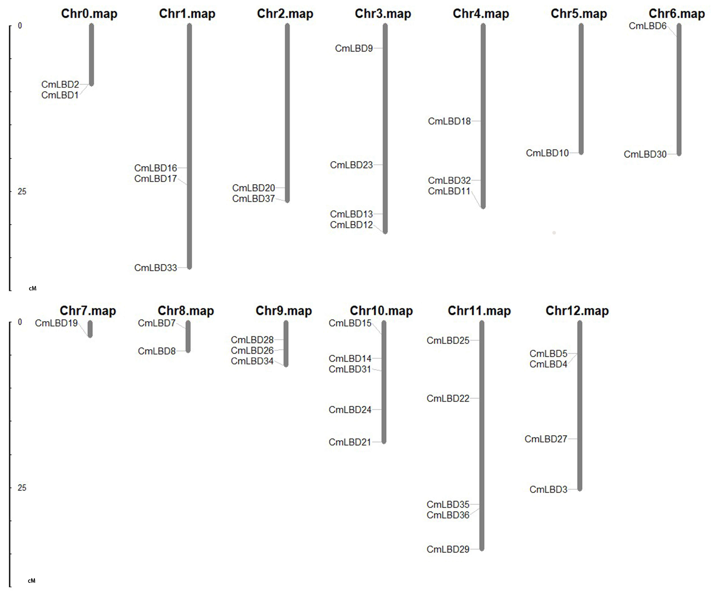
3.2. Chromosomal Distribution and Synteny Analysis of Melon LBD Gene Family

The synteny analysis diagram (Figure 1) and chromosomal location diagram (Figure 2) of the melon LBD gene family were plotted respectively. The LBD genes are unevenly distributed across 13 chromosomes, with chromosome 10 and 11 having the highest number of genes (five), and chromosomes 5, 7 having the least (one each). Only two genes are distributed on chromosomes 0, 2, 6, and 8. Homologous genes on a chromosome can be considered tandem repeat genes if their physical positions do not exceed 100 kb [15]. A tandem duplication event (CmLBD1/2) was identified in the melon genome. Additionally, there are 38 segmental duplication gene pairs present within the duplicated segments of the melon genome. These findings suggest that some CmLBDs may have originated from gene duplication, with segmental duplication events being a major driving force in the evolution of CmLBDs. Gene duplication events are common in all species, as they can generate new functional genes and drive species evolution [16,17].

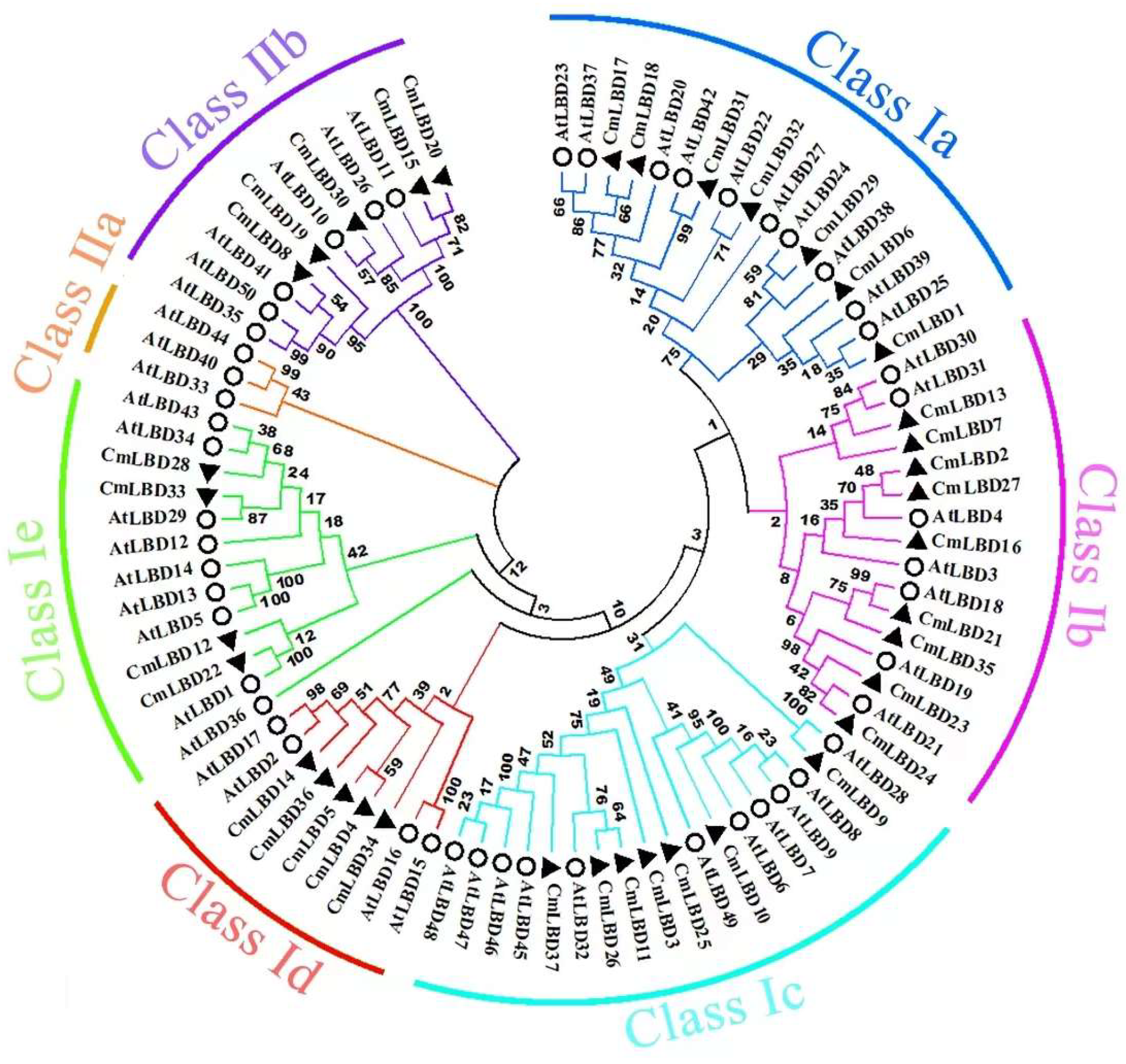
3.3. Construction of the Phylogenetic Tree of Melon and Arabidopsis LBD Gene Family Members

Based on the classification of the LBD family in Arabidopsis, the melon LBD proteins can be divided into two major classes, I and II. Class I contains five subclasses (Ia, Ib, Ic, Id, Ie), and Class II contains two subclasses (IIa, IIb) (Figure 3). Six subclasses contain members from both species, and members within each subclass may exhibit some degree of structural and functional similarity. The distribution of melon LBD genes in these seven subclasses is uneven, with the most members (nine) in Ib. There are no members of CmLBD in IIa. The neighbor-joining (NJ) phylogenetic tree analysis of melon and Arabidopsis reveals that there are five pairs of orthologous paralog genes in the melon LBD family: CmLBD4 and CmLBD5, CmLBD15 and CmLBD20, CmLBD11 and CmLBD26, CmLBD2 and CmLBD27, and CmLBD17 and CmLBD18.

3.4. Conservation Domain Analysis of Melon LBD Family Genes

We performed an alignment of the protein sequences of the 37 members of the melon LBD gene family (Figure 4). The LOB domain is mainly composed of three conserved sequences: the zinc finger-like structure (CX2CX6CX3C), the GAS region, and the leucine-zipper-like model (LX6LX3LX6L). Among these, CmLBD2 and CmLBD13 completely lack both the zinc finger-like structure and the GAS region. CmLBD34 completely lacks the leucine-zipper-like model, while CmLBD1, CmLBD19, and CmLBD22 have partially truncated leucine-zipper-like models. The leucine-zipper-like model has relatively more mutations compared to the other two conserved sequences.

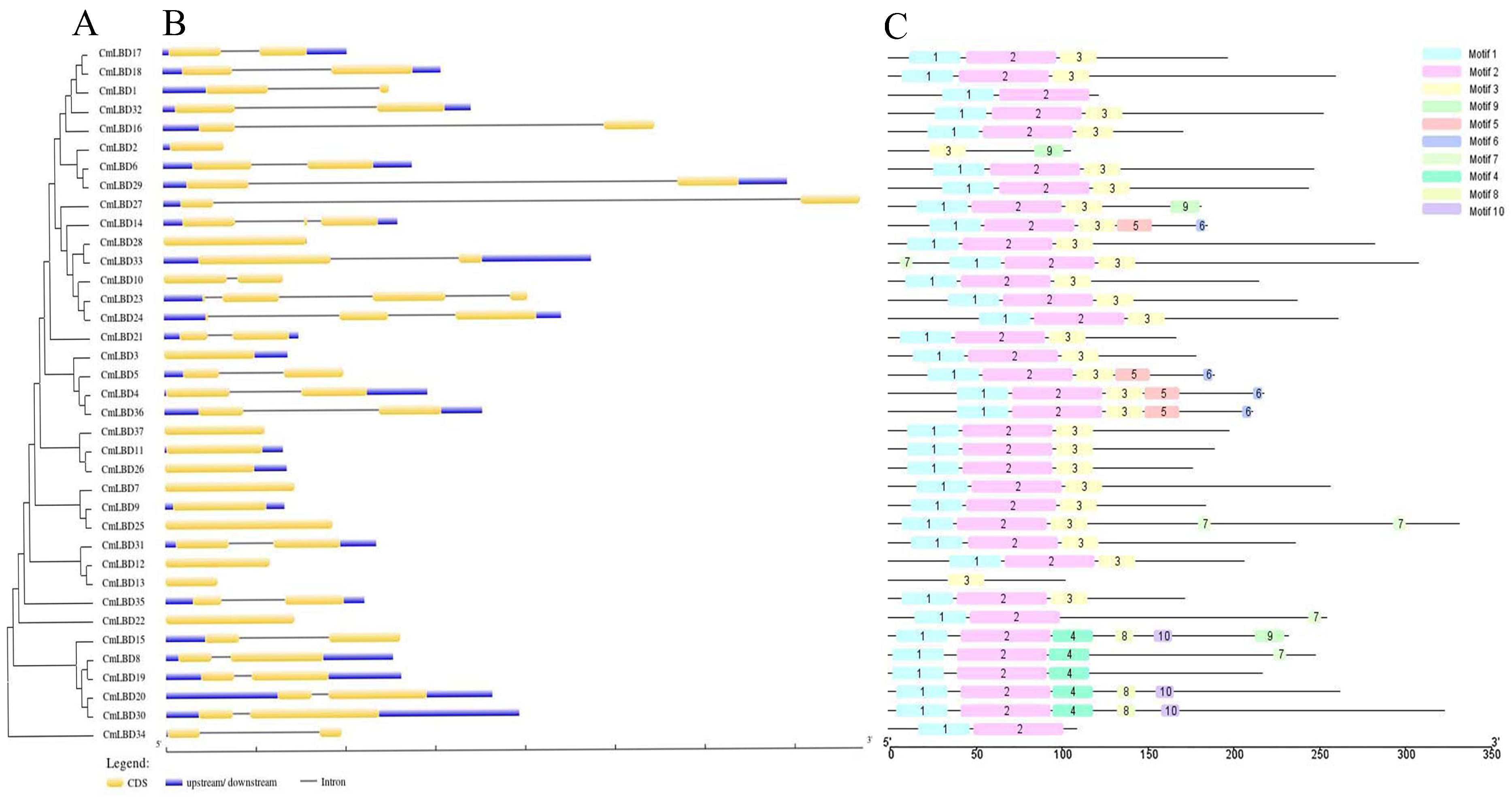
3.5. Gene Structure and Conserved Motif Analysis of Melon LBD Family Genes

The number of introns in the 37 LBD genes ranges from one to three (Figure 5B). Twelve of these genes lack introns; two genes (CmLBD14, CmLBD24) contain two introns, and only one gene (CmLBD23) contains three introns. The remaining genes all contain one intron. Genes within the same subfamily generally have similar gene structures with minor differences. For example, in Class IIb, all five genes each contain one intron.
Using the MEME website to analyze the melon LBD family protein sequences, a total of 10 conserved motifs were identified (Figure 5C). As can be seen in Figure 5, while CmLBD2 and CmLBD13 do not contain motifs 1 and 2, all of the other CmLBDs do, indicating a high degree of conservation in CmLBD. Motif 3 also appears frequently, present in 29 members, but motif 9 is only found in two transcription factors (CmLBD2 and CmLBD27). Some motifs only appear in specific subfamilies, such as motifs 8 and 10, which are only found in the IIb subfamily. Research on the melon LBD gene family reveals that genes within the same subfamily are similar in terms of gene structure and conserved sequences. This suggests that they likely have similar functions.

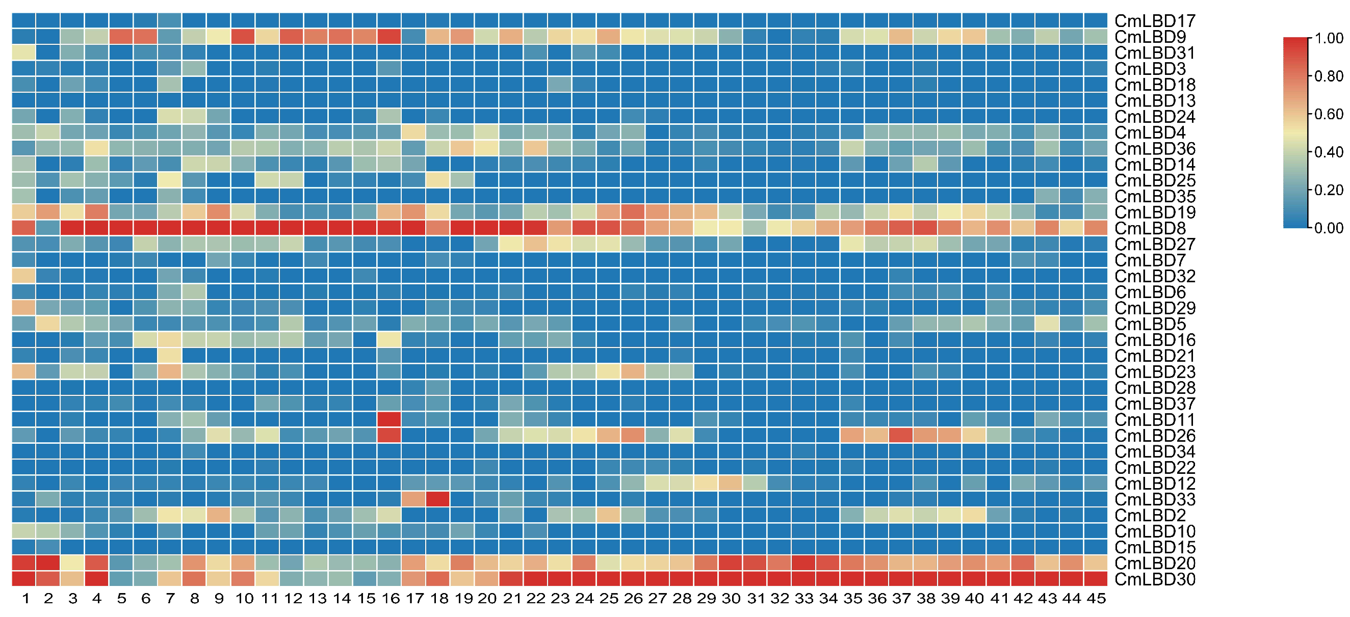
3.6. Expression Pattern Analysis of Melon LBD Family Genes

To further explore the function of LBD genes, we retrieved the expression specifics of melon LBD family genes in different tissues and at different time points through the Melonet DB website (Figure 6). The results showed that, among the 37 genes retrieved, only CmLBD13 was not expressed. The remaining genes showed significant differences in expression levels in different organs and at different times, demonstrating tissue specificity.
Genes such as CmLBD24, CmLBD8, CmLBD23, CmLBD16, CmLBD20, and CmLBD30 have higher expression levels in roots, suggesting that they may be involved in root development and affect root function. CmLBD9, CmLBD19, CmLBD8, and CmLBD2 show higher expression in stems. CmLBD9, CmLBD36, and CmLBD8 have higher expression levels in leaves. CmLBD9, CmLBD36, CmLBD8, CmLBD20, and CmLBD30 show significant expression in flowers, indicating that they may promote the formation and development of melon flowers. CmLBD9, CmLBD19, CmLBD8, CmLBD12, CmLBD20, and CmLBD30 have high expression levels in fruits. In seeds, the highest expression levels are seen in CmLBD9, CmLBD5, CmLBD20, and CmLBD30, suggesting they may promote the formation of melon seeds.
Expression analysis (Figure 6) showed that CmLBD8, CmLBD20, and CmLBD30, which are all from subfamily IIb, have higher expression levels in roots, stems, leaves, flowers, fruits, and seeds. We speculate that genes from subfamily IIb participate in the development of various organs in melons.

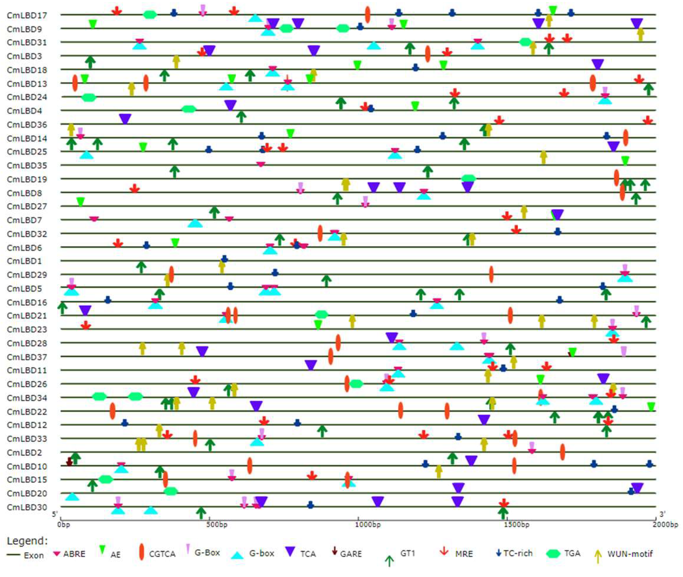
3.7. Cis-Acting Element Analysis of Melon LBD Family Genes

Cis-acting elements are non-coding DNA sequences in the gene promoter that regulate the transcription of related genes [18]. In melon LBD genes, a total of 12 cis-acting elements were identified, categorized into four classes: plant hormone response, light response, stress response, and specific binding sites for MYB (Figure 7). (1) Elements related to plant hormone responses were the most numerous, including abscisic acid-responsive elements (ABRE), methyl jasmonate(MeJA) responsiveness, salicylic acid-responsive elements (TCA), auxin-responsive elements (TGA), and gibberellin-responsive element (GARE). (2) The light response category includes light-responsive elements (G-box), a part of a module for light response (AE box), and light-responsive elements (GT1). (3) The stress response class, involved in hypoxia-specific induction, includes two elements, namely the defense and stress response elements (TC-rich), and wound response elements (WUN-motif). (4) MYB binding sites (MRE) are involved in light responses.
Among the five types of hormone response elements, ABRE has the widest distribution, across 56 sites, while the GARE-motif has the least distribution, across only two sites. ABA-responsive elements (ABRE) were found in most genes, such as CmLBD31, CmLBD7, CmLBD5, CmLBD34, CmLBD33, and CmLBD30, and were distributed across all subfamilies. Light-responsive elements (GT1), MYB binding sites involved in light response (MRE), and wound response elements (WUN) were also found in multiple CmLBD genes, and these three response elements are distributed across all subfamilies.

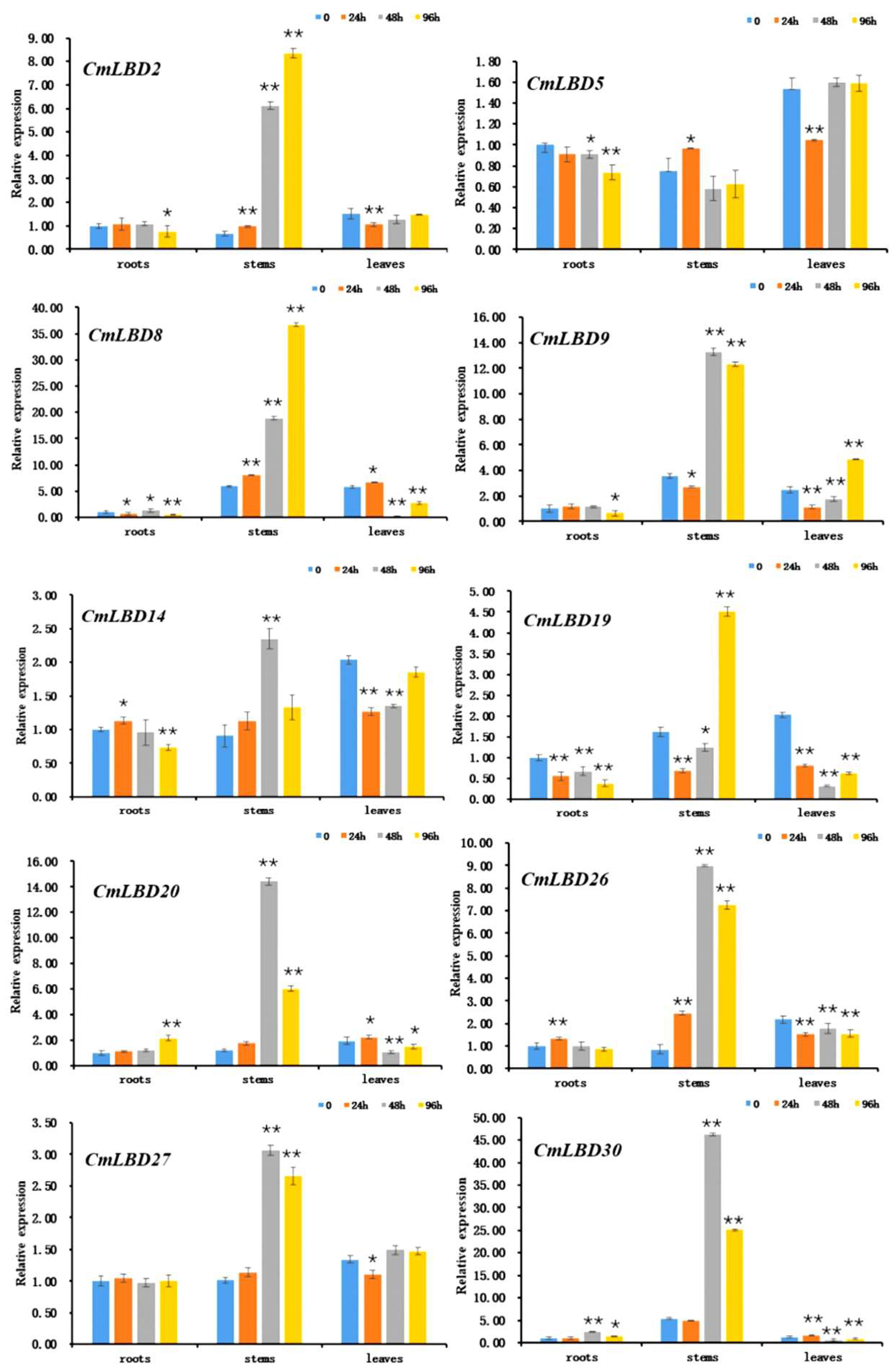
3.8. Expression of Melon LBD Family Genes in Response to Wilt Disease Pathogen

The response of melon LBD family genes to wilt disease pathogen stress was studied using qRT-PCR. The results showed (Figure 8) that 10 LBD family genes exhibited high expression in roots, stems, and leaves after inoculation with F. oxysporum. Most of these genes had significantly upregulated expression levels in stems (CmLBD2, CmLBD8, CmLBD9, CmLBD14, CmLBD19, CmLBD20, CmLBD26, CmLBD27, CmLBD30), especially at 48 h and 96 h post-inoculation, when expression levels increased by several to tens of times. Moreover, most genes showed a trend of initial increase and subsequent decrease, reaching peak expression at 48 h post-inoculation, with a few genes exhibiting a continuous increase and reaching maximum values at 96 h post-inoculation (CmLBD2, CmLBD8, CmLBD19).
Four LBD family genes had a certain degree of downregulation in roots (CmLBD2, CmLBD8, CmLBD9, CmLBD19), whereas CmLBD20 had a certain degree of upregulation. The expression of CmLBD8, CmLBD19, CmLBD20, and CmLBD30 in leaves significantly decreased with increased inoculation time, whereas CmLBD9 exhibited significant upregulation. The genes of Class II (CmLBD20, CmLBD30) responded strongly in the stems after inoculation, and also showed a certain degree of up- and downregulation in roots and leaves.

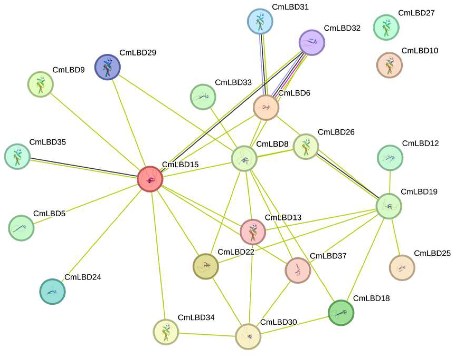
3.9. Protein Interactions

Potential interactions among CmLBD proteins were predicted using the STRING database (Figure 9). The CmLBD protein interaction network consists of 21 nodes, each communicating with other nodes. It was observed that CmLBD15 acts as a central hub in the network with numerous connections, suggesting its potential core role in regulating the network. In contrast, nodes like CmLBD5, despite having fewer connections, may still participate in specific interactions. Complex multigene interaction relationships, such as the connections between CmLBD6, CmLBD8, and CmLBD26, might indicate a synergistic function of these proteins within the network. This network diagram can assist in predicting the complexity of gene regulation and their potential roles in biological processes.

4. Discussion

LBD transcription factors are plant-specific transcription factors that play crucial roles in plant growth, development, and responses to abiotic stress. So far, 43, 35, and 44 members of the LBD transcription factor family have been identified in Arabidopsis, rice, and maize, respectively [3,5,19]. Using bioinformatics methods, this study identified 37 LBD genes in melon, unevenly distributed across 13 chromosomes. Only one LBD gene each was found on chromosomes 5 and 7, while the highest number of genes, five, was found on chromosome 10 and 11. Subcellular localization of all 37 genes was determined using the Plant-Ploc, and all were found to be located in the nucleus. Upon analyzing the structure of the LBD gene family, it was found that 32% of the genes did not contain introns, while 68% contained one to three introns. In addition, it was found that all genes except CmLBD2 and CmLBD13 contained motif 1 and motif 2. Thus, it can be seen that most LBD genes have a similar exon-intron structure and the same conserved elements, indicating that the LBD family is widely conserved in the evolutionary process.
Research has shown that the majority of LBD proteins belong to Class I. A total of 43 LBD transcription factors have been identified in the model plant Arabidopsis, 37 of which belong to Class I, and 6 to Class II. In Physcomitrella patens, a total of 31 LBD transcription factors have been identified, including 24 Class I members and 7 Class II members [20]. Among the 37 LBD genes identified in melon, 32 belong to Class I and 5 belong to Class II (Figure 3), further corroborating that the number of Class I members in the LBD gene family significantly outnumbers Class II members across different species.
Phylogenetic tree analysis indicates that each subfamily contains related genes from both melon and Arabidopsis, suggesting these genes share a common ancestral origin. The constructed phylogenetic tree reveals a close evolutionary relationship between melon and Arabidopsis, indicating the CmLBD genes are highly conserved throughout evolution [16]. The expression levels of genes located on the same branch are similar (e.g., CmLBD8 and CmLBD19, CmLBD20 and CmLBD30, CmLBD4 and CmLBD36) (Figure 3 and Figure 6), suggesting that duplication of LBD genes may primarily lead to functional redundancy. Exon/intron structure analysis the vast majority of CmLBDs contain no more than two introns, which is largely consistent with LBD genes in other plants, indicating a relatively conservative gene structure throughout evolution [21]. Gene function is closely related to the presence of conserved motifs within the protein sequence [22]. The expansion of the CmLBD gene family may primarily occur through segmental duplication, as 38 pairs of segmentally duplicated CmLBD genes were identified, while only one tandemly duplicated CmLBD gene pair (CmLBD1/2) was found, similar to other species from different taxonomic groups [21,23]. We hypothesize that during evolution, CmLBD gene family amplification was dominated by a segmental replication mechanism and supplemented by a tandem replication mechanism. This contributes to the development of new gene functions and may explain the relatively conservative number of CmLBD gene family members. Conservative motif analysis revealed that CmLBD proteins belonging to the same cluster possess similar conserved motifs. Proteins appearing in the same cluster are likely to have similar functions. Compared to other transcription factors, CmLBD8, CmLBD19, CmLBD20, and CmLBD30 not only share motif 1 and motif 2, but also uniquely possess motif 4. This finding implies these genes may play similar roles in plant physiological functions. Moreover, gene structure analysis reveals that these genes all have a single intron. These structural characteristics might reflect specific adaptations during evolution, thereby maintaining functional consistency. These genes exhibit a strong expression pattern in roots, stems, and leaves after inoculation with F. oxysporum, suggesting that motif 4 might be closely related to the function of the LBD family in resisting biotic stress. Further, we note that members of the subfamily IIb, such as CmLBD20 and CmLBD30, besides having motif 1, motif 2, and motif 4, uniquely contain motif 8 and motif 10. The high expression of these two genes in flowers, fruits, and seeds draws our attention, suggesting that motif 8 and motif 10 may be closely related to reproductive traits in melon, such as flower development, fruit maturation, and seed formation. These findings provide important insights into the diverse functions of LBD family members in plant growth, development, and response to environmental stresses.
In the LBD gene family, Class I primarily regulates growth and development, while Class II may be associated with environmental stress responses [2,16]. Our research reveals that several genes within Class II, including CmLBD8, CmLBD20, and CmLBD30, not only play significant roles in response to biotic stress, but also in the development of crucial organs such as flowers, fruits, and seeds in melon (Figure 6 and Figure 8). This enhances our understanding of the functions of the LBD family’s Class II category. Phylogenetic analysis of melon LBD proteins and those of Arabidopsis suggests that LBD genes are more evolutionarily recent, and those that cluster together in the same major class or subclass likely have similar structures and functions [24]. In Arabidopsis, AtLBD6 can inhibit the division and differentiation of cells near the mid-axial region, resulting in the formation of bilaterally symmetrical, planar leaves [25]. Thus, it is hypothesized that CmLBD10, which is homologous to AtLBD6, may have a similar function. AtLBD12 is involved in the development of the leaf [26], suggesting that CmLBD genes, which belong to the same branch, may regulate similar function.
The tissue expression patterns of genes are closely related to their functional features. In this study, the analysis of the expression patterns of melon LBD family genes revealed that genes in the Ic subclass (CmLBD9) and in the Ⅱb subclass (CmLBD8, CmLBD20, and CmLBD30) are highly expressed in roots, stems, leaves, flowers, fruits, and seeds, suggesting that they are involved in the development of various melon organs. In Carya illinoensis, LBD30 and LBD3 belong to Group II, and LBD37 belongs to Group I; these genes are not only related to nutrient accumulation in the embryo, but are also involved in the processes of cell division and organ differentiation [27]. In Capsicum annuum, Ca02g002988 has a higher expression level in roots than in other organs, while Capana00g003460, Capana01g001405, and Capana06g000315 are all expressed only in roots. These genes may have a certain association with root morphogenesis [28].
Like many transcription factors, the LBD gene family forms a variety of molecular regulatory networks, playing important roles in plant growth and development and responses to environmental stress [29]. Multiple cis-acting elements in the gene promoter region play a crucial role in signal transduction, and their synergistic interactions can regulate complex biological processes. AtLBD16 and AtLBD29 are involved in growth hormone response and lateral root formation [29,30]. AtLBD20 plays a role in the plant disease resistance process, mediated by the jasmonic acid signaling pathway [31]. Research has found that the LBD promoter contains many elements related to hormone regulation pathways, including abscisic acid, MeJA, GA, and IAA. MeJA is a plant hormone that regulates defense mechanisms and stress responses [32], while ABA is an important plant stress hormone that plays a crucial role in the salt stress signaling pathway [33]. Meanwhile, the LBD promoter also contains the base sequences of light response, stress response, and MYB specific binding sites. These different cis-acting elements together regulate the growth and development of melon, allowing it to adapt to unfavorable living environments and grow healthily.
LBD genes play an important role in plant defense responses. In the Arabidopsis LBD family, LBD20 is a F. oxysporum susceptibility gene, marking the first demonstration of a LBD gene family member’s role in biotic stress [31]. Transcriptomic data of cassava under biotic and abiotic stresses showed that MeASLBD46 and MeASLBD47 were more responsive to disease and drought, and that MeASLBD47 significantly reduced the virulence of cassava bacterial wilt (XamCHN11), according to real-time fluorescence quantitative reverse transcription PCR (qRT-PCR) and virus-induced gene silencing (VIGS) [34].
This study shows that after melon seedlings were inoculated with wilt bacteria, the expression levels of nine out of ten genes were significantly different in the stem. The significant upregulation of several LBD genes in stems suggests their involvement in the activation of defense pathways in response to pathogen invasion. After root infection by the wilt pathogen, mycelium forms and moves upward through the vascular bundle to invade the plant body. In the pathogenic mechanism of wilt disease, there are two main theories: the vascular occlusion hypothesis, and the toxin hypothesis. The vascular occlusion hypothesis believes that the blockage of xylem vessels by the pathogen is the main reason for plant wilting [35]. Our study found that compared to roots and leaves, the gene response in the stem was more intense, which may be related to the well-developed vascular bundles (xylem) in the stem, and the characteristic of wilt pathogens mainly damaging the vascular bundles (xylem). The dynamic regulation of LBD genes in the stem, reaching peak expression levels at 48 h and 96 h after inoculation, may indicate their key role in the early stages of plant-pathogen interaction. Moreover, compared to the stem, the expression patterns of LBD genes in roots and leaves showed significant differences. These results suggest that the response of melon LBD genes to wilt disease stress is tissue-specific, highlighting the complexity of LBD gene regulation in different plant organs.
Lü et al. analyzed the transcriptional situation of the non-host interaction between watermelon and F. oxysporum, finding that most genes involved in jasmonic acid (JA) biosynthesis were continuously upregulated, indicating that JA biosynthesis, along with the tryptophan-phenylalanine-lignin pathway, might play an important role in watermelon’s resistance against wilt pathogen infection [36]. Ding showed that black-seeded pumpkin primarily responds to wilt pathogen infection through disease resistance pathways mediated by ABA and JA [14]. Research by Zhou and Wu suggests that ABA signaling may play a significant role in cucumber’s defense response to F. oxysporum [37]. Studies on the gene expression profile of bananas at different infection times by wilt pathogen [38] conjecture that the infection by wilt pathogen in bananas is closely related to the JA signaling pathway.
In Arabidopsis, AtLBD20 is considered to be a susceptible gene for F. oxysporum disease, and it is involved in the plant disease response mediated by jasmonate [39]. Cis-acting element analysis showed that CmLBD2, CmLBD8, CmLBD14, CmLBD19, and CmLBD26 all contain methyl jasmonate (MeJA) responsiveness (CGTCA-motif); hence, it is inferred that these genes participate in the plant disease signaling pathway mediated by jasmonic acid methyl ester (MeJA). However, CmLBD20 and CmLBD30 do not contain CGTCA-motif, but both contain salicylic acid response elements (TCA-element), suggesting that these two genes resist wilt bacteria by participating in the signaling pathway mediated by salicylic acid (SA).
Our data indicate significant variations in the expression of the CmLBD gene at three selected time points post-inoculation (24, 48, and 96 h), providing crucial insights into the response of the melon LBD family to Fusarium infection. Nonetheless, we recognize that a more frequent analysis of time points could unveil finer details of dynamic expression patterns, which are of paramount importance for a comprehensive understanding of the melon LBD family’s reaction to infection. Therefore, future studies could consider including a broader range of time points for analysis, especially shorter intervals within the first 24 h, to delve deeper into the interactions between the host and pathogen.
Despite this study offering significant insights into the functionality of the LBD gene family in melon, particularly in resisting F. oxysporum infection, the applicability of these findings to other stress conditions or pathogens remains unclear. Future research should aim to explore the functions of LBD genes under a variety of biotic and abiotic stress conditions. Such an understanding will contribute to a multifaceted view of the roles of LBD genes in stress physiology and potentially enhance our capability to improve melon resistance through molecular breeding. Such investigations will require the integrated application of genomics, transcriptomics, and proteomics, among other approaches, to uncover the complex regulatory networks involving LBD genes.

5. Conclusions

At present, research on LBD genes of other species continues to emerge, which will help people further understand the LBD gene family. In this article, systematic analyses of the physical and chemical properties of LBD genes, chromosome positioning, collinearity, evolutionary tree, and cis-acting elements were carried out in sequence, aiming to provide a reference for more in-depth future research on the LBD gene family. In the interim, our study provides comprehensive insights into the expression profiles of melon LBD family genes in response to the wilt disease pathogen F. oxysporum. Nine genes exhibited strong expression in roots, stems, and leaves in response to pathogenic invasion. With the infection of Fusarium, the expression levels of these genes generally showed an increasing trend, among which CmLBD20 and CmLBD30 responded most intensely. It is speculated that these genes play significant biological roles during the Fusarium infection process. These results not only powerfully demonstrate that LBDs are involved in the melon’s resistance to biotic stress (Fusarium infection) but also, by analyzing the expression patterns of these ten CmLBD genes under Fusarium infection, our results provide robust molecular evidence for research into melon resistance breeding. Especially, the marked response exhibited by the CmLBD20 and CmLBD30 genes suggests they may play key roles in plant defense reactions, offering potential candidate genes for future molecular breeding efforts to improve melon’s resistance to Fusarium wilt. Our study lays a foundation for further understanding the function of the LBD gene family in the biology and pathology of melon plants and for the development of new molecular markers and creation of melon varieties with high resistance to Fusarium wilt. Overall, this research provides valuable resources and new strategic directions for improving melon quality and enhancing crop resistance to Fusarium wilt.

Author Contributions

L.Z.: analyzed the data, wrote the article, and approved the final draft. Y.C.: analyzed the data, wrote the article. Y.W.: conceptualized and analyzed data. Y.X.: prepared numbers and/or table. S.L.: designed experiments and approved the final draft. All authors have read and agreed to the published version of the manuscript.

Funding

The work was funded by Innovation and entrepreneurship training program for college students of China (Project grant: 202310482026); Sample Courses on Curriculum Thinking and Politics in Undergraduate Colleges and Universities in Henan Province in 2023 (Botany); Fine Online Open Courses in Undergraduate Colleges and Universities in Henan Province in 2021 (Botany).

Institutional Review Board Statement

Not Applicable.

Informed Consent Statement

Not Applicable.

Data Availability Statement

Transcriptome data are available through the Melonet DB (https://melonet-db.dna.affrc.g-o.jp/ap/top).

Conflicts of Interest

The author declares there is no competitive advantage.

References

  1. Ma, W.; Wu, F.; Sheng, P.; Wang, X.; Zhang, Z.; Zhou, K.; Zhang, H.; Hu, J.; Lin, Q.; Cheng, Z.; et al. Rice LBD 12-1 reduces meristem size by inhibiting ago10-hd-zip III pathway. Plant Physiol. 2017, 173, 801–811. [Google Scholar] [CrossRef] [PubMed]
  2. Majer, C.; Hochholdinger, F. Defining the boundaries: Structure and function of LOB domain proteins. Trends Plant Sci. 2010, 16, 47–52. [Google Scholar] [CrossRef] [PubMed]
  3. Shuai, B.; Reynaga-Pena, C.G.; Springer, P.S. The lateral organ boundaries gene defines a novel, plant-specific gene family. Plant Physiol. 2002, 129, 747–761. [Google Scholar] [CrossRef] [PubMed]
  4. Lee, H.W.; Kim, M.J.; Park, M.Y.; Han, K.H.; Kim, J. The conserved proline residue in the LOB domain of LBD18 is critical for DNA-binding and biological function. Mol. Plant 2013, 6, 1722–1725. [Google Scholar] [CrossRef] [PubMed]
  5. Liu, H.; Wang, S.; Yu, X.; Yu, J.; He, X.; Zhang, S.; Shou, H.; Wu, P. ARL1, a LOB-domain protein required for adventitious root formation in rice. Plant J. 2005, 43, 47–56. [Google Scholar] [CrossRef] [PubMed]
  6. Yu, J.; Xie, Q.; Li, C.; Dong, Y.; Zhu, S.; Chen, J. Comprehensive characterization and gene expression patterns of LBD gene family in Gossypium Planta. Planta 2020, 251, 81. [Google Scholar] [CrossRef] [PubMed]
  7. Wang, X.; Zhang, S.; Su, L.; Liu, X.; Hao, Y. A Genome-Wide Analysis of the LBD Gene Family in Malus domestica with a Functional Characterization of MdLBD11. PLoS ONE 2013, 8, e57044. [Google Scholar] [CrossRef] [PubMed]
  8. Chen, X.; Wang, J.; Zhao, M.; Yuan, H. Identification and expression analysis of Lateral Organ Bound Aries Domain (LBD) transcription factor genes in Fragaria vesca. Plant Sci. 2017, 23, 4700. [Google Scholar] [CrossRef]
  9. Rubin, G.; Tohge, T.; Matsuda, F.; Saito, K.; Scheible, W.R. Members of the LBD family of transcription factors repress anthocyanin synthesis and affect additional nitrogen responses in Arabidopsis. Plant Cell 2009, 21, 3567–3584. [Google Scholar] [CrossRef]
  10. Albinsky, D.; Kusano, M.; Higuchi, M.; Hayashi, N.; Kobayashi, M.; Fukushima, A.; Mori, M.; Ichikawa, T.; Matsui, K.; Kuroda, H.; et al. Metabolomic screening applied to rice FOX Arabidopsis lines leads to the identification of a gene-changing nitrogen metabolism. Mol. Plant 2010, 3, 125–142. [Google Scholar] [CrossRef]
  11. Lavenus, J.; Goh, T.; Roberts, I.; Guyomarc’hv, S.; Lucas, M.; Smet, I.D.; Fukaki, H.; Beeckman, T.; Bennett, M.; Laplaze, L. Lateral root development in Arabidopsis: Fifty shades of auxin. Trends Plant Sci. 2013, 18, 450–458. [Google Scholar] [CrossRef] [PubMed]
  12. Gil, J.F.; Liebe, S.; Thiel, H.; Lennefors, B.; Kraft, T.; Gilmer, D.; Maiss, E.; Varrelmann, M.; Savenkov, E.I. Massive up-regulation of LBD transcription factors and EXPANSINs highlights the regulatory programs of rhizomania disease. Mol. Plant Pathol. 2018, 19, 2333–2348. [Google Scholar] [CrossRef]
  13. Cao, H.; Liu, C.Y.; Liu, C.X.; Zhao, Y.L.; Xu, R.R. Genomewide analysis of the lateral organ boundaries domain gene family in Vitis vinifera. J. Genet. Genom. 2016, 95, 515–526. [Google Scholar] [CrossRef] [PubMed]
  14. Ding, Y.M. Response Mechanism of Black Seed Pumpkin to Fusarium Wilt Infection and Screening of NBS Resistance Genes; Southwest University: Chongqing, China, 2019. (In Chinese) [Google Scholar]
  15. Huang, S.X.; Gao, Y.F.; Liu, J.K.; Peng, X.L.; Niu, X.L.; Fei, Z.J.; Cao, S.Q.; Liu, Y.S. Genome-wide analysis of Wrky transcription factors in Solanum lycopersicum. Mol. Genet. Genom. 2012, 287, 495–511. [Google Scholar] [CrossRef] [PubMed]
  16. Yang, Y.; Yu, X.; Wu, P. Comparison and evolution analysis of two rice subspecies LATERAL ORGAN BOUNDARIES domain gene family and their evolutionary characterization from Arabidopsis. Mol. Phylogenet. Evol. 2006, 9, 248–262. [Google Scholar] [CrossRef] [PubMed]
  17. Taramino, G.; Sauer, M.; Stauffer, J.L.; Multani, D.; Niu, X.; Sakai, H.; Hochholdinger, F. The maize (Zea mays L.) RTCS gene encodes a LOB domain protein that is a key regulator of embryonic seminal and post-embryonic shoot-borne root initiation. Plant J. 2007, 50, 649–659. [Google Scholar] [CrossRef] [PubMed]
  18. Evans, M. The indeterminate gametophyte1 gene of maize encodes a LOB domain protein required for embryo sac and leaf development. Plant Cell 2007, 19, 46–62. [Google Scholar] [CrossRef] [PubMed]
  19. Bortiri, E.; Chuck, G.; Vollbrecht, E.; Rocheford, T.; Martienssen, R.; Hake, S. ramosa2 encodes a LATERAL ORGAN BOUNDARY domain protein that determines the fate of stem cells in branch meristems of maize. Plant Cell 2006, 18, 574–585. [Google Scholar] [CrossRef] [PubMed]
  20. Huang, X.L.; Yan, H.Q.; Liu, Y.J.; Yi, Y. Genome-wide analysis of LATERAL ORGAN BOUNDARIES DOMAIN-in Physcomitrella patens and stress responses. Genes Genom. 2020, 42, 651–662. [Google Scholar] [CrossRef]
  21. Liang, J.; Hou, Z.; Liao, J.; Qin, Y.; Wang, L.; Wang, X.; Su, W.; Cai, Z.; Fang, Y.; Aslam, M.; et al. Genome-Wide Identification and Expression Analysis of LBD Transcription Factor Genes in Passion Fruit (Passiflora edulis). Int. J. Mol. Sci. 2022, 23, 4700. [Google Scholar] [CrossRef]
  22. Sylvia, C.; Sun, J.; Zhang, Y.; Ntini, C.; Ogutu, C.; Zhao, Y.; Han, Y. Genome-wide analysis of ATP binding cassette (ABC) transporters in peach (Prunus persica) and identification of a Gene PpABCC1 involved in anthocyanin accumulation. Int. J. Mol. Sci. 2023, 24, 1931. [Google Scholar] [CrossRef] [PubMed]
  23. Wang, J.; Zhang, W.; Cheng, Y.; Feng, L. Genome-Wide Identification of LATERAL ORGAN BOUNDARIES DOMAIN (LBD) Transcription Factors and Screening of Salt Stress Candidates of Rosa rugosa Thunb. Biology 2021, 10, 992. [Google Scholar] [CrossRef] [PubMed]
  24. Wang, L.; Chen, H.; Zheng, Y.F.; Huang, R.D.; Liu, C.S. Identification of LBD gene family in Amaranthus tricolor L. based on transcriptome and expression analysis under blue light. J. China Agric. Univ. 2021, 26, 66–80. [Google Scholar] [CrossRef]
  25. Semiarti, E.; Ueno, Y.; Iwakawa, H.; Tsukaya, H.; Machida, C.; Machida, Y. The Asymmetric Leaves2 (AS2) gene of Arabidopsis thaliana regulates Lamina formation and is required for patterning of leaf venation. Prog. Biotechnol. 2001, 18, 63–68. [Google Scholar] [CrossRef]
  26. Nakazawa, M.; Ichikawa, T.; Ishikawa, A.; Kobayashi, H.; Tsuhara, Y.; Kawashima, M.; Suzuki, K.; Muto, S.; Matsui, M. Activation tagging, a novel tool to dissect the functions of a gene family. Plant J. 2003, 34, 741–750. [Google Scholar] [CrossRef] [PubMed]
  27. Huang, Y.C.; Guo, W.L.; Wang, Z.J. Bioinformatics analysis of the whole genome LBD gene family in Carya illinoensis. J. Zhejiang A F Univ. 2021, 38, 464–475. [Google Scholar] [CrossRef]
  28. Zheng, Z.F.; Zhang, Y.L.; Hu, C.; Dai, X.Z.; Liu, F.; Yuan, Z.H. Genome-wide Identification and Expressing Analysis of LBD Transcription Factors in Pepper. Acta Hortic. Sin. 2016, 43, 683–694. [Google Scholar] [CrossRef]
  29. Shi, Y.; Shen, S.Y.; Zhang, Q.Y.; Shun, Z.M.; Wu, R.L.; Guo, Y.Q. Research progress of LBD gene family. Chin. J. Cell Biol. 2019, 41, 738–745. [Google Scholar] [CrossRef]
  30. Xu, L. De novo root regeneration from leaf explants: Wounding, auxin, and cell fate transition. Curr. Opin. Plant Biol. 2018, 41, 39–45. [Google Scholar] [CrossRef]
  31. Thatcher, L.F.; Powell, J.J.; Aitken, E.A.B.; Kazan, K.; Manners, J.M. The lateral organ boundaries domain transcription factor LBD20 functions in Fusarium wilt susceptibility and Jasmonate signaling in Arabidopsis. Plant Physiol. 2012, 160, 407–418. [Google Scholar] [CrossRef]
  32. Lang, D.; Yu, X.; Jia, X.; Li, Z.X.; Zhang, X.H. Methyl jasmonate improves metabolism and growth of NaCl-stressed Glycyrrhiza uralensis seedlings. Sci. Hortic. 2020, 266, 109287. [Google Scholar] [CrossRef]
  33. Gómez, S.; Ferrieri, R.A.; Schueller, M.; Orians, C.M. Methyl jasmonate elicits rapid changes in carbon and nitrogen dynamics in tomato. New Phytol. 2010, 188, 835–844. [Google Scholar] [CrossRef] [PubMed]
  34. Mao, Y.; Abdoulaye, A.H.; Song, J.; Yao, X.; Zhang, Y.; Gao, Y.; Ye, Y.; Luo, K.; Xia, W.; Chen, Y. Genome-wide analyses of LATERAL ORGAN BOUNDARIES in cassava reveal the role of LBD47 in defence against bacterial blight. PLoS ONE 2023, 18, e0282100. [Google Scholar] [CrossRef] [PubMed]
  35. Yu, T.X.; Zhang, M.F. Research Progress on Watermelon Fusarium Wilt. China Watermelon Melon 2004, 1, 17–19. (In Chinese) [Google Scholar]
  36. Lü, G.; Guo, S.; Zhang, H.; Geng, L.; Song, F.; Fei, Z.; Xu, Y. Transcriptional profiling of watermelon during its incompatible interaction with Fusarium oxysporum f. sp. niveum. Eur. J. Plant Pathol. 2011, 131, 585–601. [Google Scholar] [CrossRef]
  37. Zhou, X.; Wu, F. Differentially expressed transcripts from cucumber (Cucumis sativus L.) root upon inoculation with Fusarium oxysporum f. sp. cucumerinum Owen. Physiol. Mol. Plant Pathol. 2009, 74, 142–150. [Google Scholar] [CrossRef]
  38. Sun, J.; Wei, D.; Qin, L.; Wei, Y.; Guo, W.; Yu, W.; Zhang, Z.; Lu, J. Application of High-Throughput Sequencing Technology in the Research of Banana Resistance to Wilt Disease. J. South. Agric. 2014, 45, 1921–1925. (In Chinese) [Google Scholar]
  39. Thatcher, L.F.; Kazan, K.; Manners, J.M. Lateral organ boundaries domain transcription factors: New roles in plant defense. Plant Signal. Behavior. 2012, 7, 1702–1704. [Google Scholar] [CrossRef]
Figure 1. Collinear analysis of LBD family genes in melon.
Genes 15 00442 g001
Figure 2. The location distribution of LBD genes in melon chromosomes.
Genes 15 00442 g002
Figure 3. The neighbor-joining (NJ) phylogenetic tree of the CmLBD gene family.
Genes 15 00442 g003
Figure 4. Conserved domain analysis of LBD family genes in melon.
Genes 15 00442 g004
Figure 5. Gene structure and phylogeny of the melon LBD family. (A) The neighbor-joining (NJ) phylogenetic tree for the CmLBD protein family. (B) Exon–intron structure of CmLBD genes. (C) Distribution of 10 motifs in 37 CmLBD proteins.
Genes 15 00442 g005
Figure 6. Expression analysis of LBD genes in melon. 1: callus, 2: dry seeds, 3: 1d-imbibed seeds, 4: 3d-imbibed seeds, 5: 7d-seedlings cotyledon, 6: 7d-seedlings hypocotyl, 7: 7d-seedlings root, 8: root, 9: middle stem, 10: upside stem, 11: shoot apex, 12: young leaves, 13: 6th leaves, 14: 9th leaves, 15: 12th leaves, 16: tendril, 17: anther male flower, 18: anther female flower, 19: petal female flower, 20: stigma female flower, 21: ovary DAF0, 22: ovary DAF2, 23: ovary DAF4, 24: fruit flesh DAF8, 25: fruit flesh DAF15, 26: fruit flesh DAF22, 27: fruit flesh DAF29, 28: fruit flesh DAF36, 29: fruit flesh DAF43, 30: fruit flesh DAF50, 31: fruit flesh post harvest 1 week, 32: fruit flesh post harvest 2 week, 33: fruit flesh post harvest 3 week, 34: fruit flesh post harvest 4 week, 35: fruit epicarp DAF8, 36: fruit epicarp DAF15, 37: fruit epicarp DAF22, 38: fruit epicarp DAF29, 39: fruit epicarp DAF36, 40: fruit epicarp DAF43, 41: fruit epicarp DAF50, 42: fruit epicarp post harvest 1 week, 43: fruit epicarp post harvest 2 week, 44: fruit epicarp post harvest 3 week, 45: fruit epicarp post harvest 4 week.
Genes 15 00442 g006
Figure 7. Cis-elements of LBD genes in melon.
Genes 15 00442 g007
Figure 8. Expression of melon LBD family genes in response to wilt bacteria. The asterisk indicates the p value in the significance test (** p < 0.01 and * p < 0.05).
Genes 15 00442 g008
Figure 9. C. melo CmLBD functional interaction networks based on Arabidopsis orthologs.
Genes 15 00442 g009
Table 1. Information regarding the LBD family in melon.
Gene NameGene SymbolProtein Length (Amino acids)Molecular
Weight (Da)
Isoelectric
Point
Instability
Index
Fat Solubility IndexMean Hydrophilic ValueSubcellular
Localization
CmLBD1MELO3C000068P111511,743.489.0655.1176.430.163Nucleus
CmLBD2MELO3C000076P19910,950.405.6761.2469.90−0.268Nucleus
CmLBD3MELO3C002290P116718,814.358.5247.7973.05−0.359Nucleus
CmLBD4MELO3C005009P120422,340.626.4172.3591.86−0.131Nucleus
CmLBD5MELO3C005013P117720,037.167.6062.0482.20−0.265Nucleus
CmLBD6MELO3C006183P123124,953.608.9380.5180.69−0.216Nucleus
CmLBD7MELO3C007098P124027,293.827.6274.9864.62−0.634Nucleus
CmLBD8MELO3C007620P123225,560.777.5866.5369.40−0.390Nucleus
CmLBD9MELO3C008309P117219,111.618.1062.8768.72−0.322Nucleus
CmLBD10MELO3C008779P120122,171.178.9654.6970.90−0.392Nucleus
CmLBD11MELO3C009989P117719,508.687.5868.2754.63−0.640Nucleus
CmLBD12MELO3C010752P119321,546.777.9951.0388.13−0.186Nucleus
CmLBD13MELO3C011135P19610,508.774.7531.2487.60−0.089Nucleus
CmLBD14MELO3C011730P117319,126.146.5679.9882.37−0.148Nucleus
CmLBD15MELO3C012250P121723,478.488.4536.6972.40−0.328Nucleus
CmLBD16MELO3C012624P116017,234.618.1472.6170.19−0.159Nucleus
CmLBD17MELO3C012741P118420,243.076.2868.1280.220.014Nucleus
CmLBD18MELO3C012908P124327,013.916.0446.6063.46−0.500Nucleus
CmLBD19MELO3C016808P120322,236.136.5056.7580.69−0.202Nucleus
CmLBD20MELO3C017305P124526,763.808.7254.1477.63−0.240Nucleus
CmLBD21MELO3C018380P115617,169.957.5261.4286.860.023Nucleus
CmLBD22MELO3C019314P123826,823.128.8846.8667.94−0.772Nucleus
CmLBD23MELO3C019995P122223,851.079.1079.3375.63−0.223Nucleus
CmLBD24MELO3C020121P124426,813.148.6176.4170.00−0.491Nucleus
CmLBD25MELO3C020944P131034,948.927.1267.3564.87−0.792Nucleus
CmLBD26MELO3C021578P116518,202.357.6650.9266.30−0.524Nucleus
CmLBD27MELO3C021789P117018,490.968.3262.5173.47−0.281Nucleus
CmLBD28MELO3C021964P126529,996.605.0055.5675.81−0.570Nucleus
CmLBD29MELO3C022505P122824,793.476.9747.7480.44−0.145Nucleus
CmLBD30MELO3C023802P130232,712.428.8651.4682.35−0.272Nucleus
CmLBD31MELO3C023880P122124,271.064.4548.4978.10−0.051Nucleus
CmLBD32MELO3C024038P123625,568.787.0645.0775.34−0.250Nucleus
CmLBD33MELO3C024387P128832,397.205.4251.1676.60−0.544Nucleus
CmLBD34MELO3C025504P110211,653.779.4352.8776.47−0.141Nucleus
CmLBD35MELO3C025701P116118,044.504.9456.3873.35−0.207Nucleus
CmLBD36MELO3C025742P119821,505.305.5975.2681.36−0.182Nucleus
CmLBD37MELO3C026269P118520,363.907.6353.8171.30−0.405Nucleus
Disclaimer/Publisher’s Note: The statements, opinions and data contained in all publications are solely those of the individual author(s) and contributor(s) and not of MDPI and/or the editor(s). MDPI and/or the editor(s) disclaim responsibility for any injury to people or property resulting from any ideas, methods, instructions or products referred to in the content.

