Non-Apoptotic Programmed Cell Death: From Ultrastructural Characterization to Emerging Therapeutic Opportunities

Highlights
- Non-apoptotic programmed cell death comprises diverse modalities such as ferroptosis, necroptosis, pyroptosis, autophagy, paraptosis, autosis, and more, with each modality characterized by unique ultrastructural hallmarks and molecular signaling pathways that distinguish them from classical apoptosis.
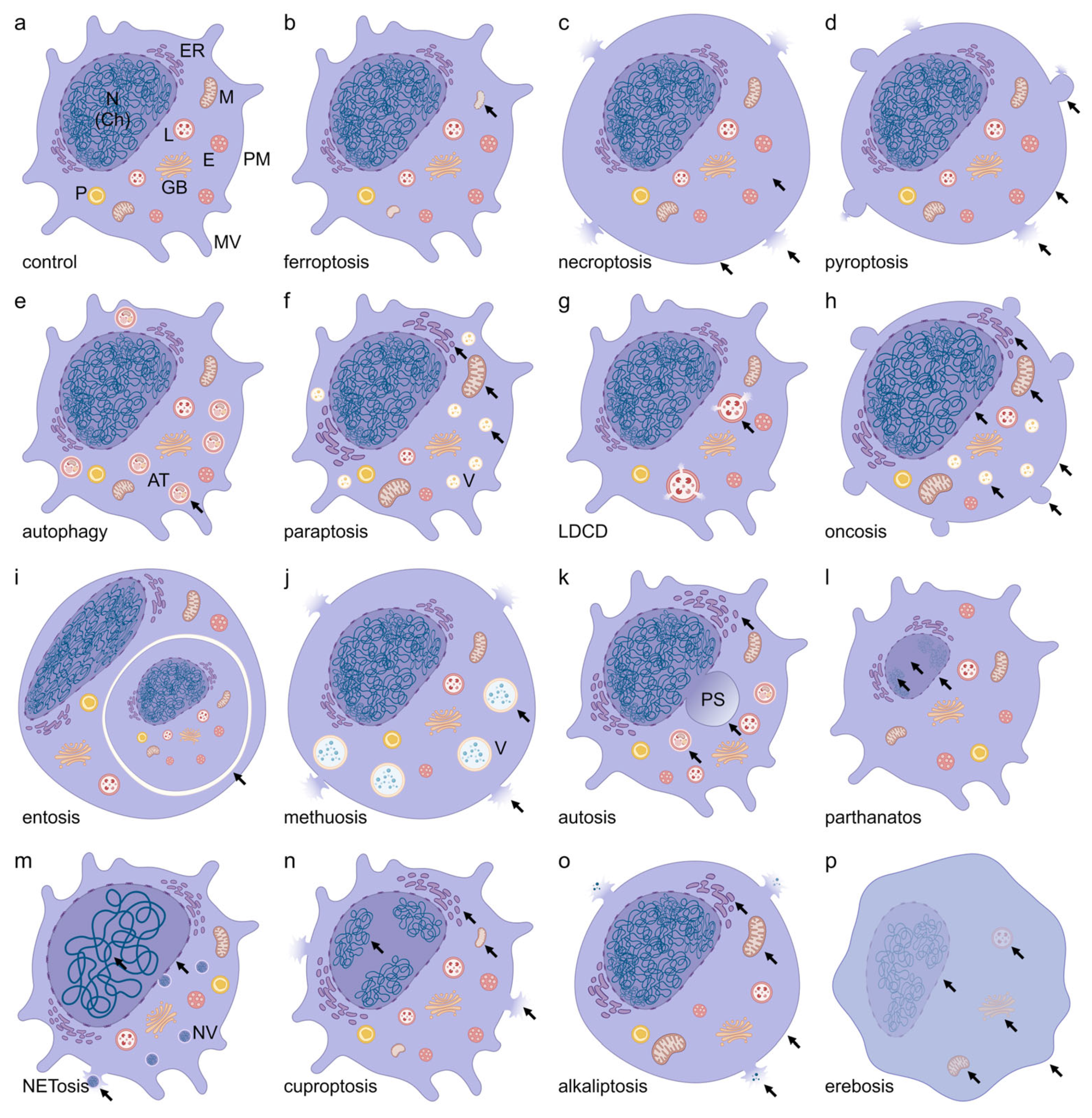
- Comprehensive morphological characterization reveals distinct intracellular features for each non-apoptotic PCD modality, ranging from mitochondrial cristae reduction in ferroptosis to perinuclear space ballooning in autosis, facilitating their identification via advanced microscopy techniques.
- Non-apoptotic PCD pathways offer dual therapeutic potential: induction strategies can overcome apoptosis resistance in cancer therapy, while inhibition approaches provide cytoprotection in neurodegenerative diseases and myocardial injury.
- Targeting non-apoptotic PCD pathways opens novel therapeutic avenues for apoptosis-resistant malignancies through pathway-specific nanomedicines and pharmacological modulators that selectively eliminate cancer cells while sparing normal tissue.
- Understanding ultrastructural hallmarks of non-apoptotic PCD enables precise mechanistic differentiation in disease models, supporting development of pathway-specific biomarkers and precision medicine approaches for cancer, neurodegeneration, and inflammatory diseases.
- The comprehensive characterization of non-apoptotic PCD modalities provides important templates to identify and manipulate these signaling pathways, potentially enabling the establishment of treatment strategies for a wide range of pathological conditions, from tumor immunity to tissue homeostasis.
Abstract
1. Introduction
2. The Importance of Non-Apoptotic Programmed Cell Death
3. Comparison Between Apoptotic and Non-Apoptotic Programmed Cell Death
4. Characterization and Pharmacological Modulation of Non-Apoptotic Programmed Cell Death

| Non-Apoptotic PCD | Year | Historical Reference | Ultrastructural Reference |
|---|---|---|---|
| Autophagy | 1859 | Anselmier 1859; de Duve 1963; Ktistakis [25] | [28] |
| Lysosome-dependent Cell Death | 1965 | de Duve [29]; Ondrouskova et al. [30] | [31] |
| Oncosis | 1910/1995 | Recklinghausen 1910 [32]; Majno et al. [33] | [34] |
| Paraptosis | 2000 | Sperandio et al. [35] | [36] |
| Pyroptosis | 2001 | Cookson et al. [37] | [38] |
| Necroptosis | 2005 | Degterev et al. [39] | [40] |
| Pyronecrosis | 2007 | Willingham et al. [41] | [42] |
| Entosis | 2007 | Overholtzer et al. [43] | [44] |
| NETosis | 2007 | Fuchs et al. [45]; Steinberg and Grinstein [46] | [47] |
| Parthanatos | 2008 | Andrabi et al. [48] | [49] |
| Methuosis | 2008 | Overmeyer et al. [50] | [51] |
| Ferroptosis | 2012 | Dixon et al. [5] | [52] |
| Autosis | 2013 | Liu et al. [53] | [54] |
| Oxeiptosis | 2018 | Holze et al. [55] | n.k. |
| Alkaliptosis | 2018 | Song et al. [56] | [56] |
| Lysozincrosis | 2021 | Du et al. [57] | n.k. |
| Cuproptosis | 2022 | Tsvetkov et al. [58] | [58] |
| Erebosis | 2022 | Ciesielski et al. [59] | [59] |
| Disulfidptosis | 2023 | Liu et al. [26] | n.k. |
4.1. Ferroptosis
4.2. Necroptosis
4.3. Pyroptosis
4.4. Autophagy
4.5. Paraptosis
| Non-Apoptotic PCD | Pharmacological Modulators (Molecular Targets) | Reference |
|---|---|---|
| Ferroptosis | RSL3 (GPX4), ML162 (GPX4), FIN56 (GPX4), erastin (SLC7A11), sorafenib (SLC7A11), sulfasalazine (SLC7A11), deferoxamine (Fe3+), deferiprone (Fe3+), deferasirox (Fe3+), ferrostatin-1 (lipidperoxide), liproxstatin-1 (lipid radicals/lipidperoxide), vitamin E (lipid radicals/lipidperoxide). | [5,65,66] |
| Necroptosis | Necrostatin-1 (RIPK1), ponatinib (RIPK1/3), pazopanib (RIPK1), GSK’872 (RIPK3), GSK’843 (RIPK3), TAK-632 (RIPK1/3), necrosulfonamide (MLKL). | [68,69,99,100,101] |
| Pyroptosis | MCC950 (NLRP3), disulfiram (GSDMD), VX-765 (Caspase-1), Ac-YVAD-cmk (Caspase-1), necrosulfonamide (GSDMD), dimethyl fumarate (GSDMD) | [81,82,83,84] |
| Autophagy | Rapamycin (mTORC1), bafilomycin A1 (V-ATPase), 3-methyladenine (Vps34), spautin-1 (USP10/13), metformin (AMPK). | [88,89,90] |
| Paraptosis | Curcumin (Proteasom), celastrol (Proteasom), elaiophylin (MAPK), paclitaxel (microtubuli), ophiobolin A (KCNMA1), cycloheximide (ribosomes). | [96,97] |
| Lysosome-Dependent Cell Death | Siramesine (sigma-2receptor), chloroquine (lysosomes), bafilomycin A1 (V-ATPase), E64d (cathepsine B, H, L), CA074Me (cathepsine B), pepstatin A (cathepsine D, E), hydroxychloroquine (lysosomes), mefloquine (lysosomes), terfenadine (lysosomes). | [102,103,104,105,106] |
| Oncosis | GNE-617 (NAMPT), artesunate (ROS), aspirin (COX-1/2), dihydrotanshinone (Porimin), solamargine (lysosomes), sanguinarine (Na+/K+-ATPase), hyperosmotic solutions (aquaporins). | [107,108,109,110] |
| Entosis | Y-27632 (ROCK), H-1152 (ROCK), E-cadherin (adherens junctions), blebbistatin (Myosin II), latrunculin B (actin), cytochalasin B (actin). | [111,112,113] |
| Methuosis | MIPP/MOMIPP (Ras/Rac1), CX-4945 (CK2), CX-5011 (CK2), DZ-514 (ROS-MKK4-p38), JH530 (ROS-MKK4-p38), 5-iodoindole (macropinocytosis), vacquinol-1 (MAP2K4). | [114,115,116,117,118,119] |
| Autosis | Digoxin (Na+/K+-ATPase), ouabain (Na+/K+-ATPase), neriifolin (Na+/K+-ATPase), Tat-Beclin 1 (GLIPR2), thapsigargin (SERCA). | [53,54,120] |
| Parthanatos | Olaparib (PARP1/2), talazoparib (PARP1/2), oxaliplatin (PARP1), PJ- 34 (PARP1/2), 3-aminobenzamide (PARP), β-lapachone (NQO1), deoxy-podophyllotoxin (PARP1). | [121] |
| NETosis | Phor-bol-12-myristate-13-acetate (PKC), ionomycin (Ca2+), A23187 (Ca2+), nigericin (NLRP3), hypochlorous acid (ROS/MPO), SCN−, SeCN−, (4-amino) TEMPO (ROS). | [122,123] |
| Cuproptosis | Elesclomol (FDX1), disulfiram (NPL4/ALDH), NSC319726 (ROS). | [124,125] |
| Lysozincrosis | ML-SAs (TRPML1), ML-SIs (TRPML1). | [57,125] |
| Disulfidptosis | BAY-876 (GLUT1), PX-12 (Trx1), PMX464 (Trx1/R), selenocystine (TrxR), auranofin (TrxR), WZ26 (TrxR1), EGCG (TrxR), DHEA (G6PD). | [126] |
| Alkaliptosis | JTC-801 (ATP6V0D1), arenobufagin (Na+/K+-ATPase). | [127] |
| Oxeiptosis | Sanguinarin (KEAP1-PGAM5-AIFM1), mitoquinon (ROS), 4-octylitaconat (NRF2), dasatinib (Src), phenethylisothiocyanate (ROS), alantolactone (NRF2), alloimperatorin (KEAP1). | [127,128,129] |
| Pyronecrosis | NLRP3 (inflammasome), lurasidone (NLRP3), paliperidone (NLRP3), E64d (cathepsin B), miraziridine A (cathepsines), cystatin (cathepsines). | [130,131] |
4.6. Lysosome-Dependent Cell Death
4.7. Oncosis
4.8. Entosis
4.9. Methuosis
4.10. Autosis
4.11. Parthanatos
4.12. NETosis
4.13. Metal-Driven Programmed Cell Death: Cuproptosis, Lysozincrosis, and Disulfidptosis
4.14. Alkaliptosis
4.15. Oxeiptosis
4.16. Erebosis
4.17. Pyronecrosis
5. Discussion
Author Contributions
Funding
Data Availability Statement
Acknowledgments
Conflicts of Interest
Abbreviations
| ACE | Angiotensin-converting enzyme |
| ACSL4 | Acyl-CoA Synthetase long-chain Family Member 4 |
| ACTB | Actin Beta |
| Ac-YVAD-cmk | Acetyl-Tyr-Val-Ala-Asp-chloromethyl ketone |
| AIF | Apoptosis-inducing factor |
| AIFM1 | Apoptosis-inducing factor mitochondria-associated 1 |
| ALDH | Aldehyde Dehydrogenase |
| AMPK | AMP-activated protein kinase |
| ASC | Apoptosis-associated speck-like protein containing a caspase-recruitment domain |
| ATP | Adenosin Triphoshpate |
| ATP1A1 | ATPase Na+/K+ transporting subunit alpha 1 |
| ATP1A3 | ATPase Na+/K+ transporting subunit alpha 3 |
| ATP6V0D1 | V-type proton ATPase subunit d 1 |
| BECN1 | Beclin 1 |
| CAPS | Cryopyrin-associated periodic syndrome |
| CK2 | Casein Kinase 2 |
| COX-1/2 | Cyclooxygenase-1/Cyclooxygenase-1 |
| CRISPR | Clustered Regularly Interspaced Short Palindromic Repeats |
| CTSB | Cathepsin B |
| DAMPs | Damage-associated molecular patterns |
| DHEA | Dehydroepiandrosterone |
| DLAT | Dihydrolipoamind S—Acetyltransferase |
| DNA | Deoxyribonucleic acid |
| EET | Eosinophil traps |
| EGCG | Epigallocatechin Gallate |
| EMT | Epithelial–mesenchymal Transition |
| ER | Endoplasmic Reticulum |
| FEGs | Free extracellular granules |
| FDA | Food and Drug Administration |
| FDX1 | Ferredoxin 1 |
| FSP1 | Ferroptosis Suppressor Protein 1 |
| GLIPR2 | GLI Pathogenesis-Related 2 |
| GLUT | Glucose Transporter type |
| GPX4 | Glutathione Peroxidase 4 |
| GSDMD | Gasdermin D |
| G6PD | Glucose-6-Phosphate Dehydrogenase |
| HCQ | Hydroxychloroquine |
| HMGB1 | High-mobility group box 1 |
| IGF1R | Insulin-like Growth Factor 1 |
| IL | Interleukin |
| JNK | c-Jun N-terminal kinase |
| KCNMA1 | Potassium Calcium-Activated Channel Subfamily M Alpha 1 |
| KEAP1 | Kelch-like ECH-associated protein 1 |
| LC3 | Microtubule-associated protein 1 light chain 3 |
| LDCD | Lysosome-dependent cell death |
| LMP | Lysosomal membrane permeabilization |
| MAPK | Mitogen-Activated Protein Kinase |
| MCU | Mitochondrial calcium uniporter |
| MIF | Migration inhibitory factor |
| MIPP | 3-(2-methyl-1H indol-3-yl)-1-(4-pyridinyl)-2-propen-1-one |
| MKK4 | Mitogen-Activated Protein Kinase 4 |
| MLKL | Mixed Lineage Kinase domain-like |
| MOMIPP | 3-(5-methoxy, 2-methyl-1H-indol-3-yl)-1-(4-pyridinyl)-2-propen-1-one |
| MPO | Myeloperoxidase |
| MPTP | Mitochondrial permeability transition pore |
| m-TORC1 | Mammalian Target of Rapamycin Complex 1 |
| MYH9 | Myosin Heavy Chain 9 |
| NAD+ | Nicotinamide Adenine Dinucleotide |
| NADPH | Nicotinamide Adenine Dinucleotide Phosphate |
| NAMPT | Nicotinamide Phosphoribosyltransferase |
| NCCD | Nomenclature Committee on Cell Death |
| NET | Neutrophil traps |
| NF-κB-CA9 | Nuclear Factor kappa B—Carbonic Anhydrase 9 |
| NINJ1 | Ninjurin-1 |
| NLR | nucleotide-binding oligomerization domain, leucine-rich repeat |
| NLRP3 | NLR family pyrin domain containing 3 |
| NPL4 | Nuclear protein localization protein 4 homolog |
| NQO1 | NAD(P)H:quinone oxidoreductase 1 |
| NRF2 | Nuclear factor erythroid-derived 2-like |
| NSA | Necrosulfonamide |
| PARP | Poly(ADP-ribose) polymerase |
| PCD | Programmed Cell Death |
| PGAM5 | Phosphorylate mutase 5 |
| PKC | Protein Kinase C |
| RIPK | Receptor-interacting Protein Kinase |
| RNAi | Ribonucleic Acid interference |
| ROCK | Rho-associated protein kinase |
| ROS | Reactive Oxygen Species |
| RSL3 | (1S,3R)-RAS-selective lethal compound 3 |
| SERCA | Sarco/Endoplasmic Reticulum ATPase |
| SHP2 | Src homology 2 domain-containing protein tyrosine phosphatase 2 |
| SLC7A11 | Solute Carrier Family 7 Member 11 |
| TCA | Tricarboxylic acid cycle |
| TEMPO | (2,2,6,6-Tetramethylpiperidin-1-yl)oxyl |
| TLR2 | Toll-like receptor 2 |
| TNF | Tumor Necrosis Factor |
| TRPML1 | Transient Receptor Potential Mucolipin 1 |
| TrxR | Thioredoxin Reductase |
| Trx-1 | Thioredoxin-1 |
| USP10/13 | Ubiquitin-specific peptidase 10/Ubiquitin-specific peptidase 13 |
References
- Galluzzi, L.; Vitale, I.; Aaronson, S.A.; Abrams, J.M.; Adam, D.; Agostinis, P.; Alnemri, E.S.; Altucci, L.; Amelio, I.; Andrews, D.W.; et al. Molecular mechanisms of cell death: Recommendations of the Nomenclature Committee on Cell Death 2018. Cell Death Differ. 2018, 25, 486–541. [Google Scholar] [CrossRef] [Scilit] [PubMed]
- Zhang, Y.; Yi, S.; Luan, M. Advances in non-apoptotic regulated cell death: Implications for malignant tumor treatment. Front. Oncol. 2025, 15, 1519119. [Google Scholar] [CrossRef] [Scilit] [PubMed]
- Chen, Y.; Guo, X.; Zeng, Y.; Mo, X.; Hong, S.; He, H.; Li, J.; Fatima, S.; Liu, Q. Oxidative stress induces mitochondrial iron overload and ferroptotic cell death. Sci. Rep. 2023, 13, 15515. [Google Scholar] [CrossRef] [Scilit] [PubMed]
- Tang, L.; Liu, S.; Li, S.; Chen, Y.; Xie, B.; Zhou, J. Induction Mechanism of Ferroptosis, Necroptosis, and Pyroptosis: A Novel Therapeutic Target in Nervous System Diseases. Int. J. Mol. Sci. 2023, 24, 10127. [Google Scholar] [CrossRef] [Scilit]
- Dixon, S.J.; Lemberg, K.M.; Lamprecht, M.R.; Skouta, R.; Zaitsev, E.M.; Gleason, C.E.; Patel, D.N.; Bauer, A.J.; Cantley, A.M.; Yang, W.S.; et al. Ferroptosis: An iron-dependent form of nonapoptotic cell death. Cell 2012, 149, 1060–1072. [Google Scholar] [CrossRef] [Scilit]
- Zhu, C.; Xu, S.; Jiang, R.; Yu, Y.; Bian, J.; Zou, Z. The gasdermin family: Emerging therapeutic targets in diseases. Signal Transduct. Target. Ther. 2024, 9, 87. [Google Scholar] [CrossRef] [Scilit]
- Wang, L.; Zhu, Y.; Zhang, L.; Guo, L.; Wang, X.; Pan, Z.; Jiang, X.; Wu, F.; He, G. Mechanisms of PANoptosis and relevant small-molecule compounds for fighting diseases. Cell Death Dis. 2023, 14, 851. [Google Scholar] [CrossRef] [Scilit]
- Teng, Y.; Xu, D.; Yang, X.; Tang, H.; Tao, X.; Fan, Y.; Ding, Y. The Emerging Roles of Pyroptosis, Necroptosis, and Ferroptosis in Non-Malignant Dermatoses: A Review. J. Inflamm. Res. 2023, 16, 1967–1977. [Google Scholar] [CrossRef] [Scilit]
- Lee, E.; Song, C.H.; Bae, S.J.; Ha, K.T.; Karki, R. Regulated cell death pathways and their roles in homeostasis, infection, inflammation, and tumorigenesis. Exp. Mol. Med. 2023, 55, 1632–1643. [Google Scholar] [CrossRef] [Scilit]
- Eskander, G.; Abdelhamid, S.G.; Wahdan, S.A.; Radwan, S.M. Insights on the crosstalk among different cell death mechanisms. Cell Death Discov. 2025, 11, 56. [Google Scholar] [CrossRef] [Scilit]
- Hajibabaie, F.; Abedpoor, N.; Mohamadynejad, P. Types of Cell Death from a Molecular Perspective. Biology 2023, 12, 1426. [Google Scholar] [CrossRef] [Scilit] [PubMed]
- Zhu, P.; Ke, Z.R.; Chen, J.X.; Li, S.J.; Ma, T.L.; Fan, X.L. Advances in mechanism and regulation of PANoptosis: Prospects in disease treatment. Front. Immunol. 2023, 14, 1120034. [Google Scholar] [CrossRef] [Scilit] [PubMed]
- Wang, X.; Hua, P.; He, C.; Chen, M. Non-apoptotic cell death-based cancer therapy: Molecular mechanism, pharmacological modulators, and nanomedicine. Acta Pharm. Sin. B 2022, 12, 3567–3593. [Google Scholar] [CrossRef] [Scilit] [PubMed]
- Pentimalli, F.; Grelli, S.; Di Daniele, N.; Melino, G.; Amelio, I. Cell death pathologies: Targeting death pathways and the immune system for cancer therapy. Genes Immun. 2019, 20, 539–554. [Google Scholar] [CrossRef] [Scilit]
- Luobin, L.; Wanxin, H.; Yingxin, G.; Qinzhou, Z.; Zefeng, L.; Danyang, W.; Huaqin, L. Nanomedicine-induced programmed cell death in cancer therapy: Mechanisms and perspectives. Cell Death Discov. 2024, 10, 386. [Google Scholar] [CrossRef] [Scilit]
- Zeng, Q.; Ma, X.; Song, Y.; Chen, Q.; Jiao, Q.; Zhou, L. Targeting regulated cell death in tumor nanomedicines. Theranostics 2022, 12, 817–841. [Google Scholar] [CrossRef] [Scilit]
- Zang, X.; Song, J.; Li, Y.; Han, Y. Targeting necroptosis as an alternative strategy in tumor treatment: From drugs to nanoparticles. J. Control. Release 2022, 349, 213–226. [Google Scholar] [CrossRef] [Scilit]
- Nafe, R.; Hattingen, E. Forms of Non-Apoptotic Cell Death and Their Role in Gliomas-Presentation of the Current State of Knowledge. Biomedicines 2024, 12, 1546. [Google Scholar] [CrossRef] [Scilit]
- Kutscher, L.M.; Shaham, S. Non-apoptotic cell death in animal development. Cell Death Differ. 2017, 24, 1326–1336. [Google Scholar] [CrossRef] [Scilit]
- Yalonetskaya, A.; Mondragon, A.A.; Hintze, Z.J.; Holmes, S.; McCall, K. Nuclear degradation dynamics in a nonapoptotic programmed cell death. Cell Death Differ. 2020, 27, 711–724. [Google Scholar] [CrossRef] [Scilit]
- Bertheloot, D.; Latz, E.; Franklin, B.S. Necroptosis, pyroptosis and apoptosis: An intricate game of cell death. Cell. Mol. Immunol. 2021, 18, 1106–1121. [Google Scholar] [CrossRef] [Scilit] [PubMed]
- Tait, S.W.; Ichim, G.; Green, D.R. Die another way-non-apoptotic mechanisms of cell death. J. Cell Sci. 2014, 127, 2135–2144. [Google Scholar] [CrossRef] [Scilit] [PubMed]
- Zhang, Z.; Wu, Y.; Liu, Y.; Zhang, J.; Zhang, Y.; Dai, Y.; Liu, C. From mechanism to application: Programmed cell death pathways in nanomedicine-driven cancer therapies. Bioact. Mater. 2025, 52, 773–809. [Google Scholar] [CrossRef] [Scilit] [PubMed]
- Motooka, Y.; Toyokuni, S. Ferroptosis As Ultimate Target of Cancer Therapy. Antioxid. Redox Signal 2023, 39, 206–223. [Google Scholar] [CrossRef] [Scilit]
- Ktistakis, N.T. In praise of M. Anselmier who first used the term “autophagie” in 1859. Autophagy 2017, 13, 2015–2017. [Google Scholar] [CrossRef] [Scilit]
- Liu, X.; Nie, L.; Zhang, Y.; Yan, Y.; Wang, C.; Colic, M.; Olszewski, K.; Horbath, A.; Chen, X.; Lei, G.; et al. Actin cytoskeleton vulnerability to disulfide stress mediates disulfidptosis. Nat. Cell Biol. 2023, 25, 404–414. [Google Scholar] [CrossRef] [Scilit]
- Lin, A.; Giuliano, C.J.; Palladino, A.; John, K.M.; Abramowicz, C.; Yuan, M.L.; Sausville, E.L.; Lukow, D.A.; Liu, L.; Chait, A.R.; et al. Off-target toxicity is a common mechanism of action of cancer drugs undergoing clinical trials. Sci. Transl. Med. 2019, 11, eaaw8412. [Google Scholar] [CrossRef] [Scilit]
- Biazik, J.; Yla-Anttila, P.; Vihinen, H.; Jokitalo, E.; Eskelinen, E.L. Ultrastructural relationship of the phagophore with surrounding organelles. Autophagy 2015, 11, 439–451. [Google Scholar] [CrossRef] [Scilit]
- De Duve, C. The separation and characterization of subcellular particles. Harvey Lect. 1965, 59, 49–87. [Google Scholar]
- Ondrouskova, E.; Soucek, K.; Horvath, V.; Smarda, J. Alternative pathways of programmed cell death are activated in cells with defective caspase-dependent apoptosis. Leuk. Res. 2008, 32, 599–609. [Google Scholar] [CrossRef] [Scilit]
- Wang, F.; Salvati, A.; Boya, P. Lysosome-dependent cell death and deregulated autophagy induced by amine-modified polystyrene nanoparticles. Open Biol. 2018, 8, 170271. [Google Scholar] [CrossRef] [Scilit] [PubMed]
- von Recklinghausen, F. Untersuchungen über Rachitis und Osteomalacie; Gustav Fischer Verlag: Jena, Germany, 1910. [Google Scholar]
- Majno, G.; Joris, I. Apoptosis, oncosis, and necrosis. An overview of cell death. Am. J. Pathol. 1995, 146, 3–15. [Google Scholar] [PubMed]
- Perrotta, I. Live and let die: Analyzing ultrastructural features in cell death. Ultrastruct. Pathol. 2025, 49, 1–19. [Google Scholar] [CrossRef] [Scilit] [PubMed]
- Sperandio, S.; de Belle, I.; Bredesen, D.E. An alternative, nonapoptotic form of programmed cell death. Proc. Natl. Acad. Sci. USA 2000, 97, 14376–14381. [Google Scholar] [CrossRef] [Scilit]
- Yokoi, K.; Yamaguchi, K.; Umezawa, M.; Tsuchiya, K.; Aoki, S. Induction of Paraptosis by Cyclometalated Iridium Complex-Peptide Hybrids and CGP37157 via a Mitochondrial Ca2+ Overload Triggered by Membrane Fusion between Mitochondria and the Endoplasmic Reticulum. Biochemistry 2022, 61, 639–655. [Google Scholar] [CrossRef] [Scilit]
- Cookson, B.T.; Brennan, M.A. Pro-inflammatory programmed cell death. Trends Microbiol. 2001, 9, 113–114. [Google Scholar] [CrossRef] [Scilit]
- Miao, R.; Jiang, C.; Chang, W.Y.; Zhang, H.; An, J.; Ho, F.; Chen, P.; Zhang, H.; Junqueira, C.; Amgalan, D.; et al. Gasdermin D permeabilization of mitochondrial inner and outer membranes accelerates and enhances pyroptosis. Immunity 2023, 56, 2523–2541.e8. [Google Scholar] [CrossRef] [Scilit]
- Degterev, A.; Huang, Z.; Boyce, M.; Li, Y.; Jagtap, P.; Mizushima, N.; Cuny, G.D.; Mitchison, T.J.; Moskowitz, M.A.; Yuan, J. Chemical inhibitor of nonapoptotic cell death with therapeutic potential for ischemic brain injury. Nat. Chem. Biol. 2005, 1, 112–119. [Google Scholar] [CrossRef] [Scilit]
- Gong, Y.N.; Guy, C.; Olauson, H.; Becker, J.U.; Yang, M.; Fitzgerald, P.; Linkermann, A.; Green, D.R. ESCRT-III Acts Downstream of MLKL to Regulate Necroptotic Cell Death and Its Consequences. Cell 2017, 169, 286–300.e16. [Google Scholar] [CrossRef] [Scilit]
- Willingham, S.B.; Bergstralh, D.T.; O’Connor, W.; Morrison, A.C.; Taxman, D.J.; Duncan, J.A.; Barnoy, S.; Venkatesan, M.M.; Flavell, R.A.; Deshmukh, M.; et al. Microbial pathogen-induced necrotic cell death mediated by the inflammasome components CIAS1/cryopyrin/NLRP3 and ASC. Cell Host Microbe 2007, 2, 147–159. [Google Scholar] [CrossRef] [Scilit]
- Willingham, S.B.; Allen, I.C.; Bergstralh, D.T.; Brickey, W.J.; Huang, M.T.; Taxman, D.J.; Duncan, J.A.; Ting, J.P. NLRP3 (NALP3, Cryopyrin) facilitates in vivo caspase-1 activation, necrosis, and HMGB1 release via inflammasome-dependent and -independent pathways. J. Immunol. 2009, 183, 2008–2015. [Google Scholar] [CrossRef] [Scilit] [PubMed]
- Overholtzer, M.; Mailleux, A.A.; Mouneimne, G.; Normand, G.; Schnitt, S.J.; King, R.W.; Cibas, E.S.; Brugge, J.S. A nonapoptotic cell death process, entosis, that occurs by cell-in-cell invasion. Cell 2007, 131, 966–979. [Google Scholar] [CrossRef] [Scilit] [PubMed]
- Bozkurt, E.; Dussmann, H.; Salvucci, M.; Cavanagh, B.L.; Van Schaeybroeck, S.; Longley, D.B.; Martin, S.J.; Prehn, J.H.M. TRAIL signaling promotes entosis in colorectal cancer. J. Cell Biol. 2021, 220, e202010030. [Google Scholar] [CrossRef] [Scilit] [PubMed]
- Fuchs, T.A.; Abed, U.; Goosmann, C.; Hurwitz, R.; Schulze, I.; Wahn, V.; Weinrauch, Y.; Brinkmann, V.; Zychlinsky, A. Novel cell death program leads to neutrophil extracellular traps. J. Cell Biol. 2007, 176, 231–241. [Google Scholar] [CrossRef] [Scilit]
- Steinberg, B.E.; Grinstein, S. Unconventional roles of the NADPH oxidase: Signaling, ion homeostasis, and cell death. Sci. STKE 2007, 2007, pe11. [Google Scholar] [CrossRef] [Scilit]
- Lassnig, S.; Hennig-Pauka, I.; Bonilla, M.C.; Morgelin, M.; Imker, R.; von Kockritz-Blickwede, M.; de Buhr, N. Impact of bronchoalveolar lavage from influenza A virus diseased pigs on neutrophil functions and growth of co-infecting pathogenic bacteria. Front. Immunol. 2024, 15, 1325269. [Google Scholar] [CrossRef] [Scilit]
- Andrabi, S.A.; Dawson, T.M.; Dawson, V.L. Mitochondrial and nuclear cross talk in cell death: Parthanatos. Ann. N. Y. Acad. Sci. 2008, 1147, 233–241. [Google Scholar] [CrossRef] [Scilit]
- Dong, K.; Yan, Y.; Lu, L.; Wang, Y.; Li, J.; Zhang, M.; Ding, J. PJ34 Protects Photoreceptors from Cell Death by Inhibiting PARP-1 Induced Parthanatos after Experimental Retinal Detachment. Curr. Eye Res. 2021, 46, 115–121. [Google Scholar] [CrossRef] [Scilit]
- Overmeyer, J.H.; Kaul, A.; Johnson, E.E.; Maltese, W.A. Active ras triggers death in glioblastoma cells through hyperstimulation of macropinocytosis. Mol. Cancer Res. 2008, 6, 965–977. [Google Scholar] [CrossRef] [Scilit]
- Overmeyer, J.H.; Young, A.M.; Bhanot, H.; Maltese, W.A. A chalcone-related small molecule that induces methuosis, a novel form of non-apoptotic cell death, in glioblastoma cells. Mol. Cancer 2011, 10, 69. [Google Scholar] [CrossRef] [Scilit]
- Li, Q.; Han, X.; Lan, X.; Gao, Y.; Wan, J.; Durham, F.; Cheng, T.; Yang, J.; Wang, Z.; Jiang, C.; et al. Inhibition of neuronal ferroptosis protects hemorrhagic brain. JCI Insight 2017, 2, e90777. [Google Scholar] [CrossRef] [Scilit] [PubMed]
- Liu, Y.; Shoji-Kawata, S.; Sumpter, R.M., Jr.; Wei, Y.; Ginet, V.; Zhang, L.; Posner, B.; Tran, K.A.; Green, D.R.; Xavier, R.J.; et al. Autosis is a Na+, K+-ATPase-regulated form of cell death triggered by autophagy-inducing peptides, starvation, and hypoxia-ischemia. Proc. Natl. Acad. Sci. USA 2013, 110, 20364–20371. [Google Scholar] [CrossRef] [Scilit] [PubMed]
- Steiner, P.; Melek, K.; Andosch, A.; Wiesbauer, L.; Madlmayr, A.; Duggan, M.; Kerschbaum, H.H.; Zierler, S. Thapsigargin triggers a non-apoptotic, caspase-independent programmed cell death in basophilic leukaemia cells. Cell Death Discov. 2025, 11, 313. [Google Scholar] [CrossRef] [Scilit] [PubMed]
- Holze, C.; Michaudel, C.; Mackowiak, C.; Haas, D.A.; Benda, C.; Hubel, P.; Pennemann, F.L.; Schnepf, D.; Wettmarshausen, J.; Braun, M.; et al. Oxeiptosis, a ROS-induced caspase-independent apoptosis-like cell-death pathway. Nat. Immunol. 2018, 19, 130–140. [Google Scholar] [CrossRef] [Scilit]
- Song, X.; Zhu, S.; Xie, Y.; Liu, J.; Sun, L.; Zeng, D.; Wang, P.; Ma, X.; Kroemer, G.; Bartlett, D.L.; et al. JTC801 Induces pH-dependent Death Specifically in Cancer Cells and Slows Growth of Tumors in Mice. Gastroenterology 2018, 154, 1480–1493. [Google Scholar] [CrossRef] [Scilit]
- Du, W.; Gu, M.; Hu, M.; Pinchi, P.; Chen, W.; Ryan, M.; Nold, T.; Bannaga, A.; Xu, H. Lysosomal Zn2+ release triggers rapid, mitochondria-mediated, non-apoptotic cell death in metastatic melanoma. Cell Rep. 2021, 37, 109848. [Google Scholar] [CrossRef] [Scilit]
- Tsvetkov, P.; Coy, S.; Petrova, B.; Dreishpoon, M.; Verma, A.; Abdusamad, M.; Rossen, J.; Joesch-Cohen, L.; Humeidi, R.; Spangler, R.D.; et al. Copper induces cell death by targeting lipoylated TCA cycle proteins. Science 2022, 375, 1254–1261. [Google Scholar] [CrossRef] [Scilit]
- Ciesielski, H.M.; Nishida, H.; Takano, T.; Fukuhara, A.; Otani, T.; Ikegawa, Y.; Okada, M.; Nishimura, T.; Furuse, M.; Yoo, S.K. Erebosis, a new cell death mechanism during homeostatic turnover of gut enterocytes. PLoS Biol. 2022, 20, e3001586. [Google Scholar] [CrossRef] [Scilit]
- Liu, J.; Kang, R.; Tang, D. Signaling pathways and defense mechanisms of ferroptosis. FEBS J. 2022, 289, 7038–7050. [Google Scholar] [CrossRef] [Scilit]
- Feng, S.; Tang, D.; Wang, Y.; Li, X.; Bao, H.; Tang, C.; Dong, X.; Li, X.; Yang, Q.; Yan, Y.; et al. The mechanism of ferroptosis and its related diseases. Mol. Biomed. 2023, 4, 33. [Google Scholar] [CrossRef] [Scilit]
- Yu, Y.; Yan, Y.; Niu, F.; Wang, Y.; Chen, X.; Su, G.; Liu, Y.; Zhao, X.; Qian, L.; Liu, P.; et al. Ferroptosis: A cell death connecting oxidative stress, inflammation and cardiovascular diseases. Cell Death Discov. 2021, 7, 193. [Google Scholar] [CrossRef] [Scilit] [PubMed]
- Stockwell, B.R. Ferroptosis turns 10: Emerging mechanisms, physiological functions, and therapeutic applications. Cell 2022, 185, 2401–2421. [Google Scholar] [CrossRef] [Scilit] [PubMed]
- Tang, D.; Chen, X.; Kang, R.; Kroemer, G. Ferroptosis: Molecular mechanisms and health implications. Cell Res. 2021, 31, 107–125. [Google Scholar] [CrossRef] [Scilit] [PubMed]
- Yang, W.S.; SriRamaratnam, R.; Welsch, M.E.; Shimada, K.; Skouta, R.; Viswanathan, V.S.; Cheah, J.H.; Clemons, P.A.; Shamji, A.F.; Clish, C.B.; et al. Regulation of ferroptotic cancer cell death by GPX4. Cell 2014, 156, 317–331. [Google Scholar] [CrossRef] [Scilit]
- Sun, S.; Shen, J.; Jiang, J.; Wang, F.; Min, J. Targeting ferroptosis opens new avenues for the development of novel therapeutics. Signal Transduct. Target. Ther. 2023, 8, 372. [Google Scholar] [CrossRef] [Scilit]
- Vanden Berghe, T.; Vanlangenakker, N.; Parthoens, E.; Deckers, W.; Devos, M.; Festjens, N.; Guerin, C.J.; Brunk, U.T.; Declercq, W.; Vandenabeele, P. Necroptosis, necrosis and secondary necrosis converge on similar cellular disintegration features. Cell Death Differ. 2010, 17, 922–930. [Google Scholar] [CrossRef] [Scilit]
- Sun, L.; Wang, H.; Wang, Z.; He, S.; Chen, S.; Liao, D.; Wang, L.; Yan, J.; Liu, W.; Lei, X.; et al. Mixed lineage kinase domain-like protein mediates necrosis signaling downstream of RIP3 kinase. Cell 2012, 148, 213–227. [Google Scholar] [CrossRef] [Scilit]
- Kaiser, W.J.; Upton, J.W.; Long, A.B.; Livingston-Rosanoff, D.; Daley-Bauer, L.P.; Hakem, R.; Caspary, T.; Mocarski, E.S. RIP3 mediates the embryonic lethality of caspase-8-deficient mice. Nature 2011, 471, 368–372. [Google Scholar] [CrossRef] [Scilit]
- Gong, Y.; Fan, Z.; Luo, G.; Yang, C.; Huang, Q.; Fan, K.; Cheng, H.; Jin, K.; Ni, Q.; Yu, X.; et al. The role of necroptosis in cancer biology and therapy. Mol. Cancer 2019, 18, 100. [Google Scholar] [CrossRef] [Scilit]
- Khoury, M.K.; Gupta, K.; Franco, S.R.; Liu, B. Necroptosis in the Pathophysiology of Disease. Am. J. Pathol. 2020, 190, 272–285. [Google Scholar] [CrossRef] [Scilit]
- Cho, Y.S. The role of necroptosis in the treatment of diseases. BMB Rep. 2018, 51, 219–224. [Google Scholar] [CrossRef] [Scilit]
- Long, X.; Zhu, N.; Qiu, J.; Yu, X.; Ruan, X.; Wang, X.; Tian, L. Necroptosis in inflammatory bowel disease: A potential effective target. J. Cent. South Univ. Med. Sci. 2022, 47, 1289–1298. [Google Scholar] [CrossRef] [Scilit]
- Piamsiri, C.; Maneechote, C.; Siri-Angkul, N.; Chattipakorn, S.C.; Chattipakorn, N. Targeting necroptosis as therapeutic potential in chronic myocardial infarction. J. Biomed. Sci. 2021, 28, 25. [Google Scholar] [CrossRef] [Scilit] [PubMed]
- Dhuriya, Y.K.; Sharma, D. Necroptosis: A regulated inflammatory mode of cell death. J. Neuroinflammation 2018, 15, 199. [Google Scholar] [CrossRef] [Scilit] [PubMed]
- Tong, X.; Tang, R.; Xiao, M.; Xu, J.; Wang, W.; Zhang, B.; Liu, J.; Yu, X.; Shi, S. Targeting cell death pathways for cancer therapy: Recent developments in necroptosis, pyroptosis, ferroptosis, and cuproptosis research. J. Hematol. Oncol. 2022, 15, 174. [Google Scholar] [CrossRef] [Scilit]
- Fink, S.L.; Cookson, B.T. Apoptosis, pyroptosis, and necrosis: Mechanistic description of dead and dying eukaryotic cells. Infect. Immun. 2005, 73, 1907–1916. [Google Scholar] [CrossRef] [Scilit]
- Broz, P. Pyroptosis: Molecular mechanisms and roles in disease. Cell Res. 2025, 35, 334–344. [Google Scholar] [CrossRef] [Scilit]
- Long, J.; Sun, Y.; Liu, S.; Yang, S.; Chen, C.; Zhang, Z.; Chu, S.; Yang, Y.; Pei, G.; Lin, M.; et al. Targeting pyroptosis as a preventive and therapeutic approach for stroke. Cell Death Discov. 2023, 9, 155. [Google Scholar] [CrossRef] [Scilit]
- Li, T.; Zheng, G.; Li, B.; Tang, L. Pyroptosis: A promising therapeutic target for noninfectious diseases. Cell Prolif. 2021, 54, e13137. [Google Scholar] [CrossRef] [Scilit]
- Hu, J.J.; Liu, X.; Xia, S.; Zhang, Z.; Zhang, Y.; Zhao, J.; Ruan, J.; Luo, X.; Lou, X.; Bai, Y.; et al. FDA-approved disulfiram inhibits pyroptosis by blocking gasdermin D pore formation. Nat. Immunol. 2020, 21, 736–745. [Google Scholar] [CrossRef] [Scilit]
- Lin, X.; Ye, H.; Siaw-Debrah, F.; Pan, S.; He, Z.; Ni, H.; Xu, Z.; Jin, K.; Zhuge, Q.; Huang, L. AC-YVAD-CMK Inhibits Pyroptosis and Improves Functional Outcome after Intracerebral Hemorrhage. BioMed Res. Int. 2018, 2018, 3706047. [Google Scholar] [CrossRef] [Scilit] [PubMed]
- Jiao, M.; Wang, J.; Liu, W.; Zhao, X.; Qin, Y.; Zhang, C.; Yin, H.; Zhao, C. VX-765 inhibits pyroptosis and reduces inflammation to prevent acute liver failure by upregulating PPARalpha expression. Ann. Hepatol. 2023, 28, 101082. [Google Scholar] [CrossRef] [Scilit] [PubMed]
- Zeng, W.; Wu, D.; Sun, Y.; Suo, Y.; Yu, Q.; Zeng, M.; Gao, Q.; Yu, B.; Jiang, X.; Wang, Y. The selective NLRP3 inhibitor MCC950 hinders atherosclerosis development by attenuating inflammation and pyroptosis in macrophages. Sci. Rep. 2021, 11, 19305. [Google Scholar] [CrossRef] [Scilit] [PubMed]
- Xia, S.; Zhang, Z.; Magupalli, V.G.; Pablo, J.L.; Dong, Y.; Vora, S.M.; Wang, L.; Fu, T.M.; Jacobson, M.P.; Greka, A.; et al. Gasdermin D pore structure reveals preferential release of mature interleukin-1. Nature 2021, 593, 607–611. [Google Scholar] [CrossRef] [Scilit]
- Kayagaki, N.; Kornfeld, O.S.; Lee, B.L.; Stowe, I.B.; O’Rourke, K.; Li, Q.; Sandoval, W.; Yan, D.; Kang, J.; Xu, M.; et al. NINJ1 mediates plasma membrane rupture during lytic cell death. Nature 2021, 591, 131–136. [Google Scholar] [CrossRef] [Scilit]
- Miller, D.R.; Cramer, S.D.; Thorburn, A. The interplay of autophagy and non-apoptotic cell death pathways. Int. Rev. Cell Mol. Biol. 2020, 352, 159–187. [Google Scholar] [CrossRef] [Scilit]
- Iwai-Kanai, E.; Yuan, H.; Huang, C.; Sayen, M.R.; Perry-Garza, C.N.; Kim, L.; Gottlieb, R.A. A method to measure cardiac autophagic flux in vivo. Autophagy 2008, 4, 322–329. [Google Scholar] [CrossRef] [Scilit]
- Liu, J.; Xia, H.; Kim, M.; Xu, L.; Li, Y.; Zhang, L.; Cai, Y.; Norberg, H.V.; Zhang, T.; Furuya, T.; et al. Beclin1 controls the levels of p53 by regulating the deubiquitination activity of USP10 and USP13. Cell 2011, 147, 223–234. [Google Scholar] [CrossRef] [Scilit]
- Lu, G.; Wu, Z.; Shang, J.; Xie, Z.; Chen, C.; Zhang, C. The effects of metformin on autophagy. Biomed. Pharmacother. 2021, 137, 111286. [Google Scholar] [CrossRef] [Scilit]
- Khandia, R.; Dadar, M.; Munjal, A.; Dhama, K.; Karthik, K.; Tiwari, R.; Yatoo, M.I.; Iqbal, H.M.N.; Singh, K.P.; Joshi, S.K.; et al. A Comprehensive Review of Autophagy and Its Various Roles in Infectious, Non-Infectious, and Lifestyle Diseases: Current Knowledge and Prospects for Disease Prevention, Novel Drug Design, and Therapy. Cells 2019, 8, 674. [Google Scholar] [CrossRef] [Scilit]
- Fan, Y.J.; Zong, W.X. The cellular decision between apoptosis and autophagy. Chin. J. Cancer 2013, 32, 121–129. [Google Scholar] [CrossRef] [Scilit] [PubMed]
- Chen, F.; Tang, H.; Cai, X.; Lin, J.; Xiang, L.; Kang, R.; Liu, J.; Tang, D. Targeting paraptosis in cancer: Opportunities and challenges. Cancer Gene Ther. 2024, 31, 349–363. [Google Scholar] [CrossRef] [Scilit] [PubMed]
- Kunst, C.; Tumen, D.; Ernst, M.; Tews, H.C.; Muller, M.; Gulow, K. Paraptosis—A Distinct Pathway to Cell Death. Int. J. Mol. Sci. 2024, 25, 11478. [Google Scholar] [CrossRef] [Scilit] [PubMed]
- Hanson, S.; Dharan, A.; Jinsha, P.V.; Pal, S.; Nair, B.G.; Kar, R.; Mishra, N. Paraptosis: A unique cell death mode for targeting cancer. Front. Pharmacol. 2023, 14, 1159409. [Google Scholar] [CrossRef] [Scilit]
- Li, G.N.; Zhao, X.J.; Wang, Z.; Luo, M.S.; Shi, S.N.; Yan, D.M.; Li, H.Y.; Liu, J.H.; Yang, Y.; Tan, J.H.; et al. Elaiophylin triggers paraptosis and preferentially kills ovarian cancer drug-resistant cells by inducing MAPK hyperactivation. Signal Transduct. Target. Ther. 2022, 7, 317. [Google Scholar] [CrossRef] [Scilit]
- Yoon, M.J.; Kang, Y.J.; Lee, J.A.; Kim, I.Y.; Kim, M.A.; Lee, Y.S.; Park, J.H.; Lee, B.Y.; Kim, I.A.; Kim, H.S.; et al. Stronger proteasomal inhibition and higher CHOP induction are responsible for more effective induction of paraptosis by dimethoxycurcumin than curcumin. Cell Death Dis. 2014, 5, e1112. [Google Scholar] [CrossRef] [Scilit]
- Xu, C.C.; Lin, Y.F.; Huang, M.Y.; Zhang, X.L.; Wang, P.; Huang, M.Q.; Lu, J.J. Paraptosis: A non-classical paradigm of cell death for cancer therapy. Acta Pharmacol. Sin. 2024, 45, 223–237. [Google Scholar] [CrossRef] [Scilit]
- Chen, X.; Zhuang, C.; Ren, Y.; Zhang, H.; Qin, X.; Hu, L.; Fu, J.; Miao, Z.; Chai, Y.; Liu, Z.G.; et al. Identification of the Raf kinase inhibitor TAK-632 and its analogues as potent inhibitors of necroptosis by targeting RIPK1 and RIPK3. Br. J. Pharmacol. 2019, 176, 2095–2108. [Google Scholar] [CrossRef] [Scilit]
- Fauster, A.; Rebsamen, M.; Huber, K.V.; Bigenzahn, J.W.; Stukalov, A.; Lardeau, C.H.; Scorzoni, S.; Bruckner, M.; Gridling, M.; Parapatics, K.; et al. A cellular screen identifies ponatinib and pazopanib as inhibitors of necroptosis. Cell Death Dis. 2015, 6, e1767. [Google Scholar] [CrossRef] [Scilit]
- Mandal, P.; Berger, S.B.; Pillay, S.; Moriwaki, K.; Huang, C.; Guo, H.; Lich, J.D.; Finger, J.; Kasparcova, V.; Votta, B.; et al. RIP3 induces apoptosis independent of pronecrotic kinase activity. Mol. Cell 2014, 56, 481–495. [Google Scholar] [CrossRef] [Scilit]
- Lam Yi, H.; Than, H.; Sng, C.; Cheong, M.A.; Chuah, C.; Xiang, W. Lysosome Inhibition by Mefloquine Preferentially Enhances the Cytotoxic Effects of Tyrosine Kinase Inhibitors in Blast Phase Chronic Myeloid Leukemia. Transl. Oncol. 2019, 12, 1221–1228. [Google Scholar] [CrossRef] [Scilit]
- Xu, Y.; Wang, J.Y.; Song, X.H.; Wei, R.L.; He, F.P.; Peng, G.P.; Luo, B.Y. Protective mechanisms of CA074-me (other than cathepsin-B inhibition) against programmed necrosis induced by global cerebral ischemia/reperfusion injury in rats. Brain Res. Bull. 2016, 120, 97–105. [Google Scholar] [CrossRef] [Scilit] [PubMed]
- Hu, P.; Wang, J.B.; Qing, Y.J.; Li, H.; Sun, W.Z.; Yu, X.X.; Hui, H.; Guo, Q.L.; Xu, J.Y. FV-429 induces autophagy blockage and lysosome-dependent cell death of T-cell malignancies via lysosomal dysregulation. Cell Death Dis. 2021, 12, 80. [Google Scholar] [CrossRef] [Scilit]
- Jung, M.; Lee, J.; Seo, H.Y.; Lim, J.S.; Kim, E.K. Cathepsin Inhibition-Induced Lysosomal Dysfunction Enhances Pancreatic Beta-Cell Apoptosis in High Glucose. PLoS ONE 2015, 10, e0116972. [Google Scholar] [CrossRef] [Scilit] [PubMed]
- Cao, M.D.; Luo, X.Y.; Wu, K.M.; He, X.X. Targeting lysosomes in human disease: From basic research to clinical applications. Signal Transduct. Target. Ther. 2021, 6, 379. [Google Scholar] [CrossRef] [Scilit] [PubMed]
- Guo, J.; Yang, W.T.; Mai, F.Y.; Liang, J.R.; Luo, J.; Zhou, M.C.; Yu, D.D.; Wang, Y.L.; Li, C.G. Unravelling oncosis: Morphological and molecular insights into a unique cell death pathway. Front. Immunol. 2024, 15, 1450998. [Google Scholar] [CrossRef] [Scilit] [PubMed]
- Du, J.H.; Zhang, H.D.; Ma, Z.J.; Ji, K.M. Artesunate induces oncosis-like cell death in vitro and has antitumor activity against pancreatic cancer xenografts in vivo. Cancer Chemother. Pharmacol. 2010, 65, 895–902. [Google Scholar] [CrossRef] [Scilit]
- Del Nagro, C.; Xiao, Y.; Rangell, L.; Reichelt, M.; O’Brien, T. Depletion of the Central Metabolite NAD Leads to Oncosis-mediated Cell Death. J. Biol. Chem. 2014, 289, 35182–35192. [Google Scholar] [CrossRef] [Scilit]
- Hasmann, M.; Schemainda, I. FK866, a highly specific noncompetitive inhibitor of nicotinamide phosphoribosyltransferase, represents a novel mechanism for induction of tumor cell apoptosis. Cancer Res. 2003, 63, 7436–7442. [Google Scholar]
- Kroemer, G.; Perfettini, J.L. Entosis, a key player in cancer cell competition. Cell Res. 2014, 24, 1280–1281. [Google Scholar] [CrossRef] [Scilit]
- Niu, Z.B.; He, M.F.; Sun, Q. Molecular mechanisms underlying cell-in-cell formation: Core machineries and beyond. J. Mol. Cell. Biol. 2021, 13, 329–334. [Google Scholar] [CrossRef] [Scilit] [PubMed]
- Kim, S.; Lee, D.; Kim, S.E.; Overholtzer, M. Entosis: The core mechanism and crosstalk with other cell death programs. Exp. Mol. Med. 2024, 56, 870–876. [Google Scholar] [CrossRef] [Scilit] [PubMed]
- D’Amore, C.; Moro, E.; Borgo, C.; Itami, K.; Hirota, T.; Pinna, L.A.; Salvi, M. “Janus” efficacy of CX-5011: CK2 inhibition and methuosis induction by independent mechanisms. Biochim. Biophys. Acta Mol. Cell Res. 2020, 1867, 118807. [Google Scholar] [CrossRef] [Scilit] [PubMed]
- Silva-Pavez, E.; Villar, P.; Trigo, C.; Caamaño, E.; Niechi, I.; Pérez, P.; Muñoz, J.P.; Aguayo, F.; Burzio, V.A.; Varas-Godoy, M.; et al. CK2 inhibition with silmitasertib promotes methuosis-like cell death associated to catastrophic massive vacuolization of colorectal cancer cells. Cell Death Dis. 2019, 10, 73. [Google Scholar] [CrossRef] [Scilit]
- Huang, W.; Sun, X.; Li, Y.; He, Z.; Li, L.; Deng, Z.; Huang, X.; Han, S.; Zhang, T.; Zhong, J.; et al. Discovery and Identification of Small Molecules as Methuosis Inducers with in vivo Antitumor Activities. J. Med. Chem. 2018, 61, 5424–5434. [Google Scholar] [CrossRef] [Scilit]
- Wang, L.; Mi, D.; Hu, J.; Liu, W.; Zhang, Y.; Wang, C.; Chen, Y.; Chen, C. A novel methuosis inducer DZ-514 possesses antitumor activity via activation of ROS-MKK4-p38 axis in triple negative breast cancer. Cancer Lett. 2023, 555, 216049. [Google Scholar] [CrossRef] [Scilit]
- He, J.; Wang, L.Z.; Mi, D.Z.; Guan, T.; Liu, W.J.; He, P.; Gu, H.J.; Li, Y.Z.; Peng, Y.R.; Jia, A.Q.; et al. Discovery of Pyrimidinediamine Derivatives as Potent Methuosis Inducers for the Treatment of Triple-Negative Breast Cancer. J. Med. Chem. 2023, 66, 7421–7437. [Google Scholar] [CrossRef] [Scilit]
- Rajasekharan, S.K.; Lee, J.H.; Ravichandran, V.; Lee, J. Assessments of iodoindoles and abamectin as inducers of methuosis in pinewood nematode. Sci. Rep. 2017, 7, 6803. [Google Scholar] [CrossRef] [Scilit]
- Shoji-Kawata, S.; Sumpter, R.; Leveno, M.; Campbell, G.R.; Zou, Z.J.; Kinch, L.; Wilkins, A.D.; Sun, Q.H.; Pallauf, K.; MacDuff, D.; et al. Identification of a candidate therapeutic autophagy-inducing peptide. Nature 2013, 494, 201–206. [Google Scholar] [CrossRef] [Scilit]
- Huang, P.; Chen, G.; Jin, W.; Mao, K.; Wan, H.; He, Y. Molecular Mechanisms of Parthanatos and Its Role in Diverse Diseases. Int. J. Mol. Sci. 2022, 23, 7292. [Google Scholar] [CrossRef] [Scilit]
- Vorobjeva, N.V.; Chernyak, B.V. NETosis: Molecular Mechanisms, Role in Physiology and Pathology. Biochemistry 2020, 85, 1178–1190. [Google Scholar] [CrossRef] [Scilit]
- Hallberg, L.A.E.; Barlous, K.; Hawkins, C.L. Antioxidant Strategies to Modulate NETosis and the Release of Neutrophil Extracellular Traps during Chronic Inflammation. Antioxidants 2023, 12, 478. [Google Scholar] [CrossRef] [Scilit] [PubMed]
- Chen, L.; Min, J.; Wang, F. Copper homeostasis and cuproptosis in health and disease. Signal Transduct. Target. Ther. 2022, 7, 378. [Google Scholar] [CrossRef] [Scilit] [PubMed]
- Mao, C.; Wang, M.; Zhuang, L.; Gan, B. Metabolic cell death in cancer: Ferroptosis, cuproptosis, disulfidptosis, and beyond. Protein Cell 2024, 15, 642–660. [Google Scholar] [CrossRef] [Scilit] [PubMed]
- Mi, T.; Kong, X.P.; Chen, M.L.; Guo, P.; He, D.W. Inducing disulfidptosis in tumors:potential pathways and significance. Medcomm 2024, 5, e791. [Google Scholar] [CrossRef] [Scilit]
- Bartoszewska, E.; Florek, K.; Zagorski, K.; Gachowska, M.; Wietrzyk, A.; Hutny, A.; Nowakowska-Toporowska, A.; Kulbacka, J. Methuosis, Alkaliptosis, and Oxeiptosis and Their Significance in Anticancer Therapy. Cells 2024, 13, 2095. [Google Scholar] [CrossRef] [Scilit]
- Pallichankandy, S.; Thayyullathil, F.; Cheratta, A.R.; Subburayan, K.; Alakkal, A.; Sultana, M.; Drou, N.; Arshad, M.; Tariq, S.; Galadari, S. Targeting oxeiptosis-mediated tumor suppression: A novel approach to treat colorectal cancers by sanguinarine. Cell Death Discov. 2023, 9, 94. [Google Scholar] [CrossRef] [Scilit]
- Chen, K.Q.; Wang, S.Z.; Lei, H.B.; Liu, X. Mini-review: Research and progress of oxeiptosis in diseases. Front. Cell Dev. Biol. 2024, 12, 1428250. [Google Scholar] [CrossRef] [Scilit]
- Alrouji, M.; Yasmin, S.; Alshammari, M.S.; Alhumaydhi, F.A.; Sharaf, S.E.; Shahwan, M.; Shamsi, A. Unveiling Cathepsin B inhibition with repurposed drugs for anticancer and anti-Alzheimer’s drug discovery. PLoS ONE 2024, 19, e0316010. [Google Scholar] [CrossRef] [Scilit]
- Mijanovic, O.; Brankovic, A.; Panin, A.N.; Savchuk, S.; Timashev, P.; Ulasov, I.; Lesniak, M.S. Cathepsin B: A sellsword of cancer progression. Cancer Lett. 2019, 449, 207–214. [Google Scholar] [CrossRef] [Scilit]
- Domagala, A.; Fidyt, K.; Bobrowicz, M.; Stachura, J.; Szczygiel, K.; Firczuk, M. Typical and Atypical Inducers of Lysosomal Cell Death: A Promising Anticancer Strategy. Int. J. Mol. Sci. 2018, 19, 2256. [Google Scholar] [CrossRef] [Scilit]
- Luke, C.J.; Markovina, S.; Good, M.; Wight, I.E.; Thomas, B.J.; Linneman, J.M.; Lanik, W.E.; Koroleva, O.; Coffman, M.R.; Miedel, M.T.; et al. Lysoptosis is an evolutionarily conserved cell death pathway moderated by intracellular serpins. Commun. Biol. 2022, 5, 47. [Google Scholar] [CrossRef] [Scilit] [PubMed]
- Serrano-Puebla, A.; Boya, P. Lysosomal membrane permeabilization as a cell death mechanism in cancer cells. Biochem. Soc. Trans. 2018, 46, 207–215. [Google Scholar] [CrossRef] [Scilit] [PubMed]
- Wang, F.; Gomez-Sintes, R.; Boya, P. Lysosomal membrane permeabilization and cell death. Traffic 2018, 19, 918–931. [Google Scholar] [CrossRef] [Scilit] [PubMed]
- Hu, M.; Carraway, K.L., 3rd. Repurposing Cationic Amphiphilic Drugs and Derivatives to Engage Lysosomal Cell Death in Cancer Treatment. Front. Oncol. 2020, 10, 605361. [Google Scholar] [CrossRef] [Scilit]
- Aits, S.; Jaattela, M. Lysosomal cell death at a glance. J. Cell Sci. 2013, 126, 1905–1912. [Google Scholar] [CrossRef] [Scilit]
- Wang, S.; Luke, C.J.; Pak, S.C.; Shi, V.; Chen, L.; Moore, J.; Andress, A.P.; Jayachandran, K.; Zhang, J.; Huang, Y.; et al. SERPINB3 (SCCA1) inhibits cathepsin L and lysoptosis, protecting cervical cancer cells from chemoradiation. Commun. Biol. 2022, 5, 46. [Google Scholar] [CrossRef] [Scilit]
- Iulianna, T.; Kuldeep, N.; Eric, F. The Achilles’ heel of cancer: Targeting tumors via lysosome-induced immunogenic cell death. Cell Death Dis. 2022, 13, 509. [Google Scholar] [CrossRef] [Scilit]
- Udayar, V.; Chen, Y.; Sidransky, E.; Jagasia, R. Lysosomal dysfunction in neurodegeneration: Emerging concepts and methods. Trends Neurosci. 2022, 45, 184–199. [Google Scholar] [CrossRef] [Scilit]
- Root, J.; Merino, P.; Nuckols, A.; Johnson, M.; Kukar, T. Lysosome dysfunction as a cause of neurodegenerative diseases: Lessons from frontotemporal dementia and amyotrophic lateral sclerosis. Neurobiol. Dis. 2021, 154, 105360. [Google Scholar] [CrossRef] [Scilit]
- Zhang, D.; Yuan, R.; Pan, J.; Fan, Q.; Sun, K.; Xu, Z.; Gao, X.; Wang, Q.; He, J.; Ye, Y.; et al. Dihydrotanshinone Triggers Porimin-Dependent Oncosis by ROS-Mediated Mitochondrial Dysfunction in Non-Small-Cell Lung Cancer. Int. J. Mol. Sci. 2023, 24, 11953. [Google Scholar] [CrossRef] [Scilit]
- Li, C.; Liu, Y.; Mao, H.; Yang, W.; Liu, S.; Shan, Y. Oncosis is the predominant type of cell death in rhabdomyolysis following exertional heat stroke. PLoS ONE 2025, 20, e0308586. [Google Scholar] [CrossRef] [Scilit] [PubMed]
- Kianfar, M.; Balcerak, A.; Chmielarczyk, M.; Tarnowski, L.; Grzybowska, E.A. Cell Death by Entosis: Triggers, Molecular Mechanisms and Clinical Significance. Int. J. Mol. Sci. 2022, 23, 4985. [Google Scholar] [CrossRef] [Scilit] [PubMed]
- Florey, O.; Kim, S.E.; Sandoval, C.P.; Haynes, C.M.; Overholtzer, M. Autophagy machinery mediates macroendocytic processing and entotic cell death by targeting single membranes. Nat. Cell Biol. 2011, 13, 1335–1343. [Google Scholar] [CrossRef] [Scilit] [PubMed]
- Krishna, S.; Overholtzer, M. Mechanisms and consequences of entosis. Cell. Mol. Life Sci. 2016, 73, 2379–2386. [Google Scholar] [CrossRef] [Scilit]
- Zeng, C.; Zeng, B.; Dong, C.; Liu, J.; Xing, F. Rho-ROCK signaling mediates entotic cell death in tumor. Cell Death Discov. 2020, 6, 4. [Google Scholar] [CrossRef] [Scilit]
- Sun, Q.; Cibas, E.S.; Huang, H.; Hodgson, L.; Overholtzer, M. Induction of entosis by epithelial cadherin expression. Cell Res. 2014, 24, 1288–1298. [Google Scholar] [CrossRef] [Scilit]
- Martins, I.; Raza, S.Q.; Voisin, L.; Dakhli, H.; Law, F.; De Jong, D.; Allouch, A.; Thoreau, M.; Brenner, C.; Deutsch, E.; et al. Entosis: The emerging face of non-cell-autonomous type IV programmed death. Biomed. J. 2017, 40, 133–140. [Google Scholar] [CrossRef] [Scilit]
- Mlynarczuk-Bialy, I.; Dziuba, I.; Sarnecka, A.; Platos, E.; Kowalczyk, M.; Pels, K.K.; Wilczynski, G.M.; Wojcik, C.; Bialy, L.P. Entosis: From Cell Biology to Clinical Cancer Pathology. Cancers 2020, 12, 2481. [Google Scholar] [CrossRef] [Scilit]
- Hamann, J.C.; Surcel, A.; Chen, R.; Teragawa, C.; Albeck, J.G.; Robinson, D.N.; Overholtzer, M. Entosis Is Induced by Glucose Starvation. Cell Rep. 2017, 20, 201–210. [Google Scholar] [CrossRef] [Scilit]
- Wu, C.; Fang, S.; Wu, L.; Mi, Z.; Yin, Y.; Liao, Y.; Zhao, Y.; Wang, T.; Na, J. Identification of the entosis-related prognostic signature and tumour microenvironment in hepatocellular carcinoma on the basis of bioinformatics analysis and experimental validation. Clin. Exp. Med. 2025, 25, 55. [Google Scholar] [CrossRef] [Scilit] [PubMed]
- Yang, X.; Tu, J.; Zang, X.; Huang, X.; Tao, Y. A bibliometric and visualization analysis of entosis research from 2007 to 2024. Front. Oncol. 2024, 14, 1424100. [Google Scholar] [CrossRef] [Scilit] [PubMed]
- Maltese, W.A.; Overmeyer, J.H. Methuosis: Nonapoptotic cell death associated with vacuolization of macropinosome and endosome compartments. Am. J. Pathol. 2014, 184, 1630–1642. [Google Scholar] [CrossRef] [Scilit] [PubMed]
- Bielsa, N.; Casasampere, M.; Abad, J.L.; Enrich, C.; Delgado, A.; Fabrias, G.; Lizcano, J.M.; Casas, J. Methuosis Contributes to Jaspine-B-Induced Cell Death. Int. J. Mol. Sci. 2022, 23, 7257. [Google Scholar] [CrossRef] [Scilit]
- Bhanot, H.; Young, A.M.; Overmeyer, J.H.; Maltese, W.A. Induction of nonapoptotic cell death by activated Ras requires inverse regulation of Rac1 and Arf6. Mol. Cancer Res. 2010, 8, 1358–1374. [Google Scholar] [CrossRef] [Scilit]
- Ye, T.; Shan, P.; Zhang, H. Progress in the discovery and development of small molecule methuosis inducers. RSC Med. Chem. 2023, 14, 1400–1409. [Google Scholar] [CrossRef] [Scilit]
- Trabbic, C.J.; Overmeyer, J.H.; Alexander, E.M.; Crissman, E.J.; Kvale, H.M.; Smith, M.A.; Erhardt, P.W.; Maltese, W.A. Synthesis and biological evaluation of indolyl-pyridinyl-propenones having either methuosis or microtubule disruption activity. J. Med. Chem. 2015, 58, 2489–2512. [Google Scholar] [CrossRef] [Scilit]
- Sander, P.; Mostafa, H.; Soboh, A.; Schneider, J.M.; Pala, A.; Baron, A.K.; Moepps, B.; Wirtz, C.R.; Georgieff, M.; Schneider, M. Vacquinol-1 inducible cell death in glioblastoma multiforme is counter regulated by TRPM7 activity induced by exogenous ATP. Oncotarget 2017, 8, 35124–35137. [Google Scholar] [CrossRef] [Scilit]
- Mao, Z.; Chai, G. Oral delivery of MOMIPP lipid nanoparticles for methuosis-induced cancer chemotherapy. Nanoscale 2025, 17, 4082–4098. [Google Scholar] [CrossRef] [Scilit]
- Liu, Y.; Levine, B. Autosis and autophagic cell death: The dark side of autophagy. Cell Death Differ. 2015, 22, 367–376. [Google Scholar] [CrossRef] [Scilit]
- Nah, J.; Zablocki, D.; Sadoshima, J. Autosis: A New Target to Prevent Cell Death. JACC Basic Transl. Sci. 2020, 5, 857–869. [Google Scholar] [CrossRef] [Scilit]
- Fernandez, A.F.; Liu, Y.; Ginet, V.; Shi, M.; Nah, J.; Zou, Z.; Zhou, A.; Posner, B.A.; Xiao, G.; Tanguy, M.; et al. Interaction between the autophagy protein Beclin 1 and Na+, K+-ATPase during starvation, exercise, and ischemia. JCI Insight 2020, 5, e133282. [Google Scholar] [CrossRef] [Scilit] [PubMed]
- Depierre, P.; Ginet, V.; Truttmann, A.C.; Puyal, J. Neuronal autosis is Na+/K+-ATPase alpha 3-dependent and involved in hypoxic-ischemic neuronal death. Cell Death Dis. 2024, 15, 363. [Google Scholar] [CrossRef] [Scilit]
- Nah, J.; Zhai, P.; Huang, C.Y.; Fernandez, A.F.; Mareedu, S.; Levine, B.; Sadoshima, J. Upregulation of Rubicon promotes autosis during myocardial ischemia/reperfusion injury. J. Clin. Investig. 2020, 130, 2978–2991. [Google Scholar] [CrossRef] [Scilit] [PubMed]
- Yang, X.; Wu, H.; Zhou, G.; Zhang, D.; Yang, Q.; Liu, Y.; Li, Y. Autosis: A new form of cell death in myocardial ischemia-reperfusion injury. Mol. Cell. Biochem. 2025, 480, 91–101. [Google Scholar] [CrossRef] [Scilit] [PubMed]
- Bai, L.; Wu, Q.; Zhang, X.; Zhao, Y. Autosis as a selective type of cell death. Front. Cell Dev. Biol. 2023, 11, 1164681. [Google Scholar] [CrossRef] [Scilit]
- Nirmala, J.G.; Lopus, M. Cell death mechanisms in eukaryotes. Cell Biol. Toxicol. 2020, 36, 145–164. [Google Scholar] [CrossRef] [Scilit]
- Zheng, D.; Liu, J.; Piao, H.; Zhu, Z.; Wei, R.; Liu, K. ROS-triggered endothelial cell death mechanisms: Focus on pyroptosis, parthanatos, and ferroptosis. Front. Immunol. 2022, 13, 1039241. [Google Scholar] [CrossRef] [Scilit]
- Park, W.; Wei, S.; Kim, B.S.; Kim, B.; Bae, S.J.; Chae, Y.C.; Ryu, D.; Ha, K.T. Diversity and complexity of cell death: A historical review. Exp. Mol. Med. 2023, 55, 1573–1594. [Google Scholar] [CrossRef] [Scilit]
- Vanden Berghe, T.; Linkermann, A.; Jouan-Lanhouet, S.; Walczak, H.; Vandenabeele, P. Regulated necrosis: The expanding network of non-apoptotic cell death pathways. Nat. Rev. Mol. Cell Biol. 2014, 15, 135–147. [Google Scholar] [CrossRef] [Scilit]
- Wang, H.; Kim, S.J.; Lei, Y.; Wang, S.; Wang, H.; Huang, H.; Zhang, H.; Tsung, A. Neutrophil extracellular traps in homeostasis and disease. Signal Transduct. Target. Ther. 2024, 9, 235. [Google Scholar] [CrossRef] [Scilit] [PubMed]
- Thiam, H.R.; Wong, S.L.; Wagner, D.D.; Waterman, C.M. Cellular Mechanisms of NETosis. Annu. Rev. Cell Dev. Biol. 2020, 36, 191–218. [Google Scholar] [CrossRef] [Scilit] [PubMed]
- Chen, Y.; Li, X.; Yang, M.; Liu, S.B. Research progress on morphology and mechanism of programmed cell death. Cell Death Dis. 2024, 15, 327. [Google Scholar] [CrossRef] [Scilit] [PubMed]
- Miyabe, Y.; Yano, M.; Fukuchi, M.; Watanabe, K.; Moritoki, Y.; Saga, T.; Takahashi, T.; Ueki, S. Neutrophil and eosinophil ETosis-mediated extracellular trap formation in a patient with chronic granulomatous disease. Allergol. Int. 2023, 72, 176–178. [Google Scholar] [CrossRef] [Scilit]
- Caruso, R.; Caruso, V.; Rigoli, L. Eosinophil cytolysis with or without ETosis in four cases of human gastric cancer: A comparative ultrastructural study. Explor. Target. Antitumor Ther. 2025, 6, 1002309. [Google Scholar] [CrossRef] [Scilit]
- Cong, Y.; Li, N.; Zhang, Z.; Shang, Y.; Zhao, H. Cuproptosis: Molecular mechanisms, cancer prognosis, and therapeutic applications. J. Transl. Med. 2025, 23, 104. [Google Scholar] [CrossRef] [Scilit]
- Wang, J.; Li, J.; Liu, J.; Chan, K.Y.; Lee, H.S.; Lin, K.N.; Wang, C.C.; Lau, T.S. Interplay of Ferroptosis and Cuproptosis in Cancer: Dissecting Metal-Driven Mechanisms for Therapeutic Potentials. Cancers 2024, 16, 512. [Google Scholar] [CrossRef] [Scilit]
- Xie, J.; Yang, Y.; Gao, Y.; He, J. Cuproptosis: Mechanisms and links with cancers. Mol. Cancer 2023, 22, 46. [Google Scholar] [CrossRef] [Scilit]
- Zhang, L.; Deng, R.; Liu, L.; Du, H.; Tang, D. Novel insights into cuproptosis inducers and inhibitors. Front. Mol. Biosci. 2024, 11, 1477971. [Google Scholar] [CrossRef] [Scilit]
- Zhang, L.; Deng, R.; Guo, R.; Jiang, Y.; Guan, Y.; Chen, C.; Zhao, W.; Huang, G.; Liu, L.; Du, H.; et al. Recent progress of methods for cuproptosis detection. Front. Mol. Biosci. 2024, 11, 1460987. [Google Scholar] [CrossRef] [Scilit]
- Liu, X.; Zhuang, L.; Gan, B. Disulfidptosis: Disulfide stress-induced cell death. Trends Cell Biol. 2024, 34, 327–337. [Google Scholar] [CrossRef] [Scilit]
- Chen, F.; Kang, R.; Liu, J.; Tang, D. Mechanisms of alkaliptosis. Front. Cell Dev. Biol. 2023, 11, 1213995. [Google Scholar] [CrossRef] [Scilit] [PubMed]
- Liu, J.; Kuang, F.; Kang, R.; Tang, D. Alkaliptosis: A new weapon for cancer therapy. Cancer Gene Ther. 2020, 27, 267–269. [Google Scholar] [CrossRef] [Scilit] [PubMed]
- Tang, D.; Kang, R.; Berghe, T.V.; Vandenabeele, P.; Kroemer, G. The molecular machinery of regulated cell death. Cell Res. 2019, 29, 347–364. [Google Scholar] [CrossRef] [Scilit] [PubMed]
- Eckhart, L.; Lippens, S.; Tschachler, E.; Declercq, W. Cell death by cornification. Biochim. Et Biophys. Acta 2013, 1833, 3471–3480. [Google Scholar] [CrossRef] [Scilit]
- Anderton, H.; Alqudah, S. Cell death in skin function, inflammation, and disease. Biochem. J. 2022, 479, 1621–1651. [Google Scholar] [CrossRef] [Scilit]
- de Vasconcelos, N.M.; Van Opdenbosch, N.; Lamkanfi, M. Inflammasomes as polyvalent cell death platforms. Cell. Mol. Life Sci. 2016, 73, 2335–2347. [Google Scholar] [CrossRef] [Scilit]
- Van Hauwermeiren, F.; Lamkanfi, M. How to dodge pyroptosis: Lessons from Shigella flexneri. Cell Res. 2022, 32, 227–228. [Google Scholar] [CrossRef] [Scilit]
- Booshehri, L.M.; Hoffman, H.M. CAPS and NLRP3. J. Clin. Immunol. 2019, 39, 277–286. [Google Scholar] [CrossRef] [Scilit]
- Peng, F.; Liao, M.; Qin, R.; Zhu, S.; Peng, C.; Fu, L.; Chen, Y.; Han, B. Regulated cell death (RCD) in cancer: Key pathways and targeted therapies. Signal Transduct. Target. Ther. 2022, 7, 286. [Google Scholar] [CrossRef] [Scilit]
- Zhang, C.; Liu, N. Ferroptosis, necroptosis, and pyroptosis in the occurrence and development of ovarian cancer. Front. Immunol. 2022, 13, 920059. [Google Scholar] [CrossRef] [Scilit]
- Saxena, R.; Welsh, C.M.; He, Y.W. Targeting regulated cell death pathways in cancers for effective treatment: A comprehensive review. Front. Cell Dev. Biol. 2024, 12, 1462339. [Google Scholar] [CrossRef] [Scilit]
- Tang, R.; Xu, J.; Zhang, B.; Liu, J.; Liang, C.; Hua, J.; Meng, Q.; Yu, X.; Shi, S. Ferroptosis, necroptosis, and pyroptosis in anticancer immunity. J. Hematol. Oncol. 2020, 13, 110. [Google Scholar] [CrossRef] [Scilit]
- Bedoui, S.; Herold, M.J.; Strasser, A. Emerging connectivity of programmed cell death pathways and its physiological implications. Nat. Rev. Mol. Cell Biol. 2020, 21, 678–695. [Google Scholar] [CrossRef] [Scilit]
- Jin, X.; Jin, W.; Tong, L.; Zhao, J.; Zhang, L.; Lin, N. Therapeutic strategies of targeting non-apoptotic regulated cell death (RCD) with small-molecule compounds in cancer. Acta Pharm. Sin. B 2024, 14, 2815–2853. [Google Scholar] [CrossRef] [Scilit]

Disclaimer/Publisher’s Note: The statements, opinions and data contained in all publications are solely those of the individual author(s) and contributor(s) and not of MDPI and/or the editor(s). MDPI and/or the editor(s) disclaim responsibility for any injury to people or property resulting from any ideas, methods, instructions or products referred to in the content. |
© 2026 by the authors. Licensee MDPI, Basel, Switzerland. This article is an open access article distributed under the terms and conditions of the Creative Commons Attribution (CC BY) license.
Share and Cite
Steiner, P.; Wiesbauer, L.; Kerschbaum, H.H.; Zierler, S. Non-Apoptotic Programmed Cell Death: From Ultrastructural Characterization to Emerging Therapeutic Opportunities. Cells 2026, 15, 111. https://doi.org/10.3390/cells15020111
Steiner P, Wiesbauer L, Kerschbaum HH, Zierler S. Non-Apoptotic Programmed Cell Death: From Ultrastructural Characterization to Emerging Therapeutic Opportunities. Cells. 2026; 15(2):111. https://doi.org/10.3390/cells15020111
Chicago/Turabian StyleSteiner, Philip, Lena Wiesbauer, Hubert H. Kerschbaum, and Susanna Zierler. 2026. "Non-Apoptotic Programmed Cell Death: From Ultrastructural Characterization to Emerging Therapeutic Opportunities" Cells 15, no. 2: 111. https://doi.org/10.3390/cells15020111
APA StyleSteiner, P., Wiesbauer, L., Kerschbaum, H. H., & Zierler, S. (2026). Non-Apoptotic Programmed Cell Death: From Ultrastructural Characterization to Emerging Therapeutic Opportunities. Cells, 15(2), 111. https://doi.org/10.3390/cells15020111