Article

Hyperparasitic Fungi against Melon Powdery Mildew Pathogens: Quantitative Analysis of Conidia Released from Single Colonies of Podosphaera xanthii Parasitised by Ampelomyces

1
Laboratory of Phytoprotection, Science and Technology, Faculty of Agriculture, Kindai University, Nara 631-8505, Japan
2
Plant Protection Institute, Centre for Agricultural Research, H-1525 Budapest, Hungary
3
Biological Evaluation Group, Agrochemicals Research Center, Research & Development Division, Mitsui Chemicals Agro, Inc., Yasu-shi 520-2362, Japan
4
Plant Center, Institute of Advanced Technology, Kindai University, Wakayama 642-0017, Japan
5
Pharmaceutical Research and Technology Institute, Kindai University, Osaka 577-8502, Japan
6
Centre for Crop Health, University of Queensland, Toowoomba, QLD 4350, Australia
7
Agricultural Technology and Innovation Research Institute, Kindai University, Nara 631-8505, Japan
*
Author to whom correspondence should be addressed.
These authors contributed equally to this work.
Agronomy 2023, 13(5), 1204; https://doi.org/10.3390/agronomy13051204
Submission received: 8 March 2023 / Revised: 19 April 2023 / Accepted: 22 April 2023 / Published: 24 April 2023

Abstract

:
In this study, we evaluated the effectiveness of hyperparasitic fungi in controlling powdery mildew (PM). In a greenhouse, we spray-inoculated single colonies of the melon PM-causing fungus Podosphaera xanthii strain KMP-6N at three different fungal developmental stages (i.e., 5, 10, and 15 days old) with spores of the hyperparasitic fungus Ampelomyces sp. strain Xs-q. After spray inoculation, we collected and counted KMP-6N conidia produced as asexual progeny from PM colonies using an electrostatic rotational spore collector. Collector insulator films were replaced at 24 h intervals until KMP-6N ceased to release additional progeny conidia. Conidial releases from each of the single Xs-q-inoculated KMP-6N colonies gradually reduced, then stopped within ca. 4 and 8 days of the first treatment in 5- and 10-day-old KMP-6N colonies, and within ca. 20 days of the second spray treatment in 15-day-old KMP-6N colonies, respectively. The total numbers of asexual progeny conidia collected from single 5-, 10-, and 15-day-old colonies were ca. 156, 1167, and 44,866, respectively. After electrostatic spore collection, conidiophores in Xs-q-uninoculated KMP-6N colonies appeared normal, whereas almost all conidiophores in 5- and 10-day-old Xs-q-inoculated KMP-6N colonies were completely deformed or collapsed due to the infection of the hyperparasitic fungus. This is the first study to apply electrostatic and digital microscopic techniques to clarify the impact of fungal hyperparasitism on mycohost survival, and, in particular, to assess quantitatively and visually the suppression of conidial release from any PM colonies infected with Ampelomyces.

1. Introduction

Powdery mildew (PM) is a serious disease affecting many crops, including cucurbits in many countries [1,2,3,4,5,6,7,8,9,10,11,12,13]. PM causes leaf damage and significantly reduces cucurbit productivity [4,9,14]. In Japan, a severe PM outbreak occurred in melon (Cucumis melo cv. ‘Earl’s Favourite’) cultivated hydroponically in a greenhouse [15]. The fungus isolated from PM-infected melon leaves was identified as Podosphaera xanthii (syn. Podosphaera fuliginea, Sphaerotheca fuliginea, Sphaerotheca fusca; anamorph: Fibroidium) based on morphological and genetic characteristics including ribosomal DNA internal transcribed spacer sequences (rDNA-ITS) [15].
Growers spray fungicides before or after PM colonies appear on host leaves, to control disease. However, frequent application can cause resistance to commercial fungicides in the fungi causing PM, as shown in diseased cucurbit plants [16,17,18,19,20,21,22]. To avoid fungicide resistance and environmental problems caused by fungicide residues, new control strategies that are independent of chemical methods are needed to control PM. The hyperparasitic fungus Ampelomyces quisqualis is a slow-growing pycnidial fungus that is widely distributed among PMs (family Erysiphaceae) [23,24,25,26] and acts as a hyperparasite of PM fungi that infect cultivated and wild plants [26,27,28,29,30] including P. xanthii Pollacci on cucumber [24,25,31,32,33,34,35,36,37] and melon [38,39]. Ampelomyces isolates have been developed as commercial biofungicide products and applied as biocontrol agents (BCAs) against PMs in various crops [29,30,40,41,42]; these agents include AQ10 (Ecogen Inc., Langhome, PA, USA), Q-fect (Green Biotech, Paju, South Korea), and Powderycare (AgriLife, Medak, India). These hyperparasitic fungi kill PM-causing fungi by invading and destroying their cytoplasm [28,43,44]. The life cycles, modes of action, and biocontrol potential of hyperparasitic fungi have been reviewed previously [45,46]. Recently, Németh et al. [47] visualised A. quisqualis transformants expressing an integrated green fluorescent protein (GFP) gene in PM-causing fungi and PM-infected leaves, and they clarified the localisation of hyperparasitic fungi in PM hyphae. In addition, Németh et al. [48] analysed the infection processes of Ampelomyces strains used as BCAs against Erysiphe neolycopersici that developed on tomato cv. ‘Moneymaker’ trichome cells using high-fidelity digital microscopy to clarify aspects of the biology and infection sites of these hyperparasitic fungi in their mycohosts.
PM-causing fungi produce asexual conidia on conidiophores, which are the source of host plant infection; conidia are dispersed by wind over large areas [49,50,51,52]. In a previous study, we collected and quantitatively analysed all progeny conidia released from single living colonies of a fungus causing melon PM throughout their lifetime under greenhouse conditions, using an electrostatic rotational spore collector consisting of a dielectrically polarised insulator drum [53]. The insulators of the collection device are electrified through dielectric polarisation caused by a charged conductor, so that the polarised dipole insulators produce a non-uniform electric field around them, creating an electrostatic force [54,55]. Because violently projected wind-dispersed fungal spores become electrically charged at the moment of release [56], conidia are attracted to both negatively and positively polarised insulator cylinders by this electrostatic force, as demonstrated previously in tomato PM [57,58]. The collection device has no detrimental effect on the survival of the fungus, even when exposed to electrostatic force throughout its lifetime; conidia collected via electrostatic force produce normally elongated hyphae and form conidiophores that produce living progeny conidia [53,59].
In this study, we applied our previously developed electrostatic spore collection system, incorporating a recent methodological advance [48], to collect all progeny conidia released from single P. xanthii KMP-6N colonies spray-inoculated with a Japanese strain of Ampelomyces, and estimated the impact of this hyperparasitic fungus on melon PM colonies through quantitative analysis of the total number of P. xanthii KMP-6N conidia attracted to the insulators. The results of this study will contribute to developing strategies for the practical application of Ampelomyces strains as BCAs against melon PMs. To our knowledge, this is the first study to apply electrostatic and digital microscopic techniques to study the impact of fungal hyperparasitism on mycohost survival and, in particular, to assess quantitatively and visually the suppression of conidial release from any PM colonies infected with Ampelomyces.

2. Materials and Methods

2.1. Plant Materials and Cultivation

Melon seeds (Cucumis melo cv. ‘Earl’s Favourite’; F1 hybrid plants derived from a ‘Natsukei-1’ × ‘Natsukei-4’ cross) were supplied by the Yuasa Experimental Farm, Kindai University (Wakayama, Japan). The seeds were placed on wet filter paper in Petri dishes and germinated for 3–4 days in a growth chamber (LH-240N; Nippon Medical and Chemical Instruments, Osaka, Japan) under continuous illumination (22.2 µmoL m−2 s−1; 380–750 nm) with white (full-spectrum) fluorescent lamps (FL40SS W/37; Mitsubishi, Tokyo, Japan) at 25 ± 2 °C. The germinated seedlings were placed on polyurethane cubic sponge supports (3 cm × 3 cm × 3 cm) that were inserted into 30 mL cylindrical plastic containers (diameter, 3 cm; length, 5 cm) containing 20 mL of hydroponic nutrient solution (4.0 mM KNO3, 1.5 mM Ca(NO3)2, 1.0 mM MgSO4, 0.66 mM NH4H2PO4, 0.057 mM FeEDTA, 0.048 mM H3BO3, and 0.009 mM MnSO4) [15] and incubated for 14 days under controlled conditions (25 ± 1 °C; 40–50% relative humidity (RH); continuous illumination at 59.5 µmoL m−2 s−1). Light intensity was measured using an LI-250A light meter (LI-COR, Tokyo, Japan) fitted with a quantum sensor that measures photosynthetically active radiation (400–700 nm).
The 14-day-old seedlings were transferred to a polystyrene plate (61.5 cm × 60.5 cm × 3.0 cm) floating in hydroponic nutrient solution in a hydroponic culture trough (67.0 cm × 65.5 cm × 21.0 cm) (Home Hyponica 303; Kyowa, Osaka, Japan) on a growing table (height, 100 cm) in a pathogen-free nursery greenhouse (10.0 m × 6.0 m; 26 ± 3 °C) [57]. The seedlings were further cultivated until used for experiments. Three plants were used to maintain the PM fungus, and 25 plants were used in experiments to collect asexual progeny conidia released from single colonies of PM-causing fungal isolates with or without hyperparasite inoculation using the electrostatic collector, as described previously [53].

2.2. Fungal Materials, Culture, Inoculation, and Incubation

2.2.1. Podosphaera xanthii KMP-6N

A single conidium from melon leaves displaying PM symptoms was isolated in 2013 in Japan. Subsequently, the isolate was identified on the morphological characteristics and sequence of the rDNA-ITS region amplified by polymerase chain reaction (PCR). The Japanese isolate of P. xanthii KMP-6N [15,52,53] was used in this study. Asexual mature conidia were collected from conidiophores on KMP-6N-infected melon leaves using a pencil-type electrostatic insulator probe. The electrified probe formed an electrostatic field around it and attracted mature conidia from conidiophores. The insulator probe consisted of an ebonite rod with a pointed tip (diameter, 4 mm; length, 7 cm; tip diameter, 5 μm); it was mounted on the micromanipulator of a KH-2700 high-fidelity digital microscope (KH-2700 DM; Hirox, Tokyo, Japan). Conidia were inoculated onto the true leaves of 14-day-old healthy melon seedlings (cv. ‘Earl’s Favourite’), as described previously [15]. The KMP-6N isolate was maintained for 14 days by incubation in an electrostatic screen (ES) chamber, which excludes airborne pathogens, installed in a greenhouse (10.0 m × 6.0 m) at 26 ± 3 °C and 30–55% RH under illumination at 190.6–400.4 µmoL m−2 s−1 [57], or in an LH-240N growth chamber at 25 ± 1 °C at 40–50% RH under continuous illumination at 22.2 µmoL m−2 s−1 [15]. A pressed KMP-6N specimen is preserved in the Herbarium Preservation Section of Kindai University (Nara, Japan).

2.2.2. Ampelomyces Strain Xs-q

Hyperparasitic fungi were isolated from the PM fungal sample (P. xanthii) collected from a naturally infected host plant (Xanthium stramonium) in November 2017 in Mie Prefecture in Japan. The strains were characterised based on morphological characteristics and sequences of rDNA-ITS regions and actin gene (ACT) fragments, and identified as Ampelomyces spp. One of the Japanese Ampelomyces sp. strains designated as Xs-q was used in this study [48]. Xs-q colonies were cultured on Czapex-Dox agar medium supplemented with 2% malt extract (MCzA; 3 g NaNO3, 1 g K2HPO4, 0.5 g KCl, 0.5 g MgSO4, 15 g agar and 20 g malt extract) and maintained at 25 ± 2 °C and continuous illumination at 22.2 µmoL m−2 s−1. The Xs-q strain was subcultured on MCzA medium every 2 months. Sporulating colonies (30 days old) were flushed with 1.0–1.5 mL of sterile distilled water and the colony was scraped with a sterile scalpel to produce spore suspensions. The concentration of the suspension was measured using a haemocytometer (Nippon Rinsho Kikai Kogyo Co. Ltd., Tokyo, Japan) and then it was diluted to 5 × 10⁵ spores mL−1. Polyoxyethylene sorbitan monolaurate (Tween 20; Nacalai Tesque, Tokyo, Japan) was added to a final concentration of 0.05%. This suspension was used for inoculations.

2.2.3. Inoculation Experiments for Observing Mycoparasitism in Melon PM

Single KMP-6N conidia were inoculated onto the leaves of 14-day-old melon seedlings using the insulator probe and incubated under pathogen-free greenhouse conditions for 5, 10, or 15 days post-inoculation (dpi). The 5-, 10-, and 15-day-old KMP-6N colonies on melon leaves (Figure 1A–C) were drenched by spraying with the density-adjusted spore suspension (5 × 10⁵ spores mL−1) of Ampelomyces strain Xs-q, and all seedlings were placed in plastic boxes (70–80% RH). Seedlings not inoculated with Xs-q were used as controls. Gauze pads soaked with sterile tap water were placed in the boxes. The boxes were closed and incubated for 10–20 days in LH-240N growth chambers at 25 ± 1 °C and 40–50% RH under continuous illumination at 22.2 µmoL m−2 s−1. Ten 14-day-old melon seedlings were used in each experiment.

2.3. Morphological Observation and Infection Processes of Ampelomyces Strain Xs-q Inoculated onto Hyphae of P. xanthii KMP-6N

Following incubation for 14 days of the KMP-6N-infected melon seedlings spray-inoculated with Ampelomyces strain Xs-q (14 dpi), the length and width of mature Xs-q pycnidia and spores were measured on glass slides under the KH-2700 DM. To facilitate release of spores from pycnidia, a drop of distilled water (10 µL) was added to the samples. Data are presented as means ± standard deviation (SD) of five replicates (20 pycnidia and 100 spores per replication). The number of spores per pycnidium was also evaluated using the KH-2700 DM. Data are presented as means ± SD of five replicates (5 pycnidia per replication).
The infection processes of Ampelomyces strain Xs-q in KMP-6N colonies were observed using the KH-2700 DM. Xs-q hyphal development was photographed for 14 days following the spray inoculation of Xs-q spores onto 10-day-old KMP-6N colonies using the 0.5” interline transfer charge-coupled device (CCD) camera of the KH-2700 DM. Ten KMP-6N conidiophores, one per melon leaf, were selected for consecutive observation of the pycnidial development of the Xs-q strain. Digital micrographs were analysed using the Adobe Photoshop v5.0 software (Adobe Systems, San Jose, CA, USA) to optimise the contrast of the images without altering the original data.

2.4. Conidial Collector and Electrostatic Spore Collection

The rotational electrostatic spore collector consisted of a copper conductor film (250 mm × 10 mm × 0.5 mm) wound around an insulated round plastic container (diameter, 8 cm; height, 5 cm), a direct current HVA 10K202NA electrostatic voltage generator (Logy Electric, Tokyo, Japan), a transparent insulator film (260 mm × 60 mm × 0.5 mm) made with polypropylene (Hapila, Tokyo, Japan), and a WH3311 timer mechanism (Matsushita Electric Works, Osaka, Japan) [53]. The conductor was connected to the negative terminal of the electrostatic voltage generator, and a current was supplied from the voltage generator to the conductor. The outer insulator film, which was negatively polarised and charged with static electricity (5.2 × 10⁻¹ nC), was placed at ca. 2 cm (Figure 2, distance A) from the apex of a fungal colony formed on a melon leaf to collect all released asexual progeny conidia, as described previously [53]. The negative charge on the outer surface of the electrified insulator film generated an electrostatic field and created an attractive force [53,57,60,61], thereby trapping KMP-6N conidia that entered the field (Figure 2). The insulator film achieved a complete rotation in 24 h at the collection site and was therefore removed from the apparatus at 24 h intervals to be replaced with a new insulator film. Then, the conidia attracted to these insulator films from each KMP-6N colony were counted.

2.5. Electrostatic Activation of the Insulator Film

The transparent insulator film was dielectrically polarised by providing impressed potential supplied from the voltage generator to the conductor film (positively on the conductor film side; negatively on the opposite conidium collection side; Figure 2). The potential of the conductor film was controlled by the voltage generator, and the potential difference (kV) between the insulator surface and ground level (i.e., the voltage) was measured using an electrostatic field meter (FMX-002; Simco, Kobe, Japan). The surface electrostatic charge of the insulator film was measured by touching the film surface with the probe (tip diameter, 50 µm) of a coulometer (NK-1001; Kasuga Denki, Kanagawa, Japan).

2.6. Consecutive Collection of KMP-6N Conidia Released from Single Ampelomyces-Treated Colonies

Experiments were conducted to estimate the number of asexual progeny conidia released from single KMP-6N colonies over periods of 3–28 days after spray inoculation with Xs-q spores. In the greenhouse, mature conidia were collected from conidiophores using the electrostatic insulator probe and transferred onto well-developed young leaves of 14-day-old melon seedlings. A melon seedling grown in an ES-chamber installed in a greenhouse (25 ± 2 °C; 30–55% RH) [57] bearing a single KMP-6N colony on a leaf (Figure 1A–C) was drenched by spraying with Xs-q spores (5 × 10⁵ spores mL−1) and placed under the electrostatic conidial collection apparatus (Figure 2). Uninoculated melon seedlings each bearing a single 5-day-old KMP-6N colony were used as controls. The collection apparatus was operated continuously throughout definite experimental periods that ranged from 5 to 32 days. The insulator film was continuously charged (5.2 × 10⁻¹ nC) until it was replaced with a new insulator film (film change duration, 30 s). A total of 5–32 films were used during each experiment. The total conidia deposited on each film were counted every 5 h after collection using the KH-2700 DM. The numbers of conidia collected per h were estimated by pooling the counts for each 60 min interval. Conidium collection experiments were conducted after a single spray inoculation of Xs-q spores onto 5-, 10-, and 15-day-old KMP-6N colonies, and during a double spray inoculation of Xs-q spores onto 15-day-old KMP-6N colonies. One PM colony per melon leaf was sampled at each colony developmental stage, with five replicates. The second spray inoculation of Xs-q spores (5 × 10⁵ spores mL−1) onto 15-day-old KMP-6N colonies was conducted at ca. 4–5 days after the first spray inoculation, by which conidial release had restarted.
Conidiophores of KMP-6N colonies on melon leaves and on the insulator films were viewed using the objective zoom lens (MX-5030RZII; ×250) of the KH-2700 DM, focused on the side of the leaf or the insulator film. Digitalised images of conidia and conidiophores were obtained using a CCD camera and adjusted using Adobe Photoshop.

2.7. Microscopic Observation of Melon PM Colonies Inoculated with Xs-q Spores

KMP-6N conidia were collected from five colonies per colony developmental stage. After the final collection, leaf segments (ca. 3 cm × 3 cm) were cut from leaves of plants inoculated with Xs-q spores. The samples were fixed and chlorophyll was removed by boiling for 1–2 min in an alcoholic lactophenol solution containing 10 mL glycerol, 10 mL phenol, 10 mL lactic acid, 10 mL distilled water, and 40 mL 99.8% ethanol, and then the fixed samples were stained with 0.1% Aniline Blue (Nacalai Tesque) dissolved in distilled water, as described previously [62]. The stained colonies were observed using a light microscope (BX-60 LM; Olympus, Tokyo, Japan) and photographed using a digital camera (EOS KISSX6i; Canon, Tokyo, Japan) mounted on the microscope. The total numbers of normal conidiophores formed in five individual colonies per colony developmental stage were calculated, and the mycelial areas of the colonies were calculated using the ImageJ software (National Institutes of Health, Bethesda, MD, USA). Data are presented as means ± SD of five replicates.

2.8. Statistical Analyses

The areas and morphological characteristics of KMP-6N colonies, and total numbers of progeny conidia collected from single KMP-6N colonies at each colony developmental stage, were analysed using the EZR v1.54 software (Jichi Medical University, Saitama, Japan). Significant differences were evaluated at a level of p < 0.05 using Tukey’s test.

3. Results

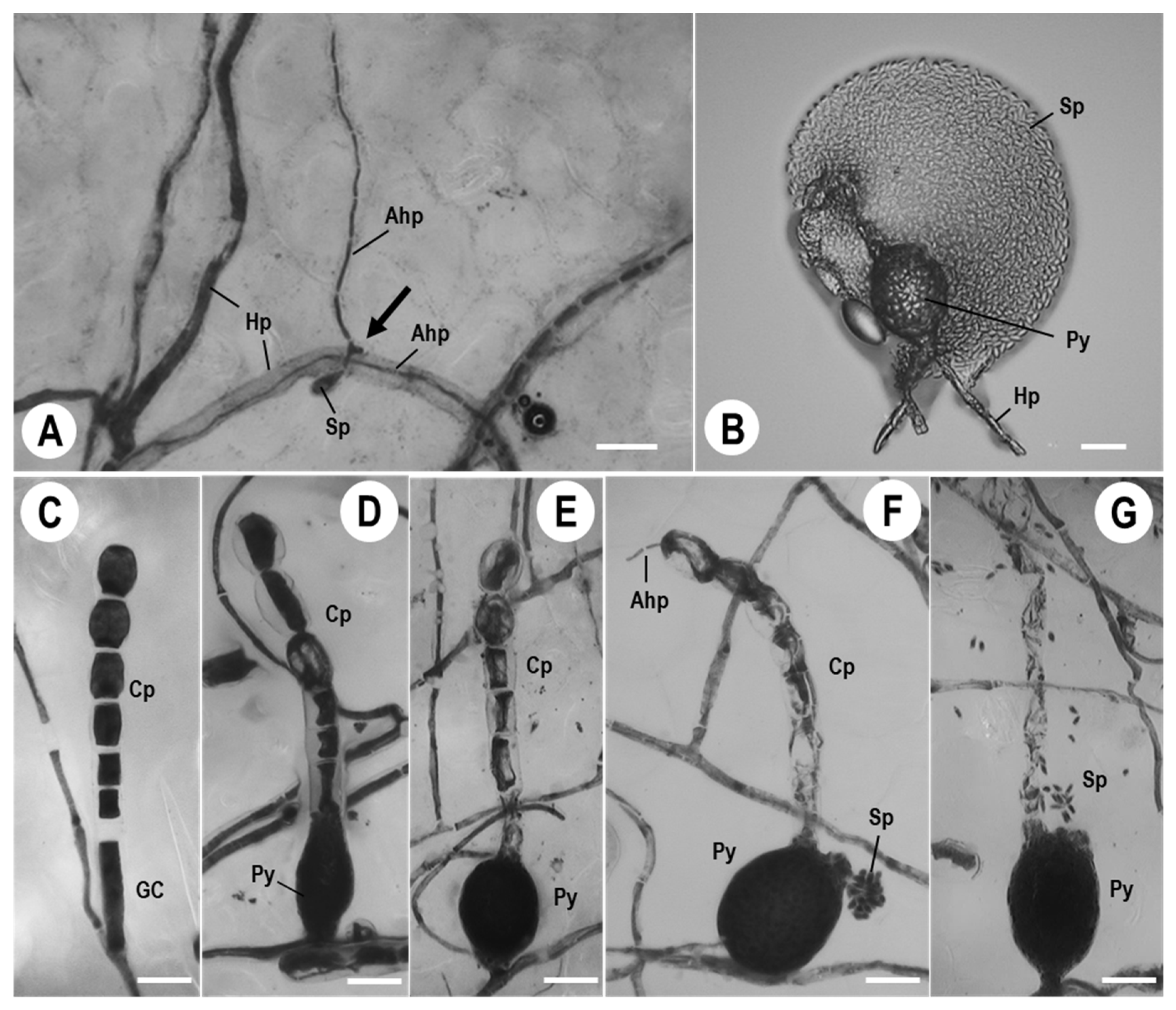
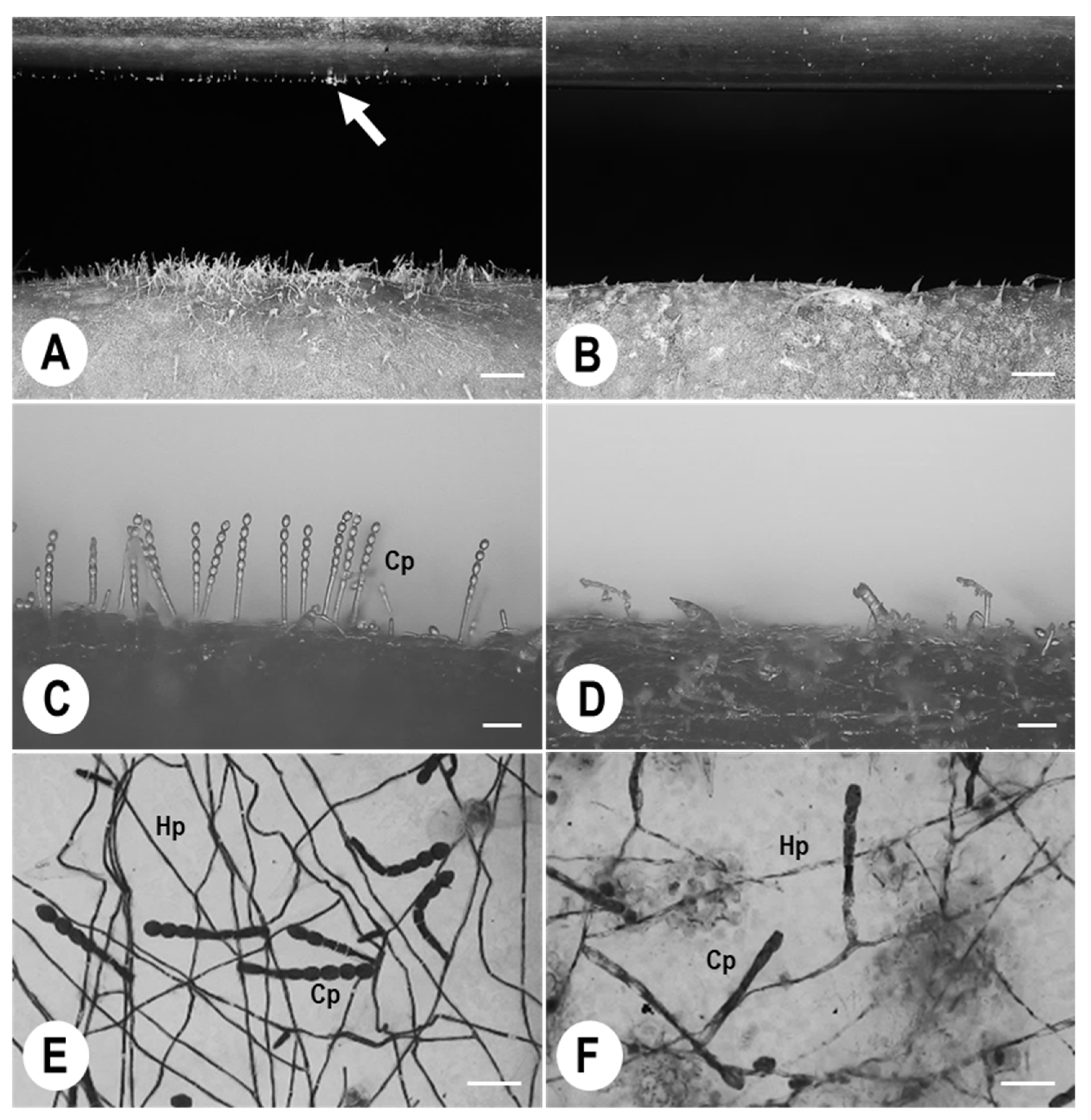
3.1. Morphological Observations of Ampelomyces Strain Xs-q in KMP-6N Colonies

We observed 5-, 10-, and 15-day-old KMP-6N colonies (Figure 1A–C) formed on melon leaves under greenhouse conditions and examined the morphology of their conidiophores (Figure 1D–F). The KMP-6N conidiophores had catenate conidia with fibrosin bodies and were produced in chains. The numbers of KMP-6N conidiophores increased as the colonies grew. After inoculation with Ampelomyces strain Xs-q, the strain grew vigorously, and elongated parasitic hyphae entered into KMP-6N hyphae (Figure 3A), followed by the complete collapse of the KMP-6N conidiophores at ca. 10–14 dpi. Xs-q pycnidia were produced in 5-, 10-, and 15-day-old KMP-6N colonies (Figure 1G–I) and changed in colour from pale yellow (immature) to black (mature) over time. The number of Xs-q pycnidia per KMP-6N colony increased with the growth stage (age) of the colony (Figure 1G–I, Table 1). At 14 dpi, Xs-q pycnidia in infected KMP-6N colonies were ovoid structures, 54.1 ± 6.4 × 37.1 ± 5.6 µm in size. Numerous Xs-q spores were released from intracellular pycnidia by the rupture of the pycnidial wall (Figure 3B). Xs-q spores with unicellular, hyaline, and ellipsoid–ovoid to doliiform morphology were 6.7 ± 0.5 × 2.8 ± 0.4 µm in size. There was an average of 1111.6 Xs-q spores per mature pycnidium, i.e., at 14 dpi, with a significant difference between 10- and 15-day-old KMP-6N colonies, but not between 5- and 10-day-old colonies (Table 1).

3.2. KMP-6N Conidiophore Morphology following Invasion by Ampelomyces Strain Xs-q

Normal KMP-6N conidiophores exhibited different morphology following invasion by Xs-q parasitic hyphae (Figure 3C). Intracellular Xs-q pycnidia were initiated (Figure 3D) and then completely formed in generative cells (GCs) of KMP-6N conidiophores (Figure 3E). Thereafter, Xs-q parasitic hyphae grew outward from conidia formed at the top of KMP-6N conidiophores in whose GCs the mature intracellular pycnidia were produced (Figure 3F), and some Xs-q spores were released from mature intracellular pycnidia in GCs (Figure 3G). Consequently, KMP-6N conidiophores collapsed completely. Histochemical staining in cells forming KMP-6N conidiophores changed greatly following invasion by parasitic Xs-q hyphae (Figure 3).

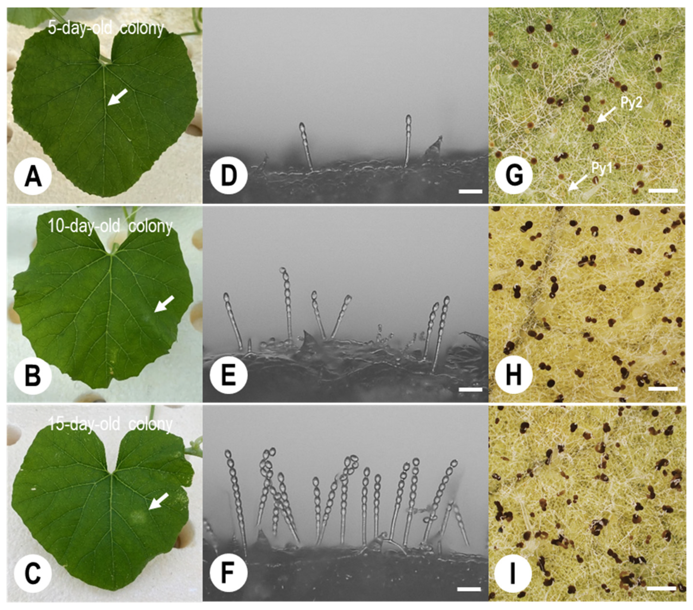
3.3. Pycnidial Development of Ampelomyces Strain Xs-q in KMP-6N Conidiophores

Normal KMP-6N conidiophores developed on melon leaves prior to inoculation with Xs-q spores (Figure 4A). After inoculation, intracellular Xs-q pycnidia initiated within GCs of the conidiophores at ca. 6–8 dpi. Single PM conidia formed at the top of the conidiophores started to degenerate at ca. 7–9 dpi (Figure 4B); then, they were completely degenerated at ca. 10–11 dpi (Figure 4C). The conidiophores collapsed completely by ca. 11–12 dpi (Figure 4D). KMP-6N hyphae containing conidiophores on melon leaves also collapsed completely. Thus, KMP-6N was unable to produce asexual progeny conidia successfully from its conidiophores after this time period. Xs-q pycnidia matured within ca. 12–14 days. Xs-q spores were released from mature intracellular pycnidia after treatment with a 10 µL drop of distilled water (Figure 4E).

3.4. Quantitative Analysis of KMP-6N Conidia Released from Parasitised PM Colonies under Greenhouse Conditions

Figure 5 shows electrostatic conidial collection from and morphological characteristics of conidiophores in KMP-6N colonies with and without Xs-q inoculation after the insulator film was negatively electrified. Progeny conidia were successfully attracted from the PM colonies after spraying with water (Figure 5A). By contrast, no progeny conidia were attracted from the colonies of 8 days after spray inoculation with Xs-q spores (8 dpi) (Figure 5B). Conidiophores in uninoculated KMP-6N colonies showed an usual morphology, forming conidial chains (Figure 5C,E), whereas those of inoculated colonies were abnormal and destroyed at 8 days after inoculation with Xs-q (8 dpi) (Figure 5D,F). We plotted the total conidia collected per 1 h from each of five KMP-6N colonies at each colony developmental stage to estimate the numbers of progeny conidia released (Figure 6). Progeny conidia were also collected from a single uninoculated 5-day-old KMP-6N colony over a period of ca. 30 days (the colony lifespan) after spraying with water as an experimental control (Figure 6A). Progeny conidia could be electrostatically collected during the daytime, but not at night (Figure 6A). No conidia could be collected from 5- and 10-day-old KMP-6N colonies after inoculation with Xs-q spores (Figure 6B,C), regardless of the colony developmental stage. Thus, conidial release from 5- and 10-day-old KMP-6N colonies decreased gradually, stopping completely by ca. 3–5 dpi and ca. 7–9 dpi, respectively (Figure 6B,C). However, in 15-day-old KMP-6N colonies, conidial releases did not stop completely after the first Xs-q spore inoculation (Figure 6D), and they began to increase again at ca. 5–6 dpi until the end of the colony lifespan was reached at ca. 22–28 days (Figure 6D). A second spray inoculation of Xs-q spores caused the number of KMP-6N conidia to decrease again, eventually stopping by ca. 16–18 days after the second inoculation (Figure 6E). The duration and total number of conidial releases by individual KMP-6N colonies throughout their lifespans are shown in Table 2. The KMP-6N colony areas and numbers of normal conidiophores per KMP-6N colony before and after one or two spray inoculations with Xs-q are also listed in Table 2. KMP-6N colony expansion ceased at ca. 4, 5, and 2 dpi in 5-, 10-, and 15-day-old colonies, respectively, after a single inoculation. KMP-6N colonies were measured after conidial releases had stopped completely; therefore, KMP-6N colonies were slightly larger in area after inoculation than before inoculation (Table 2). After electrostatic spore collection, conidiophores in uninoculated KMP-6N colonies were normal under greenhouse conditions, whereas conidiophores and hyphae in inoculated KMP-6N colonies were either deformed or collapsed, and the numbers of normal conidiophores clearly decreased (Table 2). Thus, there were no normal KMP-6N conidiophores per single 5- and 10-day-old KMP-6N colonies following a single spray inoculation of Xs-q spores, except in 15-day-old colonies. No Xs-q pycnidia formed in KMP-6N colonies at any colony developmental stage under our greenhouse conditions (low RH) throughout these experiments.

4. Discussion

Hyperparasitic fungi in the genus Ampelomyces have been used as BCAs against PM fungi in various crops worldwide [29,40,41,46], as an eco-friendly method for PM disease management. Based on morphological and molecular phylogenetic analyses, Németh et al. [48] recently identified hyperparasites isolated from different PM species in Japan as Ampelomyces spp. Spores of Japanese hyperparasitic strains produced in pycnidia, which develop intracellularly in the mycelia of PM fungi, are unicellular, hyaline, mostly guttulate, and embedded in a mucilaginous matrix within swollen ampulliform or pyriform pycnidia [48]. These strains always grew slowly, with an in vitro radial growth rate of 0.5–1.0 mm day−1 on MCzA. Thus, the morphological and physiological characteristics of the Japanese strains were clearly similar to those of previously reported A. quisqualis isolates [30,44,63,64,65,66].
To control PMs effectively with these hyperparasitic Japanese strains, we established a method for inoculating hyperparasite spores onto PM fungal colonies. The concentration of hyperparasite spores is an important factor affecting their germination and infection in pathogens, as spore germination decreases rapidly at spore concentrations of >106 spores mL−1, due to the production of self-inhibitory substances [67]. In the present study, we adjusted the spore concentration to 5 × 10⁵ spores mL−1 for spray inoculations. Our results agreed with those of Németh et al. [48], because the spores of the tested Japanese Ampelomyces strain germinated successfully at 15–20 h after spray inoculation onto PM-inoculated melon leaves at high RH.
Previous studies have reported that penetration by the hyperparasite A. quisqualis into mycohost structures can occur within 24 h [38,68], and that its hyphae of hyperparasites continue to grow internally and produce intracellular pycnidia in the mycelia or conidiophores of mycohosts at 5–8 days [24,69] or 7–10 days [42,68] after penetration. After infection, the hyperparasites further ramify throughout the mycohost hyphae, resulting in its reduced growth and eventual death [44]. Németh et al. [48] visually clarified the infection processes and conidiogenesis of a Japanese Ampelomyces strain in tomato PM colonies; the foot and GCs of PM conidiophores began to degenerate by ca. 5–6 dpi, and intracellular pycnidia of the hyperparasite strain initiated in basal cells of the conidiophores at ca. 6–8 dpi, followed by the complete collapse of the conidiophores at ca. 10–14 dpi. In the present study, we consecutively observed the degeneration and constriction of hyphae in P. xanthii, followed by intracellular pycnidial formation in the hyphae (ex. Conidiophores). The infection processes and conidiogenesis of the tested hyperparasitic Ampelomyces strain in the melon PM fungus were very similar to those of a tomato PM fungus reported by Németh et al. [48]. Interestingly, almost all intracellular pycnidia were produced in conidiophores, but not in the hyphae, of the mycohost, as observed by DM, and Ampelomyces hyphae that invaded the PM conidiophores grew out of conidia that formed at the top of the conidiophores.
In this study, we applied electrostatic and DM techniques to estimate quantitatively the control effects and infection efficiency of a Japanese hyperparasitic Ampelomyces strain against a melon PM fungus, to determine whether asexual PM progeny conidia, which are a source of host plant infection, are released from Ampelomyces-inoculated melon PM colonies at different developmental stages. Previous studies have determined the numbers of progeny conidia released by PM colonies throughout their lifespans, to clarify the conidial dispersal process using negatively polarised Insulator plates [60], probes [70], and drums [53,59]. Using a negatively polarised insulator drum, one study demonstrated that a single melon PM colony releases an average of 12.6 × 10⁴ conidia throughout its lifespan under greenhouse conditions [53]; the dielectrically polarised insulator drum only detached mature conidia from conidiophores. Interestingly, Suzuki et al. [53] observed the vigorous release of conidia from melon PM colonies in the daytime, whereas very few conidia were released at night. The conidial release from colonies also largely reflected seasonal day length and light intensity [53]. These studies demonstrate that the use of an electrostatic spore collector system is crucial for the elucidation of ecological characteristics such as conidial release from PM colonies, which otherwise might be impossible to analyse.
In the present study, we also found that PM isolate KMP-6N actively released asexual progeny conidia during the day, and few at night, from colonies with and without spray inoculation of Xs-q spores (see Figure 6). Moreover, the numbers of conidia released from 5- and 10-day-old KMP-6N colonies after spray inoculation with Xs-q spores decreased gradually and stopped completely after ca. 4 dpi and ca. 8 dpi, respectively. Thus, we were able to suppress conidial releases completely from KMP-6N colonies by the application of a Japanese Ampelomyces strain under greenhouse conditions. However, we were only able to stop progeny conidial releases completely from 15-day-old KMP-6N colonies after two spray inoculations of Xs-q spores; a single spray treatment was insufficient to achieve this. Thus, our results strongly suggest that PM colonies should be inoculated with hyperparasitic fungal spores in the early developmental stages (e.g., when colonies are 5–10 days old, or as soon as PM is detected on host leaves) to successfully control the disease.
Ampelomyces hyperparasites are transported for long distances by wind [25,71,72] and spores in pycnidia can be dispersed by rain splash [68,73] to parasitise mycohosts over broad areas. In older colonies (e.g., >15 days old) spray-inoculated with Xs-q spores, a few normal conidiophores survived due to suboptimal control with Ampelomyces, allowing melon PM fungi to scatter progeny conidia widely from the colonies. To suppress the spread of PM, it may be more effective to inoculate PM colonies with Xs-q spores at night, when conidiophores do not release progeny conidia, than during the day, when progeny conidia are actively released. Interestingly, our tested Ampelomyces strain did not produce intracellular pycnidia in PM hyphae under our greenhouse conditions (low RH) but did produce them in PM hyphae under our growth chamber conditions (high RH). It appears that the hyperparasitic Ampelomyces strain requires high-RH conditions to produce intracellular pycnidia in mycohost hyphae. Similar results were observed in previous studies [24,45,48,69].
To elucidate the process of conidial release from hyperparasitic fungus-inoculated pathogenic PM colonies, we counted the PM conidia captured by our electrostatic spore collection apparatus to estimate quantitatively the ability of a hyperparasitic fungus to control cucurbit PM disease. To our knowledge, this is the first report describing pycnidial formation by hyperparasitic fungi within cucurbit PM hyphae using a DM, to determine directly the duration of conidial release from hyperparasite-inoculated PM colonies at different colony developmental stages and apply electrostatic techniques to determine the total number of progeny conidia released from live PM colonies after inoculation with a hyperparasitic fungus. The findings of the present study will provide important insights into the ecological interactions between hyperparasitic Ampelomyces strains and PM mycohosts. Because A. quisqualis has been reported to tolerate a number of fungicides [25,32,38,69], it shows potential for application in combination with other pathogen control agents. Therefore, in a future study, we will examine the tolerance of Japanese Ampelomyces strains to commercial fungicides and focus on their use as practical BCAs against PMs.

5. Conclusions

In this study, we quantitatively and visually evaluated the suppressive effects of Ampelomyces Japanese Xs-q strain on the development of conidiophores and colonies of the melon PM isolate KMP-6N, to support the practical application of this hyperparasitic fungus as a biocontrol agent (BCA). In the first experiment, we monitored pycnidial formation of the Xs-q strain in conidiophores of the KMP-6N isolate using DM technology. Intracellular pycnidia of Xs-q were produced in KMP-6N conidiophores incubated in a growth chamber until ca. 10–14 dpi with Xs-q spores. KMP-6N conidiophores atrophied completely and did not release asexual progeny conidia. This is the first report to describe the pycnidial formation processes of a hyperparasitic fungus in melon PM conidiophores. In a second experiment, KMP-6N conidia were collected from Xs-q-inoculated KMP-6N colonies at different developmental stages under greenhouse conditions using an electrostatic spore collector. Conidial releases from 5- and 10-day-old KMP-6N colonies stopped after a single spray inoculation of Xs-q spores, whereas those from 15-day-old KMP-6N colonies did not stop. Therefore, we suggest that younger colonies should be targeted, as soon as melon PM fungi are observable on host leaves (e.g., in 5–10-day-old colonies) for the effective control of PM fungi using Ampelomyces strains. In addition, these results will lead to the establishment of ideal spray inoculation systems of hyperparasitic fungi to PM pathogens. These findings offer valuable insights into the development of hyperparasitic fungi as BCAs against PMs.

Author Contributions

Conceptualisation, Y.K., M.Z.N. and T.N.; methodology, Y.K., M.Z.N., D.S., L.K. and T.N.; software, Y.K. and Y.T.; validation, T.S., K.K., Y.M. and T.N.; formal analysis, Y.K. and T.S.; investigation, Y.K., K.N. and A.M.; resources, Y.K. and T.N.; data curation, Y.K. and T.N.; writing—original draft preparation, Y.K. and T.N.; writing—review and editing, M.Z.N., L.K. and T.N.; visualization, Y.M., M.Z.N., L.K. and T.N.; supervision, M.Z.N., L.K. and T.N.; project administration, L.K. and T.N. All authors have read and agreed to the published version of the manuscript.

Funding

This work was supported by a Grant for Agricultural Technology and Innovation Research Institute, Kindai University, by the János Bolyai Research Scholarship of the Hungarian Academy of Sciences (awarded to Márk Z. Németh; BO/00221/21/4), and by the Hungarian Scientific Research Fund (NKFIH OTKA FK142735).

Institutional Review Board Statement

Not applicable.

Informed Consent Statement

Not applicable.

Data Availability Statement

Data sets analysed during the current study are available from the current author on reasonable request.

Acknowledgments

The English in this document has been checked by at least two professional editors, both native speakers of English.

Conflicts of Interest

The authors declare no conflict of interest.

References

  1. Sowell, G., Jr. Population shift of Sphaerotheca fuliginea on musk melon. J. Am. Soc. Hortic. Sci. 1982, 112, 156–160. [Google Scholar]
  2. Reifschneider, F.J.B.; Boiteux, L.S.; Occhiena, E.M. Powdery mildew on melon (Cucumis melo) caused by Sphaerotheca fuliginea in Brazil. Plant Dis. 1985, 69, 1069–1070. [Google Scholar]
  3. Mohamed, Y.F.; Bardin, M.; Nicot, P.C.; Pitrat, M. Causal agents of powdery mildew of cucurbits in Sudan. Plant Dis. 1995, 79, 634–636. [Google Scholar] [CrossRef]
  4. Hosoya, K.; Narisawa, K.; Pitrat, M.; Ezura, H. Race identification in powdery mildew (Sphaerotheca fuliginea) on melon (Cucumis melo) in Japan. Plant Breed. 1999, 118, 259–262. [Google Scholar] [CrossRef]
  5. Del Pino, D.; Olalla, L.; Pérez-García, A.; Rivera, M.E.; García, S.; Moreno, R.; Torés, J.A. Occurrence of races and pathotypes of cucurbit powdery mildew in southeastern Spain. Phytoparasitica 2002, 30, 459–466. [Google Scholar] [CrossRef]
  6. Křístková, E.; Lebeda, A.; Sedláková, B. Virulence of Czech cucurbit powdery mildew isolates on Cucumis melo genotypes MR-1 and PI 124112. Sci. Hortic. 2004, 99, 257–265. [Google Scholar] [CrossRef]
  7. Křístková, E.; Lebeda, A.; Sedláková, B. Species spectra, distribution and host range of cucurbit powdery mildews in the Czech Republic, and in some other European and Middle Eastern countries. Phytoparasitica 2009, 37, 337–350. [Google Scholar] [CrossRef]
  8. Tomason, Y.; Gibson, P.T. Fungal characteristics and varietal reactions of powdery mildew species on cucurbits in steppes of Ukraine. Agron. Res. 2006, 4, 549–562. [Google Scholar]
  9. Pérez-García, A.; Romero, D.; Fernández-Ortuño, D.; López-Ruiz, F.; De Vicente, A.; Torés, J.A. The powdery mildew fungus Podosphaera fusca (synonym Podosphaera xanthii), a constant threat to cucurbits. Mol. Plant Pathol. 2009, 10, 153–160. [Google Scholar] [CrossRef]
  10. Hong, Y.-J.; Hossain, M.R.; Kim, H.-T.; Park, J.-I.; Nou, I.-S. Identification of two new races of Podosphaera xanthii causing powdery mildew in melon in South Korea. Plant Pathol. J. 2018, 34, 182–190. [Google Scholar] [CrossRef]
  11. Kiss, L.; Vaghefi, N.; Bransgrove, K.; Dearnaley, J.D.W.; Takamatsu, S.; Tan, Y.P.; Marston, C.; Liu, S.Y.; Jin, D.N.; Adorada, D.L.; et al. Australia: A continent without native powdery mildews? The first comprehensive catalogue indicates recent introductions and multiple host range expansion events, and leads to the re-discovery of Salmonomyces as a new lineage of the Erysiphales. Front. Microbiol. 2020, 11, 1571. [Google Scholar] [CrossRef] [PubMed]
  12. Kelly, L.A.; Vaghefi, N.; Bransgrove, K.; Fechner, N.A.; Stuart, K.; Pandey, A.K.; Sharma, M.; Németh, M.Z.; Liu, S.Y.; Tang, S.R.; et al. One crop disease, how many pathogens? Podosphaera xanthii and Erysiphe vignae sp. nov. identified as the two species that cause powdery mildew of mungbean (Vigna radiata) and black gram (V. mungo) in Australia. Phytopathology 2021, 111, 1193–1206. [Google Scholar] [CrossRef] [PubMed]
  13. Vaghefi, N.; Kusch, S.; Németh, M.Z.; Seress, D.; Braun, U.; Takamatsu, S.; Panstruga, R.; Kiss, L. Beyond nuclear ribosomal DNA sequences: Evolution, taxonomy, and closest known saprobic relatives of powdery mildew fungi (Erysiphaceae) inferred from their first comprehensive genome-scale phylogenetic analyses. Front. Microbiol. 2022, 13, 903024. [Google Scholar] [CrossRef] [PubMed]
  14. Keinath, A.P.; Wintermantel, W.M.; Zitter, T.A. Compendium of Cucurbit Diseases and Pests, 2nd ed.; The American Phytopathological Society Press: St. Paul, MN, USA, 2017. [Google Scholar]
  15. Takikawa, Y.; Nonomura, T.; Miyamoto, S.; Okamoto, N.; Murakami, T.; Matsuda, Y.; Kakutani, K.; Kusakari, S.; Toyoda, H. Digital microscopic analysis of developmental process of conidiogenesis by powdery mildew pathogens isolated from melon leaves. Phytoparasitica 2015, 43, 517–530. [Google Scholar] [CrossRef]
  16. Huggenberger, F.; Collins, M.A.; Skylakakis, G. Decreased sensitivity of Sphaerotheca fuliginea to fenarimol and other ergosterol-biosynthesis inhibitors. Crop Prot. 1984, 3, 137–149. [Google Scholar] [CrossRef]
  17. Lebeda, A.; Sedláková, B. Fungicide resistance in population of cucurbit powdery mildew. J. Plant Pathol. 2008, 90, S2.142. [Google Scholar]
  18. McGrath, M.T.; Shishkoff, N. Resistance to triadimefon and benomyl: Dynamics and impact on managing cucurbit powdery mildew. Plant Dis. 2001, 85, 147–154. [Google Scholar] [CrossRef]
  19. McGrath, M.T.; Shishkoff, N. Frist report of the cucurbit powdery mildew fungus (Podosphaera xanthii) resistant to strobilurin fungicides in the United States. Plant Dis. 2003, 87, 1007. [Google Scholar] [CrossRef]
  20. Miyamoto, T.; Hayashi, K.; Ogawara, T. First report of the occurrence of multiple resistance to Flutianil and Pyriofenone in field isolates of Podosphaera xanthii, the causal fungus of cucumber powdery mildew. Eur. J. Plant Pathol. 2020, 156, 953–963. [Google Scholar] [CrossRef]
  21. Miyamoto, T.; Hayashi, K.; Okada, R.; Wari, D.; Ogawara, T. Resistance to succinate dehydrogenase inhibitors in field isolates of Podosphaera xanthii on cucumber: Monitoring, cross-resistance patterns and molecular characterization. Pestic. Biochem. Physiol. 2020, 169, 104646. [Google Scholar] [CrossRef]
  22. Vielba-Fernández, A.; Polonio, A.; Ruiz-Jiménez, L.; de Vicente, A.; Pérez-García, A.; Fernández-Ortuño, D. Fungicide resistance in powdery mildew fungi. Microorganisms 2020, 8, 1431. [Google Scholar] [CrossRef] [PubMed]
  23. Cesati, V. Ampelomyces quisqualis Ces. Bot. Ztg. 1852, 10, 301–302. [Google Scholar]
  24. Jarvis, W.R.; Slingsby, K. The control of powdery mildew of greenhouse cucumber by water sprays and Ampelomyces quisqualis. Plant Dis. Reptr. 1977, 61, 728–730. [Google Scholar]
  25. Sundheim, L. Control of cucumber powdery mildew by the hyperparasite Ampelomyces quisquialis and fungicides. Plant Pathol. 1982, 31, 209–214. [Google Scholar] [CrossRef]
  26. Kiss, L. Natural occurrence of Ampelomyces intracellular mycoparasites in mycelia of powdery mildew fungi. New Phytol. 1998, 140, 709–714. [Google Scholar] [CrossRef]
  27. Yarwood, C.E. An overwintering pycnidial stage of Cicinnobolus. Mycologia 1939, 31, 420–422. [Google Scholar] [CrossRef]
  28. Falk, S.P.; Gadoury, D.M.; Cortesi, P.; Pearson, R.C.; Seem, R.C. Parasitism of Uncinula necator cleistothecia by the mycoparasite Ampelomyces quisqualis. Phytopathology 1995, 85, 794–800. [Google Scholar] [CrossRef]
  29. Kiss, L. A review of fungal antagonists of powdery mildews and their potential as biocontrol agents. Pest Manag. Sci. 2003, 59, 475–483. [Google Scholar] [CrossRef]
  30. Huth, L.; Ash, G.J.; Idnurm, A.; Kiss, L.; Vaghefi, N. The “bipartite” structure of the first genome of Ampelomyces quisqualis, a common hyperparasite and biocontrol agent of powdery mildews, may point to its evolutionary origin from plant pathogenic fungi. Genome Biol. Evol. 2021, 13, evab182. [Google Scholar] [CrossRef]
  31. Sztejnberg, A. Biological control of powdery mildews by Ampelomyces quisqualis. Phytopathology 1979, 69, 1047. [Google Scholar]
  32. Sundheim, L. Ampelomyces quisqualis, a hyperparasitic fungus in biological control of powdery mildews on greenhouse cucumber. Acta Hortic. 1984, 156, 229–236. [Google Scholar] [CrossRef]
  33. Sztejnberg, A.; Mazar, S. Biocontrol of cucumber and carrot powdery mildew by Ampelomyces quisqualis. Phytopathology 1985, 75, 1301–1302. [Google Scholar]
  34. Sztejnberg, A.; Galper, S.; Mazar, S.; Lisker, N. Ampelomyces quisqualis for biological and integrated control of powdery mildews in Israel. J. Phytopathol. 1989, 124, 285–295. [Google Scholar] [CrossRef]
  35. Dik, A.J.; Verhaar, M.A.; Bélanger, R.R. Comparison of three biological control agents against cucumber powdery mildew (Sphaerotheca fuliginea) in semi-commercial-scale glasshouse trials. Eur. J. Plant Pathol. 1998, 104, 413–423. [Google Scholar] [CrossRef]
  36. Shishkoff, N.; McGrath, M.T. AQ10 biofungicide combined with chemical fungicides or AddQ spray adjuvant for control of cucurbit powdery mildew in detached leaf culture. Plant Dis. 2002, 86, 915–918. [Google Scholar] [CrossRef] [PubMed]
  37. Angeli, D.; Maurhofer, M.; Gessler, C.; Pertot, I. Existence of different physiological forms within genetically diverse strains of Ampelomyces quisqualis. Phytoparasitica 2012, 40, 37–51. [Google Scholar] [CrossRef]
  38. Sztejnberg, A.; Mazar, S. Studies on the hyperparasite Ampelomyces quisqualis and preliminary trials on biological control of powdery mildew. Phytoparasitica 1983, 11, 219–220. [Google Scholar]
  39. Romero, D.; Rivera, M.E.; Cazorla, F.M.; De Vicente, A.; Pérez-García, A. Effect of mycoparasitic fungi on the development of Sphaerotheca fusca in melon leaves. Mycol. Res. 2003, 107, 64–71. [Google Scholar] [CrossRef]
  40. Park, M.-J.; Choi, Y.-J.; Hong, S.-B.; Shin, H.-D. Genetic variability and mycohost association of Ampelomyces quisqualis isolates inferred from phylogenetic analyses of ITS rDNA and actin gene sequences. Fungal Biol. 2010, 114, 235–247. [Google Scholar] [CrossRef]
  41. Siozios, S.; Tosi, L.; Ferrarini, A.; Ferrari, A.; Tononi, P.; Bellin, D.; Maurhofer, M.; Gessler, C.; Delledonne, M.; Pertot, I. Transcriptional reprogramming of the mycoparasitic fungus Ampelomyces quisqualis during the powdery mildew host-induced germination. Phytopathology 2015, 105, 199–209. [Google Scholar] [CrossRef]
  42. Legler, S.E.; Pintye, A.; Caffi, T.; Gulyás, S.; Bohár, G.; Rossi, V.; Kiss, L. Sporulation rate in culture and mycoparasitic activity, but not mycohost specificity, are the key factors for selecting Ampelomyces strains for biocontrol of grapevine powdery mildew (Erysiphe necator). Eur. J. Plant Pathol. 2016, 144, 723–736. [Google Scholar] [CrossRef]
  43. Falk, S.P.; Gadoury, D.M.; Pearson, R.C.; Seem, R.C. Partial control of grape powdery mildew by the mycoparasite Ampelomyces quisqualis. Plant Dis. 1995, 79, 483–490. [Google Scholar] [CrossRef]
  44. Hashioka, Y.; Nakai, Y. Ultrastructure of pycnidial development and mycoparasitism of Ampelomyces quisqualis parasitic on Erysiphales. Trans. Mycol. Soc. Japan 1980, 21, 329–338. [Google Scholar]
  45. Kiss, L.; Russell, J.C.; Szentiványi, O.; Xu, X.; Jeffries, P. Biology and biocontrol potential of Ampelomyces mycoparasites, natural antagonists of powdery mildew fungi. Biocont. Sci. Technol. 2004, 14, 635–651. [Google Scholar] [CrossRef]
  46. Kiss, L. Intracellular mycoparasites in action: Interactions between powdery mildew fungi and Ampelomyces. In Stress in Yeasts and Filamentous Fungi; Avery, S.V., Stratford, M., van West, P., Eds.; Academic Press: Cambridge, MA, USA, 2008; Volume 27, pp. 37–52. [Google Scholar]
  47. Németh, M.Z.; Pintye, A.; Horváth, Á.N.; Vági, P.; Kovács, G.M.; Gorfer, M.; Kiss, L. Green fluorescent protein transformation sheds more light on a widespread mycoparasitic interaction. Phytopathology 2019, 109, 1404–1416. [Google Scholar] [CrossRef] [PubMed]
  48. Németh, M.Z.; Mizuno, Y.; Kobayashi, H.; Seress, D.; Shishido, N.; Kimura, Y.; Takamatsu, S.; Suzuki, T.; Takikawa, Y.; Kakutani, K.; et al. Ampelomyces strains isolated from diverse powdery mildew hosts in Japan: Their phylogeny and mycoparasitic activity, including timing and quantifying mycoparasitism of Pseudoidium neolycopersici on tomato. PLoS ONE 2021, 16, e0251444. [Google Scholar] [CrossRef]
  49. Aylor, D.E. The role of intermittent wind in the dispersal of fungal pathogens. Annu. Rev. Phytopathol. 1990, 28, 73–92. [Google Scholar] [CrossRef]
  50. Brown, J.K.M.; Hovmøller, M.S. Aerial dispersal of pathogens on the global and continental scales and its impact on plant disease. Science 2002, 297, 537–541. [Google Scholar] [CrossRef]
  51. Nonomura, T.; Matsuda, Y.; Yamashita, S.; Akahoshi, H.; Takikawa, Y.; Kakutani, K.; Toyoda, H. Natural woody plant, Mallotus japonicus, as an ecological partner to transfer different pathotypic conidia of Oidium neolycopersici to greenhouse tomatoes. Plant Protect. Sci. 2013, 49, S33–S40. [Google Scholar] [CrossRef]
  52. Suzuki, T.; Iwasaki, S.; Hisazumi, H.; Miyamoto, A.; Ogami, H.; Takikawa, Y.; Kakutani, K.; Matsuda, Y.; Nonomura, T. Inhibitory effects of blue light-emitting diode irradiation on Podosphaera xanthii conidial release and infection of melon seedlings. Agriculture 2022, 12, 198. [Google Scholar] [CrossRef]
  53. Suzuki, T.; Nakamura, R.; Takagi, N.; Takikawa, Y.; Kakutani, K.; Matsuda, Y.; Matsui, K.; Nonomura, T. Quantitative analysis of the lifelong production of conidia released from single colonies of Podosphaera xanthii on melon leaves using electrostatic techniques. Austral. Plant Pathol. 2019, 48, 297–307. [Google Scholar] [CrossRef]
  54. Griffith, W.T. Electrostatic phenomena. In The Physics of Everyday Phenomena: A Conceptual Introduction to Physics; Bruflodt, D., Loehr, B.S., Eds.; McGraw-Hill: New York, NY, USA, 2004; pp. 232–252. [Google Scholar]
  55. Halliday, D.; Resnick, R.; Walker, J. Electric charge. In Fundamentals of Physics; Johnson, S., Ford, E., Eds.; John Wiley & Sons: New York, NY, USA, 2005; pp. 561–579. [Google Scholar]
  56. Leach, C.M. An electrostatic theory to explain violent spore liberation by Drechslera turcica and other fungi. Mycologia 1976, 68, 63–86. [Google Scholar] [CrossRef]
  57. Matsuda, Y.; Ikeda, H.; Moriura, N.; Tanaka, N.; Shimizu, K.; Oichi, W.; Nonomura, T.; Kakutani, K.; Kusakari, S.; Higashi, K.; et al. A new spore precipitator with polarized dielectric insulators for physical control of tomato powdery mildew. Phytopathology 2006, 96, 967–974. [Google Scholar] [CrossRef] [PubMed]
  58. Shimizu, T.; Matsuda, Y.; Nonomura, T.; Ikeda, H.; Tamura, N.; Kusakari, S.; Kimbara, J.; Toyoda, H. Dual protection of hydroponic tomatoes from rhizosphere pathogens Ralstonia solanacearum and Fusarium oxysporum f. sp. radicis-lycopersici and airborne conidia of Oidium neolycopersici with an ozone-generative electrostatic spore precipitator. Plant Pathol. 2007, 56, 987–997. [Google Scholar] [CrossRef]
  59. Ayabe, S.; Kimura, Y.; Umei, N.; Takikawa, Y.; Kakutani, K.; Matsuda, Y.; Nonomura, T. Real-time collection of conidia released from living single colonies of Podosphaera aphanis on strawberry leaves under natural conditions with electrostatic techniques. Plants 2022, 11, 3453. [Google Scholar] [CrossRef]
  60. Moriura, N.; Matsuda, Y.; Oichi, W.; Nakashima, S.; Hirai, T.; Nonomura, T.; Kakutani, K.; Kusakari, S.; Higashi, K.; Toyoda, H. An apparatus for collecting total conidia of Blumeria graminis f. sp. hordei from leaf colonies using electrostatic attraction. Plant Pathol. 2006, 55, 367–374. [Google Scholar] [CrossRef]
  61. Suzuki, T.; Nishimura, S.; Yagi, K.; Nakamura, R.; Takikawa, Y.; Matsuda, Y.; Kakutani, K.; Nonomura, T. Effects of light quality on conidiophore formation of the melon powdery mildew pathogen Podosphaera xanthii. Phytoparasitica 2018, 94, 1105–1110. [Google Scholar] [CrossRef]
  62. Sameshima, T.; Kashimoto, K.; Kida, K.; Matsuda, Y.; Nonomura, T.; Kakutani, K.; Nakata, K.; Kusakari, S.; Toyoda, H. Cytological events in tomato leaves inoculated with conidia of Blumeria graminis f. sp. hordei and Oidium neolycopersici KTP-01. J. Gen. Plant Pathol. 2004, 70, 7–10. [Google Scholar] [CrossRef]
  63. Kiss, L. Graminicolous powdery mildew fungi as new natural hosts of Ampelomyces mycoparasites. Can. J. Bot. 1997, 75, 680–683. [Google Scholar] [CrossRef]
  64. Kiss, L.; Nakasone, K.K. Ribosomal DNA internal transcribed spacer sequences do not support the species status of Ampelomyces quisqualis, a hyperparasite of powdery mildew fungi. Curr. Genet. 1998, 33, 362–367. [Google Scholar] [CrossRef]
  65. Ranković, B. Hyperparasites of the genus Ampelomyces on powdery mildew fungi in Serbia. Mycopathologia 1997, 139, 157–164. [Google Scholar] [CrossRef] [PubMed]
  66. Pintye, A.; Ropars, J.; Harvey, N.; Shin, H.D.; Leyronas, C.; Nicot, P.C.; Giraud, T.; Kiss, L. Host phenology and geography as drivers of differentiation in generalist fungal mycoparasites. PLoS ONE 2015, 10, e0120703. [Google Scholar] [CrossRef]
  67. Gu, Y.H.; Ko, W.H. Water agarose medium for studying factors affecting germination of conidia of Ampelomyces quisqualis. Mycol. Res. 1997, 101, 422–424. [Google Scholar] [CrossRef]
  68. Sundheim, L.; Krekling, T. Host-parasite relationships of the hyperparasite Ampelomyces quisqualis and its powdery mildew host Sphaerotheca fuliginea. Phytopath. Z. 1982, 104, 202–210. [Google Scholar] [CrossRef]
  69. Philipp, W.-D.; Crüger, G. Parasitismus von Ampelomyces quisqualis auf echten mehltaupilzen an gurken und anderen gemüsearten. Z. Pflanzenkrankh. Pflanzenschutz 1979, 86, 129–142. [Google Scholar]
  70. Nonomura, T.; Matsuda, Y.; Xu, L.; Kakutani, K.; Takikawa, Y.; Toyoda, H. Collection of highly germinative pseudochain conidia of Oidium neolycopersici from conidiophores by electrostatic attraction. Mycol. Res. 2009, 113, 364–372. [Google Scholar] [CrossRef] [PubMed]
  71. Speer, E.O. Ampelomyces cesati (Fungi, Sphaeropsidales). Taxon 1978, 27, 549–562. [Google Scholar] [CrossRef]
  72. Kiss, L.; Pintye, A.; Zséli, G.; Jankovics, T.; Szentiványi, O.; Hafez, Y.M.; Cook, R.T.A. Microcyclic conidiogenesis in powdery mildews and its association with intracellular parasitism by Ampelomyces. Eur. J. Plant Pathol. 2010, 126, 445–451. [Google Scholar] [CrossRef]
  73. Zeng, L.F.; Jiang, Y.C. An investigation of parasitic fungi on Erysiphe spp. RoPP 1986, 65, 419. [Google Scholar]
Figure 1. Photographs and micrographs of Podosphaera xanthii Pollacci KMP-6N colonies and conidiophores on melon leaves spray-inoculated with spores of the Japanese Ampelomyces strain Xs-q. (AC) Single 5-day-old (A), 10-day-old (B), and 15-day-old (C) KMP-6N colonies were prepared by inoculating KMP-6N conidia onto leaves of melon seedlings. Arrows show the growth of single KMP-6N colonies on melon leaves at different fungal developmental stages. (DF) KMP-6N conidiophores in single 5-day-old (D), 10-day-old (E), and 15-day-old (F) KMP-6N colonies observed using a digital microscope (KH-2700 DM). KMP-6N conidiophores had normal catenate conidia, forming chains. (GI) KH-2700 DM images of pycnidia of the Xs-q strain produced in plastic boxes at 70–80% relative humidity (RH) under growth chamber conditions at 10 days post-inoculation (dpi) onto 5-day-old (G), 10-day-old (H), and 15-day-old (I) KMP-6N colonies. The Xs-q pycnidia changed in colour from pale yellow to black as they matured; Py1 and Py2 indicate immature and mature pycnidia, respectively. Bars represent 60 µm (DF) and 300 µm (GI).
Agronomy 13 01204 g001
Figure 2. An electrostatic spore collector system for collecting progeny conidia released from KMP-6N colonies with and without spray inoculation with hyperparasite spores (cross-sectional view). The electrostatic voltage generator produced a negative charge, which was transferred to the conductor film, inducing a positive image charge on the surface of the insulator film. Dielectric polarisation produced a negative surface charge on the opposite side of the insulator film, and an electrostatic field formed around the dielectrically polarised insulator film. The dielectrically polarised insulated plastic container was timed to revolve once every 24 h. KMP-6N progeny conidia from normal conidiophores were collected on the insulator film by electrostatic attraction. The insulator film (5.2 × 10⁻¹ nC) was placed at <2 cm from a KMP-6N colony on a melon leaf (distance A).
Agronomy 13 01204 g002
Figure 3. Infection of the Japanese Ampelomyces strain Xs-q in P. xanthii KMP-6N colonies. (A) Light micrograph of Xs-q hyphae that invaded and grew vigorously into KMP-6N hyphae. (B) Digital micrograph of Xs-q spores released from a pycnidium after treatment with a 10 µL drop of distilled water. (CG) Morphological changes in KMP-6N conidiophores parasitised by Xs-q. (C) Normal KMP-6N conidiophore. (D) Xs-q pycnidium initiated in generative cell (GC) of a KMP-6N conidiophore. (E) Mature intracellular pycnidium produced in a GC of a KMP-6N conidiophore. (F) The hyperparasitic Xs-q strain produced intracellular pycnidia in GCs, and Xs-q hyphae grew out from conidial cells at the apex of the KMP-6N conidiophore. (G) Abundant Xs-q spores released from mature pycnidia. KMP-6N conidiophores atrophied and collapsed without releasing progeny conidia. Light micrographs were taken at 0 (C), 6 (D), 8 (E), 10 (F), and 12 days (G) after spray inoculation with Xs-q spores onto 10-day-old KMP-6N mycelia. Bars represent 10 µm (A) and 20 µm (BG). Ahp, Xs-q hypha; Cp, KMP-6N conidiophore; Hp, KMP-6N hypha; Sp, Xs-q spore; Py, Xs-q pycnidium.
Agronomy 13 01204 g003
Figure 4. Pycnidial development of the Xs-q strain in KMP-6N conidiophores. A KMP-6N mycelium formed on a melon leaf at 10 dpi with a KMP-6N conidium using a micromanipulation technique. Digital micrographs were taken from the same view at 0 (A), 10 (B), 11 (C), 12 (D), and 14 days (E) after spray inoculation of Xs-q spores onto 10-day-old KMP-6N mycelium. KMP-6N conidiophores (Cp) that formed near a trichome cell (Tc) on a melon leaf atrophied completely after invasion by Xs-q hyphae into KMP-6N hyphae (Hp). Xs-q pycnidia (Py) were successfully produced in generative cells (GC) of KMP-6N conidiophores at high RH (80–90%). Mature pycnidia released abundant progeny spores (Sp). Arrow indicates atrophied conidial cells at the top of KMP-6N conidiophores. Bars represent 20 µm.
Agronomy 13 01204 g004
Figure 5. Micrographs of conidiophores and colonies in Xs-q-uninoculated (A,C,E) or Xs-q-inoculated (B,D,F) 10-day-old KMP-6N colonies after electrostatic attraction of conidia to the insulator film under greenhouse conditions. (A,B) Digital micrographs showing the attraction of progeny conidia released from Xs-q-uninoculated (A) and Xs-q-inoculated (B) KMP-6N colonies to an electrostatic insulator film. An insulator film carrying a charge of 1.0 nC was placed at 2000 µm from the apex of the conidiophore. Micrographs were taken at 8 days after the start of electrostatic spore collection. Conidia (arrow) were attracted to the insulator film in (A), whereas no conidia were captured in (B). (C,D) Digital micrographs showing Xs-q-uninoculated (C) and Xs-q-inoculated (D) KMP-6N colonies at 8 days after the electrostatic collector was applied. Conidiophores (Cp) were normal and developed conidia in chains in (C) but developed abnormally and atrophied completely without forming Xs-q pycnidia in (D). (E,F) Light micrographs showing KMP-6N hyphae (Hp) and conidiophores (Cp) in histochemically stained Xs-q-uninoculated (E) and Xs-q-inoculated (F) KMP-6N colonies at 8 days after the electrostatic insulator was applied. Conidiophores (Cp) were normal with the development of conidial cell chains in (E), but were atrophied completely without forming Xs-q pycnidia in (F). Bars represent 500 µm (A,B) and 60 µm (CF).
Agronomy 13 01204 g005
Figure 6. Numbers of progeny conidia collected from P. xanthii Pollacci KMP-6N colonies without (A) or with (BE) spray inoculation with Xs-q spores on melon leaves. Electrostatic conidial collection was conducted during definite periods until conidial release stopped from uninoculated 5-day-old (A) or spray-inoculated 5-day-old (B), 10-day-old (C), and 15-day-old KMP-6N colonies with single Xs-q spore application (D) or double Xs-q spore application (E). Data are plotted for 1 h periods. Open arrows indicate no conidia were released from KMP-6N colonies; the day on which KMP-6N conidial releases stopped completely is indicated. Black and grey arrows indicate first and second spray inoculation, respectively.
Agronomy 13 01204 g006
Table 1. Numbers of Ampelomyces strain Xs-q pycnidia formed per Podosphaera xanthii KMP-6N colony and of Xs-q spores produced per mature pycnidium at different KMP-6N colony developmental stages.
ColonyTimes of Spray InoculationNumbers of Pycnidia in a Single Colony xNumbers of Spores Produced in a Mature Pycnidium y
5 days oldOnce 313.7 ± 125.6 a745.3 ± 287.3 a
10 days oldOnce1602.1 ± 220.5 b851.0 ± 372.3 a
15 days oldOnce2321.0 ± 399.5 c1738.5 ± 494.2 b
x Xs-q pycnidia were counted in each powdery mildew KMP-6N colony at 14 days after spray inoculation with Xs-q spores. y Xs-q spores in newly formed mature pycnidia were counted 14 days post-inoculation (dpi). Different letters in each column indicate significant differences (p < 0.05, Tukey’s test).
Table 2. Development of Xs-q-uninoculated and Xs-q spray-inoculated KMP-6N colonies on melon leaves throughout the conidiation period, assessed by directly counting KMP-6N progeny conidia continuously collected on electrostatically activated insulator films under greenhouse conditions.
ColonyTimes of Spray InoculationColony Area (cm2)Number of Normal Conidiophores in a Single ColonyDuration of Conidial Secession (Day) Total Conidia Collected from Xs-q-Inoculated Colonies y
Before InoculationAfter Inoculation wBefore InoculationAfter Inoculation x
5 days oldOne0.02 ± 0.02 a0.04 ± 0.02 a12.0 ± 10.6 a0 a4.6 ± 0.9 a156.2 ± 108.3 a
10 days oldOne0.32 ± 0.13 b0.46 ± 0.14 b752.1 ± 170.4 b0 a8.2 ± 0.8 b1167.0 ± 745.9 b
15 days oldOne0.98 ± 0.14 c1.21 ± 0.15 c1130.7 ± 145.9 c864.8 ± 91.2 b24.6 ± 3.0 c61,530.4 ± 8785.3 c
15 days oldTwice0.94 ± 0.18 c1.10 ± 0.15 c1255.3 ± 179.4 c654.4 ± 45.6 c20.4 ± 1.1 d44,866.4 ± 7654.7 d
Uninoculated zOne (water)0.05 ± 0.02 a2.00 ± 0.16 d15.4 ± 4.0 a1409.0 ± 100.1 d28.2 ± 2.2 c124,761.0 ± 12,157.4 e
w KMP-6N colonies were measured when conidial releases stopped completely after spray inoculation with Xs-q spores. x Normal conidiophores per KMP-6N colony were counted when conidial releases stopped completely after spray inoculation with Xs-q spores. y Total KMP-6N conidia collected using the electrostatic spore collector were summed for each colony throughout its lifespan. z Single colonies were spray-inoculated with water, as a control. Electrostatic spore collection was conducted throughout the lifespan of each KMP-6N colony. Data were analysed after conidial releases from the colony had stopped completely. Different letters in each column indicate significant differences (p < 0.05, Tukey’s test).
Disclaimer/Publisher’s Note: The statements, opinions and data contained in all publications are solely those of the individual author(s) and contributor(s) and not of MDPI and/or the editor(s). MDPI and/or the editor(s) disclaim responsibility for any injury to people or property resulting from any ideas, methods, instructions or products referred to in the content.

