Error in Figure
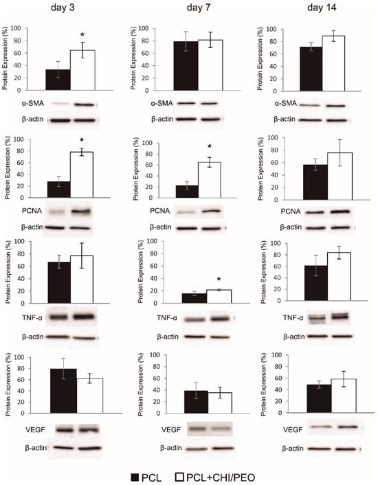
In the original publication, the authors claimed that Figure 6 reporting Western blot data was erroneous as published. Specifically, the PCNA band on day 3 was found to be duplicated with PCNA band on day 7. We replaced the image from PCNA on day 3. The corrected Figure 6 (PCNA bands) appears below. The authors state that the scientific conclusions are unaffected. This correction was approved by the Academic Editor. The original publication has also been updated [1].

Figure 6.
Western blot analysis and densitometric analysis of smooth muscle actin (α-SMA), PCNA, Tumor Necrosis Factor (TNF-α), and VEGF observed in the excision lesions of mice topically treated with PCL or PCL+CHI/PEO membrane on days 3, 7, and 14. The results were expressed as mean ± standard deviation. (*) p < 0.05 indicates statistically significant differences between treatments according to the Student’s t-test. (n = 4–6). Protein expression levels were standardized against the internal β-actin expression levels of each sample.
Reference
- Zanchetta, F.C.; Trinca, R.B.; Gomes Silva, J.L.; Breder, J.d.S.C.; Cantarutti, T.A.; Consonni, S.R.; Moraes, Â.M.; Pereira de Araújo, E.; Saad, M.J.A.; Adams, G.G.; et al. Effects of Electrospun Fibrous Membranes of PolyCaprolactone and Chitosan/Poly(Ethylene Oxide) on Mouse Acute Skin Lesions. Polymers 2020, 12, 1580. [Google Scholar] [CrossRef] [PubMed]
Disclaimer/Publisher’s Note: The statements, opinions and data contained in all publications are solely those of the individual author(s) and contributor(s) and not of MDPI and/or the editor(s). MDPI and/or the editor(s) disclaim responsibility for any injury to people or property resulting from any ideas, methods, instructions or products referred to in the content. |
© 2024 by the authors. Licensee MDPI, Basel, Switzerland. This article is an open access article distributed under the terms and conditions of the Creative Commons Attribution (CC BY) license (https://creativecommons.org/licenses/by/4.0/).