Article

Controlled Synthesis and Growth Mechanism of Two-Dimensional Zinc Oxide by Surfactant-Assisted Ion-Layer Epitaxy

Center on Nanoenergy Research, Guangxi Key Laboratory for Relativistic Astrophysics, School of Physical Science and Technology, Guangxi University, Nanning 530004, China
*
Author to whom correspondence should be addressed.
Crystals 2023, 13(1), 5; https://doi.org/10.3390/cryst13010005
Submission received: 30 November 2022 / Revised: 14 December 2022 / Accepted: 16 December 2022 / Published: 20 December 2022

Abstract

:
Two-dimensional (2D) zinc oxide (ZnO) has attracted much attention for its potential applications in electronics, optoelectronics, ultraviolet photodetectors, and resistive sensors. However, little attention has been focused on the growth mechanism, which is highly desired for practical applications. In this paper, the growth mechanism of 2D ZnO by surfactant-assisted ion-layer epitaxy (SA-ILE) is explored by controlling the amounts of surfactant, temperature, precursor concentration, and growth time. It is found that the location and the number of nucleation sites at the initial stages are restricted by the surfactant, which absorbs Zn 2 + ions via electrostatic attraction at the water-air interface. Then, the growth of 2D ZnO is administered by the temperature, precursors, and growth time. In other words, the temperature is connected with the diffusion of solute ions and the number of nucleation sites. The concentration of precursors determines the solute ions in solution, which plays a dominant role in the growth rate of 2D ZnO, while growth time affects the nucleation, growth, and dissolution processes of ZnO. However, if the above criteria are exceeded, the nucleation sites significantly increase, resulting in multiple 2D ZnO with tiny size and multilayers. By optimizing the above parameters, 2D ZnO nanosheets with a size as large as 20 μm are achieved with 10 × 10−5 of the ratio of sodium oleyl sulfate to Zn 2 + , 70 °C, 50 mM of precursor concentration, and 50 min of growth time. 2D ZnO sheets, are confirmed by scanning electron microscope (SEM), energy-dispersive X-ray spectrometer (EDS), X-ray photoelectron spectroscopy (XPS), and Raman spectrum. Our work might guide the development of SA-ILE and pave the platform for practical applications of 2D ZnO on photodetectors, sensors, and resistive switching devices.

1. Introduction

Zinc oxide (ZnO) has received considerable attention for its excellent properties of wide-bandgap (3.3 eV) [1], high exciton binding energy (60 meV) [2], non-centrosymmetric crystal structure [3], and high electron mobility [4,5], presenting wide applications in photodetectors [6,7,8,9], gas sensors [10,11,12,13,14], photoelectric catalytic devices [15,16,17,18,19,20], piezoelectric devices [21,22,23,24,25], resistive sensors [26,27,28], and short-channel high-performance transistors [29]. However, compared with zero-dimensional (0D) and one-dimensional (1D) ZnO, the development of two-dimensional (2D) ZnO has been deserted, though the high surface area and tunable bandgap promote the exploitation of 2D ZnO in new fields [30,31,32,33,34,35,36].
2D ZnO possesses a great deal of excellent performance beyond bulk, 1D, and 0D ZnO. For example, decreasing the thickness of 2D ZnO increases the bandgap due to the quantum confinement effect, yielding promising potential in deep-ultraviolet photodetectors and photocatalysis [31,35]. The mechanical stability is also better than 1D ZnO, overcoming the low durability of piezoelectric nanogenerators based on 1D ZnO [37]. Notably, additional non-centrosymmetric structure appears when the thickness of 2D ZnO is reduced to a few atomic layers [38]. That is, piezoelectric performance is largely improved with a higher output voltage and current. Despite the enormous potential applications of 2D ZnO, development is slow due to the immature growth of 2D ZnO.
Various methods have been tried to grow 2D ZnO. Magnetron sputtering and atomic layer deposition were first developed to grow 2D ZnO, but the excessive power produced lots of defects and a small size of monocrystalline ZnO [39,40,41,42,43,44]. Subsequently, laser deposition was exploited, and the thickness of 2D ZnO was lowered to 5 nm, but with poor crystal quality and morphology [45,46]. Surfactant-assisted ion-layer epitaxy (SA-ILE) was proposed and rapidly disseminated to tackle the challenges of tiny size and irregular shape. 2D ZnO nanosheets with a regular shape and thickness of 2 nm were first realized at the water-air interface, revealing a novel approach to obtain 2D materials from non-van der Waals solids [47]. The method was subsequently extended to the water-air interface to develop 2D ZnO with high Zn vacancy concentrations and strong ferromagnetism [48]. Later, different kinds of anionic surfactants were used to modulate the growth of 2D ZnO, including sodium oleyl sulfate (SOS) [28] and sodium dodecyl sulfate (SDS) [35]. Thus, SA-ILE is considered a simple and feasible method to grow 2D ZnO with a lower layer number and higher crystal quality. However, the growth mechanism of 2D ZnO by SA-ILE has not been reported in detail, which is highly desired for practical applications.
Herein, the nucleation and growth of 2D ZnO by SA-ILE is investigated by exploring the factors of amount of surfactants, temperature, precursor concentration, and growth time. Actually, the nucleation and location of 2D ZnO are determined by the amount and distribution of surfactants, which absorb Zn 2 + ions with electrostatic interaction between Zn 2 + and air. Then, the growth of 2D ZnO is influenced by Zn 2 + / OH   at the water-air interface. The temperature shows an important effect on its growth due to the temperature dependence of the chemical reaction rate and the nucleation potential. Moreover, the precursor concentration decides the quantity of zinc ion at the water-air interface. It is noteworthy that the thickness of 2D ZnO will increase greatly and the size will decrease rapidly as the precursor concentration increases. The growth time affects the amounts of Zn 2 + / OH   attracted to the water-air interface, hence, the size and thickness of 2D ZnO. However, excessive growth time leads to the dissolution of 2D ZnO, resulting in the generation of irregular, thick, and small ZnO. Finally, by optimizing the mentioned parameters, 2D ZnO nanosheets with regular triangle morphology and high surface cleanliness were synthesized at the water-air interface, which were verified by scanning electron microscope (SEM), as well as Raman and X-ray photoelectron spectroscopy (XPS) spectra. This work lays a foundation for understanding the mechanism of 2D ZnO by SA-ILE and provides a road for improvement and optimization of 2D ZnO in the application of deep-ultraviolet photodetectors, 2D spintronics, and piezoelectric devices.

2. Materials and Methods

2.1. Preparation of 2D ZnO Nanosheets

For a typical synthesis process, precursor with the ratio 1:1 of Zn(NO3)2·6H2O and hexamethylenetetramine (HMT) was dissolved in deionized water (50 mM, 17 mL). Then, surfactants of sodium oleyl sulfate, dissolved in chloroform (0.1 vol%, 30 μL), were added at the water-air interface formed after 20 min. Subsequently, the solution was heated at 70 °C for 50 min, and 2D ZnO would appear and grow in the water-air interface after completion of the reaction. SiO2/Si substrate was used to scoop the nanosheets from the water–air interface for subsequent characterization. Due to the small volume of the surfactant and the large water-air interface, the surfactant could be separated discretely.

2.2. Materials Characterization

The morphology of the synthesized nanosheets was investigated by scanning electron microscope (SEM, Carl Zeiss, Jena, Germany, Sigma 500) and optical microscope (OM, OLYMPUS, BX43F). The chemical bonding state was investigated by X-ray Photoelectron Spectroscopy (XPS, Thermo Fisher Scientific, Waltham, MA, USA, ESCALAB 250Xi). The Raman spectra were recorded with Raman spectroscopy with a 532 nm laser (Raman, HORIBA Jobin Yvon, Paris, France, LABRAM HR EVO). The characterizations for all of the materials were performed at room temperature.

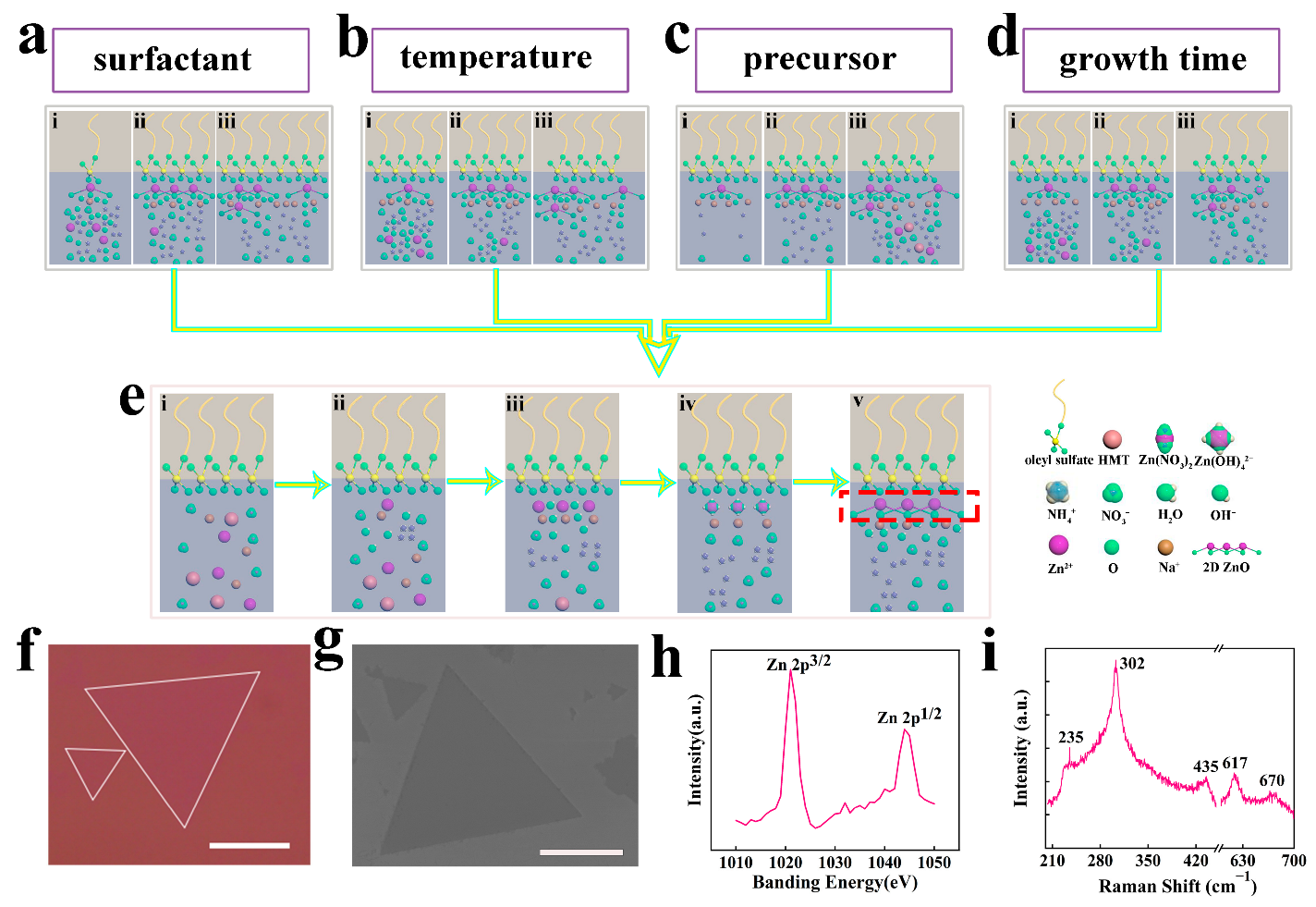
3. Results and Discussion

For a typical synthesis process of 2D ZnO by SA-ILE, precursor was dissolved in deionized water. Then, surfactant was added at the water-air interface after 20 min. Subsequently, the solution was heated at a certain temperature for a long growth time, and 2D ZnO grew in the water-air interface. In this synthesis process, the precursor, surfactant, growth temperature, and time play important roles for the 2D ZnO. Thus, we will discussion the factors of precursor, surfactant, growth temperature, and time of the growth of 2D ZnO as following.

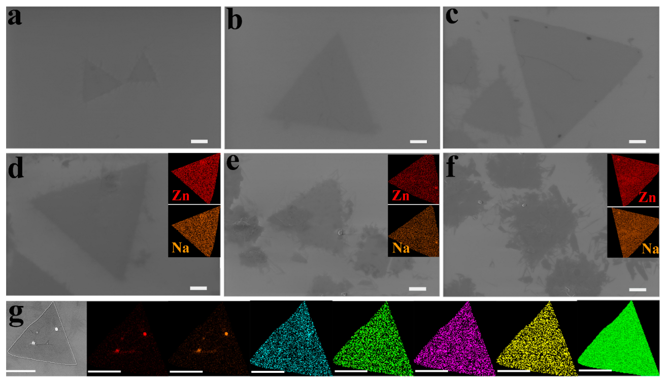
3.1. Effect of the Ratio of Sodium Oleyl Sulfate to Z n 2 + on 2D ZnO Growth

Surfactant is first chosen to enable the growth of 2D ZnO because it provides the dominant roles for nucleation and growth. Sodium oleyl sulfate is adopted as a surfactant for its excellent dispersion performance at the water-air interface. The amounts of surfactants are increased gradually. When the ratio of sodium oleyl sulfate to Zn 2 + is lower than 6 × 10⁻⁵, no obvious ZnO is present. Once increased, the ratio of sodium oleyl sulfate to Zn 2 + to 6 × 10⁻⁵, scattered 2D ZnO with a size around 2.1 μm first appears at the position of the surfactant location with triangle morphology (Figure 1a), indicating the nucleation and growth of ZnO. Then, the 2D ZnO grows, increasing the ratio to 10 × 10−5 (Figure 1b,c). The size of 2D ZnO is around 8.4 μm, indicating the growth rate is 4 times the ratio of 10 × 10−5. However, the size reduces a great deal while increasing the ratio of sodium oleyl sulfate to Zn 2 + after 10 × 10−5, accompanied by a rough surface and increased thickness of ZnO (Figure 1d–f). As the increment of the ratio of sodium oleyl sulfate to Zn 2 + , the group of oleyl sulfate becomes crumpled at the water-air interface due to the strong intermolecular repulsion [49]. In addition, random nucleation and stacking appear on the surface of the 2D ZnO, resulting in rough surfaces and boundaries. The rough surface was analyzed by energy-dispersive X-ray spectroscopic (EDS). The EDS mapping images show the uniform distribution of the Cl, N, S, and O elements (Figure 1g), while the prominent points at the surface of the ZnO are identified as Zn 2 + and Na + (Insets in Figure 1d–f), which are the nucleation sites for the next layer. The larger amount of surfactants, the greater the number of nucleation sites. As a result, the number of ZnO increases, but the size reduces and the thickness increases. Therefore, the surfactant determines the location of nucleation and the number of nucleation sites.

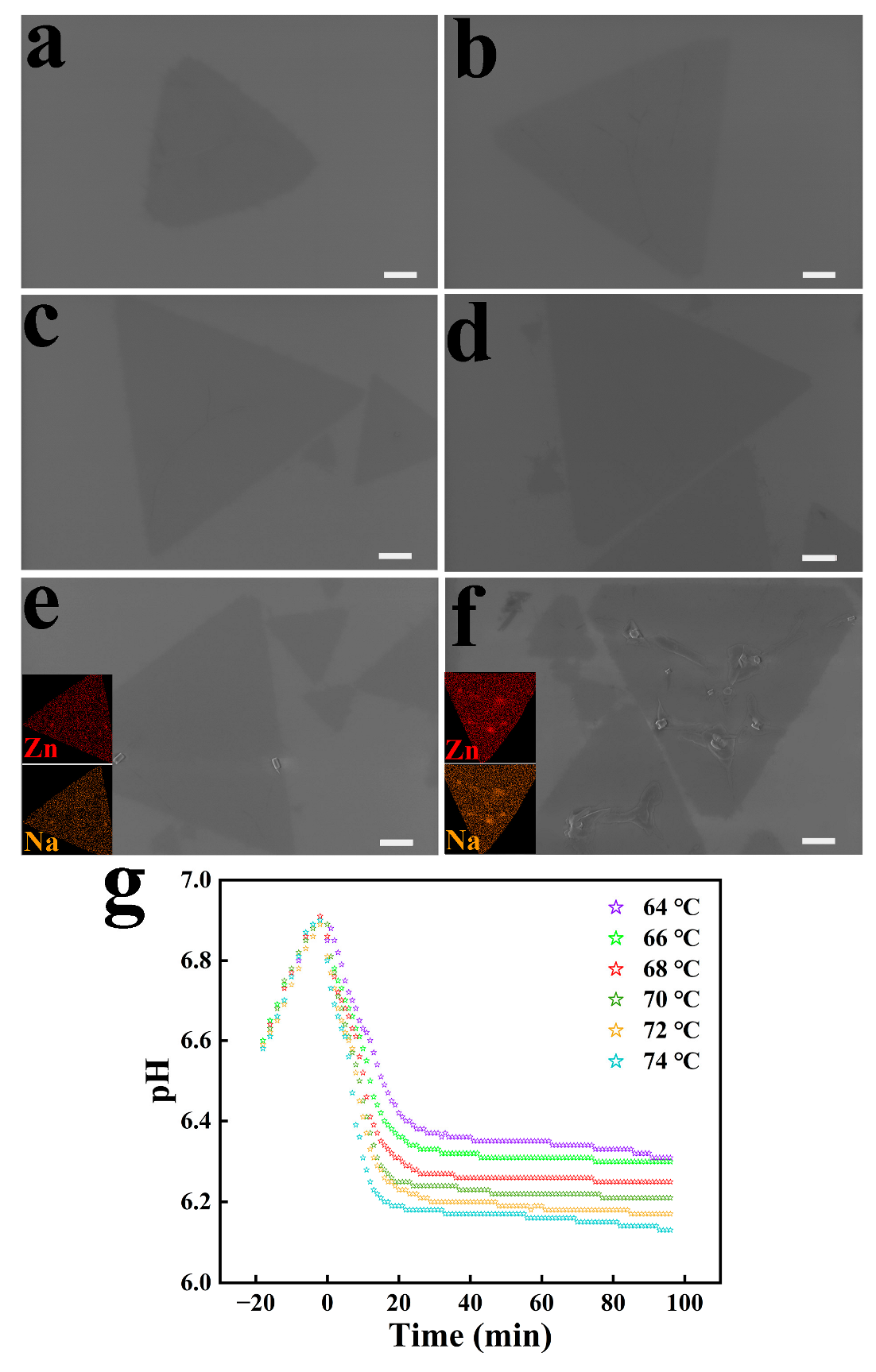
3.2. Effect of Temperature on 2D ZnO Growth

Temperature is an important factor for the growth of 2D ZnO because the reaction ratio and growth are closely related to temperature. When increasing the temperature from 64 °C to 70 °C, the size of the 2D ZnO enlarged from 4 μm to 10 μm (Figure 2a–d). Then, the rough surface with prominent points appeared while increasing the temperature further (Figure 2e,f). The increased size with increasing temperature is derived from the fast response time to form the nucleation of 2D ZnO, as in Equations (1)–(6).
  Zn NO 3 2   ·   6 H 2 O + H 2 O     Zn 2 + + 2 NO 3 + 7 H 2 O
CH 2 6 N 4 + 6 H 2 O     6 HCHO + 4 NH 3
NH 3 + H 2 O     NH 4 + + OH  
Zn 2 + + 2 OH       Zn OH 2
Zn OH 2 + 2 OH       Zn ( OH ) 4 2
  Zn ( OH ) 4 2     ZnO + H 2 O + 2 OH  
When increasing the temperature, the chemical reactions of Equations (1)–(5) are accelerated because the promoted diffusive transfer of the Zn 2 + / OH   to the water-air interface for the higher temperature accelerates the molecular motion. As a result, Zn ( OH ) 4 2 is quickly produced, prompting the nucleation growth of 2D ZnO for the decomposition of Zn ( OH ) 4 2 , as in Equation (6). The accelerated chemical reactions can be confirmed by pH with increasing temperature (Figure 2g). Because the fast conformation of Zn ( OH ) 4 2 , leads to a more rapid consumption of OH   in the solution, pH will be reduced with increasing temperature. Figure 2g shows that the pH values are reduced and rapidly decrease with the increment of the temperature, indicating the accelerated chemical reactions with increasing temperature. Thus, the size of 2D ZnO gradually increased with the temperature from 64 °C to 70 °C. Furthermore, the sharp triangle angle changed with decreasing pH, and a truncated triangle emerged. The reason for this might be the imbalance between the zinc and the hydroxide at the water-air interface for the decrease of the concentration of OH   . Meanwhile, the rough surface with prominent points is connected with the increasing nucleation of the 2D ZnO, again with increasing temperature. On one hand, classical nucleation theory predicts that as the temperature increases, the nucleation potential barrier decreases [50], causing an increase in the number of nuclei of 2D ZnO. On the other hand, the high temperature also promotes the diffusive transfer of Zn 2 + / OH   and nucleation on the 2D ZnO surface. The nucleation on the 2D ZnO surface is presented by EDS mapping images (Insets in Figure 2e,f), and the nucleation on 2D ZnO results in a rough surface. Therefore, temperature administrates the diffusive transfer of Zn 2 + / OH   , nucleation, and the growth of the 2D ZnO.

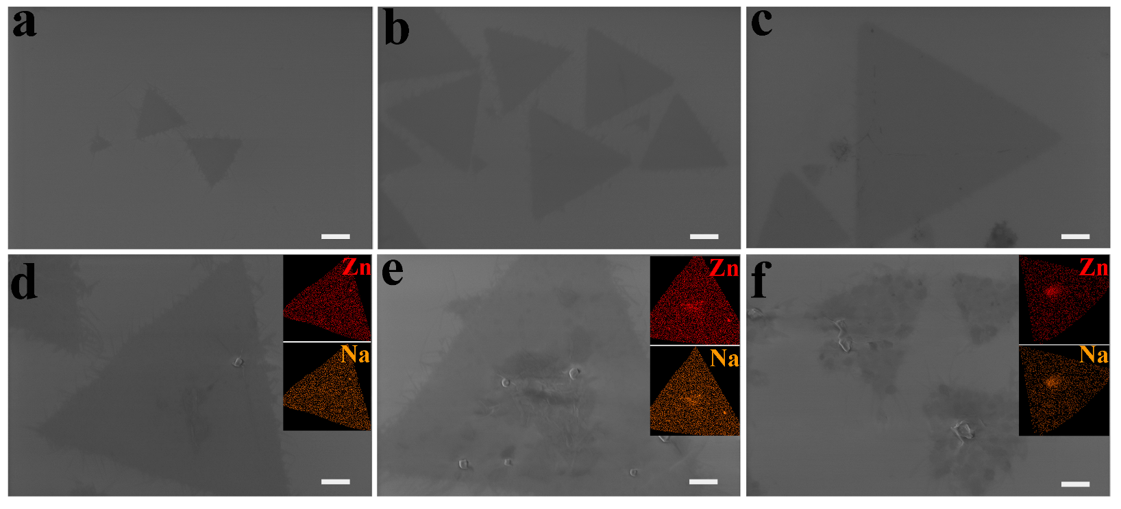
3.3. Effect of Precursor Concentration on 2D ZnO Growth

According to chemical reaction kinetics, the rate of the radical reaction is proportional to the concentration of each reactant. Therefore, precursor concentration should play an important role in the growth of 2D ZnO. Thus, we investigated the effect of precursor concentration on the growth of 2D ZnO to further clarify the growth mechanism of 2D ZnO. The shape of 2D ZnO was triangular at the precursor concentration of 40 mM, but the 2D ZnO nanosheets are sparsely distributed (Figure 3a). This phenomenon is attributed to the lower precursor concentration, which causes a lower zinc concentration. Furthermore, fewer zinc ions are attracted to the water-air interface by the oleyl sulfate anions, which is not conducive to the nucleation and growth of 2D ZnO. When the precursor concentration was increased from 40 mM to 50 mM, the size of the 2D ZnO gradually increased (Figure 3b,c). The increment of precursor concentration induces an increase of Zn 2 + / OH   in the solution, promoting the diffusive transfer of Zn 2 + / OH   from the solution to the water-air interface. Thus, the growth rate of 2D ZnO is accelerated and the size of the 2D ZnO increases. Subsequently, when the precursor concentration is incremented from 50 mM to 60 mM, the size of the 2D ZnO was seen to increase further, accompanied by the surface stacking of the 2D ZnO (Figure 3d,e), i.e., originated from nucleation on the 2D ZnO surface with higher precursor concentration and Zn 2 + / OH   in the solution. Finally, the size is reduced and the layering of 2D ZnO is increased with increasing precursor of more than 65 mM (Figure 3f). The excessive precursor induces the rapid spread of Zn 2 + / OH   from the solution to the water-air interface and a massive amount of nucleation of the 2D ZnO, leading to a small size. The larger number of Zn 2 + / OH   also prompts the nucleation on the first layer of 2D ZnO, resulting in a thick and irregular shape. In a word, the concentration of the precursor controls the amount of Zn 2 + / OH   at the water-air interface, as well as the size and thickness of the 2D ZnO.

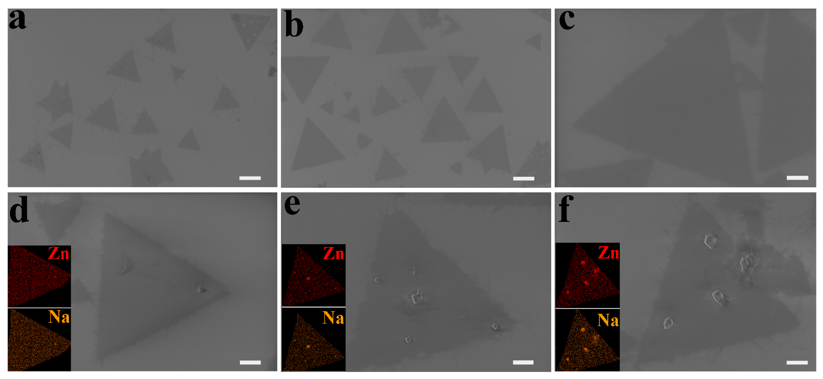
3.4. Effect of Growth Time on 2D ZnO Growth

In order to clarify the growth mechanism of 2D ZnO, it is particularly important to investigate the effect of different growth times on 2D ZnO. The size of the 2D ZnO increased continuously when the growth time was incremented from 40 min to 50 min (Figure 4a–c). As the reaction time increased, the number of Zn 2 + attracted to the water-air interface by the anionic surfactant increased, leading to the increment of Zn ( OH ) 4 2 . Then, the Zn ( OH ) 4 2 gradually decomposed, and ZnO formed and grew at the water-air interface, causing the enlarged size of the 2D ZnO. However, when increasing the growth time from 55 min, a rough surface with prominent points appeared, and the dissolving phenomenon emerged (Figure 4d–f). The rough surface could also be confirmed as clusters of Zn 2 + and Na + by the EDS mapping images (Insets in Figure 4d–f), which can be seen as the nucleation of the next layer of 2D ZnO. The dissolving phenomenon of the 2D ZnO might be connected to the following reasons. Due to the amphoteric character and large surface area of 2D ZnO, the 2D ZnO cannot maintain stability in alkaline solutions for a long time and start to dissolve with increasing time [51]. Another possible scenario is poor crystalline quality, which accelerates the dissolving process [52]. In a word, a suitable growth time promotes the gathering of Zn ( OH ) 4 2 and the growth of 2D ZnO. However, a long growth time will induce dissolution of 2D ZnO.

3.5. Characterization

Thus, the growth process of 2D ZnO, modulated by the surfactant, temperature, precursor, and growth time, can be concluded as follows (Table 1 and Figure 5a–e). Firstly, the surfactant dissolves to form a group of oleyl sulfate, which cannot be dissolved in the water and floats on the solution’s surface. The ionized surfactants are negatively charged, which absorb Zn 2 + at the water-air interface. Then, Zn 2 + ions react to yield Zn ( OH ) 4 2 and 2D ZnO. Thus, the location of the surfactant determines the sites, number of nucleation sites, and growth of the 2D ZnO. Secondly, temperature is connected with the conformation rate of Zn ( OH ) 4 2 . The lower the temperature, the slower the diffusion rate of the Zn 2 + / OH   and the formation of Zn ( OH ) 4 2 , and vice versa. Moreover, the higher temperature also causes a low potential barrier of nucleation, resulting in a large number of nucleation sites of 2D ZnO. However, the excessive number of nucleation sites will lead to a small size of 2D ZnO. Thirdly, the concentration of precursor controls the amount of Zn 2 + / OH   in the solution, which dominates the rates of diffusion and the transfer of Zn 2 + / OH   to the water-air interface by chemical gradient, as well as the rates of the nucleation and growth of 2D ZnO. Additionally, an excessive concentration of precursor induces rapid diffusion and transfer of Zn 2 + / OH   , which generates a great deal of new nucleation sites and a small size of 2D ZnO. Fourthly, the growth time influences the amount of Zn 2 + / OH   at the water-air interface. A suitable growth time encourages continuous growth and a large size of 2D ZnO. However, growth time that is too long will result in the dissolution of 2D ZnO.
By carefully controlling the factors of surfactant, temperature, precursor, and growth time, the 2D ZnO nanosheets with a smooth surface and a regular triangle shape are achieved. As can be seen from the optical and SEM images, the triangle shape and high cleanliness of the 2D ZnO are present (Figure 5f,g). The crystal structure can be characterized by XPS and Raman spectra. Figure 5h shows 2 peaks centered around 1021 eV and 1044 eV, corresponding to the binding energies for Zn 2p3/2 and Zn 2p1/2, respectively, revealing that the Zn element is in a +2 oxidation state [53]. The Raman spectrum gives 5 peaks, centered around 235 cm 1 , 302 cm 1 , 435 cm 1 , 617 cm 1 , and 670 cm 1 , respectively. The peaks centered at 235 cm 1 may correspond to undesired adulteration [54]. The peaks at 302 cm 1 , 617 cm 1 , and 670 cm 1 originate from symmetric stretching vibration, E1 (LO) modes, and the TA + LO intrinsic mode of ZnO [55,56]. The peak centered at 435 cm 1 corresponds to the E2 (high) of ZnO, corresponding to the energy band characteristic peak of wurtzite ZnO and confirming the formation of wurtzite ZnO (Figure 5i) [57,58].

4. Conclusions

In summary, we have achieved the large size, triangle shape, and high cleanliness of 2D ZnO by precisely controlling the concentration of the surfactant, precursor, temperature, and growth time. The surfactant administrates the location of the nucleation and the number of nucleation sites. With the ratio of sodium oleyl sulfate to Zn 2 + increasing from 6 × 10⁻⁵ to 10 × 10−5, the size of the 2D ZnO grows from 2.1 μm to 8.4 μm. However, the size reduces a great deal while increasing the ratio, accompanied by rough surface and increased thickness of the ZnO. The temperature controls the diffusive transfer of Zn 2 + / OH   , nucleation, and the growth of the 2D ZnO. The size of the 2D ZnO increases, and the surface is smooth, with the temperature increased from 64 °C to 70 °C. However, truncated triangles appeared as the temperature increases higher than 70 °C. The concentration of the precursor dominates the amount of Zn 2 + / OH   at the water-air interface, as well as the size and thickness of the 2D ZnO. The size of the 2D ZnO grows from 1.8 μm to 13 μm as the precursor concentration increases from 40 mM to 60 mM. Yet, the rough surface emerges once it exceeds 50 mM. The suitable growth time promotes the gathering of Zn ( OH ) 4 2 and the growth of the 2D ZnO. The size of the 2D ZnO increases with the growth time from 40 min to 50 min. However, long growth time will induce dissolution of 2D ZnO. Finally, the best parameters for the growth of 2D ZnO is 10 × 10−5 of the ratio of sodium oleyl sulfate to Zn 2 + , 70 °C, 50 mM of precursor concentration, and 50 min of growth time. Our work clarifies the mechanism of the SA-ILE growth of 2D ZnO, promotes the further development of SA-ILE, and paves the way for applications of 2D ZnO in ultraviolet photodetectors, 2D spintronics, and piezoelectric devices.

Author Contributions

P.C. and C.H. conceived the ideas. C.H. performed the synthesis. C.H., Q.S., Z.C., D.W., Z.T., Y.L. and Y.H. conducted the characterization. C.H. and P.C. analyzed the results. C.H. and P.C. write the manuscript. All authors have read and agreed to the published version of the manuscript.

Funding

The work is supported by the National Natural Science Foundation of China (Nos. 11704081) and Guangxi Natural Science Foundation (Nos. 2020GXNSFAA297182, 2020GXNSFAA297041, 2017GXNSFBA198229).

Data Availability Statement

Not applicable.

Conflicts of Interest

The authors declare no conflict of interest.

References

  1. Izaki, M.; Chizaki, R.; Saito, T.; Murata, K.; Sasano, J.; Shinagawa, T. Hybrid ZnO/Phthalocyanine Photovoltaic Device with Highly Resistive ZnO Intermediate Layer. ACS Appl. Mater. Interfaces 2013, 5, 9386–9395. [Google Scholar] [CrossRef] [PubMed]
  2. Cole, J.J.; Wang, X.; Knuesel, R.J.; Jacobs, H.O. Patterned Growth and Transfer of ZnO Micro and Nanocrystals with Size and Location Control. Adv. Mater. 2008, 20, 1474–1478. [Google Scholar] [CrossRef]
  3. Lee, P.-C.; Hsiao, Y.-L.; Dutta, J.; Wang, R.-C.; Tseng, S.-W.; Liu, C.-P. Development of porous ZnO thin films for enhancing piezoelectric nanogenerators and force sensors. Nano Energy 2020, 82, 105702. [Google Scholar] [CrossRef]
  4. Hsu, Y.F.; Xi, Y.Y.; Tam, K.H.; Djurišić, A.B.; Luo, J.; Ling, C.C.; Cheung, C.K.; Ng, A.M.C.; Chan, W.K.; Deng, X.; et al. Undoped p-Type ZnO Nanorods Synthesized by a Hydrothermal Method. Adv. Funct. Mater. 2008, 18, 1020–1030. [Google Scholar] [CrossRef]
  5. Kang, Z.; Gu, Y.; Yan, X.; Bai, Z.; Liu, Y.; Liu, S.; Zhang, X.; Zhang, Z.; Zhang, X.; Zhang, Y. Enhanced photoelectrochemical property of ZnO nanorods array synthesized on reduced graphene oxide for self-powered biosensing application. Biosens. Bioelectron. 2015, 64, 499–504. [Google Scholar] [CrossRef] [PubMed]
  6. Ji, Y.; Wu, L.; Liu, Y.; Yang, Y. Chemo-phototronic effect induced electricity for enhanced self-powered photodetector system based on ZnO nanowires. Nano Energy 2021, 89, 106449. [Google Scholar] [CrossRef]
  7. Wang, P.; Wang, Y.; Ye, L.; Wu, M.; Xie, R.; Wang, X.; Chen, X.; Fan, Z.; Wang, J.; Hu, W. Ferroelectric Localized Field-Enhanced ZnO Nanosheet Ultraviolet Photodetector with High Sensitivity and Low Dark Current. Small 2018, 14, 1800492. [Google Scholar] [CrossRef]
  8. Meng, J.; Li, Q.; Huang, J.; Pan, C.; Li, Z. Self-powered photodetector for ultralow power density UV sensing. Nano Today 2022, 43, 101399. [Google Scholar] [CrossRef]
  9. Peng, Y.; Lu, J.; Wang, X.; Ma, W.; Que, M.; Chen, Q.; Li, F.; Liu, X.; Gao, W.; Pan, C. Self-powered high-performance flexible GaN/ZnO heterostructure UV photodetectors with piezo-phototronic effect enhanced photoresponse. Nano Energy 2022, 94, 106945. [Google Scholar] [CrossRef]
  10. Hoffmann, M.W.G.; Mayrhofer, L.; Casals, O.; Caccamo, L.; Hernandez-Ramirez, F.; Lilienkamp, G.; Daum, W.; Moseler, M.; Waag, A.; Shen, H.; et al. A highly selective and self-powered gas sensor via organic surface functionalization of p-Si/n-ZnO diodes. Adv. Mater. 2014, 26, 8017–8022. [Google Scholar] [CrossRef]
  11. Shan, H.; Liu, C.; Liu, L.; Wang, L.; Zhang, X.; Chi, X.; Bo, X.; Wang, K. Excellent ethanol sensor based on multiwalled carbon nanotube-doped ZnO. Chin. Sci. Bull. 2014, 59, 374–378. [Google Scholar] [CrossRef]
  12. Yuan, H.; Aljneibi, S.A.A.A.; Yuan, J.; Wang, Y.; Liu, H.; Fang, J.; Tang, C.; Yan, X.; Cai, H.; Gu, Y.; et al. ZnO Nanosheets Abundant in Oxygen Vacancies Derived from Metal-Organic Frameworks for ppb-Level Gas Sensing. Adv. Mater. 2019, 31, e1807161. [Google Scholar] [CrossRef] [PubMed]
  13. Fan, C.; Shi, J.; Zhang, Y.; Quan, W.; Chen, X.; Yang, J.; Zeng, M.; Zhou, Z.; Su, Y.; Wei, H.; et al. Fast and recoverable NO2 detection achieved by assembling ZnO on Ti3C2Tx MXene nanosheets under UV illumination at room temperature. Nanoscale 2022, 14, 3441–3451. [Google Scholar] [CrossRef] [PubMed]
  14. Li, D.L.; Lu, J.F.; Zhang, X.J.; Peng, X.L.; Li, J.; Yang, Y.T.; Hong, B.; Wang, X.Q.; Jin, D.F.; Jin, H.X. Reversible switching from P- to N-Type NO2 sensing in ZnO Rods/rGO by changing the NO2 concentration, temperature, and doping ratio. J. Phys. Chem. C 2022, 126, 14470–14478. Available online: https://pubs.acs.org/doi/full/10.1021/acs.jpcc.2c03616 (accessed on 29 November 2022). [CrossRef]
  15. Park, S.J.; Das, G.S.; Schuett, F.; Adelung, R.; Mishra, Y.K.; Tripathi, K.M.; Kim, T. Visible-light photocatalysis by car-bon-nano-onion-functionalized ZnO tetrapods: Degradation of 2,4-dinitrophenol and a plant-model-based ecological assess-ment. NPG Asia Mater. 2019, 11. [Google Scholar] [CrossRef] [Green Version]
  16. Wang, F.; Zhao, D.; Xu, Z.; Zheng, Z.; Zhang, L.; Shen, D. Monochromatic visible light-driven photocatalysis realized on 2D ZnO shell arrays. J. Mater. Chem. A 2013, 1, 9132–9137. [Google Scholar] [CrossRef]
  17. Song, S.; Song, H.; Li, L.; Wang, S.; Chu, W.; Peng, K.; Meng, X.; Wang, Q.; Deng, B.; Liu, Q.; et al. A selective Au-ZnO/TiO2 hybrid photocatalyst for oxidative coupling of methane to ethane with dioxygen. Nat. Catal. 2022, 5, 1032–1042. [Google Scholar] [CrossRef]
  18. Peng, F.; Lin, J.; Li, H.; Liu, Z.; Su, Q.; Wu, Z.; Xiao, Y.; Yu, H.; Zhang, M.; Wu, C.; et al. Design of piezoelectric ZnO based catalysts for ammonia production from N2 and H2O under ultrasound sonication. Nano Energy 2022, 95, 107020. [Google Scholar] [CrossRef]
  19. Liu, W.; Liu, H.; Liu, Y.; Dong, Z.; Luo, L. Surface Plane Effect of ZnO on the Catalytic Performance of Au/ZnO for the CO Oxidation Reaction. J. Phys. Chem. C 2022, 126, 14155–14162. [Google Scholar] [CrossRef]
  20. Gleißner, R.; Noei, H.; Chung, S.; Semione, G.D.L.; Beck, E.E.; Dippel, A.-C.; Gutowski, O.; Gizer, G.; Vonk, V.; Stierle, A. Copper Nanoparticles with High Index Facets on Basal and Vicinal ZnO Surfaces. J. Phys. Chem. C 2021, 125, 23561–23569. [Google Scholar] [CrossRef]
  21. Kim, H.; Yun, S.; Kim, K.; Kim, W.; Ryu, J.; Nam, H.G.; Han, S.M.; Jeon, S.; Hong, S. Breaking the elastic limit of piezoelectric ceramics using nanostructures: A case study using ZnO. Nano Energy 2020, 78, 105259. [Google Scholar] [CrossRef]
  22. Yin, B.; Qiu, Y.; Zhang, H.; Lei, J.; Chang, Y.; Ji, J.; Luo, Y.; Zhao, Y.; Hu, L. Piezoelectric performance enhancement of ZnO flexible nanogenerator by a NiO-ZnO p-n junction formation. Nano Energy 2015, 14, 95–101. [Google Scholar] [CrossRef]
  23. Park, J.; Ghosh, R.; Song, M.S.; Hwang, Y.; Tchoe, Y.; Saroj, R.K.; Ali, A.; Guha, P.; Kim, B.; Kim, S.-W.; et al. Individually addressable and flexible pressure sensor matrixes with ZnO nanotube arrays on graphene. NPG Asia Mater. 2022, 14, 40. [Google Scholar] [CrossRef]
  24. Yu, Q.; Ge, R.; Wen, J.; Du, T.; Zhai, J.; Liu, S.; Wang, L.; Qin, Y. Highly sensitive strain sensors based on piezotronic tunneling junction. Nat. Commun. 2022, 13, 778. [Google Scholar] [CrossRef]
  25. An, C.; Qi, H.; Wang, L.; Fu, X.; Wang, A.; Wang, Z.L.; Liu, J. Piezotronic and piezo-phototronic effects of atomically-thin ZnO nanosheets. Nano Energy 2020, 82, 105653. [Google Scholar] [CrossRef]
  26. Yalishev, V.; Yuldashev, S.U.; Kim, Y.S.; Park, B.H. The role of zinc vacancies in bipolar resistance switching of Ag/ZnO/Pt memory structures. Nanotechnology 2012, 23, 375201. [Google Scholar] [CrossRef]
  27. Wang, X.; Qian, H.; Guan, L.; Wang, W.; Xing, B.; Yan, X.; Zhang, S.; Sha, J.; Wang, Y. Influence of metal electrode on the performance of ZnO based resistance switching memories. J. Appl. Phys. 2017, 122, 154301. [Google Scholar] [CrossRef]
  28. Yin, X.; Wang, Y.; Chang, T.; Zhang, P.; Li, J.; Xue, P.; Long, Y.; Shohet, J.L.; Voyles, P.M.; Ma, Z.; et al. Memristive Behavior Enabled by Amorphous–Crystalline 2D Oxide Heterostructure. Adv. Mater. 2020, 32, 2000801. [Google Scholar] [CrossRef]
  29. Wang, L.; Liu, S.; Gao, G.; Pang, Y.; Yin, X.; Feng, X.; Zhu, L.; Bai, Y.; Chen, L.; Xiao, T.; et al. Ultrathin Piezotronic Transistors with 2 nm Channel Lengths. ACS Nano 2018, 12, 4903–4908. [Google Scholar] [CrossRef]
  30. Jang, E.S.; Won, J.-H.; Hwang, S.-J.; Choy, J.-H. Fine tuning of the face orientation of ZnO crystals to optimize their photo-catalytic activity. Adv. Mater. 2006, 18, 3309–3312. [Google Scholar] [CrossRef]
  31. Gupta, S.P.; Pawbake, A.S.; Sathe, B.R.; Late, D.J.; Walke, P.S. Superior humidity sensor and photodetector of mesoporous ZnO nanosheets at room temperature. Sens. Actuators B Chem. 2019, 293, 83–92. [Google Scholar] [CrossRef]
  32. Wang, L.; Liu, S.; Zhang, Z.; Feng, X.; Zhu, L.; Guo, H.; Ding, W.; Chen, L.; Qin, Y.; Wang, Z.L. 2D piezotronics in atomically thin zinc oxide sheets: Interfacing gating and channel width gating. Nano Energy 2019, 60, 724–733. [Google Scholar] [CrossRef]
  33. Gao, J.; Meng, H.; Hu, Q.; Chang, J.; Feng, L. 2D ZIF-derived ZnO nanosheets—An example for improving semiconductor metal oxide detector performance in gas chromatography through material design strategy. Sens. Actuators B Chem. 2019, 307, 127580. [Google Scholar] [CrossRef]
  34. Ren, K.; Yu, J.; Tang, W. Two-dimensional ZnO/BSe van der waals heterostructure used as a promising photocatalyst for water splitting: A DFT study. J. Alloys Compd. 2019, 812, 152049. [Google Scholar] [CrossRef]
  35. Yu, H.; Liao, Q.; Kang, Z.; Wang, Z.; Liu, B.; Zhang, X.; Du, J.; Ou, Y.; Hong, M.; Xiao, J.; et al. Atomic-thin ZnO sheet for visible-blind ultraviolet photodetection. Small 2020, 16, 2005520. [Google Scholar] [CrossRef]
  36. Kumar, M.; Bhatt, V.; Kim, J.; Abhyankar, A.C.; Chung, H.-J.; Singh, K.; Bin Cho, Y.; Yun, Y.J.; Lim, K.S.; Yun, J.-H. Holey engineered 2D ZnO-nanosheets architecture for supersensitive ppm level H2 gas detection at room temperature. Sens. Actuators B Chem. 2021, 326, 128839. [Google Scholar] [CrossRef]
  37. Rafique, S.; Kasi, A.K.; Aminullah; Kasi, J.K.; Bokhari, M.; Shakoor, Z. Fabrication of Br doped ZnO nanosheets piezoelectric nanogenerator for pressure and position sensing applications. Curr. Appl. Phys. 2020, 21, 72–79. [Google Scholar] [CrossRef]
  38. Mahmood, N.; Khan, H.; Tran, K.; Kuppe, P.; Zavabeti, A.; Atkin, P.; Ghasemian, M.B.; Yang, J.; Xu, C.; Tawfik, S.A.; et al. Maximum piezoelectricity in a few unit-cell thick planar ZnO—A liquid metal-based synthesis approach. Mater. Today 2021, 44, 69–77. [Google Scholar] [CrossRef]
  39. Samavati, A.; Nur, H.; Ismail, A.F.; Othaman, Z. Radio frequency magnetron sputtered ZnO/SiO2/glass thin film: Role of ZnO thickness on structural and optical properties. J. Alloys Compd. 2016, 671, 170–176. [Google Scholar] [CrossRef]
  40. Akhtaruzzaman; Hossain, M.I.; Islam, M.A.; Shahiduzzaman; Muhammad, G.; Hasan, A.K.M.; Tsang, Y.H.; Sopian, K. Nanophotonic-structured front contact for high-performance perovskite solar cells. Sci. China Mater. 2022, 65, 1727–1740. [Google Scholar] [CrossRef]
  41. Walter, T.N.; Lee, S.; Zhang, X.; Chubarov, M.; Redwing, J.M.; Jackson, T.N.; Mohney, S.E. Atomic layer deposition of ZnO on MoS2 and WSe2. Appl. Surf. Sci. 2019, 480, 43–51. [Google Scholar] [CrossRef]
  42. Wang, Y.; Kang, K.-M.; Kim, M.; Park, H.-H. Film thickness effect in c-axis oxygen vacancy-passivated ZnO prepared via atomic layer deposition by using H2O2. Appl. Surf. Sci. 2020, 529, 147095. [Google Scholar] [CrossRef]
  43. Lu, J.; Wang, W.; Liang, J.; Lan, J.; Lin, L.; Zhou, F.; Chen, K.; Zhang, G.; Shen, M.; Li, Y. Contact Resistance Reduction of Low Temperature Atomic Layer Deposition ZnO Thin Film Transistor Using Ar Plasma Surface Treatment. IEEE Electron Device Lett. 2022, 43, 890–893. [Google Scholar] [CrossRef]
  44. Parmar, D.H.; Pina, J.M.; Zhu, T.; Vafaie, M.; Atan, O.; Biondi, M.; Najjariyan, A.M.; Hoogland, S.; Sargent, E.H. Controlled crystal plane orientations in the ZnO transport layer enable high-responsivity, low-dark-current infrared photodetectors. Adv. Mater. 2022, 34, 2200321. [Google Scholar] [CrossRef] [PubMed]
  45. Kraemer, A.; Engel, S.; Sangiorgi, N.; Sanson, A.; Bartolome, J.F.; Graef, S.; Mueller, F.A. ZnO thin films on single carbon fibres fabricated by Pulsed Laser Deposition (PLD). Appl. Surf. Sci. 2017, 399, 282–287. [Google Scholar] [CrossRef]
  46. Li, B.; Ding, L.; Gui, P.; Liu, N.; Yue, Y.; Chen, Z.; Song, Z.; Wen, J.; Lei, H.; Zhu, Z.; et al. Pulsed Laser Deposition Assisted van der Waals Epitaxial Large Area Quasi-2D ZnO Single-Crystal Plates on Fluorophlogopite Mica. Adv. Mater. Interfaces 2019, 6, 1901156. [Google Scholar] [CrossRef]
  47. Wang, F.; Seo, J.-H.; Luo, G.; Starr, M.B.; Li, Z.; Geng, D.; Yin, X.; Wang, S.; Fraser, D.G.; Morgan, D.; et al. Na-nometre-thick single-crystalline nanosheets grown at the water-air interface. Nat. Commun. 2016, 7, 10444. [Google Scholar] [CrossRef] [PubMed]
  48. Yin, X.; Wang, Y.; Jacobs, R.; Shi, Y.; Szlufarska, I.; Morgan, D.; Wang, X. Massive Vacancy Concentration Yields Strong Room-Temperature Ferromagnetism in Two-Dimensional ZnO. Nano Lett. 2019, 19, 7085–7092. [Google Scholar] [CrossRef]
  49. Yin, X.; Shi, Y.; Wei, Y.; Joo, Y.; Gopalan, P.; Szlufarska, I.; Wang, X. Unit Cell Level Thickness Control of Single-Crystalline Zinc Oxide Nanosheets Enabled by Electrical Double-Layer Confinement. Langmuir 2017, 33, 7708–7714. [Google Scholar] [CrossRef] [Green Version]
  50. Kim, S.; Iida, K.; Kuromiya, Y.; Seto, T.; Higashi, H.; Otani, Y. Effect of Nucleation Temperature on Detecting Molecular Ions and Charged Nanoparticles with a Diethylene Glycol-Based Particle Size Magnifier. Aerosol Sci. Technol. 2014, 49, 35–44. [Google Scholar] [CrossRef]
  51. Mao, J.; Li, J.-J.; Ling, T.; Liu, H.; Yang, J.; Du, X.-W. Facile synthesis of zinc hydroxide carbonate flowers on zinc oxide nanorods with attractive luminescent and optochemical performance. Nanotechnology 2011, 22, 245607. [Google Scholar] [CrossRef] [PubMed]
  52. Li, P.; Liu, H.; Lu, B.; Wei, Y. Formation Mechanism of 1D ZnO Nanowhiskers in Aqueous Solution. J. Phys. Chem. C 2010, 114, 21132–21137. [Google Scholar] [CrossRef]
  53. Fang, M.; Liu, Z.W. Controllable size and photoluminescence of ZnO nanorod arrays on Si substrate prepared by micro-wave-assisted hydrothermal method. Ceram. Int. 2017, 43, 6955–6962. [Google Scholar] [CrossRef]
  54. Luo, C.-Q.; Ho, L.-P.; Ling, F.C.-C. The origin of additional modes in Raman spectra of ZnO:Sb films. Physica B 2020, 593, 412256. [Google Scholar] [CrossRef]
  55. Das, P.K.; Biswal, R.; Choudhary, R.J.; Sathe, V.; Ganesan, V.; Khan, S.A.; Mishra, N.C.; Mallick, P. Effect of 120 MeV Au9+ ion irradiation on the structure and surface morphology of ZnO/NiO heterojunction. Surf. Interface Anal. 2018, 50, 954–961. [Google Scholar] [CrossRef]
  56. Sharma, G.N.; Dutta, S.; Singh, S.K.; Chatterjee, R. Growth and optical properties of nano-textured (110) Pb(Zr0.52Ti0.48)O3/(001) ZnO hetero-structure on oxidized silicon substrate. J. Mater. Sci.-Mater. Electron. 2017, 28, 5058–5062. [Google Scholar] [CrossRef]
  57. Husain, S.; Alkhtaby, L.A.; Giorgetti, E.; Zoppi, A.; Miranda, M.M. Effect of Mn doping on structural and optical properties of sol gel derived ZnO nanoparticles. J. Lumin. 2013, 145, 132–137. [Google Scholar] [CrossRef]
  58. Zamiri, R.; Kaushal, A.; Rebelo, A.; Ferreira, J. Er doped ZnO nanoplates: Synthesis, optical and dielectric properties. Ceram. Int. 2014, 40, 1635–1639. [Google Scholar] [CrossRef]
Figure 1. Controlling the growth of 2D ZnO by the amount of surfactant. SEM images of 2D ZnO synthesized with the ratio of sodium oleyl sulfate to Zn 2 + is around (a) 6 × 10⁻⁵, (b) 8 × 10⁻⁵, (c) 10 × 10−5, (df) 11 × 10−5, 13 × 10−5, and 14 × 10−5. Insets are the corresponding EDS mapping images of Zn and Na elements. Scale bar is 1 μm. (g) SEM image and EDS mapping images for Zn, Na, C, Cl, N, S, and O elements, respectively. Scale bar is 2.5 μm.
Crystals 13 00005 g001
Figure 2. Controlling the growth of 2D ZnO by temperature. SEM images of 2D ZnO synthesized with the temperature at (a) 64 °C, (b) 66 °C, (c) 68 °C, (d) 70 °C, (e,f) 72 °C and 74 °C. Insets are the corresponding EDS mapping images of Zn and Na elements. Scale bar is 1 μm. (g) Dependence of pH values on times with different temperatures.
Crystals 13 00005 g002
Figure 3. Controlling the growth of 2D ZnO by precursor concentration. SEM images of 2D ZnO synthesized with precursor concentration of (a) 40 mM, (b) 45 mM, (c) 50 mM, (df) 55 mM, 60 mM and 65 mM. Insets are the corresponding EDS mapping images of Zn and Na elements. Scale bar is 1 μm.
Crystals 13 00005 g003
Figure 4. Controlling the growth of 2D ZnO by growth time. Controlling the growth of 2D ZnO with a time of (a) 40 min, (b) 45 min, (c) 50min, (df) 55 min, 60 min and 65 min. Insets are the corresponding EDS mapping images of Zn and Na elements. Scale bar is 1 μm.
Crystals 13 00005 g004
Figure 5. Mechanism analysis of 2D ZnO synthesis by SA-ILE. Schematic diagram of the synthesis of 2D ZnO while controlling (a) the ratio of sodium oleyl sulfate to Zn 2 + (i): lower than 10 × 10−5, (ii): 10 × 10−5, (iii): higher than 10 × 10−5, (b) temperature (i): lower than 70 °C, (ii): 70 °C, (iii): higher than 70 °C. (c) precursor (i): lower than 50 mM, (ii): 50 mM, (iii): higher than 60 mM and (d) growth time (i): lower than 50 min, (ii): 50 min, (iii): higher than 50 min. (e) Diagram illustrates the nucleation and growth of 2D ZnO. The red dotted box indicated the formation of 2D ZnO at the water-air interface. (f) Optical image of 2D ZnO. Scale bar is 5 μm. (g) SEM image of 2D ZnO. Scale bar is 5 μm. (h) XPS spectrum of 2D ZnO. (i) Raman spectrum of 2D ZnO.
Crystals 13 00005 g005
Table 1. Effect of the above parameters on 2D ZnO growth.
ParametersValueSize (μm)Morphology
The ratio of sodium oleyl sulfate to Zn 2 + 6 × 10−52.1triangle
8 × 10−56.5triangle
10 × 10−58.4triangle
11 × 10−57.2triangle + rough
13 × 10−54.6triangle + rough
14 × 10−53.7triangle + rough
Temperature (°C)645.2triangle
667.5triangle
687.6triangle
708.5triangle
727.8triangle + rough
747.6truncated triangle + rough
precursor
concentration (mM)
401.8triangle
453.5triangle
508.6triangle
5510.1triangle + rough
6013triangle + rough
653.8triangle + rough
Growth time (min)401.5triangle
452.1triangle
508.9triangle
557.7triangle + rough
607.3triangle + rough
657.2triangle + rough
Disclaimer/Publisher’s Note: The statements, opinions and data contained in all publications are solely those of the individual author(s) and contributor(s) and not of MDPI and/or the editor(s). MDPI and/or the editor(s) disclaim responsibility for any injury to people or property resulting from any ideas, methods, instructions or products referred to in the content.

