Article

Fabrication of In(P)As Quantum Dots by Interdiffusion of P and As on InP(311)B Substrate

National Institute of Information and Communications Technology (NICT) 4-2-1 Nukuikitamachi, Koganei, Tokyo 184-8795, Japan
*
Author to whom correspondence should be addressed.
Crystals 2020, 10(2), 90; https://doi.org/10.3390/cryst10020090
Submission received: 29 January 2020 / Revised: 2 February 2020 / Accepted: 3 February 2020 / Published: 5 February 2020
(This article belongs to the Section Inorganic Crystalline Materials)

Abstract

:
Herein, we report on our investigation of a fabrication scheme for self-assembled quantum dots (QDs), which is another type of Stranski–Krastanow (S–K) growth mode. The In(P)As QD structure was formed by the irradiation of As flux on an InP(311)B surface in a molecular beam epitaxy system controlled by substrate temperature and irradiation duration. These QDs show photoluminescence at around 1500 nm, which is suitable for fiber optic communication systems. The QDs formed by this structure had high As composition because they had size, density, and emission wavelength similar to those of QDs grown by the usual S–K growth mode.

1. Introduction

Semiconductor quantum dots (QDs) have attracted much attention because they can confine electrons and holes three dimensionally. Therefore, QDs were expected to be used in high performance semiconductor lasers and to be applied in quantum information and communication technologies [1,2,3,4,5]. The technique for self-assembling QDs is widely used for application to optical devices (such as semiconductor laser diodes) using the Stranski–Krastanow (S–K) growth mode because these applications do not require control of a QD position. In the crystal growth of a compound semiconductor, especially lattice-mismatched-material systems, the strained layer on the substrate forms a QD structure that is determined by the total energy stabilization including strain energy and surface energy [6,7]. In systems of III-V materials, techniques for self-assembling of InAs QDs on GaAs or InP substrate have been well investigated [8,9,10,11]. Control of the size, density, and emission wavelength of the QDs is achieved by optimizing the crystal growth condition. In the growth of self-assembled QDs, a molecular beam of group III atoms and group V atoms are usually supplied simultaneously. On the other hand, there are some special growth techniques such as droplet epitaxy [12,13], in which only group III atoms are supplied to the substrate at low temperature followed by irradiation with group V atoms at high temperature. Droplet epitaxy has an advantage because the nanoscale droplets of group III atoms form independently of lattice mismatch, which allowed us to fabricate QDs in lattice-matched-material systems. In addition, we found that, during the formation of QDs irradiated only by group V atoms, growth occurred on the substrate by molecular beam epitaxy (MBE), in which the group V atoms on the substrate were replaced by the irradiating atoms [14]. It is believed that the replacement of group V atoms occurred at equilibrium, so that high-quality crystals formed. However, detailed research has not been done because changing the group V atoms during irradiation is not easy due to the high vapor pressure of this group. In this study, we investigated a technique for the growth of QDs using interdiffusion of arsenic atoms into an InP(311)B substrate via a process called interdiffusion epitaxy (IDE).

2. Experiments

The samples were fabricated using solid-source molecular beam epitaxy (MBE; Veeco Gen 930). Semi-insulating substrates of InP(311)B were used for this study. A large amount of atom migration was observed on the (311)B surface [15,16]. Interdiffusion also occurred easily on this surface as compared to a (001) surface [14]. The supply of group V atoms was provided by a valved cracker cell, which could rapidly change the group V equivalent beam pressure. The surfaces of the InP(311)B substrates were cleaned by increasing the substrate temperature to 500 °C under irradiation with a P beam (2 × 10⁻⁶ Torr) in the growth chamber. After thermal cleaning, the substrate temperature was set to 470 °C and the InP buffer layer was grown. Then the substrate temperature was set to each process temperature, and irradiation with As was conducted. Change of the surface morphology was monitored by reflection of high-energy electron diffraction (RHEED) in situ. After growth of the QDs, the surface morphology was evaluated using atomic force microscopy (AFM) in air. The samples used for photoluminescence (PL) of the InP embedding layer were grown at 430 °C after the As irradiation process. PL measurement was carried out using a second harmonic line of a YAG laser (532 nm), a 320 mm monochromator, and an InGaAs array photodetector at room temperature.

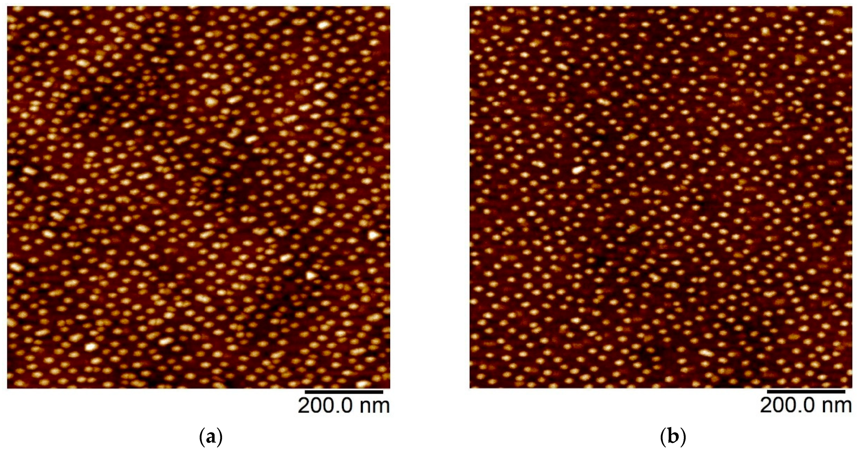
3. Results and Discussion

Figure 1a shows the RHEED pattern after growth of the InP buffer layer along the [01−1] direction. The RHEED pattern shows a streak pattern, which means that a clean, flat surface was obtained after growth of the buffer layer. First, we examined the As irradiation process at 470 °C. Figure 1b,c show the change of the RHEED pattern, of which the streak pattern was weakened after starting irradiation with the As beam. A spotty pattern appeared at around 180 s, as shown in Figure 1b. After irradiating with As for 240 s, a clear spot pattern was observed, as shown in Figure 1c. These results indicate that a crystalline three-dimensional nanostructure was formed, because metallic or amorphous surfaces show a halo pattern with RHEED. Therefore, the surface phosphorus of the substrate was exchanged with irradiating arsenic atoms, and interdiffusion occurred during this process [14]. This is quite different from the process used for the InP(001) substrate. On the InP(001) surface, one or two monolayer phosphorus atoms were exchanged with arsenic, and a stabilized flat surface was obtained [17,18]. On the other hand, more than two monolayer phosphorus atoms should be exchanged to form a three-dimensional structure in our case because large mass transport occurred along the normal direction on the (311)B surface [15,16]. The incorporated As atoms form an InPAs layer with high As composition, which generates a strain field. This strain is the driving force that forms the structure of the QDs. This is the reason for the use of the (311)B surface in our process. Next, we examined the same process at different substrate temperatures during which the As irradiation time was fixed to 240 s. Figure 1d,e show the RHEED pattern after 240 s of As irradiation at 450 °C and 430 °C, respectively. In the process at 450 °C, the RHEED pattern changed from a streak to a spotty pattern. Although it was similar to the one formed during the process at 470 °C, the three-dimensional structure on the surface was expected to be smaller at 450 °C than at 470 °C. In the process at 430 °C, the RHEED streak pattern was maintained or changed to slightly streaky, which indicates that the surface was flat and that there was insignificant formation of a three-dimensional structure.
To investigate the surface morphology, AFM measurement was conducted after the processing at three temperatures. In this case, we kept the substrate in the growth chamber for 300 s after setting the substrate temperature at 430 °C for each process because this better adapted the growth process of the cap layer for the PL samples. Figure 2a shows an AFM image after As irradiation at 470 °C for 240 s. Three-dimensional QD structures were observed on the surface, which is consistent with RHEED measurements. The average lateral size (L) along the [−233] direction, the height (H), and the density (D) were 35.7 nm, 4.51 nm, and 8.96 × 10¹⁰/cm−2, respectively. The histogram of the lateral size and height are shown in Figure 3a. The size and density of this sample were almost the same as for InAs QDs grown on InP(311)B substrate with an ordinary scheme [19,20,21]. Although it is difficult to determine accurately the composition of the thin surface layer, these QDs were expected to be formed as InPAs with high As composition (nearly InAs). In the AFM image, some larger islands were observed that were considered to be formed by the coalescence of smaller QDs. However, the fluctuation of size was not very large; it remained around 17%. Figure 2b and Figure 3b show AFM images of the surface morphology after 240 s of As irradiation at 450 °C and a histogram of QD size, respectively. In this sample, the surface morphology observed was similar to the one processed at 470 °C. The lateral size, height, and density were 31.7 nm, 3.70 nm, and 8.04 × 10¹⁰/cm2, respectively, slightly smaller than those of the sample at 470 °C. Their smaller size is consistent for spotty patterns of RHEED. In this sample, fewer coalescent islands were observed than in the sample processed at 470 °C. In contrast, the sample processed at 430 °C showed different surface morphology and size distribution, as shown in Figure 2c and Figure 3c. It is believed that the small change in the RHEED pattern mentioned above indicates that these small QD structures were formed with As irradiation at 430 °C. The average lateral size, height, and density were 25.8 nm, 1.82 nm, and 4.80 × 10¹⁰/cm2, respectively. Since the size distribution in Figure 3c shows a significant shift to the smaller region, it is clear that the exchange reaction and interdiffusion of P and As were not promoted as well in this temperature range. In addition, the height distribution shows a bimodal shape. The larger peak (around 4 nm) is similar to the ones for the samples processed at 470 °C and 450 °C. Therefore, the group with greater height was distinct from the coalescent islands observed in Figure 2a,b. This is related to the self-size-limiting phenomena that occur during formation of QDs [22,23]. In [18,19], the size of InAs QDs was limited under low As pressure or low growth rates, which can be considered a quasi-equilibrium condition even for the non-equilibrium growth technique of MBE. In our case, a better equilibrium condition was achieved because we only supplied As atoms and exchanged P and As at the surface. However, the P and As atoms exchanged under our conditions did not achieve states of complete equilibrium, so that large coalescent islands were observed in the samples processed at 470 °C and 450 °C. These results show that the P and As exchange and interdiffusion process is strongly dependent on the substrate temperature.
Next, we evaluated the surface morphology by changing the As irradiation time at 470 °C. Two samples were fabricated using As irradiation for 120 s and 480 s. The other process was the same as for the sample with As irradiation for 240 s at 470 °C, as shown in Figure 2a. The AFM image and histogram of lateral size and height are shown in Figure 2d and Figure 3d for 120 s of As irradiation, and in Figure 2e and Figure 3e for 480 s of As irradiation. For the sample with 120 s of As irradiation, the average size, height, and density were 33.0 nm, 4.03 nm, and 6.84 × 10¹⁰/cm2, respectively, which were slightly smaller than that of the sample with 240 s of As irradiation. The distributions of the lateral size and height were similar to those of the sample with 240 s of As irradiation, and shifted to smaller regions, which indicated that the formation of QDs was proceeding in this stage. In addition, the coalescent islands were not found in this region. On the other hand, the sample with 480 s of As irradiation shows many coalescent islands. The average size, height, and density were 38.6 nm, 5.26 nm, and 1.01 × 10¹¹/cm2, respectively. The size distribution became quite broad, which means that both the nucleation of small QDs and the formation of large islands by coalescence occurred in this stage. This is also supported by the shift of average size to larger regions and by the increase in the QD density. Therefore, the size limiting effect was not very large in this temperature region.
Finally, we evaluated the optical properties of the observed QD by PL measurement at room temperature. All QDs were embedded within 100 nm thick InP at 430 °C. The results are shown in Figure 4, where parts (a) to (e) show the spectra for the samples with As irradiation for (a) 240 s at 470 °C, (b) 240 s at 450 °C, (c) 240 s at 430 °C, (d) 120 s at 470 °C, and (e) 480 s at 470 °C. These correspond to the AFM images in Figure 2a–e. The sample with 240 s of As irradiation at 470 °C shows strong emission (even at room temperature) around 1500 nm, which is a higher intensity when compared with conventional S–K QDs. Therefore, the crystal quality of these quantum dots was very high. The wetting layer emission could not be detected as it was expected to appear in a shorter wavelength region (out of measurement range). The sample with 240 s of As irradiation in Figure 2b at 450 °C, and c at 430 °C, shows shorter wavelength emission at 1346 nm and 1259 nm, which is consistent with the smaller QDs observed in these samples, as compared to the sample processed at 470 °C. The full widths at half maxima (FWHMs) of these spectra were (a) 95 meV, (b) 82 meV, and (c) 102 meV. The wider spectrum was observed in the sample processed at 430 °C where the size distribution of QDs was wide. The samples processed at 470 °C for 120 s and 480 s showed emission centered at (d) 1446 nm and (e) 1608 nm, respectively. In this case, the center of the emission wavelength shifted depending on the average size of the QDs. The FWHM of each spectrum was (d) 93 meV and (e) 93 meV. Furthermore, we could not observe the emission of the wetting layer (WL) in any of the samples, which either implies that the WL did not form or that it had vanished during the embedding process. Although the FWHM of spectrum (d) had almost the same size distribution of QDs as in the sample processed for 240 s at 470 °C, the FWHM of spectrum (e) was also the same, in spite of the wide size distribution in this sample. The reason for this is believed to be carrier transfer from smaller QDs to larger QDs. In this sample, a highly packed structure was observed, as shown in Figure 2e. The smaller QDs nucleated between larger QDs, so that the carriers in the smaller QDs moved to neighboring larger QDs and were emitted from there. In these cases, higher energy emission was suppressed so that the FWHM became smaller [24]. Although the details of carrier dynamics still need to be investigated, it was found that this technique can be used to control the emission wavelength over a wide range, from 1346 nm to 1608 nm. Therefore, wide-range optical devices with these QDs could be realized using this technique after optimization. The PL results also show that these QDs were composed of InPAs with a high As composition (more than 90%) because the InAs QDs formed by the S–K growth at 470 °C showed emission wavelength centered at 1599 nm.

4. Summary

In summary, we fabricated self-assembled In(P)As QDs using IDE. The three-dimensional QD structures were obtained by only irradiating As flux onto an InP(311)B substrate at substrate temperatures ranging from 430 °C to 470 °C. The QD size and density were controlled by changing the substrate temperature and duration of As irradiation. The QDs obtained by IDE show strong PL at room temperature, controllable over a range of 300 nm, in the region of 1500 nm.

Author Contributions

K.A. conceived and design the study, performed the experiments and wrote the paper; A.M., T.U. and N.Y. reviewed manuscript and edited the manuscript. All authors have read and agree to the published version of the manuscript.

Funding

This research was funded by the program of MEXT Grant-in-Aid for Scientific Research on Innovative Areas “Science of hybrid quantum systems”, a CREST project (JPMJCR17N2) funded by Japan Science and Technology Agency, and a research project “R&D to Expand Radio Frequency Resources” by the Ministry of Internal Affairs and Communications.

Acknowledgments

This work was conducted as part of MEXT Grant-in-Aid for Scientific Research on Innovative Areas “Science of hybrid quantum systems”, a CREST project (JPMJCR17N2) funded by Japan Science and Technology Agency, and a research project “R&D to Expand Radio Frequency Resources” by the Ministry of Internal Affairs and Communications.

Conflicts of Interest

The authors declare no conflict of interest.

References

  1. Arakawa, Y.; Sakaki, H. Multidimensional quantum well laser and temperature dependence of its threshold current. Appl. Phys. Lett. 1982, 40, 939–941. [Google Scholar] [CrossRef]
  2. Shoji, H.; Mukai, K.; Ohtsuka, N.; Sugawara, M.; Uchida, T.; Ishikawa, H. Lasing at three-dimensionally quantum-confined sublevel of self-organized In/sub 0.5/Ga/sub 0.5/As quantum dots by current injection. IEEE Photon. Technol. Lett. 1995, 7, 1385–1387. [Google Scholar] [CrossRef]
  3. Akiyama, T.; Kuwatsuka, H.; Simoyama, T.; Nakata, Y.; Mukai, K.; Sugawara, M.; Wada, O.; Ishikawa, H. Nonlinear gain dynamics in quantum-dot optical amplifiers and its application to optical communication devices. IEEE J. Quantum Electron. 2001, 37, 1059–1065. [Google Scholar] [CrossRef]
  4. Sugawara, M.; Hatori, N.; Ishida, M.; Ebe, H.; Arakawa, Y.; Akiyama, T.; Otsubo, K.; Yamamoto, T.; Nakata, Y. Recent progress in self-assembled quantum-dot optical devices for optical telecommunication: Temperature-insensitive 10 Gb s−1 directly modulated lasers and 40 Gb s−1 signal-regenerative amplifiers. J. Phys. D 2005, 38, 2126–2134. [Google Scholar] [CrossRef]
  5. Huber, D.; Reindl, M.; Aberl, J.; Rastelli, A.; Trotta, R. Semiconductor quantum dots as an ideal source of polarization-entangled photon pairs on-demand: A review. J. Opt. 2018, 20, 073002. [Google Scholar] [CrossRef] [Green Version]
  6. Goldstein, L.; Glas, F.; Marzin, J.Y.; Charasse, M.N.; le Roux, G. Growth by molecular beam epitaxy and characterization of InAs/GaAs strained-layer superlattices. Appl. Phys. Lett. 1985, 47, 1099–1101. [Google Scholar] [CrossRef]
  7. Shchukin, V.A.; Ledentsov, N.N.; Kop’ev, P.S.; Bimberg, D. Spontaneous ordering of arrays of coherent strained islands. Phys. Rev. Lett. 1995, 75, 2968–2971. [Google Scholar] [CrossRef]
  8. Solomon, G.S.; Trezza, J.A.; Harris, J.S. Substrate temperature and monolayer coverage effects on epitaxial ordering of InAs and InGaAs islands on GaAs. Appl. Phys. Lett. 1995, 66, 991–993. [Google Scholar] [CrossRef]
  9. Lei, W.; Tan, H.H.; Jagadish, C. Formation and shape control of InAsSb/InP (001) nanostructures. Appl. Phys. Lett. 2009, 95, 013108. [Google Scholar] [CrossRef] [Green Version]
  10. Lei, W.; Tan, H.H.; Jagadish, C. Effect of matrix material on the morphology and optical properties of InP-based InAsSb nanostructures. Appl. Phys. Lett. 2009, 95, 143124. [Google Scholar] [CrossRef]
  11. Lei, W.; Chen, Y.H.; Wang, Y.L.; Huang, X.Q.; Zhao, C.; Liu, J.Q.; Xu, B.; Jin, P.; Zeng, Y.P.; Wang, Z.G. Optical properties of self-assembled InAs/InAlAs/InP quantum wires with different InAs deposited thickness. J. Cryst. Growth 2006, 286, 23–27. [Google Scholar] [CrossRef]
  12. Koguchi, N.; Takahashi, S.; Chikyow, T. New MBE growth method for InSb quantum-well boxes. J. Cryst. Growth 1991, 111, 688–692. [Google Scholar] [CrossRef]
  13. Mano, T.; Mitsuishi, K.; Nakayama, Y.; Noda, T.; Sakoda, K. Structural properties of GaAs nanostructures formed by a supply of intense As4 flux in droplet epitaxy. Appl. Surf. Sci. 2008, 254, 7770–7773. [Google Scholar] [CrossRef]
  14. Kawamura, T.; Akahane, K.; Okada, Y.; Kawabe, M. Growth Mechanism of Surface Dots Self-Assembled on InP(311)B Substrate. J. Appl. Phys. 1999, 38, L720–L723. [Google Scholar] [CrossRef]
  15. Nötzel, R.; Temmyo, J.; Tamamura, T. Self-organized growth of strained InGaAs quantum disks. Nature 1994, 369, 131–133. [Google Scholar] [CrossRef]
  16. Temmyo, J.; Nötzel, R.; Tamamura, T. Semiconductor nanostructures formed by the Turing instability. Appl. Phys. Lett. 1997, 71, 1086–1088. [Google Scholar] [CrossRef]
  17. Davies, G.J.; Heckingbottom, R.; Ohno, H.; Wood, C.E.C.; Calawa, A.R. Arsenic stabilization of InP substrates for growth of GaxIn1−xAs layers by molecular beam epitaxy. Appl. Phys. Lett. 1980, 37, 290–292. [Google Scholar] [CrossRef]
  18. Moison, J.M.; Bensoussan, M.; Houzay, F. Epitaxial regrowth of an InAs surface on InP: An example of artificial surfaces. Phys. Rev. B 1986, 34, 2018. [Google Scholar] [CrossRef]
  19. Akahane, K.; Ohtani, N.; Okada, Y.; Kawabe, M. Fabrication of ultra-high density InAs-stacked quantum dots by strain-controlled growth on InP (3 1 1) B substrate. J. Cryst. Growth 2002, 245, 31–36. [Google Scholar] [CrossRef]
  20. Akahane, K.; Yamamoto, N.; Kawanishi, T. Fabrication of ultra-high-density InAs quantum dots using the strain-compensation technique. Phys. Status Solidi A 2011, 208, 425–428. [Google Scholar] [CrossRef]
  21. Akahane, K.; Yamamoto, N. Fabrication of low-density self-assembled InAs quantum dots on InP (311) B substrate by molecular beam epitaxy. J. Cryst. Growth 2013, 378, 450–453. [Google Scholar] [CrossRef]
  22. Kaizu, T.; Yamaguchi, K. Self size-limiting process of InAs quantum dots grown by molecular beam epitaxy. Jpn. J. Appl. Phys. 2001, 40, 1885. [Google Scholar] [CrossRef]
  23. Kaizu, T.; Yamaguchi, K. Facet formation of uniform InAs quantum dots by molecular beam epitaxy. Jpn. J. Appl. Phys. 2003, 42, 4166. [Google Scholar] [CrossRef]
  24. Xu, Z.Y.; Lu, Z.D.; Yang, X.P.; Yuan, Z.L.; Zheng, B.Z.; Xu, J.Z. Carrier relaxation and thermal activation of localized excitons in self-organized InAs multilayers grown on GaAs substrates. Phys. Rev. B 1996, 54, 11528. [Google Scholar] [CrossRef] [PubMed] [Green Version]
Figure 1. Reflection of high-energy electron diffraction (RHEED) pattern for InP(311)B substrate (a) after growth of the InP buffer layer, (b) after irradiation of As for 180 s at 470 °C, (c) for 240 s at 470 °C, (d) for 240 s at 450 °C, and (e) for 240 s at 430 °C.
Crystals 10 00090 g001aCrystals 10 00090 g001b
Figure 2. Atomic force microscopy (AFM) images for samples irradiated with As for (a) 240 s at 470 °C, (b) 240 s at 450 °C, (c) 240 s at 430 °C, (d) 120 s at 470 °C, and (e) 480 s at 470 °C.
Crystals 10 00090 g002aCrystals 10 00090 g002b
Figure 3. Histograms of lateral size and height of quantum dots (QDs): irradiated with As for (a) 240 s at 470 °C, (b) 240 s at 450 °C, (c) 240 s at 430 °C, (d) 120 s at 470 °C, and (e) 480 s at 470 °C.
Crystals 10 00090 g003aCrystals 10 00090 g003b
Figure 4. Photoluminescence (PL) spectra of QDs obtained by interdiffusion epitaxy (IDE): irradiated with As for (a) 240 s at 470 °C, (b) 240 s at 450 °C, (c) 240 s at 430 °C, (d) 120 s at 470 °C, and (e) 480 s at 470 °C.
Crystals 10 00090 g004

