
Application of High Potential Electrophoretic Particles Modified with High Ionization Mono Ionic Liquid for Electrophoretic Displays

 , ,
, ,
Abstract
:1. Introduction
2. Experimental
2.1. Materials and Methods
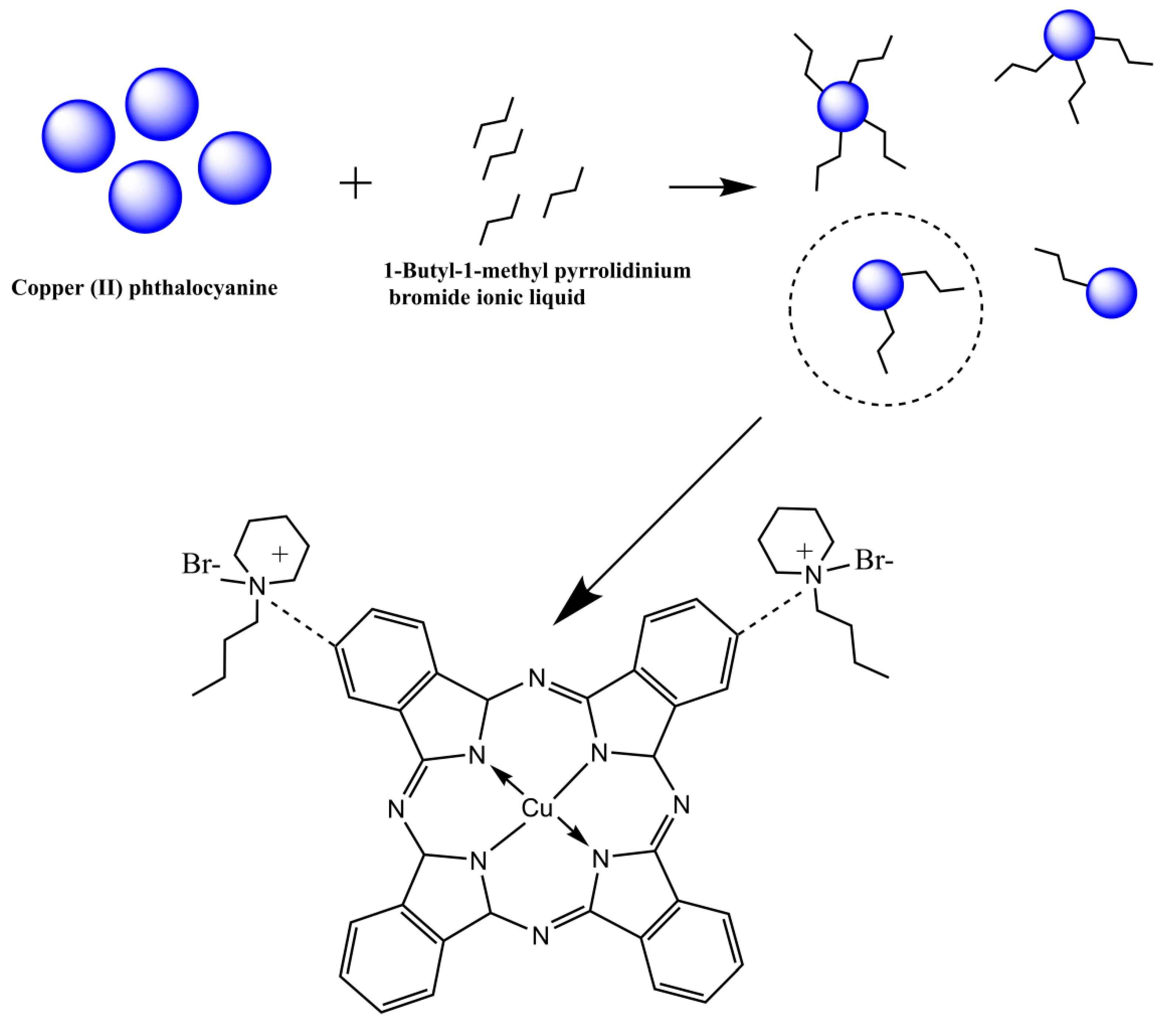
2.2. Preparation of CuPc with Ionic Liquids
2.3. Preparation of Blue Electrophoretic Dispersion
2.4. Preparation of Blue and White Dual-Color Electrophoretic Dispersion
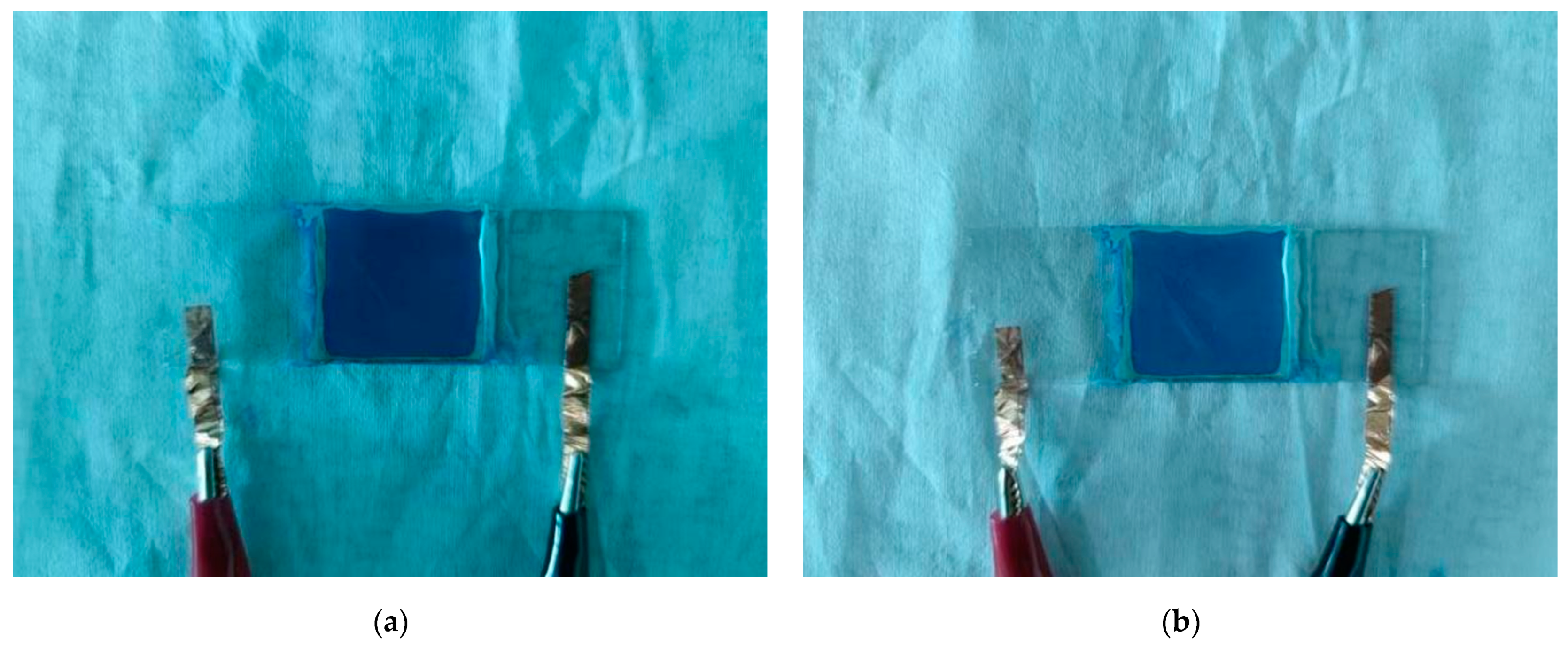
2.5. Preparation of a Blue and White Dual-Color EPD Cell
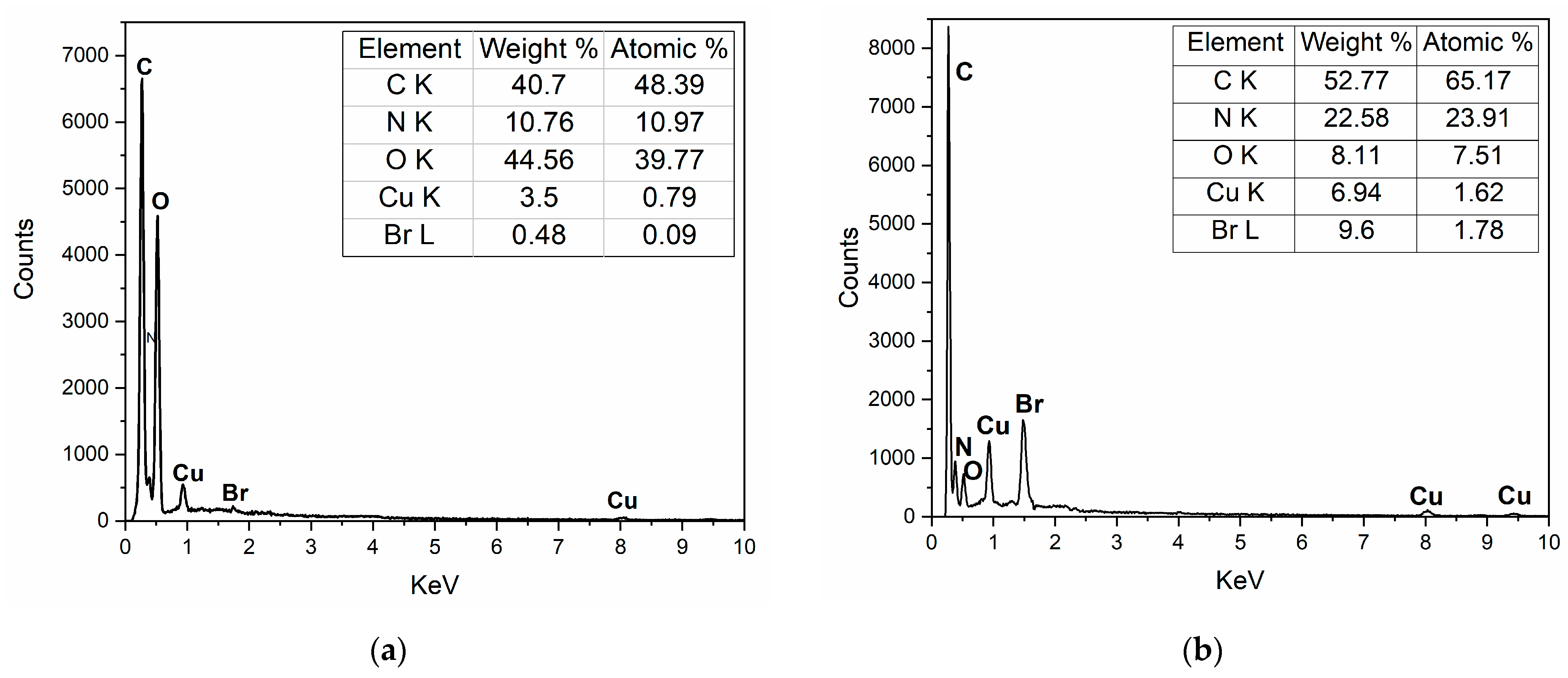
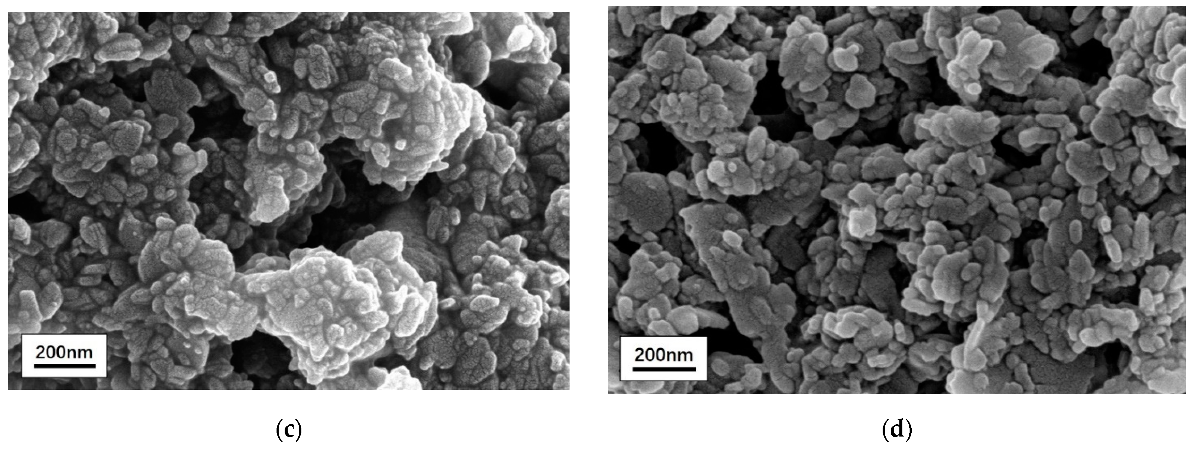
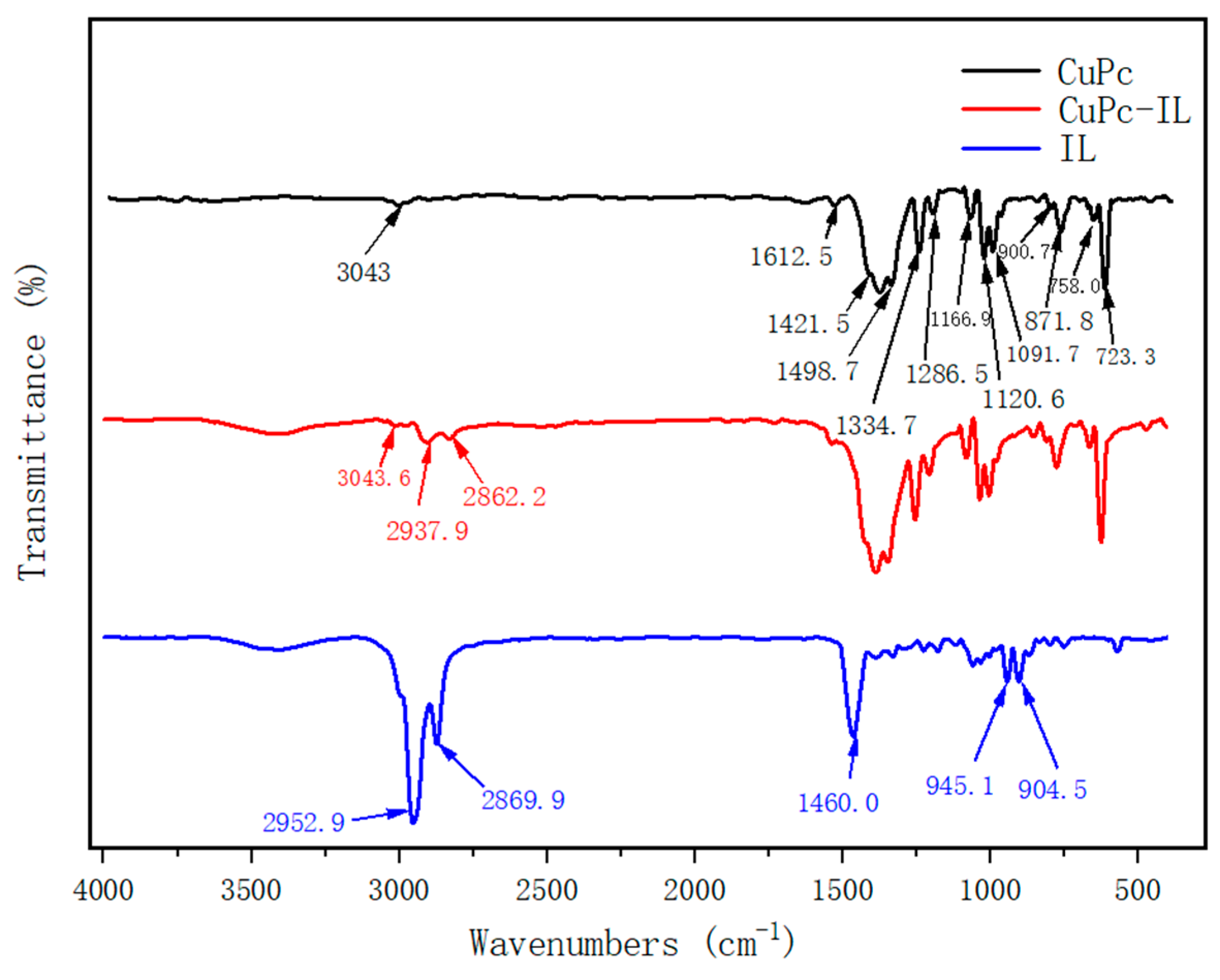
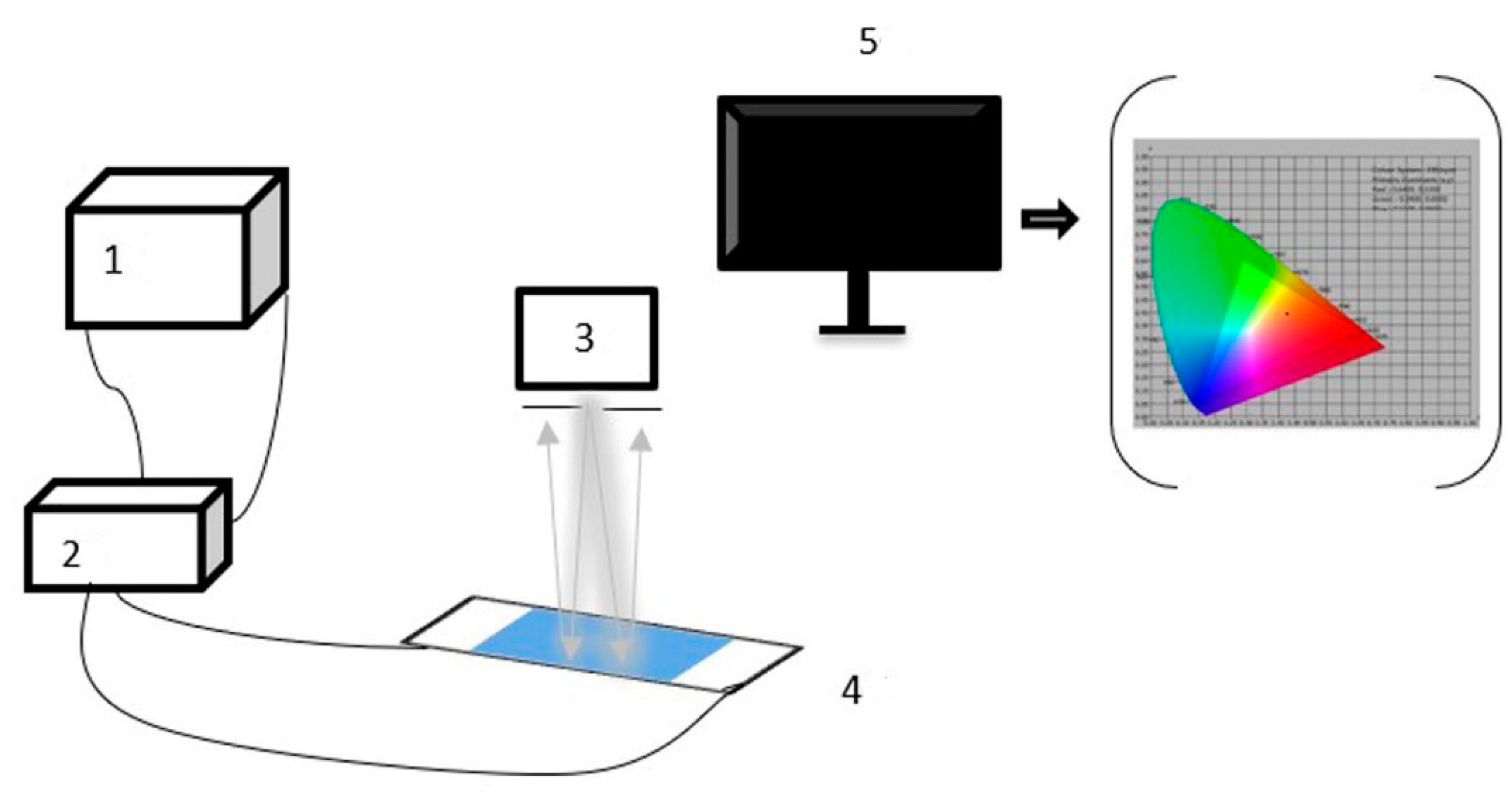
2.6. Instruments and Characterization
3. Results and Discussion
3.1. Material Modification
3.2. Electrophoretic Properties
4. Conclusions
Author Contributions
Funding
Institutional Review Board Statement
Data Availability Statement
Conflicts of Interest
References
- Chen, Y. Flexible active-matrix electronic ink display. Nature 2003, 423, 136. [Google Scholar] [CrossRef] [PubMed]
- Graham-Rowe, D. Electronic paper rewrites the rulebook for displays. Nat. Photonics 2007, 1, 248–251. [Google Scholar] [CrossRef]
- Wen, Z.Q.; Feng, Y.Q.; Li, X.G.; Li, X.X.; Bai, Y.; Tang, Q.M.; Gao, Y.D. Fabrication of diarylide yellow pigments/modified SiO2 core–shell hybrid composite particles for electrophoretic displays. Curr. Appl. Phys. 2012, 12, 259–265. [Google Scholar] [CrossRef]
- Kim, M.K.; Kim, C.A.; Ahn, S.D.; Kang, S.R.; Suh, K.S. Density compatibility of encapsulation of white inorganic TiO2 particles using dispersion polymerization technique for electrophoretic display. Synth. Met. 2004, 146, 197–199. [Google Scholar] [CrossRef]
- Comiskey, B.; Albert, J.D.; Yoshizawa, H.; Jacobson, J. An electrophoretic ink for all-printed reflective electronic displays. (cover story). Nature 1998, 394, 253–255. [Google Scholar] [CrossRef]
- Yi, Z.C.; Zeng, W.B.; Ma, S.M.; Feng, H.Q.; Zeng, W.J.; Shen, S.T.; Shui, L.L.; Zhou, G.F.; Zhang, C.F. Design of Driving Waveform Based on a Damping Oscillation for Optimizing Red Saturation in Three-Color Electrophoretic Displays. Micromachines 2021, 12, 162. [Google Scholar] [CrossRef]
- Zeng, W.J.; Yi, Z.C.; Zhou, X.C.; Zhao, Y.M.; Zhou, G.F. Design of Driving Waveform for Shortening Red Particles Response Time in Three-Color Electrophoretic Displays. Micromachines 2021, 12, 578. [Google Scholar] [CrossRef] [PubMed]
- Kim, C.A.; Joung, M.J.; Ahn, S.D.; Kim, G.H.; Kang, S.Y.; You, I.K.; Oh, J.; Myoung, H.J.; Baek, K.H.; Suh, K.S. Microcapsules as an electronic ink to fabricate color electrophoretic displays. Synth. Met. 2005, 151, 181–185. [Google Scholar] [CrossRef]
- Cho, S.H.; Kwon, Y.R.; Kim, S.K.; Noh, C.H.; Lee, J.Y. Electrophoretic Display of surface modified TiO2 driven by Poly (3,4-ethylenedioxythiophene) Electrode. Polym. Bull. 2007, 59, 331–338. [Google Scholar] [CrossRef]
- Wen, T.; Meng, X.W.; Li, Z.Y.; Ren, J.; Tang, F.Q. Pigment-based tricolor ink particles via mini-emulsion polymerization for chromatic electrophoretic displays. J. Mater. Chem. 2010, 20, 8112–8117. [Google Scholar] [CrossRef]
- Meng, X.W.; Tang, F.Q.; Peng, B.; Ren, J. Monodisperse Hollow Tricolor Pigment Particles for Electronic Paper. Nanoscale Res. Lett. 2009, 5, 174–179. [Google Scholar] [CrossRef] [PubMed] [Green Version]
- Dai, R.Y.; Wu, G.; Chen, H.Z. Stable titanium dioxide grafted with poly [N-(p-vinyl benzyl) phthalimide] composite particles in suspension for electrophoretic displays. Colloid Polym. Sci. 2011, 289, 401–407. [Google Scholar] [CrossRef]
- Serment, B.; Brochon, C.; Hadziioannou, G.; Buffière, S.; Demourgues, A.; Gaudon, M. The versatile co2+/co3+ oxidation states in cobalt alumina spinel: How to design strong blue nanometric pigments for color electrophoretic display. RSC Adv. 2009, 9, 34125–34135. [Google Scholar] [CrossRef] [Green Version]
- Zhang, T.Y.; Zhou, C.L. Properties of copper phthalocyanine blue (C.I. Pigment Blue 15:3) treated with poly(ethylene glycol)s. Dye. Pigment. 1997, 35, 123–130. [Google Scholar]
- Wang, J.P.; Zhao, X.P.; Guo, H.L.; Zheng, Q. Preparation and response behavior of blue electronic ink microcapsules. Opt. Mater. 2008, 30, 1268–1272. [Google Scholar] [CrossRef]
- Wang, C.L.; Wang, L.L.; Huang, Y.; Nan, X.Y.; Fan, Q.G.; Shao, J.Z. Preparation and characterization of Phthalocyanine Blue encapsulated with silane coupling agent for blue light curable inkjet printing of textiles. Dye. Pigment. 2017, 139, 453–459. [Google Scholar] [CrossRef]
- Fu, S.; Zhang, K.; Zhhang, M.J.; Tian, L. Encapsulated phthalocyanine blue pigment with polymerisable dispersant for inkjet printing inks. Pigm. Resin. Technol. 2012, 41, 3–8. [Google Scholar] [CrossRef]
- Li, G.X.; Qin, S.C.; Feng, Y.Q.; Fang, S.; Meng, S.X. Preparation and Characterization of Microcapsule-Encapsulated Colored Electrophoretic Fluid in Trifluorotoluene System for Electrophoretic Display. J. Disp. Technol. 2016, 12, 1145–1151. [Google Scholar] [CrossRef]
- Li, D.; Le, Y.; Hou, X.Y.; Chen, J.F.; Shen, Z.G. Colored nanoparticles dispersions as electronic inks for electrophoretic display. Synth. Met. 2011, 161, 1270–1275. [Google Scholar] [CrossRef]
- Han, J.J.; Zhang, W.H.; Li, X.G.; Sun, C.; Shao, J.Z.; Feng, Y.Q. Encapsulation of modified copper phthalocyanine (CuPc) via miniemulsion polymerisation for electrophoretic display. Mater. Res. Innov. 2014, 19, 24–27. [Google Scholar] [CrossRef]
- Duan, Y.D.; Fu, N.Q.; Fang, Y.Y.; Li, X.N.; Liu, Q.P.; Zhou, X.W.; Lin, Y. Synthesis and formation mechanism of mesoporous TiO2 microspheres for scattering layer in dye-sensitized solar cells. Electrochim. Acta 2013, 113, 109–116. [Google Scholar] [CrossRef]
- Badila, M.; Hebraud, A.; Brochon, C.; Hadziioannou, G. Design of colored multilayered electrophoretic particles for electronic inks. ACS Appl. Mater. Interfaces 2011, 3, 3602–3610. [Google Scholar] [CrossRef]
- Yin, P.P.; Wu, G.; Dai, R.Y.; Qin, W.L.; Wang, M.; Chen, H.Z. Fine encapsulation of dual-particle electronic ink by incorporating block copolymer for electrophoretic display application. J. Colloid Interface Sci. 2012, 388, 67–73. [Google Scholar] [CrossRef] [PubMed]
- Guo, Q.; Lee, J.H.; Singh, V.; Behrens, S.H. Surfactant mediated charging of polymer particles in a nonpolar liquid. J. Colloid Interface Sci. 2013, 392, 83–89. [Google Scholar] [CrossRef] [PubMed]
- Sun, C.; Feng, Y.Q.; Zhang, B.; Li, X.G.; Shao, J.Z.; Han, J.J.; Chen, X. Preparation and application of microcapsule-encapsulated color electrophortic fluid in Isopar M system for electrophoretic display. Opt. Mater. 2013, 35, 1410–1417. [Google Scholar] [CrossRef]
- Meischein, M.; Fork, M.; Ludwig, A. On the Effects of Diluted and Mixed Ionic Liquids as Liquid Substrates for the Sputter Synthesis of Nanoparticles. Nanomaterials 2020, 10, 525. [Google Scholar] [CrossRef] [Green Version]
- Wolny, A.; Chrobok, A. Ionic Liquids for Development of Heterogeneous Catalysts Based on Nanomaterials for Biocatalysis. Nanomaterials 2021, 11, 2030. [Google Scholar] [CrossRef]
- Julio, A.; Caparica, R.; Costa Lima, S.A.; Fernandes, A.S.; Rosado, C.; Prazeres, D.M.F.; Reis, S.; Santos de Almeida, T.; Fonte, P. Ionic Liquid-Polymer Nanoparticle Hybrid Systems as New Tools to Deliver Poorly Soluble Drugs. Nanomaterials 2019, 9, 1148. [Google Scholar] [CrossRef] [Green Version]
- Correia, D.M.; Fernandes, L.C.; Fernandes, M.M.; Hermenegildo, B.; Meira, R.M.; Ribeiro, C.; Ribeiro, S.; Reguera, J.; Lanceros-Mendez, S. Ionic Liquid-Based Materials for Biomedical Applications. Nanomaterials 2021, 11, 2401. [Google Scholar] [CrossRef]
- Atta, A.M.; Ezzat, A.O.; Moustafa, Y.M.; Sabeela, N.I.; Tawfeek, A.M.; Al-Lohedan, H.A.; Hashem, A.I. Synthesis of New Magnetic Crosslinked Poly (Ionic Liquid) Nanocomposites for Fast Congo Red Removal from Industrial Wastewater. Nanomaterials 2019, 9, 1286. [Google Scholar] [CrossRef] [Green Version]
- Shi, T.; Livi, S.; Duchet, J.; Gerard, J.F. Ionic Liquids-Containing Silica Microcapsules: A Potential Tunable Platform for Shaping-Up Epoxy-Based Composite Materials? Nanomaterials 2020, 10, 881. [Google Scholar] [CrossRef]
- Zornio, C.F.; Livi, S.; Duchet-Rumeau, J.; Gerard, J.F. Ionic Liquid-Nanostructured Poly(Methyl Methacrylate). Nanomaterials 2019, 9, 1376. [Google Scholar] [CrossRef] [PubMed] [Green Version]
- Migliorini, L.; Santaniello, T.; Borghi, F.; Saettone, P.; Comes Franchini, M.; Generali, G.; Milani, P. Eco-Friendly Supercapacitors Based on Biodegradable Poly(3-Hydroxy-Butyrate) and Ionic Liquids. Nanomaterials 2020, 10, 2062. [Google Scholar] [CrossRef] [PubMed]
- Kholghi Eshkalak, S.; Khatibzadeh, M.; Kowsari, E.; Chinnappan, A.; Ramakrishna, S. Application of ionic liquids as charge control agents of pigments and preparation of microcapsules as electronic inks through electrospraying. Opt. Mater. 2018, 84, 73–81. [Google Scholar] [CrossRef]
- Rajmohan, K.S.; Raghuram, C. Enhanced nitrate reduction with copper phthalocyanine-coated carbon nanotubes in a solid polymer electrolyte reactor. J. Appl. Electrochem. 2016, 47, 63–74. [Google Scholar] [CrossRef]
- Liu, M.D.; Jia, K.; Liu, X.B. Preparation of hybrid colloidal graphite-copper phthalocyanine and their utilization in polymer composites with enhanced thermal conductivity and mechanical properties. J. Polym. Res. 2014, 21, 570. [Google Scholar] [CrossRef]
- Wang, Y.; Zhang, Z.; Chen, Q.; Ye, C.H.; Zhang, J.H.; Gao, Q.G.; Liu, L.M.; Yang, J.J.; Pan, X.J.; Miao, Y.; et al. A novel modification of copper (II) phthalocyanine particles towards electrophoretic displays. Micromachines 2022, 13, 880. [Google Scholar] [CrossRef] [PubMed]
- Filip, S.; Filip, B.; Matthias, M.; Kristiaan, N. Charging mechanism in colloidal particles leading to a linear relation between charge and size. Phys. Rev. E 2007, 75, 31405. [Google Scholar]
- Pugh, R.J.; Fowkes, F.M. The dispersibility and stability of carbon black in media of low dielectric constant. 2. Sedimentation volume of concentrated dispersions, adsorption and surface calorimetry studies. Colloids Surf. 1984, 9, 33–46. [Google Scholar] [CrossRef]


| Material | CuPc | CuPc-IL |
|---|---|---|
| Zeta Potential (mV) | +30.50 | +60.27 |
| Ionic Liquids | Chemical Constitution | Side Chain Length | Zeta Potential(mV) |
|---|---|---|---|
| 1-butyl-3-methylimidazolium bromide | 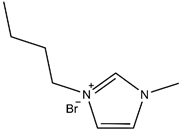 | 4C | 41.60 |
| 1-butyl-1-methylpyrrolidinium bromide | 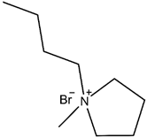 | 4C | 49.91 |
| 1-butyl-1-methylpiperidinium bromide | 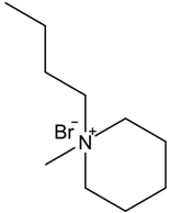 | 4C | 60.27 |
Publisher’s Note: MDPI stays neutral with regard to jurisdictional claims in published maps and institutional affiliations. |
© 2022 by the authors. Licensee MDPI, Basel, Switzerland. This article is an open access article distributed under the terms and conditions of the Creative Commons Attribution (CC BY) license (https://creativecommons.org/licenses/by/4.0/).
Share and Cite
Zhang, Z.; Wang, Y.; Chen, Q.; Gao, Q.; Liu, L.; Yang, J.; Pan, X.; Miao, Y.; Chi, F. Application of High Potential Electrophoretic Particles Modified with High Ionization Mono Ionic Liquid for Electrophoretic Displays. Micromachines 2022, 13, 1235. https://doi.org/10.3390/mi13081235
Zhang Z, Wang Y, Chen Q, Gao Q, Liu L, Yang J, Pan X, Miao Y, Chi F. Application of High Potential Electrophoretic Particles Modified with High Ionization Mono Ionic Liquid for Electrophoretic Displays. Micromachines. 2022; 13(8):1235. https://doi.org/10.3390/mi13081235
Chicago/Turabian StyleZhang, Zhi, Yao Wang, Qun Chen, Qingguo Gao, Liming Liu, Jianjun Yang, Xinjian Pan, Yu Miao, and Feng Chi. 2022. "Application of High Potential Electrophoretic Particles Modified with High Ionization Mono Ionic Liquid for Electrophoretic Displays" Micromachines 13, no. 8: 1235. https://doi.org/10.3390/mi13081235
APA StyleZhang, Z., Wang, Y., Chen, Q., Gao, Q., Liu, L., Yang, J., Pan, X., Miao, Y., & Chi, F. (2022). Application of High Potential Electrophoretic Particles Modified with High Ionization Mono Ionic Liquid for Electrophoretic Displays. Micromachines, 13(8), 1235. https://doi.org/10.3390/mi13081235