Article

Prevalence, Enterotoxigenic Potential and Antimicrobial Resistance of Staphylococcus aureus and Methicillin-Resistant Staphylococcus aureus (MRSA) Isolated from Algerian Ready to Eat Foods

by
Omar Amine Mekhloufi
1,
Daniele Chieffi
2,*,
Abdelhamid Hammoudi
1,
Sid Ahmed Bensefia
3,
Francesca Fanelli
2 and
Vincenzina Fusco
2
1
Laboratory of Hygiene and Animal Pathology, Institute of Veterinary Sciences, University of Tiaret, Tiaret 14000, Algeria
2
National Research Council of Italy, Institute of Sciences of Food Production (CNR-ISPA), 70126 Bari, Italy
3
Food, Water and Environmental Bacteriology Laboratory, Pasteur Institute of Algeria, Algiers 16000, Algeria
*
Author to whom correspondence should be addressed.
Toxins 2021, 13(12), 835; https://doi.org/10.3390/toxins13120835
Submission received: 29 September 2021 / Revised: 28 October 2021 / Accepted: 17 November 2021 / Published: 24 November 2021
(This article belongs to the Section Bacterial Toxins)

Abstract

:
Staphylococcus aureus causes a foodborne intoxication due to the production of enterotoxins and shows antimicrobial resistance, as in the case of methicillin-resistant strains (MRSA). Herein, we analyzed 207 ready-to-eat foods collected in Algeria, reporting a S. aureus prevalence of 23.2% (48/207) and respective loads of coagulase positive staphylococci (CPS) ranging from 1.00 ± 0.5 to 5.11 ± 0.24 Log CFU/g. The 48 S. aureus isolates were widely characterized by staphylococcal enterotoxin gene (SEg)-typing and 16S-23S rDNA intergenic spacer region (ISR)-PCR, as well as by detecting tst and mecA genes, genetic determinants of toxic shock syndrome toxin-1 and methicillin resistance, respectively. We found that the S. aureus isolates belonged to seven different SEg-types harboring the following combinations of genes: (1) selW, selX; (2) egc (seG, seI, seM, seN, seO), selW, selX; (3) seA, seH, seK, seQ, selW, selX; (4) seB, selW, selX; (5) seD, selJ, seR, selW, selX; (6) seH, selW, selX, selY; and (7) seA, egc, selW, selX, while among these, 2.1% and 4.2% were tst- and mecA- (staphylococcal chromosomal cassette mec-type IV) positive, respectively. Selected strains belonging to the 12 detected ISR-types were resistant towards antimicrobials including benzylpenicillin, ofloxacin, erythromycin, lincomycin, tetracyclin, kanamycin, oxacillin, and cefoxitin; 8.3% (1/12) were confirmed as MRSA and 16.7% (2/12) were multidrug resistant. The present study shows the heterogeneity of the S. aureus population in Algerian ready-to-eat foods as for their toxigenic potential and antimicrobial resistance, shedding the light on the quality and safety related to the consume of ready-to-eat foods in Algeria.
Key Contribution: The consumption of Algerian ready-to-eat foods may represent a risk for the consumer’s health due to the presence of enterotoxigenic and antimicrobial-resistant S. aureus strains. To our knowledge, we performed the widest genetic characterization conducted so far in Algeria to investigate the enterotoxigenic potential of foodborne S. aureus isolates.

1. Introduction

Food-associated health problems, caused by food contamination or malnutrition, have a worldwide impact on public health and economy [1]. Particularly, foodborne diseases (FBDs) represent a major issue for public health, and there is a growing number of outbreaks and sporadic cases of disease associated with various type of foods [2,3,4]. Furthermore, FBDs have a significant economic impact on both the industry and the public health system in terms of loss of productivity, cost of treatment, and food safety governance [5]. On top of that, FBDs are responsible for nearly 600 million illnesses and 420,000 deaths each year [6].
Staphylococcus aureus is one of the major pathogens of humans. It causes various diseases including staphylococcal food poisoning (SFP), toxic shock syndrome and other systemic diseases, bacteremia, pneumonia, and skin and soft tissue infections, to cite a few [7,8]. Its pathogenicity is promoted via several virulence factors, such as staphylococcal enterotoxins (SEs), toxic shock syndrome toxin-1, haemolysins, and fibronectin-binding proteins [9]. This microorganism is not only considered as the most pathogenic of staphylococci but, as reported by Havelaar et al. [10] and Fusco et al. [11], it is also acknowledged among the top 10 causes of bacterial FBDs, therefore playing a major role in terms of food safety [12].
Staphylococcal enterotoxins (SEs), produced by enterotoxigenic S. aureus strains, are a super family of pyrogenic exotoxins that share structural and functional similarities, causing vomiting, diarrhea, and cramps upon ingestion. To date, at least 28 SEs and staphylococcal enterotoxin-like toxins (SEls) have been identified [13]. SE and SEls are globular, single polypeptides with molecular weights ranging from 22 to 29 kDa. They can be encoded in prophages, plasmids, or chromosomal pathogenicity islands. The location of the se and sel genes on mobile genetic elements presents an additional risk factor in S. aureus food intoxication, due to possible horizontal gene transfer [14].
Foods that have been frequently incriminated in SFP include meat and meat products, poultry and egg products, milk and dairy products, salads, bakery products, particularly cream-filled pastries and cakes, and sandwich fillings [15]. However, according to le Loir et al. [16], foods implicated with SFP vary from country to country, particularly due to variation in consumption and food habits. In particular, food contamination with S. aureus and its SEs is mainly due to its ability to enter the food chain through contaminated raw material, inappropriate handling of processed food, and failure to maintain the cold chain [17].
Moreover, S. aureus is a well-known bacterium that develops antibiotic resistance [18] due to its ability to acquire a variety of resistance mechanisms towards antimicrobial agents [19], such as the resistance to methicillin. The latter mainly depends on the acquisition of the staphylococcal chromosomal cassette mec (SCCmec) harboring the mecA gene that encodes for penicillin-binding protein 2a (PBP2a) [20] that has low affinity for β-lactam antibiotics. In recent years, methicillin-resistant S. aureus (MRSA) strains have been recovered from several animal-derived foods such as poultry, pork, and beef meats, suggesting that foods may serve as a reservoir and source of MRSA [21]. Apart from direct transmission between humans and animals, the latter being considered natural reservoirs of this organism, transmission of MRSA might occur via exposure to, or ingestion of, contaminated foods [22,23]. Therefore, S. aureus and MRSA are considered a significant public health concern given their ability to contaminate foods and to colonize and infect both humans and animals [23,24].
Various ready-to-eat products are becoming increasingly popular in developing countries, particularly in the metropolitan areas. The prevalence and characteristics of enterotoxigenic S. aureus in ready-to-eat food products has been studied in different parts of the world [25,26,27]; however, as highlighted by Lozano et al. [28], studies on foodborne S. aureus in Africa are limited and, to date, mainly restricted to few countries, such as South Africa, which in general is reported to contribute to the most part of the African investigations, followed by Egypt and Nigeria [29]. Moreover, the available studies are focused primarily on the assessment of the prevalence of S. aureus and corresponding loads in various foods [28], while a further characterization is not systematically addressed. Thus, the information on enterotoxigenic S. aureus and MRSA from food in the African continent is, to date, fragmented and still incomplete. In order to overcome these limits, the present study aims to investigate the prevalence, the enterotoxigenic potential, and the antimicrobial resistance of S. aureus in ready-to-eat foods sampled in Algeria, Africa.

2. Results

2.1. Prevalance of Staphylococcus aureus in Algerian Ready to Eat Foods

In this study, S. aureus was found in 23.2% (48/207) of ready-to-eat foods sampled in Algeria. High prevalence was found in meat/fish-based foods (38.2%, 21/55), followed by vegetable-based foods (22.2%, 16/72), cereals (17.6%, 3/17), pastries (16.3%, 7/43), and various foods (milk- and egg-based foods) (5.0%, 1/20) (Table 1).
The coagulase positive staphylococci (CPS) counts in the S. aureus positive samples showed a median of 3.38 ± 0.94 Log CFU (colony forming unit)/g, ranging between 1.00 ± 0.5 and 5.11 ± 0.24 Log CFU/g as minimum and maximum counts, respectively. Pastries showed the highest CPS counts (between 2.84 ± 0.11 and 5.11 ± 0.24 Log CFU/g; median: 3.57 ± 0.80 Log CFU/g), while various foods (milk- and egg-based foods) showed the lowest (1.70 ± 0.10 Log CFU/g) (Table 1).

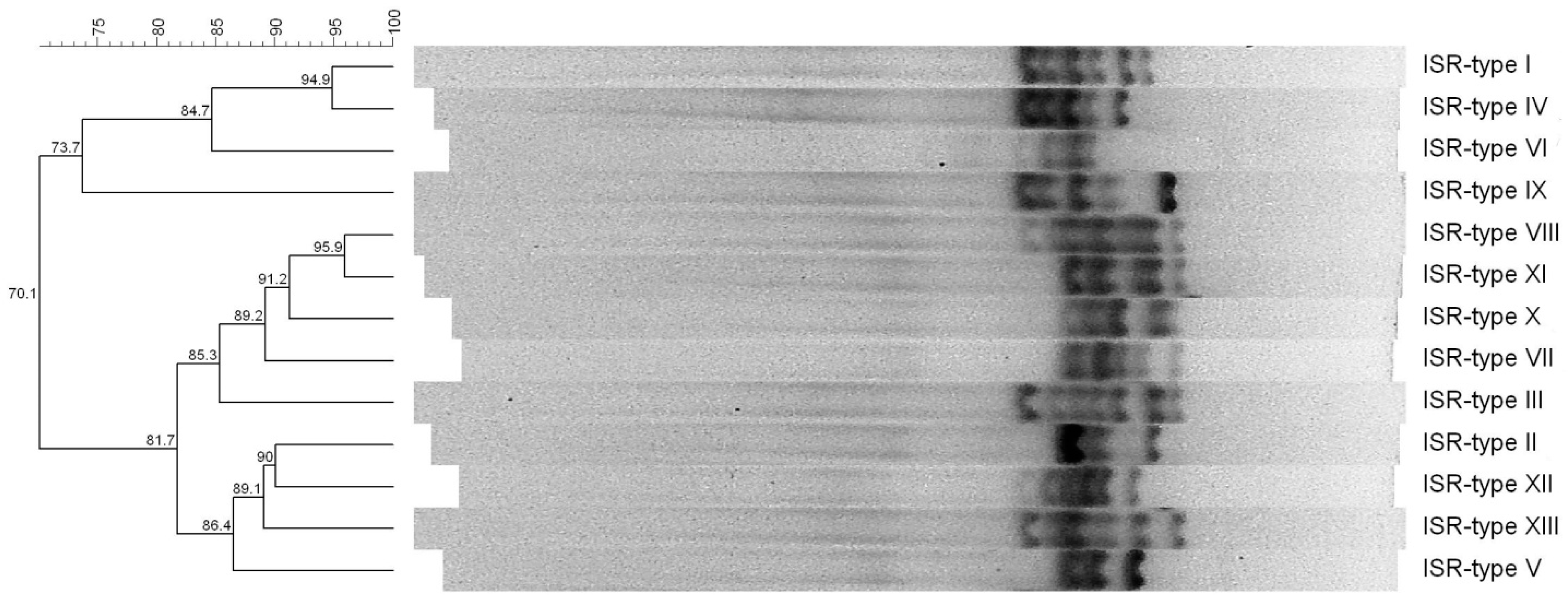
2.2. 16S-23S rDNA Intergenic Spacer Region (ISR)-PCR, Staphylococcal Enterotoxin Gene (SEg)-Typing and Presence of tst and mecA Genes

The 48 S. aureus isolated from the ready-to-eat foods sampled in Algeria are listed in Table 2.
The 16S-23S rDNA intergenic spacer region (ISR)-PCR allowed us to identify 12 different ISR-types, as shown in Figure 1.
Thirty-nine point six percent of the S. aureus (19/48) belonged to ISR-type I; 12.5% (6/48) to ISR-type III; 10.4% (5/48) to ISR-type IV and VII, respectively; 8.3% (4/48) to ISR-type IX; and 4.2% (2/48) to ISR-type II and X, respectively, while 2.1%, of the S. aureus isolates belonged to ISR-types V, VI, VIII, XI, and XII, which were represented by one isolate each (1/48, respectively) (Table 2).
The staphylococcal enterotoxin gene (SEg)-typing allowed us to detect seven different SEg-types (Table 2). The most prevalent was SEg-type 1 (selW, selX) detected in 52.1% (25/48) of the S. aureus that belonged to three different ISR-types (I, IV, and VI). It was followed by SEg-type 2 [egc (seG, seI, seM, seN, seO), selW, selX] (16.7%) (8/48) observed in S. aureus belonging to ISR-types II, V, IX, and XII, SEg-type 3 (seA, seH, seK, seQ, selW, selX) (12.5%) (6/48) in S. aureus belonging to ISR-types III, SEg-type 4 (seB, selW, selX) (10.4%) (5/48) in S. aureus belonging to ISR-type VII, and SEg-type 6 (seH, selW, selX, selY) (4.2%) (2/48) in S. aureus belonging to ISR-type X. The least prevalent were SEg-types 5 (seD, selJ, seR, selW, selX) and 7 [seA, egc (seG, seI, seM, seN, seO), selW, selX] (2.1%) (1/48), which were detected in S. aureus belonging to ISR-type VIII and XI, respectively (Table 2).
One out of the 48 S. aureus isolates (2.1%) that belonged to ISR-type XII was positive for the tst gene, and two S. aureus (4.2%) belonging to ISR-type X were positive for the mecA gene (Table 2).
It is noteworthy that S. aureus isolates belonging to the same ISR-type harbored the same genes as detected by PCRs (Table 2).
In regard to the prevalence of the se and sel genes, selW and selX were detected in all the isolates (48/48), followed by the enterotoxin gene cluster (egc) and its related genes (seG, seI, seM, seN, seO) (18.8%, 9/48), seH (16.7%, 8/48), seA (14.6%, 7/48), seK and seQ (12.5%, 6/48 respectively), seB (10.4%, 5/48), selY (4.2%, 2/48), and seD, selJ, and seR (2.1%, 1/48, respectively).
Lastly, the distribution of the S. aureus in relation to their genotypes and the corresponding sources of isolation is displayed in Table 3.

2.3. Characterization of the Staphylococcal Chromosomal Cassette mec (SCCmec)

The multiplex PCR used for the characterization of the staphylococcal chromosomal cassette mec (SCCmec) [30] showed that the two mecA positive isolates (Table 2) harbored the SCCmec-type IV as identified by the presence of a band of 342 bp (while the band of 162 bp indicates the presence of the mecA gene) (Figure 2).

2.4. Antimicrobial Resistance

The antimicrobial susceptibility test showed that 8 out of the 12 (66.7%) selected S. aureus strains (one for each ISR-type) isolated from ready-to-eat foods from Algeria were resistant to one antibiotic, i.e., benzylpenicillin, while three strains (25.0%) were resistant towards three to four antimicrobials (Table 4). Four resistance profiles were observed (Table 4). The highest prevalence of resistance was recorded for benzylpenicillin (10/12) (83.3%), while lower resistance was recorded towards tetracycline and kanamycin (2/12 strains, respectively) (16.7%, respectively) and towards ofloxacin, erythromycin, lincomycin, oxacillin, and cefoxitin (1/12 strains, respectively) (8.3%, respectively) (Table 4). One strain (1/12) (8.3%) (SA46) was considered MRSA, while two strains (2/12) (16.7%) (SA02, SA18) were multidrug-resistant.

3. Discussion

In the present study, we found S. aureus in Algerian ready-to-eat foods with a prevalence of 23.2%. Other studies analyzed the presence of this microorganism in ready-to-eat foods in African countries, but only fragmented and incomplete information about the enterotoxigenic S. aureus and MRSA isolated from African ready-to-eat foods is available so far. Similar to our findings, Chaalal et al. [31] reported an overall S. aureus prevalence of 23.8% in pastries and cooked dishes sampled from supermarkets and university cities in Western Algeria, and Titouche et al. [32], in the same country of the aforementioned study, found a prevalence of 14.46% in pastries collected from several market points (in Tizi Ouzou area), while no S. aureus contamination was found by these authors in the sampled sandwiches [32]. A prevalence of 33.26% was reported in ready-to-eat meat products collected in all the provinces of South Africa [33], while in Egypt (in Benha city), a prevalence of 50.8% was reported for ready-to-eat meat products sampled from restaurants and street vendors [34], and in Nigeria (in Port Harcourt Metropolis), 100% of the analyzed street ready-to-eat meals were reported to be contaminated by S. aureus [35].
As shown by these studies, the prevalence of S. aureus contamination in ready-to-eat foods may greatly vary and, as highlighted by some authors [32,36], such differences are related to several factors that include the source (e.g., street-vendors or shops) and type of samples (e.g., animal- or non-animal-derived foods), the sample size, the accuracy of the identification method (based on cultivation characteristics, biochemical tests, or molecular biology techniques), the manufacturing procedures (e.g., involving bactericidal temperatures or not), and the overall hygienic measures that are implemented during the preparation and handling of the foods. In particular, S. aureus, being a commensal bacterium present on the skin, the nose, and mucous membranes of animals and humans [28], may contaminate food especially when poor hygienic practices and conditions exist. It is noteworthy that, beyond the contamination that may originate from animals at the primary production stage, food handlers are recognized as the main source of food contamination with S. aureus [31,32,37], and Sezer et al. [38] found that the 79% of the food handlers employed in a catering establishment were carriers of S. aureus. Nevertheless, it should be taken into account that contamination may also happen and spread by cross-contamination among foods and/or surfaces in the environment surrounding the manufacturing and storage of the ready-to-eat foods, since S. aureus may survive on inanimate surfaces for prolonged times [39], especially in cases of improper or ineffective sanitizing procedures [40].
When contamination occurs, S. aureus may replicate, and its load may increase in food. S. aureus is mostly a coagulase positive bacterium, and according to the Interministerial Decree of the Algerian Republic Official Journal No 39 (published on 2 July 2017) establishing the microbiological criteria for food products, the limits of CPS in ready-to-eat foods are set to values equal to 102 CFU/g (“m” value), below which the product quality is considered satisfactory, and 103 CFU/g (“M” value), above which the product quality is considered unacceptable. In the ready-to-eat products sampled in this study, we found CPS loads, above the “M” value, in four of the five analyzed food categories (meat/fish-based foods, vegetable-based foods, pastries, and cereals), in which the highest CPS counts reached values higher than 4 and 5 Log CFU/g. Additionally, according to the Decree, samples in which CPS counts are superior or equal to 105 CFU/g are considered as toxic. Therefore, these findings shed light on the quality and safety of the Algerian retail ready-to-eat foods.
Some other studies assessed CPS loads, even reported as S. aureus counts, in ready-to-eat foods in the African continent. Although Mahami et al. [41] found low contamination of cooked and smoked sausages, reporting S. aureus loads ranging from 1.85 to 2.15 Log CFU/g in samples purchased from a factory in Ghana (in Accra city) and less than 1 Log CFU/g in samples from a shopping mall, Oguttu et al. [42], analyzing ready-to-eat chicken sold in informal markets in South Africa (in Tshwane Metropolitan City), reported the presence of unsatisfactory quality samples with loads of S. aureus greater than 3 Log CFU/g, and an overall mean of 3.6 Log CFU/g. Similarly, Shiningeni et al. [43] found ready-to-eat beef and chicken meats of unsatisfactory quality, with loads of S. aureus equal or greater than 3 Log CFU/g, that were purchased from street vendors in Namibia (Windhoek city). These authors reported that, in relation to the vending sites, the mean counts in the ready-to-eat meat samples ranged from 0 to 3.46 Log CFU/g, and the highest load was 5.12 Log CFU/g [43].
Since the issue regarding S. aureus and corresponding loads in foods is related to the possible contamination of food by staphylococcal enterotoxins, that in very round figures may occur at hazardous levels if S. aureus load reaches around 5 Log CFU/g [40], a great importance was given in this study to the investigation of the enterotoxigenic potential of the S. aureus we isolated, and to the best of our knowledge this is the first study in Algeria in which the presence of a total of 27 se and sel genes was sought in foodborne S. aureus.
We found that the 48 S. aureus belonged to seven different SEg-types harboring the following combination of se and sel genes: (1) selW, selX; (2) egc (seG, seI, seM, seN, seO), selW, selX; (3) seA, seH, seK, seQ, selW, selX; (4) seB, selW, selX; (5) seD, selJ, seR, selW, selX; (6) seH, selW, selX, selY; and (7) seA, egc (seG, seI, seM, seN, seO), selW, selX. Previous studies detecting a high number of se and sel genes (17 to 27 genes) found 6 different genotypes in 53 S. aureus from raw milk [13], 11 different genotypes in 50 S. aureus from raw minced meat and sausages [44], and 120 enterotoxin gene patterns in a group comprising 568 S. aureus from humans, animals, foods, and the environment [45]. Our study corroborates these findings and indicates the heterogeneity of the enterotoxigenic potential that can be encountered in S. aureus isolates from ready-to-eat foods in Algeria.
In our study, we found that 100% (48/48) of the analyzed S. aureus harbored selW and selX. These are located in the chromosome and are reported as highly prevalent genes; indeed, they were detected in more than 92.0% and in more than 79.0% of the S. aureus analyzed in previous studies, respectively [46,47]. The recent identification of this and other new se and sel genes indicates that the pathogenic potential of S. aureus may be greater than previously thought [48]. Moreover, based on the findings of Aung et al. [46], it was suggested that selW, and to a greater extent selX, may play a universal role in the virulence of S. aureus [46].
The enterotoxin gene cluster (egc) was found in the 18.8% (9/48) of the S. aureus herein analyzed. This cluster, located in a variable genomic island (vSaβ) inserted in the chromosome [37], has been similarly reported with a prevalence of 16.2% [49] and of 26.4% [13] in S. aureus from various sources, including food. In our study, the presence of the egc was confirmed by the detection, in the same strains, of the egc encoded genes seG, seI, seM, seN, and seO. Various types of egc exist in S. aureus (egc1 to egc5) and, considering our findings, the strains herein analyzed may harbor egc1 [50] or the newly described egc5 [13]. It should be specified that these two egc types, beyond harboring the enterotoxin genes we detected (seG, seI, seM, seN, seO), might also include two pseudogenes (ψent1-ψent2) (egc1) or selU2 (egc5) [13,50].
Since selU2 results from a single adenine deletion in the ψent1-ψent2 region (being just a 1 nucleotide frameshift of the ψent1-ψent2 sequence that deletes the stop codon of pseudogene ψent1) [51], the design of primers able to specifically detect ψent1-ψent2 or selU2 is not achievable, making impracticable their easy and affordable detection by a PCR screening such as the one we performed.
The seH gene was detected in the S. aureus herein analyzed with a prevalence of 16.7% (8/48). This gene, located on a presumptive transposon [52], was reported with lower prevalence in some previous studies, being in the range of 4.05–4.8% in S. aureus from food and other sources [32,45], while a higher prevalence, more similar to our findings, and ranging between 24.6 and 28.0%, was reported by other authors analyzing S. aureus from raw milk, raw meat, and ready-to-eat foods [13,44,53].
The egc-encoded enterotoxins are reported with increasingly probability to have a role in staphylococcal foodborne poisoning (SFP) [54,55], and the seH encoded enterotoxin (SEH) is the first non-classical SE that has been reported to have caused SFP outbreaks [56,57], highlighting therefore the relevance of the detection of these genes in foodborne S. aureus.
When the classical se genes are considered, in our study we found seA, seB, and seD with a prevalence of 14.6% (7/48), 10.4% (5/48), and 2.1% (1/48), respectively. The presence of these genes, whose encoded enterotoxins are the most frequently reported causes of SFP outbreaks [58], is frequently investigated, and in African countries prevalences ranging between 6.7–90.0%, 0.0–18.3%, and 0.0–8.0% for each of them have been reported in S. aureus isolated from food products [32,59,60].
In our investigation, we also detected selJ, seK, seQ, seR, and selY. In general, scarce knowledge is available on the prevalence of these new se and sel genes in foodborne S. aureus. This study helps to improve this paucity of data, representing one of the few African reports available to date. Interestingly, we observed that some of the above-mentioned genes were found in association in the S. aureus herein analyzed, in particular, selJ and seR along with seD, and seK and seQ along with seA. The first association is carried on plasmids (pIB485-like), and the second is carried on prophages [37], and both of these associations are being found in S. aureus isolates from SFP outbreaks [61,62,63,64]. Considering that the emetic properties of the encoded SEs (SEK, SEQ, and SER) have been demonstrated [65,66], these new se genes may also have a role in the genesis of staphylococcal intoxication that to date is still not fully understood.
tst and mecA genes were found at low prevalence in the S. aureus analyzed in our study (2.1% (1/48) and 4.2% (2/48), respectively), and they were not in association in the same strains. These genes, being the genetic determinants of toxic shock syndrome toxin-1 (tst), which causes a range of systemic diseases, and methicillin resistance (mecA), which confers a general resistance to β-lactam antibiotics in MRSA, represent important virulence and antimicrobial resistance attributes that can worsen clinical conditions when S. aureus infections occur.
In general, a low prevalence of tst gene in foodborne S. aureus has also been reported in previous studies conducted in various parts of the world including African countries [31,67,68,69]. Although the association of tst and mecA genes has been found in S. aureus isolated especially from clinical samples [70,71], our findings resemble those of Chaalal et al. [31] that recently analyzed S. aureus from food products in Algeria detecting a low prevalence of tst gene (3.2%) that was found only in mecA-negative isolates.
Nevertheless, the presence of mecA has been reported in foodborne S. aureus isolates in Algeria, with a prevalence of 3.03 [72], 4.81 [32], and 16.9% [31], and, additionally, in the aforementioned studies, an overall low prevalence of MRSA in the analyzed foods has been reported, which is consistent with our findings [31,32,72]. The massive and inappropriate use of antimicrobials in veterinary and human medicine is considered the main cause for the emergence of antimicrobial-resistant strains [73]; however, the prevalence of MRSA in food is described to still be low and reported in the range of 1.6–6.4% in investigations also conducted in other countries [74].
Transmission of MRSA occurs between animals and humans but, notably, food contamination may serve as a vehicle to increase the dissemination of MRSA [73]. The community-acquired MRSA (CA-MRSA) but also the livestock-acquired MRSA (LA-MRSA) frequently carry the SCCmec-type IV [75,76], and they principally spread in human community settings and livestock populations. The detection of the SCCmec-type IV in our S. aureus isolates tentatively suggests their origin and, consistently with our findings, it was also the prevalent SCCmec-type found in the MRSA isolated from food in previous studies [31,77,78,79].
Interestingly, the ability of MRSA strains to act as a foodborne pathogens has been reported [80], and the first foodborne gastrointestinal illness outbreak caused by an enterotoxigenic MRSA strain has been described in 2002 [81]. However, to date, the actual MRSA involvement in SFP (cases or outbreaks) is not precisely elucidated, and a general underestimation is suggested [80]. Our mecA-SCCmec-type IV positive isolates harbored seH, selW, selX, and selY genes (SEg-type 6) suggesting their potential role as SFP agents, especially considering that the seH encoded enterotoxin (SEH) has been already reported to have caused SFP outbreaks [56,57], as mentioned above. Additionally, the association of seH and mecA gene is known and has been previously described [52]. Moreover, our results also corroborate those of previous studies that detected se and sel genes in MRSA isolated from food products [32,77,79,82].
To further characterize our S. aureus isolates, we employed ISR-PCR, previously used also by other authors as a practical tool for S. aureus genotyping [13,83]. Interestingly, unlike results previously reported by Chieffi et al. [13], we observed that ISR-PCR had a discriminatory power higher than SEg-typing since ISR-PCR enabled us to detect 12 ISR-types despite the seven SEg-types. On the other hand, we observed that S. aureus belonging to the same ISR-type harbored the same genes as detected by PCRs, corroborating the aforementioned study that reported that strains belonging to the same ISR-type showed the same SEg-type.
Therefore, ISR-PCR allowed us to select 12 representative genetically diverse S. aureus strains that, when tested for antimicrobial resistance, showed four patterns of resistance.
In particular, the antimicrobial susceptibility test allowed us to confirm one selected strain (1/12, 8.3%) as MRSA that is in agreement with the detection in the same strain of the mecA gene. Moreover, two selected strains (2/12, 16.7%) were classified as multidrug-resistant, being resistant to three (i.e., benzylpenicillin, kanamycin, and tetracycline) and four (i.e., ofloxacin, erythromycin, lincomycin, and tetracycline) antimicrobials belonging to different classes.
Multidrug-resistant S. aureus isolated from food products were detected also in previous studies conducted in Africa and in other parts of the world, whose reported prevalence ranged between 15.38–33.3% [31,32,84] and 10.4–57.5% [85,86], respectively. Such findings represent a threat to public health since the assortment of effective antimicrobials to treat S. aureus infections is reduced. Moreover, as for MRSA, these multidrug strains also harbored se and sel genes belonging to SEg-type 2 [egc (seG, seI, seM, seN, seO), selW, selX] and SEg-type 4 (seB, selW, selX), being therefore enterotoxigenic strains with the potential to cause SFP. Interestingly, the presence in food products of multidrug-resistant S. aureus strains carrying se and sel genes has also been reported by other authors [87].
The resistance to penicillin is reported with high prevalence; indeed, from 60% to more than 90% of foodborne S. aureus isolates showed penicillin resistance in many studies [31,32,85,86,88], which is consistent with the high prevalence of resistance observed also in our investigation (10/12, 83.3%).
We found that few isolates were resistant to tetracycline and kanamycin (2/12, 16.7%) and to ofloxacin, erythromycin, and lincomycin (1/12, 8.3%). Resistance to these antimicrobials has been reported also in previous studies with various prevalences of 17.31–54.1% [32,85,86,89], 10.2–32.6% [31,89], 0.0–12.4% [31,32], 5.77–52.1% [31,32,84,85,86,89], and 17.6% [31], respectively.
Some antimicrobials have an important role in the clinical practice, and among these, vancomycin is the drug of choice to treat serious infections caused by MRSA [90]. All our isolates were vancomycin-susceptible, which is consistent with other investigations that are reporting the absence of resistance to vancomycin in foodborne S. aureus [31,84,86,89].

4. Conclusions

The information on foodborne S. aureus in Africa is, to date, fragmented and still incomplete, calling for studies that can cover this lack of data from most of its countries. Herein, the detailed molecular characterization, in particular aimed to investigate the actual enterotoxigenic potential of the analyzed S. aureus isolates, along with the findings of MRSA and multidrug-resistant strains, highlight the pathogenicity as well as the heterogeneity of S. aureus population in Algerian ready-to-eat foods. Additionally, the finding of generally high CPS loads in the analyzed ready-to-eat foods, with respect to the microbiological criteria established by the Algerian legislation, draws attention to the quality and safety of such foods in Algeria.
Therefore, S. aureus contamination represents a current risk for consumers’ health, and ready-to-eat foods seem to still be far from the relevant Algerian standard, but we should point out that better hygienic practices during the manufacture of ready-to-eat foods may be an affordable strategy to successfully address these current issues.

5. Materials and Methods

5.1. Sampling

A total of 207 samples of ready-to-eat foods were randomly collected from hotels, restaurants, fast foods, and pizzerias in Algiers, capital of Algeria, during 2018 and 2019. Those samples included 55 meat-based foods, 72 vegetable-based foods, 43 pastries, 17 cereals, and 20 various foods (milk- and egg-based foods). The samples were transported on ice to the laboratory and analyzed immediately.

5.2. Isolation of Presumptive Staphylococcus aureus

The analysis was carried out using 25 g of homogenized food in 225 mL of pre-enrichment diluent tryptone-salt broth (Oxoid, Dardilly, France) using a Stomacher-type homogenizer. Further decimal dilutions were carried up to 10−5. Thereafter, the corresponding dilutions were plate-counted in accordance with the standard reference culture method recommended by the International Organization for Standardization [91] for the enumeration of coagulase-positive staphylococci, using Baird Parker with egg yolk emulsion (BPEY) incubated at 37 °C for 24–48 h. From each food sample processed, one presumptive Staphylococcus aureus colony was subcultured on BPEY (Oxoid, Dardilly, France) and purified by repeated streaking. The pure cultures were stored at −80 °C in brain heart infusion broth (BHI; Conda Pronadisa, Madrid, Spain), amended with 0.6% yeast extract (Biolife Italiana, Milano, Italy) added with 20% glycerol.

5.3. DNA Extraction

The presumptive S. aureus pure cultures were cultivated in BHI broth (Oxoid, Dardilly, France) amended with 0.6% yeast extract (Biolife Italiana, Milano, Italy) and incubated at 37 °C for 24 h. Five hundred microliters of each broth culture were centrifuged at 12,000 rpm for 90 s, and the resulting pellets were washed with 1 mL of sterilized distilled water. DNA was extracted with InstaGene Matrix (Bio-Rad, Hercules, CA, USA) following the manufacturer’s instructions. Three to five microliters of the resulting DNA solutions were used for the conventional polymerase chain reaction (PCR) protocols, while two microliters were used for the real time PCR protocols, as described below.

5.4. Identification of Staphylococcus aureus

The identification of Staphylococcus aureus isolates was carried out by a species-specific simplex polymerase chain reaction (PCR) targeting the nuc gene [92]. Briefly, each reaction mixture contained 3 µL of the extracted DNA, 0.4 µM of each primer, 2.5 mM of MgCl2 (Promega, Madison, WI, USA), 0.1 mM of each deoxynucleotide triphosphates (Promega, Madison, WI, USA), 0.8 U of GoTaq G2 hot start polymerase (Promega, Madison, WI, USA), 1X reaction buffer (Promega, Madison, WI, USA), and nucelase-free water to a final volume of 25 µL. Thermocycling conditions were the following: 94 °C for 2 min; 37 cycles of 94 °C for 1 min, 55 °C for 30 s, 72 °C for 1 min. 30 s.; final extension of 72 °C for 5 min. Amplicons were separated by electrophoresis in TAE buffer at 100V on agarose gel (1.5% w/v). S. aureus DSM20231T was used as positive control.

5.5. Staphylococcal Enterotoxin Gene (SEg-) Typing and Real Time PCR of the Enterotoxin Gene Cluster (egc)

Conventional and real time PCR assays targeting the classical (seA to seE) and the newly described (seG to selZ, sel27 and sel28) se and sel genes were carried out following the protocols described by Chieffi et al. [13], while the presence of the egc was assessed using the SYBR Green real-time PCR protocol described by Fusco et al. [11]. Amplicons obtained by conventional PCR were separated on agarose gel by electrophoresis in TAE buffer at 100V. S. aureus strains used as controls in the PCR assays are reported in Table S1 [13,50,93,94,95,96,97].

5.6. 16S-23S rDNA Intergenic Spacer Region PCR (ISR-PCR)

16S-23S rDNA intergenic spacer region PCR (ISR-PCR) was carried out using 5 µL of each S. aureus DNA following the protocol described by Chieffi et al. [13]. DNA of S. aureus DSM20231T was also included in the PCR reaction as control reference strain. The resulting patterns were visualized as described by Fusco et al. [98], on agarose gel (1.7% w/v) electrophoresized in TAE buffer at 60 V for 6.5 h. Two patterns were considered different ISR-types if one or more DNA bands differed in size. The relevant analysis was carried out to construct an UPGMA (unweighted pair group method with arithmetic mean) dendrogram using the Bionumerics software version 5.1 (AppliedMaths, Sint Martens Latem, Belgium) [99], setting the Pearson correlation as a fingerprint similarity coefficient and choosing 1.0% position tolerance and 0.0% optimization as position tolerance settings.

5.7. Detection of tst and mecA Genes and Characterization of the Staphylococcal Chromosomal Cassette mec (SCCmec)

Five µL of S. aureus DNA were employed in the multiplex PCR protocol described by Oliveira and de Lencastre [30] for the detection of the mecA gene and the characterization of the SCCmec as well. The resulting amplicons were separated, loading 2 µL of the reaction mixture on agarose gel (2% w/v) and performing electrophoresis in TAE buffer at 100 V for 1 h 50 min. The tst gene was detected as described by Johnson et al. [100] with minor modifications in the reaction mixture that was prepared as follows: 3 µL of the extracted DNA, 0.4 µM of each primer, 2.5 mM of MgCl2 (Promega, Madison, WI, USA), 0.25 mM of each deoxynucleotide triphosphate (Promega, Madison, WI, USA), 1 U of GoTaq G2 hot start polymerase (Promega, Madison, WI, USA), 1X reaction buffer (Promega, Madison, WI, USA), and nucelase-free water to a final volume of 25 µL. Amplicons were separated by electrophoresis in TAE buffer at 100 V on agarose gel (1.3% w/v). S. aureus strains used as controls in the PCR assays are reported in Table S1 [13,50,93,94,95,96,97].

5.8. Antimicrobial Susceptibility Testing

Twelve S. aureus strains belonging to the different ISR-types were selected for testing (Table 4).
The pure cultures of each strain were cultured in Brain Heart Infusion (BHI) agar (Conda Pronadisa, Madrid, Spain) and incubated at 37 °C for 24 h. Suspensions of 0.5 McFarland were prepared from these cultures.
Antimicrobial susceptibility was performed by Vitek 2 (bioMérieux Inc., Durham, NC, USA) using AST-P631 cards. The cards were inoculated with the prepared suspensions and loaded into the Vitek 2 automated reader. Results were interpreted by the Vitek 2 Advanced Expert System software according to the reference criteria of CLSI and EUCAST [101,102]. S. aureus DSM 20231T was used as control. Strains resistant to cefoxitin and oxacillin were considered MRSA, and strains resistant to ≥3 antimicrobials of different classes were considered multidrug-resistant. The following antimicrobial agents were included: benzylpenicillin, oxacillin, cefoxitin, gentamicin, kanamycin, tobramycin, ofloxacin, erythromycin, lincomycin, clindamycin, pristinamycin, linezolid, teicoplanin, vancomycin, tetracycline, fosfomycin, nitrofurantoin, fusidic acid, rifampicin, and co-trimoxazole.

Supplementary Materials

The following are available online at https://www.mdpi.com/article/10.3390/toxins13120835/s1, Table S1: S. aureus strains used as controls in conventional and real time PCR assays in the present study.

Author Contributions

V.F. conceived the work. V.F., O.A.M., and D.C. interpreted the data and wrote the manuscript. O.A.M. performed sampling, enumeration, and isolation of S. aureus and antimicrobial susceptibility testing. O.A.M. and D.C. carried out the PCR-based identification, staphylococcal enterotoxin gene typing, real-time PCR of the egc, ISR typing, detection of mecA and tst genes, and characterization of SCCmec of S. aureus isolates. A.H., S.A.B. and F.F. performed writing—review and editing. All the authors contributed to the revision of the manuscript. All authors have read and agreed to the published version of the manuscript.

Funding

This research received no external funding.

Conflicts of Interest

The authors declare no conflict of interest.

References

  1. Andjelković, U.; Šrajer Gajdošik, M.; Gašo-Sokač, D.; Martinović, T.; Josić, D. Foodomics and Food Safety: Where We Are. Food Technol. Biotechnol. 2017, 55, 290–307. [Google Scholar] [CrossRef]
  2. Jung, Y.; Jang, H.; Matthews, K.R. Effect of the food production chain from farm practices to vegetable processing on outbreak incidence. Microb. Biotechnol. 2014, 7, 517–527. [Google Scholar] [CrossRef] [PubMed]
  3. World Health Organization. WHO Estimates of the Global Burden of Food Borne Diseases; Foodborne Disease Burden Epidemiology Reference Group (2007–2015); World Health Organization: Geneva, Switzerland, 2015; 255p. [Google Scholar]
  4. Flynn, K.; Pérez Villarreal, B.; Barranco, A.; Belc, N.; Björnsdóttir, B.; Fusco, V.; Rainieri, S.; Smaradóttir, S.E.; Smeu, I.; Teixeira, P.; et al. An introduction to current food safety needs. Trends Food Sci. Technol. 2019, 84, 1–3. [Google Scholar] [CrossRef] [Green Version]
  5. McLinden, T.; Sargeant, J.M.; Thomas, M.K.; Papadopoulos, A.; Fazil, A. Component costs of foodborne illness: A scoping review. BMC Public Health 2014, 14, 509. [Google Scholar] [CrossRef] [PubMed] [Green Version]
  6. Franz, C.M.A.P.; den Besten, H.M.W.; Bohnlein, C.; Gareis, M.; Zwietering, M.H.; Fusco, V. Microbial food safety in the 21st century: Emerging challenges and foodborne pathogenic bacteria. Trends Food Sci Technol. 2018, 81, 155–158. [Google Scholar] [CrossRef]
  7. Foster, T.J. The Staphylococcus aureus “superbug”. J. Clin. Investig. 2004, 114, 1693–1696. [Google Scholar] [CrossRef] [PubMed] [Green Version]
  8. Tong, S.Y.C.; Davis, J.S.; Eichenberger, E.; Holland, T.L.; Fowler, V.G., Jr. Staphylococcus aureus infections: Epidemiology, pathophysiology, clinical manifestations, and management. Clin. Microbiol. Rev. 2015, 28, 603–661. [Google Scholar] [CrossRef] [Green Version]
  9. Puah, S.M.; Chua, K.H.; Tan, J.A. Virulence factors and antibiotic susceptibility of Staphylococcus aureus isolates in ready-to-eat foods: Detection of S. aureus contamination and a high prevalence of virulence genes. Int. J. Environ. Res. Public Health 2016, 13, 199. [Google Scholar] [CrossRef] [PubMed] [Green Version]
  10. Havelaar, A.H.; Kirk, M.D.; Torgerson, P.R.; Gibb, H.J.; Hald, T.; Lake, R.J.; Praet, N.; Bellinger, D.C.; de Silva, N.R.; Gargouri, N.; et al. World Health Organization global estimates and regional comparisons of the burden of foodborne disease in 2010. PLoS Med. 2015, 12, e1001923. [Google Scholar] [CrossRef] [Green Version]
  11. Fusco, V.; Quero, G.M.; Morea, M.; Blaiotta, G.; Visconti, A. Rapid and reliable identification of Staphylococcus aureus harbouring the enterotoxin gene cluster (egc) and quantitative detection in raw milk by real time PCR. Int. J. Food Microbiol. 2011, 144, 528–537. [Google Scholar] [CrossRef]
  12. Leyral, G.; Vierling, E. Microbiologie et Toxicologie des Aliments: Hygiène et Sécurité Alimentaires, 4th ed.; Doin: Bordeaux, France, 2007; 290p. [Google Scholar]
  13. Chieffi, D.; Fanelli, F.; Cho, G.-S.; Schubert, J.; Blaiotta, G.; Franz, C.M.A.P.; Bania, J.; Fusco, V. Novel insights into the enterotoxigenic potential and genomic background of Staphylococcus aureus isolated from raw milk. Food Microbiol. 2020, 90, 103482. [Google Scholar] [CrossRef]
  14. Cafini, F.; Le Thuy, N.T.; Román, F.; Prieto, J.; Dubrac, S.; Msadek, T.; Morikawa, K. Methodology for the Study of Horizontal Gene Transfer in Staphylococcus aureus. J. Vis. Exp. 2017, 121, 55087. [Google Scholar] [CrossRef]
  15. Rajkovic, A.; Jovanovic, J.; Monteiro, S.; Decleer, M.; Andjelkovic, M.; Foubert, A.; Beloglazova, N.; Tsilla, V.; Sas, B.; Madder, A.; et al. Detection of toxins involved in foodborne diseases caused by Gram-positive bacteria. Compr. Rev. Food Sci. Food Saf. 2020, 19, 1605–1657. [Google Scholar] [CrossRef] [PubMed]
  16. Le Loir, Y.; Baron, F.; Gautier, M. Staphylococcus aureus and food poisoning. Genet. Mol. Res. 2003, 2, 63–76. [Google Scholar] [PubMed]
  17. Gomes, B.C.; Franco, B.D.G.M.; De Martinis, E.C.P. Microbiological food safety issues in Brazil: Bacterial pathogens. Foodborne Pathog. Dis. 2013, 10, 197–205. [Google Scholar] [CrossRef] [PubMed]
  18. Chambers, H.F.; Deleo, F.R. Waves of resistance: Staphylococcus aureus in the antibiotic era. Nat. Rev. Microbiol. 2009, 7, 629–641. [Google Scholar] [CrossRef] [PubMed]
  19. Shlaes, D.M.; Projan, S.J. Antimicrobial resistance versus the discovery and development of newantimicrobials. In BT—Antimicrobial Drug Resistance: Mechanisms of Drug Resistance; Mayers, D.L., Ed.; Humana Press: Totowa, NJ, USA, 2009; pp. 43–50. [Google Scholar]
  20. Fishovitz, J.; Hermoso, J.A.; Chang, M.; Mobashery, S. Penicillin-binding protein 2a of methicillin-resistant Staphylococcus aureus. IUBMB Life 2014, 66, 572–577. [Google Scholar] [CrossRef] [PubMed] [Green Version]
  21. Wang, X.; Li, G.; Xia, X.; Yang, B.; Xi, M.; Meng, J. Antimicrobial susceptibility and molecular typing of methicillin-resistant Staphylococcus aureus in retail foods in Shaanxi, China. Foodborne Pathog. Dis. 2014, 11, 281–286. [Google Scholar] [CrossRef] [PubMed]
  22. Islam, M.A.; Parveen, S.; Rahman, M.; Huq, M.; Nabi, A.; Khan, Z.U.M.; Ahmed, N.; Wagenaar, J.A. Occurrence and Characterization of Methicillin Resistant Staphylococcus aureus in Processed Raw Foods and Ready-to-Eat Foods in an Urban Setting of a Developing Country. Front. Microbiol. 2019, 10, 503. [Google Scholar] [CrossRef]
  23. Fusco, V.; Chieffi, D.; Fanelli, F.; Logrieco, A.F.; Cho, G.-S.; Kabisch, J.; Böhnlein, C.; Franz, C.M.A.P. Microbial quality and safety of milk and milk products in the 21st century. Compr. Rev. Food Sci. Food Saf. 2020, 19, 2013–2049. [Google Scholar] [CrossRef] [PubMed]
  24. Petinaki, E.; Spiliopoulou, L. Methicillin-resistant Staphylococcus aureus among companion and food-chain animals: Impact of human contacts. Clin. Microbiol. Infect. 2012, 18, 626–634. [Google Scholar] [CrossRef] [Green Version]
  25. Chiang, Y.C.; Liao, W.W.; Fan, C.M.; Pai, W.Y.; Chiou, C.S.; Tsen, H.Y. PCR detection of Staphylococcal enterotoxins (SEs) N, O, P, Q, R, U, and survey of SE types in Staphylococcus aureus isolates from food-poisoning cases in Taiwan. Int. J. Food Microbiol. 2008, 15, 66–73. [Google Scholar] [CrossRef] [PubMed]
  26. Chomvarin, C.; Chantarasuk, Y.; Srigulbutr, S.; Chareonsudjai, S.; Chaicumpar, K. Enteropathogenic bacteria and enterotoxin-producing Staphylococcus aureus isolated from ready-to-eat foods in Khon Kaen, Thailand. Southeast Asian J. Trop. Med. Public Health 2006, 37, 983–990. [Google Scholar] [PubMed]
  27. Oh, S.K.; Lee, N.; Cho, Y.S.; Shin, D.B.; Choi, S.Y.; Koo, M. Occurrence of toxigenic Staphylococcus aureus in ready-to-eat food in Korea. J. Food Prot. 2007, 70, 1153–1158. [Google Scholar] [CrossRef]
  28. Lozano, C.; Gharsa, H.; Ben Slama, K.; Zarazaga, M.; Torres, C. Staphylococcus aureus in animals and food: Methicillin resistance, prevalence and population structure. A review in the African Continent. Microorganisms 2016, 4, 12. [Google Scholar] [CrossRef] [PubMed] [Green Version]
  29. Paudyal, N.; Anihouvi, V.; Hounhouigan, J.; Matsheka, M.I.; Sekwati-Monang, B.; Amoa-Awua, W.; Atter, A.; Ackah, N.B.; Mbugua, S.; Asagbra, A.; et al. Prevalence of foodborne pathogens in food from selected African countries—A meta-analysis. Int. J. Food Microbiol. 2017, 249, 35–43. [Google Scholar] [CrossRef]
  30. Oliveira, D.C.; de Lencastre, H. Multiplex PCR strategy for rapid identification of structural types and variants of the mec element in methicillin-resistant Staphylococcus aureus. Antimicrob. Agents Chemother. 2002, 46, 2155–2161. [Google Scholar] [CrossRef] [PubMed] [Green Version]
  31. Chaalal, W.; Chaalal, N.; Bourafa, N.; Kihal, M.; Diene, S.M.; Rolain, J.-M. Characterization of Staphylococcus aureus Isolated from Food Products in Western Algeria. Foodborne Pathog. Dis. 2018, 15, 353–360. [Google Scholar] [CrossRef] [PubMed]
  32. Titouche, Y.; Houali, K.; Ruiz-Ripa, L.; Vingadassalon, N.; Nia, Y.; Fatihi, A.; Cauquil, A.; Bouchez, P.; Bouhier, L.; Torres, C.; et al. Enterotoxin genes and antimicrobial resistance in Staphylococcus aureus isolated from food products in Algeria. J. Appl. Microbiol. 2020, 129, 1043–1052. [Google Scholar] [CrossRef] [PubMed]
  33. Madoroba, E.; Magwedere, K.; Chaora, N.S.; Matle, I.; Muchadeyi, F.; Mathole, M.A.; Pierneef, R. Microbial communities of meat and meat products: An exploratory analysis of the product quality and safety at selected enterprises in South Africa. Microorganisms 2021, 9, 507. [Google Scholar] [CrossRef]
  34. Saad, M.S.; Fatin, S.H.; Fahim, A.S.; Marionette, Z.N.; Marwa, Z.S. Prevalence of methicillin-resistant Staphylococcus aureus in some ready-to-eat meat products. Am. J. Biomed. Sci. Res. 2019, 4, 461–465. [Google Scholar] [CrossRef]
  35. Ire, S.F.; Imuh, V.T. Bacteriological quality evaluation and safety of randomly selected ready-to-eat foods sold in Port Harcourt city, Nigeria. J. Appl. Life Sci. Int. 2016, 7, 1–10. [Google Scholar] [CrossRef]
  36. Abdeen, E.; Mousa, W.; Abdelsalam, S.; Heikal, H.; Shawish, R.; Nooruzzaman, M.; Soliman, M.; Batiha, G.; Hamad, A.; Abdeen, A. Prevalence and Characterization of Coagulase Positive Staphylococci from Food Products and Human Specimens in Egypt. Antibiotics 2021, 10, 75. [Google Scholar] [CrossRef] [PubMed]
  37. Argudín, M.Á.; Mendoza, M.C.; Rodicio, M.R. Food Poisoning and Staphylococcus aureus Enterotoxins. Toxins 2010, 2, 1751–1773. [Google Scholar] [CrossRef] [PubMed]
  38. Sezer, Ç.; Özgür, Ç.; Aksem, A.; Leyla, V. Food handlers: A bridge in the journey of enterotoxigenic MRSA in food. J. Verbr. Lebensm. 2015, 10, 123–129. [Google Scholar] [CrossRef]
  39. Kadariya, J.; Smith, T.C.; Thapaliya, D. Staphylococcus aureus and staphylococcal food-borne disease: An ongoing challenge in public health. BioMed Res. Int. 2014, 827965. [Google Scholar] [CrossRef] [Green Version]
  40. Prado Martin, J.G.; de Oliveira, E.; Silva, G.; da Fonseca, C.R.; Morales, C.B.; Souza Pamplona Silva, C.; Miquelluti, D.L.; Porto, E. Efficiency of a cleaning protocol for the removal of enterotoxigenic Staphylococcus aureus strains in dairy plants. Int. J. Food Microbiol. 2016, 238, 295–301. [Google Scholar] [CrossRef] [PubMed]
  41. Mahami, T.; Amafu-Dey, H.; Odonkor, S. Microbial food safety risk: Cooked and smoked sausages as a potential source. Int. J. Biol. Pharm. Allied Sci. 2012, 1, 99–107. [Google Scholar]
  42. Oguttu, J.W.; McCrindle, C.M.E.; Makita, K.; Grace, D. Investigation of the food value chain of ready-to-eat chicken and the associated risk for staphylococcal food poisoning in Tshwane Metropole, South Africa. Food Control 2014, 45, 87–94. [Google Scholar] [CrossRef] [Green Version]
  43. Shiningeni, D.; Chimwamurombe, P.; Shilangale, R.; Misihairabgwi, J. Prevalence of pathogenic bacteria in street vended ready-to-eat meats in Windhoek, Namibia. Meat Sci. 2019, 148, 223–228. [Google Scholar] [CrossRef]
  44. Bania, J.; Dabrowska, A.; Bystron, J.; Korzekwa, K.; Chrzanowska, J.; Molenda, J. Distribution of newly described enterotoxin-like genes in Staphylococcus aureus from food. Int. J. Food Microbiol. 2006, 108, 36–41. [Google Scholar] [CrossRef] [PubMed]
  45. Chao, G.; Bao, G.; Cao, Y.; Yan, W.; Wang, Y.; Zhang, X.; Zhou, L.; Wu, Y. Prevalence and diversity of enterotoxin genes with genetic background of Staphylococcus aureus isolates from different origins in China. Int. J. Food Microbiol. 2015, 211, 142–147. [Google Scholar] [CrossRef] [PubMed]
  46. Aung, M.S.; Urushibara, N.; Kawaguchiya, M.; Ito, M.; Habadera, S.; Kobayashi, N. Prevalence and Genetic Diversity of Staphylococcal Enterotoxin (-Like) Genes sey, selw, selx, selz, sel26 and sel27 in Community-Acquired Methicillin-Resistant Staphylococcus aureus. Toxins 2020, 12, 347. [Google Scholar] [CrossRef] [PubMed]
  47. Wilson, G.J.; Tuffs, S.W.; Wee, B.A.; Seo, K.S.; Park, N.; Connelley, T.; Guinane, C.M.; Morrison, W.I.; Fitzgerald, J.R. Bovine Staphylococcus aureus superantigens stimulate the entire T cell repertoire of cattle. Infect. Immun. 2018, 86, e00505-18. [Google Scholar] [CrossRef] [Green Version]
  48. Bianchi, D.; Gallina, S.; Bellio, A.; Chiesa, F.; Civera, T.; Decastelli, L. Enterotoxin gene profiles of Staphylococcus aureus isolated from milk and dairy products in Italy. Lett. Appl. Microbiol. 2014, 58, 190–196. [Google Scholar] [CrossRef]
  49. Song, M.; Shi, C.; Xu, X.; Shi, X. Molecular typing and virulence gene profiles of enterotoxin gene cluster (egc)-positive Staphylococcus aureus isolates obtained from various food and clinical specimens. Foodborne Pathog. Dis. 2016, 13, 592–601. [Google Scholar] [CrossRef]
  50. Jarraud, S.; Peyrat, M.A.; Lim, A.; Tristan, A.; Bes, M.; Mougel, C.; Etienne, J.; Vandenesch, F.; Bonneville, M.; Lina, G. egc, a highly prevalent operon of enterotoxin gene, forms a putative nursery of superantigens in Staphylococcus aureus. J. Immunol. 2001, 166, 669–677. [Google Scholar] [CrossRef] [PubMed] [Green Version]
  51. Collery, M.M.; Smyth, C.J. Rapid differentiation of Staphylococcus aureus isolates harbouring egc loci with pseudogenes ψent1 and ψent2 and the selu or seluv gene using PCR-RFLP. J. Med. Microbiol. 2007, 56, 208–216. [Google Scholar] [CrossRef] [PubMed] [Green Version]
  52. Noto, M.J.; Archer, G.L. A subset of Staphylococcus aureus strains harboring staphylococcal cassette chromosome mec (SCCmec) type IV is deficient in CcrAB-mediated SCCmec excision. Antimicrob. Agents Chemother. 2006, 50, 2782–2788. [Google Scholar] [CrossRef] [PubMed] [Green Version]
  53. Yang, X.; Yu, S.; Wu, Q.; Zhang, J.; Wu, S.; Rong, D. Multilocus sequence typing and virulence-associated gene profile analysis of Staphylococcus aureus isolates from retail ready-to-eat food in China. Front. Microbiol. 2018, 9, 197. [Google Scholar] [CrossRef] [PubMed]
  54. Johler, S.; Giannini, P.; Jermini, M.; Hummerjohann, J.; Baumgartner, A.; Stephan, R. Further evidence for staphylococcal food poisoning outbreaks caused by egc-encoded enterotoxins. Toxins 2015, 7, 997–1004. [Google Scholar] [CrossRef] [PubMed]
  55. Umeda, K.; Nakamura, H.; Yamamoto, K.; Nishina, N.; Yasufuku, K.; Hirai, Y.; Hirayama, T.; Goto, K.; Hase, A.; Ogasawara, J. Molecular and epidemiological characterization of staphylococcal foodborne outbreak of Staphylococcus aureus harboring seg, sei, sem, sen, seo, and selu genes without production of classical enterotoxins. Int. J. Food Microbiol. 2017, 256, 30–35. [Google Scholar] [CrossRef]
  56. Ikeda, T.; Tamate, N.; Yamaguchi, K.; Makino, S. Mass outbreak of food poisoning disease caused by small amounts of staphylococcal enterotoxins A and H. Appl. Environ. Microbiol. 2005, 71, 2793–2795. [Google Scholar] [CrossRef] [PubMed] [Green Version]
  57. Jørgensen, H.J.; Mathisen, T.; Lovseth, A.; Omoe, K.; Qvale, K.S.; Loncarevic, S. An outbreak of staphylococcal food poisoning caused by enterotoxin H in mashed potato made with raw milk. FEMS Microbiol. Lett. 2005, 252, 67–72. [Google Scholar] [CrossRef] [PubMed] [Green Version]
  58. Benkerroum, N. Staphylococcal enterotoxins and enterotoxin-like toxins with special reference to dairy products: An overview. Crit. Rev. Food Sci. Nutr. 2017, 58, 1943–1970. [Google Scholar] [CrossRef]
  59. Bissong, M.E.A.; Tahnteng, B.F.; Ateba, C.N.; Akoachere, J.-F.T.K. Pathogenic Potential and Antimicrobial Resistance Profile of Staphylococcus aureus in Milk and Beef from the Northwest and Southwest Regions of Cameroon. BioMed Res. Int. 2020, 2020, 1–12. [Google Scholar] [CrossRef]
  60. Omwenga, I.; Aboge, G.O.; Mitema, E.S.; Obiero, G.; Ngaywa, C.; Ngwili, N.; Wamwere, G.; Wainaina, M.; Bett, B. Staphylococcus aureus enterotoxin genes detected in milk from various livestock species in northern pastoral region of Kenya. Food Control 2019, 103, 126–132. [Google Scholar] [CrossRef]
  61. Chen, Q.; Xie, S. Genotypes, enterotoxin gene profiles, and antimicrobial resistance of Staphylococcus aureus associated with foodborne outbreaks in Hangzhou, China. Toxins 2019, 11, 307. [Google Scholar] [CrossRef] [Green Version]
  62. Ciupescu, L.-M.; Auvray, F.; Nicorescu, I.M.; Meheut, T.; Ciupescu, V.; Lardeux, A.-L.; Tanasuica, R.; Hennekinne, J.-A. Characterization of Staphylococcus aureus strains and evidence for the involvement of non-classical enterotoxin genes in food poisoning outbreaks. FEMS Microbiol. Lett. 2018, 365, fny139. [Google Scholar] [CrossRef]
  63. Denayer, S.; Delbrassinne, L.; Nia, Y.; Botteldoorn, N. Food-Borne outbreak investigation and molecular typing: High diversity of Staphylococcus aureus strains and importance of toxin detection. Toxins 2017, 9, 407. [Google Scholar] [CrossRef] [Green Version]
  64. Sato’o, Y.; Omoe, K.; Naito, I.; Ono, H.K.; Nakane, A.; Sugai, M.; Yamagishi, N.; Hu, D. Molecular epidemiology and identification of a Staphylococcus aureus clone causing food poisoning outbreaks in Japan. J. Clin. Microbiol. 2014, 5, 2637–2640. [Google Scholar] [CrossRef] [PubMed] [Green Version]
  65. Omoe, K.; Hu, D.-L.; Ono, H.K.; Shimizu, S.; Takahashi-Omoe, H.; Nakane, A.; Uchiyama, T.; Shinagawa, K.; Imanishi, K. Emetic potentials of newly identified staphylococcal enterotoxin-like toxins. Infect. Immun. 2013, 81, 3627–3631. [Google Scholar] [CrossRef] [Green Version]
  66. Ono, H.K.; Omoe, K.; Imanishi, K.; Iwakabe, Y.; Hu, D.L.; Kato, H.; Saito, N.; Nakane, A.; Uchiyama, T.; Shinagawa, K. Identification and characterization of two novel staphylococcal enterotoxins, types S and T. Infect. Immun. 2008, 76, 4999–5005. [Google Scholar] [CrossRef] [PubMed] [Green Version]
  67. Aydin, A.; Sudagidan, M.; Muratoglu, K. Prevalence of staphylococcal enterotoxins, toxin genes and genetic-relatedness of foodborne Staphylococcus aureus strains isolated in the Marmara Region of Turkey. Int. J. Food Microbiol. 2011, 148, 99–106. [Google Scholar] [CrossRef] [PubMed] [Green Version]
  68. Jans, C.; Merz, A.; Johler, S.; Younan, M.; Tanner, S.A.; Kaindi, D.W.M.; Wangoh, J.; Bonfoh, B.; Meile, L.; Tasara, T. East and West African milk products are reservoirs for human and livestock-associated Staphylococcus aureus. Food Microbiol. 2017, 65, 64–73. [Google Scholar] [CrossRef]
  69. Wongboot, W.; Chomvarin, C.; Namwat, W. Phenotypic and genotypic detection of enterotoxins, toxic shock syndrome toxin-1 and of methicillin resistance in Staphylococcus aureus isolated from retail ready-to-eat foods in Northeastern Thailand. Southeast Asian J. Trop. Med. Public Health 2015, 46, 97–104. [Google Scholar] [PubMed]
  70. Durand, G.; Bes, M.; Meugnier, H.; Enright, M.C.; Forey, F.; Liassine, N.; Wenger, A.; Kikuchi, K.; Lina, G.; Vandenesch, F.; et al. Detection of new methicillin-resistant Staphylococcus aureus clones containing the toxic shock syndrome toxin 1 gene responsible for hospital- and community-acquired infections in France. J. Clin. Microbiol. 2006, 44, 847–853. [Google Scholar] [CrossRef] [Green Version]
  71. Sultan, A.M.; Nabiel, Y. Association of tsst-1 and pvl with mecA genes among clinical Staphylococcus aureus isolates from a tertiary care hospital. J. Pure Appl. Microbiol. 2019, 13, 855–864. [Google Scholar] [CrossRef] [Green Version]
  72. Achek, R.; Hotzel, H.; Cantekin, Z.; Nabi, I.; Hamdi, T.M.; Neubauer, H.; El-Adawy, H. Emerging of antimicrobial resistance in staphylococci isolated from clinical and food samples in Algeria. BMC Res. Notes 2018, 11, 663. [Google Scholar] [CrossRef]
  73. Oniciuc, E.-A.; Nicolau, A.I.; Hernández, M.; Rodríguez-Lázaro, D. Presence of methicillin-resistant Staphylococcus aureus in the food chain. Trends Food Sci. Technol. 2017, 61, 49–59. [Google Scholar] [CrossRef]
  74. Crago, B.; Ferrato, C.; Drews, S.J.; Svenson, L.W.; Tyrrell, G.; Louie, M. Prevalence of Staphylococcus aureus and methicillin-resistant S. aureus (MRSA) in food samples associated with foodborne illness in Alberta, Canada from 2007 to 2010. Food Microbiol. 2012, 32, 202–205. [Google Scholar] [CrossRef] [PubMed]
  75. Mairi, A.; Touati, A.; Lavigne, J.-P. Methicillin-Resistant Staphylococcus aureus ST80 Clone: A systematic review. Toxins 2020, 12, 119. [Google Scholar] [CrossRef] [PubMed] [Green Version]
  76. Monecke, S.; Slickers, P.; Gawlik, D.; Müller, E.; Reissig, A.; Ruppelt-Lorz, A.; Cortez de Jäckel, S.; Feßler, A.T.; Frank, M.; Hotzel, H.; et al. Variability of SCCmec elements in livestock-associated CC398 MRSA. Vet. Microbiol. 2018, 217, 36–46. [Google Scholar] [CrossRef]
  77. Aung, K.T.; Hsu, L.Y.; Koh, T.H.; Hapuarachchi, H.C.; Chau, M.L.; Gutiérrez, R.A.; Ng, L.C.; Hapuarachchi, H.C. Prevalence of methicillin-resistant Staphylococcus aureus (MRSA) in retail food in Singapore. Antimicrob. Resist. Infect. Control 2017, 6, 94. [Google Scholar] [CrossRef] [Green Version]
  78. Liao, F.; Gu, W.; Yang, Z.; Mo, Z.; Fan, L.; Guo, Y.; Fu, X.; Xu, W.; Li, C.; Dai, J. Molecular characteristics of Staphylococcus aureus isolates from food surveillance in southwest China. BMC Microbiol. 2018, 18, 91. [Google Scholar] [CrossRef] [PubMed]
  79. Rodríguez-Lázaro, D.; Oniciuc, E.-A.; García, P.G.; Gallego, D.; Fernández-Natal, I.; Dominguez-Gil, M.; Eiros-Bouza, J.M.; Wagner, M.; Nicolau, A.I.; Hernández, M. Detection and characterization of Staphylococcus aureus and methicillin-resistant S. aureus in foods confiscated in EU Borders. Front. Microbiol. 2017, 8, 1344. [Google Scholar] [CrossRef]
  80. Sergelidis, D.; Angelidis, A. Methicillin-resistant Staphylococcus aureus: A controversial food-borne pathogen. Lett. Appl. Microbiol. 2017, 64, 409–418. [Google Scholar] [CrossRef] [PubMed] [Green Version]
  81. Jones, T.F.; Kellum, M.E.; Porter, S.S.; Bell, M.; Schaffner, W. An outbreak of community-acquired foodborne illness caused by methicillin-resistant Staphylococcus aureus. Emerg. Infect. Dis. 2002, 8, 82–84. [Google Scholar] [CrossRef] [PubMed]
  82. Pu, S.; Wang, F.; Ge, B. Characterization of toxin genes and antimicrobial susceptibility of Staphylococcus aureus isolates from Louisiana retail meats. Foodborne Pathog. Dis. 2011, 8, 299–306. [Google Scholar] [CrossRef]
  83. Cremonesi, P.; Zottola, T.; Locatelli, C.; Pollera, C.; Castiglioni, B.; Scaccabarozzi, L.; Moroni, P. Identification of virulence factors in 16S-23S rRNA intergenic spacer genotyped Staphylococcus aureus isolated from water buffaloes and small ruminants. J. Dairy Sci. 2013, 96, 7666–7674. [Google Scholar] [CrossRef]
  84. Govender, V.; Madoroba, E.; Magwedere, K.; Fosgate, G.; Kuonza, L. Prevalence and risk factors contributing to antibiotic-resistant Staphylococcus aureus isolates from poultry meat products in South Africa, 2015–2016. J. S. Afr. Vet. Assoc. 2019, 90, a1738. [Google Scholar] [CrossRef] [Green Version]
  85. Ge, B.; Mukherjee, S.; Hsu, C.-H.; Davis, J.A.; Tran, T.T.T.; Yang, Q.; Abbott, J.W.; Ayers, S.L.; Young, S.R.; Crarey, E.T.; et al. MRSA and multidrug-resistant Staphylococcus aureus in U.S. retail meats, 2010–2011. Food Microbiol. 2017, 62, 289–297. [Google Scholar] [CrossRef]
  86. Wang, W.; Baloch, Z.; Jiang, T.; Zhang, C.; Peng, Z.; Li, F.; Fanning, S.; Ma, A.; Xu, J. Enterotoxigenicity and antimicrobial resistance of Staphylococcus aureus isolated from retail food in China. Front. Microbiol. 2017, 8, 2256. [Google Scholar] [CrossRef] [PubMed]
  87. Ma, Y.; Zhao, Y.; Tang, J.; Tang, C.; Chen, J.; Liu, J. Antimicrobial susceptibility and presence of resistance & enterotoxins/enterotoxinlikes genes in Staphylococcus aureus from food. CyTA J. Food 2018, 16, 76–84. [Google Scholar] [CrossRef]
  88. Abdalrahman, L.S.; Stanley, A.; Wells, H.; Fakhr, M.K. Isolation, Virulence, and Antimicrobial Resistance of Methicillin-Resistant Staphylococcus aureus (MRSA) and Methicillin Sensitive Staphylococcus aureus (MSSA) Strains from Oklahoma Retail Poultry Meats. Int. J. Environ. Res. Public Health 2015, 12, 6148–6161. [Google Scholar] [CrossRef] [PubMed]
  89. Kim, Y.H.; Kim, H.S.; Kim, S.; Kim, M.; Kwak, H.S. Prevalence and characteristics of antimicrobial-resistant Staphylococcus aureus and methicillin-resistant Staphylococcus aureus from retail meat in Korea. Food Sci. Anim. Resour. 2020, 40, 758–771. [Google Scholar] [CrossRef]
  90. Foster, T.J. Antibiotic resistance in Staphylococcus aureus. Current status and future prospects. FEMS Microbiol. Rev. 2017, 41, 430–449. [Google Scholar] [CrossRef] [PubMed]
  91. Anonymous. Microbiology of Food and Animal Feeding Stuffs—Horizontal Method for the Enumeration of Coagulase-Positive Staphylococci (Staphylococcus aureus and Other Species)—Part 1: Technique Using Baird-Parker Agar Medium; ISO: London, UK, 1999; ISO 6888-1. [Google Scholar]
  92. Brakstad, O.G.; Aasbakk, K.; Maeland, J.A. Detection of Staphylococcus aureus by polymerase chain reaction amplification of the nuc gene. J. Clin. Microbiol. 1992, 30, 1654–1660. [Google Scholar] [CrossRef] [PubMed] [Green Version]
  93. Blaiotta, G.; Ercolini, D.; Pennacchia, C.; Fusco, V.; Casaburi, A.; Pepe, O.; Villani, F. PCR detection of staphylococcal enterotoxin genes in Staphylococcus spp. strains isolated from meat and dairy products. Evidence for new variants of seG and seI in S. aureus AB-8802. J. Appl. Microbiol. 2004, 97, 719–730. [Google Scholar] [CrossRef]
  94. Sergeev, N.; Volokhov, D.; Chizhikov, V.; Rasooly, A. Simultaneous analysis of multiple staphylococcal enterotoxin genes by an oligonucleotide microarray assay. J. Clin. Microbiol. 2004, 42, 2134–2143. [Google Scholar] [CrossRef] [Green Version]
  95. Collery, M.M.; Smyth, D.S.; Tumilty, J.J.; Twohig, J.M.; Smyth, C.J. Associations between enterotoxin gene cluster types egc1, egc2 and egc3, agr types, enterotoxin and enterotoxin-like gene profiles, and molecular typing characteristics of human nasal carriage and animal isolates of Staphylococcus aureus. J. Med. Microbiol. 2009, 58, 13–25. [Google Scholar] [CrossRef] [PubMed] [Green Version]
  96. Schubert, J.; Podkowik, M.; Bystron, J.; Bania, J. Production of staphylococcal enterotoxins in microbial broth and milk by Staphylococcus aureus strains harboring seh gene. Int. J. Food Microbiol. 2016, 235, 36–45. [Google Scholar] [CrossRef] [PubMed]
  97. Thomas, D.Y.; Jarraud, S.; Lemercier, B.; Cozon, G.; Echasserieau, K.; Etienne, J.; Gougeon, M.-L.; Lina, G.; Vandenesch, F. Staphylococcal enterotoxin-like toxins U2 and V, two new staphylococcal superantigens arising from recombination within the enterotoxin gene cluster. Infect. Immun. 2006, 74, 4724–4734. [Google Scholar] [CrossRef] [PubMed] [Green Version]
  98. Fusco, V.; Quero, G.M.; Stea, G.; Morea, M.; Visconti, A. Novel PCR-based identification of Weissella confusa using an AFLP-derived marker. Int. J. Food Microbiol. 2011, 145, 437–443. [Google Scholar] [CrossRef]
  99. Di Lena, M.; Quero, G.M.; Santovito, E.; Verran, J.; De Angelis, M.; Fusco, V. A selective medium for isolation and accurate enumeration of Lactobacillus casei-group members in probiotic milks and dairy products. Int. Dairy J. 2015, 47, 27–36. [Google Scholar] [CrossRef]
  100. Johnson, W.M.; Tyler, S.D.; Ewan, E.P.; Ashton, F.E.; Pollard, D.R.; Rozee, K.R. Detection of genes for enterotoxins, exfoliative toxins, and toxic shock syndrome toxin 1 in Staphylococcus aureus by the polymerase chain reaction. J. Clin. Microbiol. 1991, 29, 426–430. [Google Scholar] [CrossRef] [Green Version]
  101. CLSI (Clinical and Laboratory Standards Institute). Performance Standards for Antimicrobial Susceptibility Testing; Twenty-Second Informational Supplement; CLSI Document M100-S22; Clinical and Laboratory Standards Institute: Wayne, PA, USA, 2012. [Google Scholar]
  102. EUCAST (The European Committee on Antimicrobial Susceptibility Testing). Breakpoint Tables for Interpretation of MICs and Zone Diameters, Version 2.0. 2012. Available online: http://www.eucast.org/clinical_breakpoints/ (accessed on 27 October 2021).
Figure 1. UPGMA (unweighted pair group method with arithmetic mean) dendrogram of the different ISR-types of the S. aureus isolated from Algerian ready-to-eat foods (ISR-types I to XII), compared to S. aureus type strain DSM 20231 (ISR-type XIII).
Toxins 13 00835 g001
Figure 2. Characterization by multiplex PCR [30] of the staphylococcal chromosomal cassette mec (SCCmec) in mecA positive isolates from Algerian ready-to-eat foods. Lane 1: positive control (MRSA strain IMM1-T002 01-04 kindly provided by Prof. Karsten Becker, University Hospital Münster, Institute of Medical Microbiology, Münster, Germany); Lane 2: negative control using nuclease free water instead of DNA in PCR reaction mixture; Lane 3: S. aureus SA46; Lane 4: S. aureus SA17; M: DNA ladder (DM2300 ExcelBand™ 100 bp + 3K, Smobio Technology Inc., Taiwan).
Toxins 13 00835 g002
Table 1. Prevalence of S. aureus in ready-to-eat foods sampled in Algeria and related counts of coagulase positive staphylococci.
Food CategoryNumber of SamplesS. aureus Positive Samples (% of Positive Samples)CPS Count aCPS Count Range b
Meat/fish-based foods5521 (38.2%)3.48 ± 0.861.40 ± 0.12–4.49 ± 0.26
Vegetable-based foods7216 (22.2%)3.19 ± 1.031.00 ± 0.5–4.43 ± 0.15
Pastries437 (16.3%)3.57 ± 0.802.84 ± 0.11–5.11 ± 0.24
Cereals173 (17.6%)2.40 ± 1.102.08 ± 0.16–4.13 ± 0.14
Various foods (milk- and egg-based foods)201 (5.0%)1.70 ± 0.10
Total20748 (23.2%)3.38 ± 0.941.00 ± 0.5–5.11 ± 0.24
a Coagulase positive staphylococci count expressed as Log CFU/g ± standard deviation in S. aureus positive samples. The count is expressed as median ± standard deviation for the categories “meat/fish-based foods”, “vegetable-based foods”, “pastries”, and “cereals” where more than one S. aureus positive sample was found, and “total”. b Minimum and maximum counts of coagulase positive staphylococci found in S. aureus positive samples, expressed as Log CFU/g ± standard deviation.
Table 2. S. aureus isolated from Algerian ready-to-eat foods and corresponding genotypes, compared to S. aureus type strain DSM 20231.
IsolateOriginISR-TypeSEg-Type atstmecA (SCCmec Type)
SA01Potato in sauce bI1--
SA06Chicken cI1--
SA11Couscous with meat cI1--
SA13Rice dI1--
SA23Minced meat cI1--
SA33Mashed potatoes bI1--
SA41Salad bI1--
SA43Rice dI1--
SA44Salad bI1--
SA50Salad bI1--
SA53Minced meat cI1--
SA54Beet salad bI1--
SA58Meat cI1--
SA73Salad bI1--
SA78Lentil soup bI1--
SA83Beans bI1--
SA84Chicken cI1--
SA86Sausages cI1--
SA87Pastry eI1--
SA07Meat cIV1--
SA22Salad bIV1--
SA38Spaghetti with tomato sauce bIV1--
SA39Vegetable and meat sauce cIV1--
SA49Potato bIV1--
SA10Meat cVI1--
SA02Meat cII2--
SA30Meat cII2--
SA08Meat cV2--
SA19Pastry eIX2--
SA24Lentil soup bIX2--
SA34Meat cIX2--
SA37Meat cIX2--
SA82Sautéed beef with potato cXII2+-
SA03Salad bIII3--
SA04Meat cIII3--
SA09Pastry eIII3--
SA29Meat cIII3--
SA31Salad bIII3--
SA72Pastry eIII3--
SA05Pastry eVII4--
SA18Pizza dVII4--
SA55Pastry eVII4--
SA56Pastry eVII4--
SA59Russian salad bVII4--
SA20Meat cVIII5--
SA17Turkey pieces cX6-+ (SCCmec IV)
SA46Braised beef cX6-+ (SCCmec IV)
SA51Fermented milk fXI7--
DSM 20231THuman pleural fluidXIII1--
a SEg-type: 1 (selW, selX); 2 [egc (seG, seI, seM, seN, seO), selW, selX]; 3 (seA, seH, seK, seQ, selW, selX); 4 (seB, selW, selX); 5 (seD, selJ, seR, selW, selX); 6 (seH, selW, selX, selY); 7 [seA, egc (seG, seI, seM, seN, seO), selW, selX]; b vegetable-based foods; c meat/fish-based foods; d cereals; e pastries; f various foods (milk- and egg-based foods).
Table 3. Distribution of S. aureus isolated from Algerian ready-to-eat foods based on their genotypes.
ISR-TypeSEg-Type atstmecA (SCCmec Type)Origin and Number of S. aureus Isolates (% Out of the Total for Each Genotype)Total (% of Total Isolates)
Meat/Fish-Based FoodsVegetable-Based FoodsPastriesCerealsVarious Foods (Milk- and Egg-Based Foods)
I1--7 (36.8%)9 (47.4%)1 (5.3%)2 (10.5%) 19 (39.6%)
IV1--2 (40.0%)3 (60.0%) 5 (10.4%)
VI1--1 (100%) 1 (2.1%)
II2--2 (100%) 2 (4.2%)
V2--1 (100%) 1 (2.1%)
IX2--2 (50.0%)1 (25.0%)1 (25.0%) 4 (8.3%)
XII2+-1 (100%) 1 (2.1%)
III3--2 (33.3%)2 (33.3%)2 (33.3%) 6 (12.5%)
VII4-- 1 (20.0%)3 (60.0%)1 (20.0%) 5 (10.4%)
VIII5--1 (100%) 1 (2.1%)
X6-+ (SCCmec IV)2 (100%) 2 (4.2%)
XI7-- 1 (100%)1 (2.1%)
Total (% of total isolates)21 (43.8%)16 (33.3%)7 (14.6%)3 (6.2%)1 (2.1%)48 (100%)
a SEg-type: 1 (selW, selX); 2 [egc (seG, seI, seM, seN, seO), selW, selX]; 3 (seA, seH, seK, seQ, selW, selX); 4 (seB, selW, selX); 5 (seD, selJ, seR, selW, selX); 6 (seH, selW, selX, selY); 7 [seA, egc (seG, seI, seM, seN, seO), selW, selX].
Table 4. Antimicrobial susceptibility of the selected S. aureus strains belonging to the different ISR-types, compared to S. aureus type strain DSM 20231.
ClassAntimicrobialsS. aureus Strains
SA01SA02SA04SA07SA08SA10SA18SA20SA24SA46SA51SA82DSM 20231T
β-LactamsPRSRRRRRRRRSRS
OXASSSSSSSSSRSSS
FOXSSSSSSSSSRSSS
AminoglycosidesGENSSSSSSSSSSSSS
KASSSSSSRSSRSSS
TOBSSSSSSSSSSSSS
QuinolonesOFSRSSSSSSSSSSS
MacrolidesERYSRSSSSSSSSSIS
LincosamidesLSRSSSSSSSSSSS
CLISSSSSSSSSSSSS
StreptograminsPRISSSSSSSSSSSSS
OxazolidinonesLZSSSSSSSSSSSSS
GlycopeptidesTEISSSSSSSSSSSSS
VANSSSSSSSSSSSSS
TetracyclinesTESRSSSSRSSSSSS
FosfomycinsFOSSSSSSSSSSSSSS
NitrofuransNITSSSSSSSSSSSSS
SteroidalFASSISSSSSSSSSS
AnsamycinsRIFSSSSSSSSSSSSS
Folate pathway inhibitorsCOTSSSSSSSSSSSSS
Resistance profile aPOF, ERY, L, TEPPPPP, KA, TEPPP, OXA, FOX, KA- bP- b
P: Benzylpenicillin, OXA: Oxacillin, FOX: Cefoxitin, GEN: Gentamicin, KA: Kanamycin, TOB: Tobramycin, OF: Ofloxacin, ERY: Erythromycin, L: Lincomycin, CLI: Clindamycin, PRI: Pristinamycin, LZ: Linezolid, TEI: Teicoplanin, VAN: Vancomycin, TE: Tetracyclin, FOS: Fosfomycin, NIT: Nitrofurantoin, FA: Fusidic acid, RIF: Rifampicin, COT: Co-trimoxazole; R: resistant; S: susceptible; I: intermediate; a antimicrobials to which the tested S. aureus strains are R; b sensitive to all tested antimicrobials.
Publisher’s Note: MDPI stays neutral with regard to jurisdictional claims in published maps and institutional affiliations.

