Article

First Evidence of Microplastic Presence in Bed Load Sediments of a Small Urban Stream in Warsaw

1
Department of Water Engineering and Applied Geology, Warsaw University of Life Sciences–SGGW, Nowoursynowska 159, 02-776 Warsaw, Poland
2
Water Center, Warsaw University of Life Sciences–SGGW, Ciszewskiego 6, 02-776 Warsaw, Poland
*
Author to whom correspondence should be addressed.
Sustainability 2022, 14(23), 16017; https://doi.org/10.3390/su142316017
Submission received: 20 October 2022 / Revised: 23 November 2022 / Accepted: 28 November 2022 / Published: 30 November 2022

Abstract

:
Microplastics (MPs) are considered as a contaminant of widespread global concern. Due to their small size, MPs become bioavailable to many types of organisms and affect them. However, there is still little known about MP release on land, storage in soils and sediments, or their transport by runoff in rivers. Thus, the aim of this work was to present the results of the first, initial investigation on microplastic presence in an urban stream located in Warsaw, Poland. A simple and relatively inexpensive procedure that leads step-by-step to the detection of microplastics in bed load sediment is presented. It consists of sampling, sieving, density separation, organic matter reduction, and Nile Red staining. The presence of MP in the channel of Służew Creek was confirmed. The estimated amount of particles ranged from 191 to 279 pieces per 30 g of bed load sediment for the selected sampling sites. The number of particles seemed to increase with the catchment area. There is a need for further broad research focusing, among others, on the standardization of methods and laboratory procedures leading to microplastic detection.

1. Introduction

Microplastics (MPs) are small plastic (i.e., polyethylene, polypropylene, polyvinylchloride, polystyrene etc.) fibers and particles that originate from everyday use objects and are less than 5 mm in size (in the largest dimension). These microplastics are considered a contaminant of widespread global concern because millions of tons of plastic reach the oceans and seas via streams every year [1]. Due to their small size, MPs become bioavailable to many types of organisms living in or close to aquatic ecosystems including zooplankton, fishes, birds, and mammals [2,3,4]. Among others, plastic fibers may affect the organisms’ mortality, reproduction, or behavior [5]. MP fibers are also transferred through the food chain and are therefore equally dangerous to other animals and consequently to humans [6]. The major interest of the previous research on MPs has been given to its occurrence and impact on the marine environment [7,8,9,10]. Thus, still little is known about MP release on land, storage in soils and sediments, or their transport by runoff in rivers [11]. For instance, Enders et al. [12] found correlations between the high-density polymer and the sediment grain size in sediment samples taken from the Warnow estuarine (Germany). Such research, although very valuable and interesting, is still rare. The issue of the microplastic cycle in local rivers needs to be explored further, as plastics and microplastics are expected to continue to be present in the environment for many years. In addition, the removal of MPs from the environment and rivers is hampered by their very long biodegradability time [13]. Therefore, research on their removal from rivers and other aquatic systems is also of high importance. The chronological reconstruction of MP in sediment core samples was carried out by Uddin et al. [14]. These authors stated decreases of MP abundance with depth in the sediment cores.
The bed load sediment generally moves near the streambed, nevertheless, occurrences of high-intensity flows can momentarily maintain the bed load in suspension [15]. Both plastic as well as excessive sediment loads are treated as pollutants that may strongly deteriorate the environment. Although their origin may differ, the mechanism of their transport in rivers remains similar [16]. Moreover, a coupled interaction between the sediment and MPs may occur and river pollution by MPs may progress in rivers with poor water quality [17].
The aim of this work was to present the results of the first, initial investigation on microplastic presence in an urban stream located in Warsaw, Poland. This area is under the hydro-meteorological monitoring of Warsaw University of Life Sciences (see Section 2.2 for details). The samples were collected at four different locations to potentially observe the effect of the catchment size and its management of the amount of plastic. As a source of microplastics, we selected the bed load sediment. This is easy for sampling and thus allows for frequent repetition and comparison of the obtained results. MPs have been found in many remote parts around the world, among others, in mountain catchments [18,19], Amazon rivers [20,21], arctic aquatic environments [22], and on the coastlines and beaches of small, tropical islands [23]. The presence of MPs in reservoir sediments has been already earlier confirmed by Di and Wang [24] and Jiang et al. [25], which suggests that MPs can also be found in sediments transported in rivers and in deposits, as proposed in this work. The novelty of the presented paper can be found in a few aspects. First, the subject of the work (i.e., microplastics are quite a new type of pollutant present in the environment for a relatively short time). Thus far, more attention has been paid to the pollution of the marine environment, and less to soil and sediment. The authors also attempted to link the abundance of microplastics with the characteristics of the study catchment. Moreover, research on plastic detection and identification are still being developed. There are many approaches and ideas. Here, the authors present and perform a simple and relatively not expensive procedure that leads step-by-step to the detection of microplastics in the bed load sediment. The present work provides scientists with some practical tips and insights related to MP pollution detection in bed load sediment.

2. Materials and Methods

2.1. Overview of the Applied Procedure

The methodology presented in this work results from a literature review and the authors’ own lab experience and equipment, which is at the disposal of the research team. It fits the specific type of sediment (i.e., bed load), compounded mainly of mineral matter with a small share of organic material. Figure 1 presents the general scheme of sample processing. The procedure leading to microplastic identification consists of the following steps: (1) sampling and initial processing; (2) density separation; (3) organic matter reduction; and (4) Nile Red staining and microplastic identification.

2.2. Study Catchment and Sampling Cross Sections

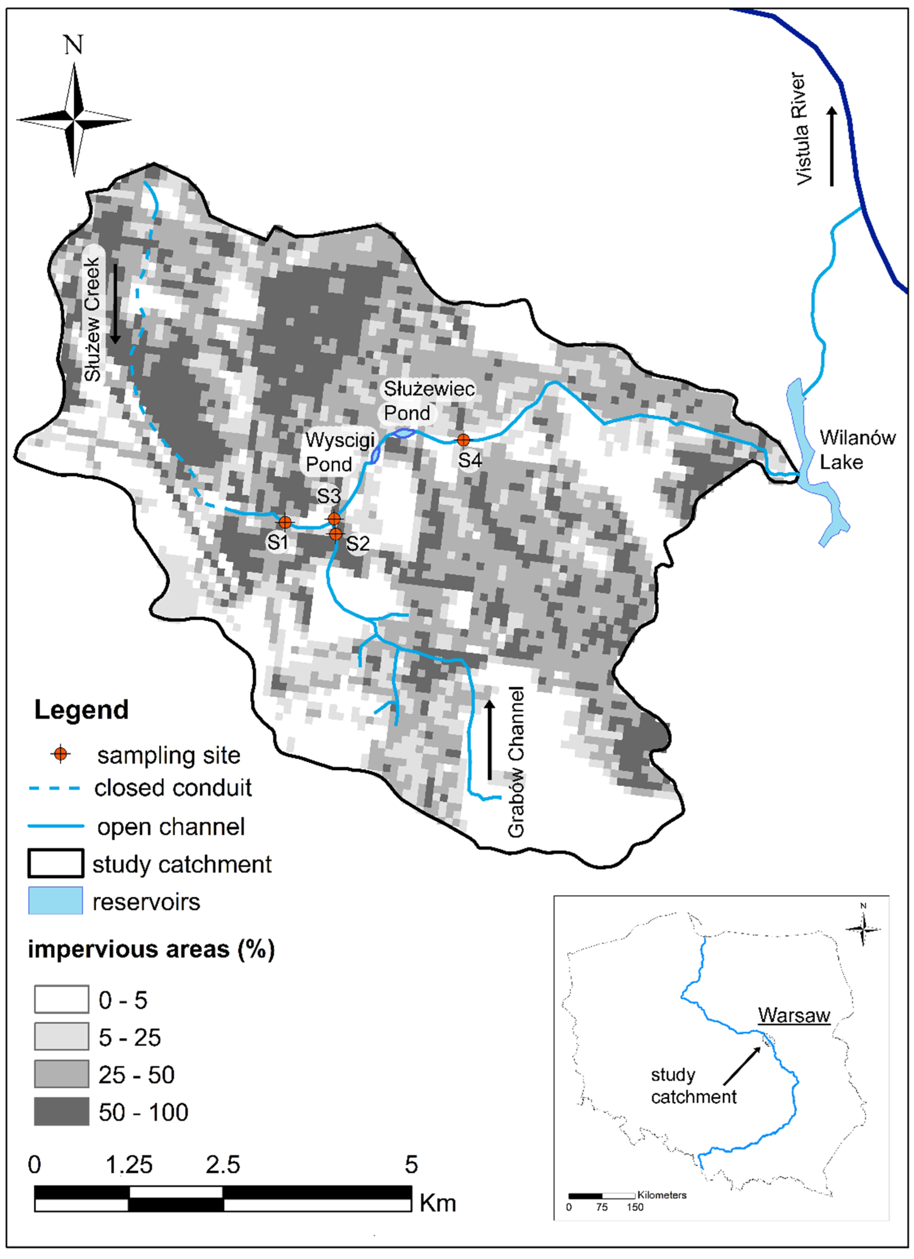
The catchment of Służew Creek located in Warsaw, Poland is under hydro-meteorological monitoring by the Warsaw University of Life Sciences [26,27,28]. Urbanization significantly affects the study area for a long time. Already at the end of the 17th century, the course of the main stream was changed [29]. The runoff has been directed through an artificial channel to Wilanów Lake (see Figure 2). In the 1970s and 1980s, the management of the catchment was changed from agricultural to urban. The number of sealed areas increased and the stream channel has been regulated. Currently, the issues related to the growing flood risk [30] and the deteriorating quality of the runoff [31] are becoming the greatest challenge to be faced by the local authorities and community. Microplastic contamination has not yet been investigated in this area.
As sampling sites, four specific cross sections were selected, see Figure 2 for the detailed locations. At point S1, the Służew Creek starts to flow in an open channel. The catchment up to this cross section (11.9 km2) is highly urbanized, the stream flows under the airport, and its source is also built up. The second sample (S2) was taken from the Grabów Channel, which drains the water from suburbs of Warsaw. The catchment until the cross section (14.0 km2) is less urbanized and covered by single-family houses, farmlands, wastelands, forests, and scrubs. The Grabów Channel joins Służew Creek. The sampling, S3, is located downstream of the junction of these two watercourses. Furthermore, the Służew Creek flows through two retention ponds. The fourth sample was taken downstream of them (catchment area equals 40.5 km2). Thus, we would like to check if their influence on the presence of microplastics can be observed.
The main idea of sampling is to take the mixture of sediment and microplastic from the upper layer (up to 5 cm) of the bottom sediment. High-density plastic particles settle down or are transported together with coarse sediment. Thus, their highest concentration may occur in the top layer of the sediment. For instance, in the case of beach sediment, samples are often taken with the use of a shovel from a small quadratic area of about 0.25–0.4 m2 [32,33]. This method is hard to apply in a stream as digging under flowing water results in the resuspension of sediment and dilution of the sample. Instead of a shovel, we used a core sampler (⌀ = 5 cm) and put it multiple times into the bottom (to a depth of 5 cm), in the central part of the channel, next to each other to cover the area of 0.2 m2. All samples were taken in one day, within three hours. Values for the water stages and flows at cross sections were close to the annual average (non-flood conditions). Collected samples were taken to the laboratory for further analysis.

2.3. Initial Processing

As part of the preprocessing step, the samples were dried for two days. To prevent plastic degradation or sticking to the sediment, the temperature was lower (70 °C) from the standard 105 °C. Furthermore, dry samples were sieved through a 1 mm sieve to separate large size particles (gravel, crushed glass, fragments of branches and leaves including plastic). After usage, the sieve was rinsed with pure water and dried. Each time, the sieve was visually inspected for any plastic particles larger than 1 mm. The rinse water was added to the appropriate sample and evaporated. This is where the first stage of the study was completed.

2.4. Density Separation

In this approach, materials of different densities (mineral and organic matter, plastic) are put into a liquid of intermediate density and the less dense material (organic matter and plastic) floats and separates out from the more dense (mineral sediment) settling [34]. This step aims to remove mineral matter from the sample. A significant share of it impedes the detection of microplastic. For the case study, the mineral matter accounted for over 97% mass of the collected bed load. Therefore, it is very important to make this step.
As a liquid, different salt solutions of varied densities can be applied. The most widely used are sodium chloride, NaCl [35,36], sodium iodate, NaI [34,37], and zinc chloride, ZnCl2 [38,39]. The first is the cheapest and most commonly available, and its saturated solution has a density of about 1.18 g·cm−3. The remaining two are much more expensive but give higher densities (i.e., up to 1. 8 g·cm−3 for saturated solutions). Please note that zinc chloride is toxic to the environment.
The density of plastic differs widely, depending on the type of the polymer. In general, it ranges from 0.85 g·cm−3 for polypropylene up to 1.45 g·cm−3 for polyvinyl chloride [34]. However, most polymers (PP, LDPE, PE, HDPE, PS, nylon) have a density lower than 1.15 g·cm−3, while polyethylene terephthalate (1.38 g·cm−3) and polyvinyl chloride differed significantly from the rest.
Considering the above information, we propose the use of a liquid for density separation of the 40% calcium chloride, CaCl2 solution. It has several advantages as it is as cheap as sodium chloride and is highly soluble in water and is commonly available. The density of the obtained solution was established at 1.37 g·dm−3. It allows for the recovery of all low- and medium-density polymers. At the same time, information on some high-density polymers may be lost. Eighty grams of salt were added to 100 mL of pure water and stirred. The solution was left to cool down to room temperature (21 °C) as the dissolving heat is released. Afterward, it was filtered to remove any undissolved particles. The density of liquid was estimated with use of the pycnometric method.
Density separation was carried out with the Sediment-Microplastic Isolation (SMI) unit. The concept of this device was developed by Coppock et al. [40]. It consists of two pipes connected with a ball valve (Figure 2). When the valve is open, high-density particles may settle, while those with a light density will float to the surface. After closing the valve, the supernatant can be collected. The SMI was filled with 30 g of dried sediments and the previously prepared salt solution. The mixture was shaken for several minutes, after that, the unit was left motionless for one day and the valve remained open. Next, the valve was closed, and the supernatant was poured into a clean beaker to separate the collected material from the liquid filtration. Furthermore, the glass fiber filters were carefully rinsed with pure water in order to transfer all of the captured particles to the beaker. Water was evaporated and in the beaker, only low-density particles were left. This is where the density separation method ends.

2.5. Organic Matter Reduction

This stage focuses on removing the organic content from the material obtained in the previous step. Organic matter may be easily mistaken for microplastic particles, leading to an overestimation of its amount. Moreover, when reducing organic matter it is important to preserve the plastic decomposition at the same time. Thus, mineralization at high temperatures or in concentrated acid is not allowed. To solve this issue, many authors have used different approaches and there is no single dedicated method. However, the type of sample impacts the method selected. In the case of animal samples (i.e., tissues of dead clams or fishes), the treatment of the sample with sodium hydroxide, KOH, at a specific concentration, temperature, and amount of time was established as an efficient method [22,41]. For soil or sediment samples, 30% oxidant peroxide, H2O2, or Fenton’s reagent (i.e., H2O2) with the addition of ferrous iron, Fe(II), are often applied [35,42,43]. It has also been proven that Fenton’s reagent is efficient in reducing algae mass, while complex structures, (i.e., wood (composed of lignin)) are more resistant to it [44]. Thus, recent research proposes the use of sodium hypochlorite, NaClO to remove the vegetal matter [45]. It is commonly used in the water treatment or paper industry and has a high ability to decompose lignin and cellulose. Here, it should also be mentioned that apart from chemical methods, biological digestion with the use of enzymes may also be applied [46,47].
In this work, to reduce organic matter, sodium hypochlorite was used. It is commonly available, low cost, and is less dangerous than Fenton’s reagent (highly caustic, may boil rapidly when overheated). It also does not require strictly defined activation and action conditions such as enzymes. The applied procedure of reduction is based on the work of Monteiro et al. [45]. To the beaker with a low-density material (resulting from the previous step), 100 mL of 10% NaClO solution was added. The beaker was covered with aluminum foil and put into a heater for 15 h at 50 °C. Under these conditions, the decomposition of organic matter was conducted.

2.6. Nile Red Staining and Microplastic Identification

Nile Red (NR) is a fluorescent, hydrophobic dye that was initially used to detect lipids in biological samples [48]. Recently, it has been commonly applied to identify microplastics [49,50,51]. The dye binds to the polymers and makes them fluorescent when exposed to the light of a specific wavelength. Prata et al. [50,52] proved through a series of tests, that stained particles exposed to blue light (470 nm) and observed (photographed) under orange filter may be easily distinguished and classified as plastic. The image showing bright, glowing particles can be processed by special software.
Nevertheless, it should also be mentioned that Nile Red may adsorb on organic matter and cause a fluorescence similar to that of plastic. Therefore, it is extremely important to remove it according to the previous stage (see, Section 2.5).
The Nile Red solution was prepared by diluting 1 mg Nile Blue A Oxazone (powder) in 100 mL of pure ethanol, which is the frequently used proportion [50,53,54]. To prepare the stock solution, acetone may also be used [39,55], but not water, as the dye is hydrophobic.
Particles that remained in the beaker (after organic matter reduction) were transferred to the filter. A few drops of NR solution were spread on the filter to stain the microplastics. To avoid background fluorescence, glass fiber filters were used instead of those made of cellulose. Stained filters were left for 10 min at 60 °C to dry and let the dye adsorb on plastic.
Furthermore, filters were moved to a dark room, exposed to blue light and photographed under an orange filter. When not analyzed, the samples stayed covered with aluminum foil to avoid accidental contamination (i.e., through air dust). As a source of blue light, we applied commercial hunting flashlights. Pictures were taken with the use of a digital camera equipped with a macro lens (Nikkon D300s, Tokyo, Japan). The camera was set to automatic mode. After that, the stained filters were observed under a fluorescence microscope (Opta-Tech MN-800FL). This step allowed for additional verification of bright particles. Under magnification (40× or 100×), it is possible to distinguish between plastic and organic material (if there is any left after reduction). Microscopic pictures of single, selected particles were taken. Additional, selected stained and unstained plastics were photographed, which could be used as reference material in the future.
Finally, the captured images were analyzed using ImageJ 1.53v software [56], which is a public domain Java image processing program. The image threshold option was used to extract bright microplastic particles from the dark background. Automatic particle counting was run. As a result of image processing, the number of particles and their surface was obtained. Thus, the step of identifying microplastics in the collected samples was completed.

3. Results and Discussion

Thee sieving method (steal sieve of 1 mm mesh size) was applied to separate the largest pieces of plastic. Such particles differ significantly in terms of color and shine, thus they can be easily recognized and picked up manually. Moreover, they melt when exposed to high temperatures (flame or hot needle). These plastics have been found in three out of four locations (see Table 1 (columns 2 and 3) for details). The image of the particles is also included in the Supplementary Materials (Figure S1). In the case of the sample taken at site number S1 (the stream begins to flow through an open channel), no plastic particles were found on the sieve. The greatest number of particles was confirmed in the tributary from the suburban area (S2). As the two channels merged, the number of particles decreased (S3), and it continued to decline downstream of two reservoirs (S4). These results may suggest that the presence of the largest particles will most likely depend on the management of the catchment area and the number of tributaries. The reservoirs can partially retain the transported material and thus improve the quality of the runoff.
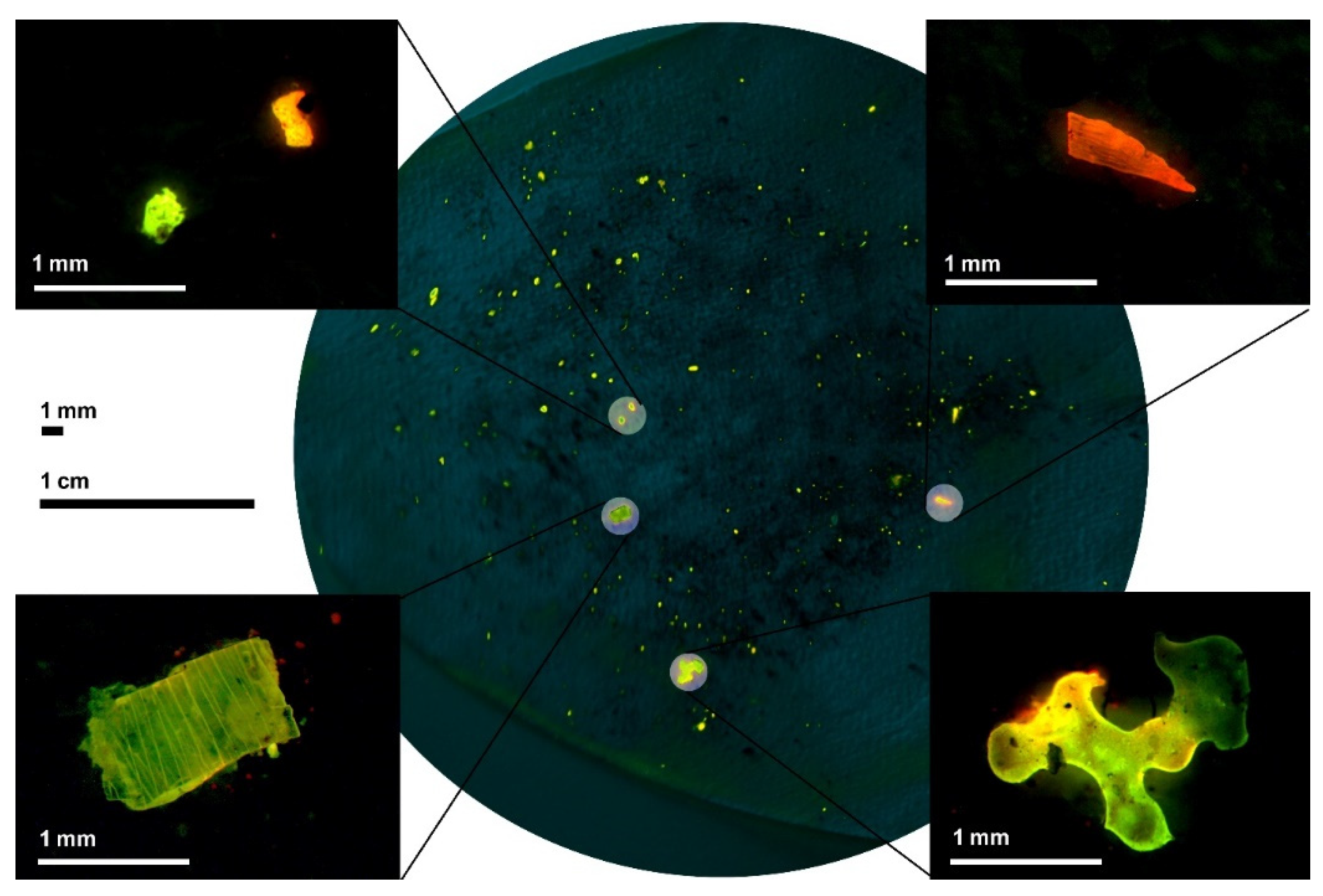
Smaller particles were captured on the filter and stained. Figure 3 presents the macro- and microphotography of microplastic particles found at sampling site no. S1. Photos for other samples are included in the Supplementary Materials (Figures S2–S4). The macrophotography showed all particles, which is useful for automatic counting. However, details on the shape and structure of the particles are missing. Here, one pixel of image corresponded to almost 200 µm2. Moreover, there is always a risk that individual brighter pixels will be incorrectly recognized by the program as plastic. This may happen on the edges of the filter that reflect light in a different way. Due to technical limitations, it is also not possible to recognize particles smaller than 0.014 mm (corresponding to the pixel size). Thus, a threshold for single pixels was set and were not taken into account. Moreover, microscopic photos of the selected particles were made. Based on these, the difference in shapes and structure as well as the colors and intensities of fluorescence may be stated. Under the microscope, one can also distinguish between plastic and organic matter. Figure S5 (Supplementary Materials) presents the results obtained due to omitting organic matter reduction, whereas on the filter, both plastic and organic matter were found.
Table 1 (columns 2 and 3) also summarizes the estimated amounts of the finest microplastics captured on filters. The numbers were relatively similar and ranged from 185 up to 276 pieces per sample (30 g of dry bed load sediment). The smallest amount of these particles were found in the outflow from the suburban, less urbanized catchment (sampling point no. S2). As before, it seems to be a noticeable effect of the reservoirs to reduce the amount of microplastics (a decline between points S3 and S4). In the case of fine particles, catchment management and land use are probably not the only factors determining their quantity in the channel. For example, another source may be rainfall and its spatial distribution over the catchment. Moreover, it seems that as the catchment area increases, the number of particles also rise. Since all identified particles were less than 5 mm in size, columns 3 and 4 were added together, and the total number is presented in column 5 of Table 1. The median surfaces describe the size of the indicated particles (Table 1, column 6). It is a simplified measurement, based on plan view. For the first three samples, it appears to be similar while for the last of them (no. S4), they were about twice as large. At this stage, it is difficult to point out the cause of these differences or similarities.
There are many factors that can affect whether or not particles accumulate in a given location, starting with their type, source, moving media, or weather conditions. Patchaiyappan et al. [57] established the average abundance of microplastics to 227.9 ± 91.4 pieces per hundred grams of street dust for an urban area in India (Chennai). This is about three times lower than estimated in our work. However, we note the type and structure of the investigated samples. Street dust is much finer than bed load, so by the same mass, the volume of these two will be different. Thus, a direct comparison can be misleading. Sekudewicz et al. [58] stated that the abundance of MPs in the sediments of the Vitula River in Warsaw, varied from 190 to 580 items·kg−1. These investigations, however, were performed on a different date (2018) and weather conditions than ours.
The fluoresce color for microplastic particles ranges from yellow-green to dark red, and the intensity of specific colors may also vary widely. These two are related among others to the type of the polymer (density, structure, polarity), its original color, and exposure time to environmental conditions (virgin or weathered). Moreover, the way the experiment was carried out or technical equipment may also affect the obtained results (i.e., the blue light is not the only one that induces fluorescence) [39]. Currently, the identification of polymers based on their color or luminous intensity is a challenge. Studies on microplastic detection with the use of Nile Red focus primarily on its quantification. Thus, there are still not enough reference images. This will probably change in the future if aa global database of such pictures with dedicated software is developed. To support this action, in the Supplementary Materials (S6–S12), the authors provide microscopic photos of the stained, virgin polymers as well as some natural fibers that could be used as reference material.
Microplastic particles may also be identified by using Raman or Fourier transform infrared (FTIR) spectroscopy [59]. These two are much more advanced than Red Nile staining. The Raman spectroscopy measures the light scattering after exciting the sample for a laser. The spectrum of Raman radiation is often unique to a given material. By comparing it with the pattern, it is possible to recognize the structure and type of the particle [60]. The FTIR method uses infrared radiation. It estimates how much light remains after absorption. This phenomenon is also typical for a given molecule. Shim et al. [61] stated that the recovery rate of polyethylene spiked to natural sand in the NR staining method was not significantly differed with FTIR identification. Patchaiyappan [62], with the use of Raman spectroscopy, confirmed that 78 out of 84 particles selected for analysis were plastics. Nevertheless, there is still a need for research focusing on methodology development [59]. In this work, further investigations with the use of spectroscopic methods were impossible as we did not have the appropriate equipment. The effectiveness of microscopic and staining methods have been confirmed by many authors (e.g., [51,62,63]). At the same time, others have pointed out the possibility of misidentification, especially in the case of nanoplastics [64,65]. The authors agree with the statement that coupling Nile Red staining with the spectroscopy method could be useful to improve the time and cost efficiency [41].
The topic of microplastic pollution has attracted more interest among researchers and the general public. The number of works summarizing the current state of knowledge, but also proposing novel solutions is still growing. It is obvious that not all of them may be tested in a single attempt. Despite this, we tried to collect the most frequently mentioned ideas and approaches and applied them in practice. However, our experience also confirmed that there is still a lot to study and improve in this area. Among others, there is a need to standardize the applied laboratory procedures for specific types of samples (i.e., river sediment, stormwater runoff, street dust, beach sediment, animal samples, etc.). Second, microplastics research with the use of Nile Red staining should be devoted to both quantitative and qualitative analysis. For this purpose, more reference data must be provided. Raman and FTIR spectroscopy could play a key role in verifying the results obtained with the use of Nile Red staining. In this respect, broad cooperation between research institutions will also be expected. In term of investigations conducted in the study catchment, there is a need to expand the scope of analyses by detailed recognition of factors influencing the presence of microplastics in the channel (i.e., sediment grain size, flow conditions in the channel, point source of pollution, weather conditions), taking other types of samples (i.e., storm runoff) and in greater numbers throughout the catchment area.

4. Conclusions

This work focused on stating the presence of microplastics in the bed load sediment of a small urban catchment in Warsaw. The samples were collected at four separate locations to potentially observe the effect of the catchment size and its management on the amount of plastics. This is also the first, initial research for the selected study site. Based on the conducted research, the following conclusions may be drawn:
  • Microplastics were present in the channel of Służew Creek in Warsaw, where the estimated number of particles ranged from 191 to 279 pieces per 30 g of bed load sediment for the selected sampling sites;
  • The presence of the largest particles (more than 1 mm in size) most likely depends on the management of the catchment area and the number of tributaries, while the abundance of the finest particles (less than 1 mm in size) could also be determined by the meteorological conditions;
  • Small reservoirs may reduce the load of particles and thus enhance the quality of urban runoff;
  • The number of particles seems to increase in the catchment area and factors influencing microplastic accumulation in the study catchment should be investigated in detail in further works;
  • There is a need for further broad research focusing among others on: (i) the standardization of methods and laboratory procedures (leading to microplastic detection) in relation to the type of sample, and (ii) thee identification of specific polymers and the verification of obtained results with the use of the NR staining method.

Supplementary Materials

The following supporting information can be downloaded at: https://www.mdpi.com/article/10.3390/su142316017/s1, Figures S1–S9. Figure S1. Macro photo of microplastic particles, captured on sieve. Sampling sites: S2, S3 and S4; the background mesh is 1 mm. Figure S2. Macro (in the center) and microscopic (in the corners) photos of stained microplastic particles, captured on filter and observed under blue (470 nm) light through orange filter. Sampling site S2. Figure S3. Macro (in the center) and microscopic (in the corners) photos of stained microplastic particles, captured on filter and observed under blue (470 nm) light through orange filter. Sampling site S3. Figure S4. Macro (in the center) and microscopic (in the corners) photos of stained microplastic particles, captured on filter and observed under blue (470 nm) light through orange filter. Sampling site S4. Figure S5. Macro (in the center) and microscopic (in the corners) photos of stained particles, captured on filter and observed under blue (470 nm) light trough orange filter. In this sample, the organic matter reduction stage was omitted. On the left—organic matter, on the right—plastic. Number of recognized particles amounts to 404 pieces. Sampling site S2. Figure S6. Fragments of virgin polyethylene, (PET) coming from bottle caps. Microscopic photos (taken under white light) of PET in differ colors: (a) green, (b) blue, (c) red, (d) yellow, (e) white. Macro photography (bottom part) taken under various conditions. Figure S7. Fragments of virgin expanded polystyrene (Styrofoam, ESP). Microscopic photos (taken under white light) of EPS (on the left). Macro photography (on the right) taken under various conditions. Figure S8. Fragments of virgin nylon (mesh). Microscopic photos (taken under white light) of mesh (on the left). Macro photography (on the right) taken under various conditions. Figure S9. Fragments of virgin polyvinyl chloride (PVC, transparent). Microscopic photos (taken under white light) of PVC (on the left). Macro photography taken under various conditions. Figure S10. Reed leaf fragment. Microscopic photos (taken under white light) of the leaf (on the left). Macro photography (on the right) taken under various conditions. Figure S11. White cotton fiber. Microscopic photos (taken under white light) of the fiber (on the left). Macro photography (on the right) taken under various conditions. Figure S12. Cellulose filter (A) and glass fiber filters (B). Microscopic photos (taken under white light) of the filters (on the left). Macro photography (on the right) taken under various conditions.

Author Contributions

A.K. designed the study and wrote the first draft of the manuscript. A.H. and L.H. reviewed the manuscript. All authors have read and agreed to the published version of the manuscript.

Funding

This research received no external funding.

Institutional Review Board Statement

Not applicable.

Informed Consent Statement

Not applicable.

Data Availability Statement

Not applicable.

Conflicts of Interest

The authors declare no conflict of interest.

References

  1. Jambeck, J.R.; Geyer, R.; Wilcox, C.; Siegler, T.R.; Perryman, M.; Andrady, A.; Narayan, R.; Law, K.L. Plastic Waste Inputs from Land into the Ocean. Science 2015, 347, 768–771. [Google Scholar] [CrossRef]
  2. Sun, X.; Liang, J.; Zhu, M.; Zhao, Y.; Zhang, B. Microplastics in Seawater and Zooplankton from the Yellow Sea. Environ. Pollut. 2018, 242, 585–595. [Google Scholar] [CrossRef] [PubMed]
  3. Nelms, S.E.; Barnett, J.; Brownlow, A.; Davison, N.J.; Deaville, R.; Galloway, T.S.; Lindeque, P.K.; Santillo, D.; Godley, B.J. Microplastics in Marine Mammals Stranded around the British Coast: Ubiquitous but Transitory? Sci. Rep. 2019, 9, 1075. [Google Scholar] [CrossRef] [PubMed] [Green Version]
  4. Barboza, L.G.A.; Lopes, C.; Oliveira, P.; Bessa, F.; Otero, V.; Henriques, B.; Raimundo, J.; Caetano, M.; Vale, C.; Guilhermino, L. Microplastics in Wild Fish from North East Atlantic Ocean and Its Potential for Causing Neurotoxic Effects, Lipid Oxidative Damage, and Human Health Risks Associated with Ingestion Exposure. Sci. Total Environ. 2020, 717, 134625. [Google Scholar] [CrossRef] [PubMed]
  5. de Sá, L.C.; Oliveira, M.; Ribeiro, F.; Rocha, T.L.; Futter, M.N. Studies of the Effects of Microplastics on Aquatic Organisms: What Do We Know and Where Should We Focus Our Efforts in the Future? Sci. Total Environ. 2018, 645, 1029–1039. [Google Scholar] [CrossRef]
  6. Campanale, C.; Massarelli, C.; Savino, I.; Locaputo, V.; Uricchio, V.F. A Detailed Review Study on Potential Effects of Microplastics and Additives of Concern on Human Health. Int. J. Environ. Res. Public Health 2020, 17, 1212. [Google Scholar] [CrossRef] [PubMed] [Green Version]
  7. Guzzetti, E.; Sureda, A.; Tejada, S.; Faggio, C. Microplastic in Marine Organism: Environmental and Toxicological Effects. Environ. Toxicol. Pharmacol. 2018, 64, 164–171. [Google Scholar] [CrossRef]
  8. Alimba, C.G.; Faggio, C. Microplastics in the Marine Environment: Current Trends in Environmental Pollution and Mechanisms of Toxicological Profile. Environ. Toxicol. Pharmacol. 2019, 68, 61–74. [Google Scholar] [CrossRef]
  9. Chatterjee, S.; Sharma, S. Microplastics in Our Oceans and Marine Health. Field Actions Sci. Rep. J. Field Actions 2019, 19, 54–61. [Google Scholar]
  10. Jamieson, A.J.; Brooks, L.S.R.; Reid, W.D.K.; Piertney, S.B.; Narayanaswamy, B.E.; Linley, T.D. Microplastics and Synthetic Particles Ingested by Deep-Sea Amphipods in Six of the Deepest Marine Ecosystems on Earth. R. Soc. Open Sci. 2019, 6, 180667. [Google Scholar] [CrossRef] [Green Version]
  11. Nizzetto, L.; Bussi, G.; Futter, M.N.; Butterfield, D.; Whitehead, P.G. A Theoretical Assessment of Microplastic Transport in River Catchments and Their Retention by Soils and River Sediments. Environ. Sci Process. Impacts 2016, 18, 1050–1059. [Google Scholar] [CrossRef] [PubMed]
  12. Enders, K.; Käppler, A.; Biniasch, O.; Feldens, P.; Stollberg, N.; Lange, X.; Fischer, D.; Eichhorn, K.-J.; Pollehne, F.; Oberbeckmann, S.; et al. Tracing Microplastics in Aquatic Environments Based on Sediment Analogies. Sci. Rep. 2019, 9, 15207. [Google Scholar] [CrossRef] [PubMed] [Green Version]
  13. Klein, S.; Dimzon, I.K.; Eubeler, J.; Knepper, T.P. Analysis, Occurrence, and Degradation of Microplastics in the Aqueous Environment. In Freshwater Microplastics: Emerging Environmental Contaminants? Wagner, M., Lambert, S., Eds.; The Handbook of Environmental Chemistry; Springer International Publishing: Cham, Switzerland, 2018; pp. 51–67. ISBN 978-3-319-61615-5. [Google Scholar]
  14. Uddin, S.; Fowler, S.W.; Uddin, M.F.; Behbehani, M.; Naji, A. A Review of Microplastic Distribution in Sediment Profiles. Mar. Pollut. Bull. 2021, 163, 111973. [Google Scholar] [CrossRef] [PubMed]
  15. Frey, P.; Church, M. Bedload: A Granular Phenomenon. Earth Surf. Process. Landf. 2011, 36, 58–69. [Google Scholar] [CrossRef]
  16. Liro, M.; Emmerik, T.V.; Wyżga, B.; Liro, J.; Mikuś, P. Macroplastic Storage and Remobilization in Rivers. Water 2020, 12, 2055. [Google Scholar] [CrossRef]
  17. Kataoka, T.; Nihei, Y.; Kudou, K.; Hinata, H. Assessment of the Sources and Inflow Processes of Microplastics in the River Environments of Japan. Environ. Pollut. 2019, 244, 958–965. [Google Scholar] [CrossRef]
  18. Allen, S.; Allen, D.; Phoenix, V.R.; Le Roux, G.; Durántez Jiménez, P.; Simonneau, A.; Binet, S.; Galop, D. Atmospheric Transport and Deposition of Microplastics in a Remote Mountain Catchment. Nat. Geosci. 2019, 12, 339–344. [Google Scholar] [CrossRef] [Green Version]
  19. Simpson, M.D.; Nicholson, K.N.; Neumann, K. Exploratory Study of Microplastics in the Eastern Himalayas of Nepal. AGU Fall Meet. Abstr. 2019, 2019, 52. [Google Scholar]
  20. Pegado, T.d.S.S.; Schmid, K.; Winemiller, K.O.; Chelazzi, D.; Cincinelli, A.; Dei, L.; Giarrizzo, T. First Evidence of Microplastic Ingestion by Fishes from the Amazon River Estuary. Mar. Pollut. Bull. 2018, 133, 814–821. [Google Scholar] [CrossRef]
  21. Gerolin, C.R.; Pupim, F.N.; Sawakuchi, A.O.; Grohmann, C.H.; Labuto, G.; Semensatto, D. Microplastics in Sediments from Amazon Rivers, Brazil. Sci. Total Environ. 2020, 749, 141604. [Google Scholar] [CrossRef]
  22. Lusher, A.L.; Tirelli, V.; O’Connor, I.; Officer, R. Microplastics in Arctic Polar Waters: The First Reported Values of Particles in Surface and Sub-Surface Samples. Sci. Rep. 2015, 5, 14947. [Google Scholar] [CrossRef] [PubMed] [Green Version]
  23. Bosker, T.; Guaita, L.; Behrens, P. Microplastic Pollution on Caribbean Beaches in the Lesser Antilles. Mar. Pollut. Bull. 2018, 133, 442–447. [Google Scholar] [CrossRef]
  24. Di, M.; Wang, J. Microplastics in Surface Waters and Sediments of the Three Gorges Reservoir, China. Sci. Total Environ. 2018, 616–617, 1620–1627. [Google Scholar] [CrossRef] [PubMed]
  25. Jiang, C.; Yin, L.; Wen, X.; Du, C.; Wu, L.; Long, Y.; Liu, Y.; Ma, Y.; Yin, Q.; Zhou, Z.; et al. Microplastics in Sediment and Surface Water of West Dongting Lake and South Dongting Lake: Abundance, Source and Composition. Int. J. Environ. Res. Public Health 2018, 15, 2164. [Google Scholar] [CrossRef] [PubMed] [Green Version]
  26. Banasik, K.; Krajewski, A.; Sikorska, A.; Hejduk, L. Curve Number Estimation for a Small Urban Catchment from Recorded Rainfall-Runoff Events. Arch. Environ. Prot. 2014, 40, 75–86. [Google Scholar] [CrossRef] [Green Version]
  27. Krajewski, A.; Sikorska-Senoner, A.E.; Hejduk, A.; Hejduk, L. Variability of the Initial Abstraction Ratio in an Urban and an Agroforested Catchment. Water 2020, 12, 415. [Google Scholar] [CrossRef] [Green Version]
  28. Krajewski, A.; Sikorska-Senoner, A.E. Suspended Sediment Routing through a Small On-Stream Reservoir Based on Particle Properties. J. Soils Sediments 2021, 21, 1523–1538. [Google Scholar] [CrossRef]
  29. Starzyński, J. Wilanów: Dzieje Budowy Pałacu Za Jana III; PWN: Warszawa, Poland, 1976. [Google Scholar]
  30. Krajewski, A.; Wasilewicz, M.; Sikorska, A.; Banasik, K. Operation of Detention Pond in Urban Area—Example of Wyścigi Pond in Warsaw. In Environmental Engineering V; Pawłowska, M., Pawłowski, L., Eds.; CRC Press: Leiden, The Netherlands, 2017; ISBN 978-1-315-28197-1. [Google Scholar]
  31. Wojtkowska, M.; Karwowska, E.; Chmielewska, I.; Bekenova, K.; Wanot, E. Copper and Cadmium in Bottom Sediments Dredged from Wyścigi Pond, Warsaw, Poland--Contamination and Bioaccumulation Study. Environ. Monit. Assess. 2015, 187, 737. [Google Scholar] [CrossRef] [Green Version]
  32. Khuyen, V.T.K.; Le, D.V.; Fischer, A.R.; Dornack, C. Comparison of Microplastic Pollution in Beach Sediment and Seawater at UNESCO Can Gio Mangrove Biosphere Reserve. Glob. Chall. 2021, 5, 2100044. [Google Scholar] [CrossRef]
  33. Schröder, K.; Kossel, E.; Lenz, M. Microplastic Abundance in Beach Sediments of the Kiel Fjord, Western Baltic Sea. Environ. Sci. Pollut. Res. 2021, 28, 26515–26528. [Google Scholar] [CrossRef]
  34. Quinn, B.; Murphy, F.; Ewins, C. Validation of Density Separation for the Rapid Recovery of Microplastics from Sediment. Anal. Methods 2017, 9, 1491–1498. [Google Scholar] [CrossRef] [Green Version]
  35. Masura, J.; Baker, J.; Foster, G.; Courtney, A. Laboratory Methods for the Analysis of Microplastics in the Marine Environment; NOAA Marine Debris Division: Silver Spring, MD, USA, 2015.
  36. Hitchcock, J.N. Storm Events as Key Moments of Microplastic Contamination in Aquatic Ecosystems. Sci. Total Environ. 2020, 734, 139436. [Google Scholar] [CrossRef]
  37. Narmadha, V.; Jose, J.; Patil, S.; Farooqui, M.O.; Srimuruganandam, B.; Saravanadevi, S.; Krishnamurthi, K. Assessment of Microplastics in Roadside Suspended Dust from Urban and Rural Environment of Nagpur, India. Int. J. Environ. Res. 2020, 14, 629–640. [Google Scholar] [CrossRef]
  38. Maes, T.; Jessop, R.; Wellner, N.; Haupt, K.; Mayes, A.G. A Rapid-Screening Approach to Detect and Quantify Microplastics Based on Fluorescent Tagging with Nile Red. Sci. Rep. 2017, 7, 44501. [Google Scholar] [CrossRef] [PubMed] [Green Version]
  39. Vermeiren, P.; Muñoz, C.; Ikejima, K. Microplastic Identification and Quantification from Organic Rich Sediments: A Validated Laboratory Protocol. Environ. Pollut. 2020, 262, 114298. [Google Scholar] [CrossRef]
  40. Coppock, R.L.; Cole, M.; Lindeque, P.K.; Queirós, A.M.; Galloway, T.S. A Small-Scale, Portable Method for Extracting Microplastics from Marine Sediments. Environ. Pollut. 2017, 230, 829–837. [Google Scholar] [CrossRef] [PubMed] [Green Version]
  41. Prata, J.C.; da Costa, J.P.; Fernandes, A.J.S.; da Costa, F.M.; Duarte, A.C.; Rocha-Santos, T. Selection of Microplastics by Nile Red Staining Increases Environmental Sample Throughput by Micro-Raman Spectroscopy. Sci. Total Environ. 2021, 783, 146979. [Google Scholar] [CrossRef]
  42. Liu, F.; Vianello, A.; Vollertsen, J. Retention of Microplastics in Sediments of Urban and Highway Stormwater Retention Ponds. Environ. Pollut. 2019, 255, 113335. [Google Scholar] [CrossRef]
  43. Thomas, D.; Schütze, B.; Heinze, W.M.; Steinmetz, Z. Sample Preparation Techniques for the Analysis of Microplastics in Soil—A Review. Sustainability 2020, 12, 9074. [Google Scholar] [CrossRef]
  44. Prata, J.C.; da Costa, J.P.; Girão, A.V.; Lopes, I.; Duarte, A.C.; Rocha-Santos, T. Identifying a Quick and Efficient Method of Removing Organic Matter without Damaging Microplastic Samples. Sci. Total Environ. 2019, 686, 131–139. [Google Scholar] [CrossRef]
  45. Monteiro, S.S.; Rocha-Santos, T.; Prata, J.C.; Duarte, A.C.; Girão, A.V.; Lopes, P.; Cristovão, T.; da Costa, J.P. A Straightforward Method for Microplastic Extraction from Organic-Rich Freshwater Samples. Sci. Total Environ. 2022, 815, 152941. [Google Scholar] [CrossRef] [PubMed]
  46. Lavoy, M.; Crossman, J. A Novel Method for Organic Matter Removal from Samples Containing Microplastics. Environ. Pollut. 2021, 286, 117357. [Google Scholar] [CrossRef] [PubMed]
  47. Mbachu, O.; Jenkins, G.; Pratt, C.; Kaparaju, P. Enzymatic Purification of Microplastics in Soil. MethodsX 2021, 8, 101254. [Google Scholar] [CrossRef]
  48. Greenspan, P.; Mayer, E.P.; Fowler, S.D. Nile Red: A Selective Fluorescent Stain for Intracellular Lipid Droplets. J. Cell. Biol. 1985, 100, 965–973. [Google Scholar] [CrossRef] [Green Version]
  49. Erni-Cassola, G.; Gibson, M.I.; Thompson, R.C.; Christie-Oleza, J.A. Lost, but Found with Nile Red: A Novel Method for Detecting and Quantifying Small Microplastics (1 Mm to 20 Μm) in Environmental Samples. Environ. Sci. Technol. 2017, 51, 13641–13648. [Google Scholar] [CrossRef] [PubMed] [Green Version]
  50. Prata, J.C.; Alves, J.R.; da Costa, J.P.; Duarte, A.C.; Rocha-Santos, T. Major Factors Influencing the Quantification of Nile Red Stained Microplastics and Improved Automatic Quantification (MP-VAT 2.0). Sci. Total Environ. 2020, 719, 137498. [Google Scholar] [CrossRef]
  51. Shruti, V.C.; Pérez-Guevara, F.; Roy, P.D.; Kutralam-Muniasamy, G. Analyzing Microplastics with Nile Red: Emerging Trends, Challenges, and Prospects. J. Hazard Mater. 2022, 423, 127171. [Google Scholar] [CrossRef] [PubMed]
  52. Prata, J.C.; Reis, V.; Matos, J.T.V.; da Costa, J.P.; Duarte, A.C.; Rocha-Santos, T. A New Approach for Routine Quantification of Microplastics Using Nile Red and Automated Software (MP-VAT). Sci. Total Environ. 2019, 690, 1277–1283. [Google Scholar] [CrossRef]
  53. Scircle, A.; Cizdziel, J.V. Detecting and Quantifying Microplastics in Bottled Water Using Fluorescence Microscopy: A New Experiment for Instrumental Analysis and Environmental Chemistry Courses. J. Chem. Educ. 2020, 97, 234–238. [Google Scholar] [CrossRef]
  54. Wiggin, K.J.; Holland, E.B. Validation and Application of Cost and Time Effective Methods for the Detection of 3–500 μm Sized Microplastics in the Urban Marine and Estuarine Environments Surrounding Long Beach, California. Mar. Pollut. Bull. 2019, 143, 152–162. [Google Scholar] [CrossRef]
  55. Godoy, V.; Prata, J.C.; Blázquez, G.; Almendros, A.I.; Duarte, A.C.; Rocha-Santos, T.; Calero, M.; Martín-Lara, M.Á. Effects of Distance to the Sea and Geomorphological Characteristics on the Quantity and Distribution of Microplastics in Beach Sediments of Granada (Spain). Sci. Total Environ. 2020, 746, 142023. [Google Scholar] [CrossRef] [PubMed]
  56. NIH—National Institutes of Health. Available online: https://imagej.nih.gov/ij/ (accessed on 22 June 2022).
  57. Patchaiyappan, A.; Dowarah, K.; Zaki Ahmed, S.; Prabakaran, M.; Jayakumar, S.; Thirunavukkarasu, C.; Devipriya, S.P. Prevalence and Characteristics of Microplastics Present in the Street Dust Collected from Chennai Metropolitan City, India. Chemosphere 2021, 269, 128757. [Google Scholar] [CrossRef]
  58. Sekudewicz, I.; Dąbrowska, A.M.; Syczewski, M.D. Microplastic Pollution in Surface Water and Sediments in the Urban Section of the Vistula River (Poland). Sci. Total Environ. 2021, 762, 143111. [Google Scholar] [CrossRef] [PubMed]
  59. Xu, J.-L.; Thomas, K.V.; Luo, Z.; Gowen, A.A. FTIR and Raman Imaging for Microplastics Analysis: State of the Art, Challenges and Prospects. TRAC Trends Anal. Chem. 2019, 119, 115629. [Google Scholar] [CrossRef]
  60. Hengstmann, E.; Fischer, E.K. Nile Red Staining in Microplastic Analysis-Proposal for a Reliable and Fast Identification Approach for Large Microplastics. Environ. Monit. Assess. 2019, 191, 612. [Google Scholar] [CrossRef]
  61. Shim, W.J.; Song, Y.K.; Hong, S.H.; Jang, M. Identification and Quantification of Microplastics Using Nile Red Staining. Mar. Pollut. Bull. 2016, 113, 469–476. [Google Scholar] [CrossRef] [PubMed]
  62. Patchaiyappan, A.; Ahmed, S.Z.; Dowarah, K.; Jayakumar, S.; Devipriya, S.P. Occurrence, Distribution and Composition of Microplastics in the Sediments of South Andaman Beaches. Mar. Pollut. Bull. 2020, 156, 111227. [Google Scholar] [CrossRef]
  63. Meyers, N.; Catarino, A.I.; Declercq, A.M.; Brenan, A.; Devriese, L.; Vandegehuchte, M.; De Witte, B.; Janssen, C.; Everaert, G. Microplastic Detection and Identification by Nile Red Staining: Towards a Semi-Automated, Cost- and Time-Effective Technique. Sci. Total Environ. 2022, 823, 153441. [Google Scholar] [CrossRef]
  64. Mariano, S.; Tacconi, S.; Fidaleo, M.; Rossi, M.; Dini, L. Micro and Nanoplastics Identification: Classic Methods and Innovative Detection Techniques. Front. Toxicol. 2021, 3, 636640. [Google Scholar] [CrossRef]
  65. Uddin, S.; Fowler, S.W.; Habibi, N.; Behbehani, M. Micro-Nano Plastic in the Aquatic Environment: Methodological Problems and Challenges. Animals 2022, 12, 297. [Google Scholar] [CrossRef]
Figure 1. The scheme of the bed load sample processing leading to microplastic identification.
Sustainability 14 16017 g001
Figure 2. Locality map of the Służew Creek catchment and sampling sites.
Sustainability 14 16017 g002
Figure 3. Macro-(in the center) and microscopic (in the corners) photos of the stained microplastic particles captured on filter and observed under blue (470 nm) light through an orange filter. Sampling site S1, channel of the Służew Creek. The contrast has been increased to make the photo easier to read.
Sustainability 14 16017 g003
Table 1. The estimated amounts of microplastics found in the bed load sediment of the Służew Creek catchment.
No.Name/Location of the Sampling SiteNumber of Microplastic Particles Captured on 1 mm SieveNumber of Microplastic Particles Captured on Filter (Pieces/30 g of Bed Load Sediment)Total Number of Microplastic Particles
(Pieces/30 g of Bed Load Sediment)
Median Particle Surface in the Sample (mm2)
23456
1S1,
Służew Creek
02132130.0057
2S2,
Grabów Channel
61851910.0076
3S3,
Służew Creek
32762790.0062
4S4,
Służew Creek
12492500.0123
Average32312330.00795
Publisher’s Note: MDPI stays neutral with regard to jurisdictional claims in published maps and institutional affiliations.