Abstract
(1) Background: This study aimed to compare patient-reported outcome measures when accelerating en masse retraction between the piezocision procedure and the subsequent application of low-level laser therapy (FC+LLLT), with the piezocision alone (FC), and in a control group. (2) Methods: A three-arm randomized controlled trial (RCT) was conducted involving 60 patients (41 females and 19 males) with Class II division I malocclusion. The en masse retraction was performed using NiTi closed coil springs attached to miniscrews. The LLLT was performed using an 808 nm Ga-Al-As diode laser. Patient responses regarding pain, discomfort, swelling, and chewing difficulties were reported at ten assessment points. (3) Results: The greatest pain levels were observed 24 h after the application of force during the first and third months of retraction. The mean pain, discomfort, swelling, and chewing difficulties were significantly smaller in the control group than in the FC and FC+LLLT groups. High satisfaction levels were reported in all three groups (p < 0.05). (4) Conclusions: The accelerated en masse retraction via piezocision, followed by a small course of LLLT, was accompanied by significantly fewer pain, discomfort, and chewing difficulties than the control group. LLLT is a valuable addition to piezocision, with an improved patient experience.
1. Introduction
The duration of orthodontic treatment presents a significant challenge for both orthodontists and patients, and even deters some patients from undergoing treatment [1]. Shortening the duration of orthodontic treatment is a primary goal for both orthodontists and patients, especially adult patients [2], as prolonged treatment can lead to adverse effects such as root resorption, periodontal disease, and dental caries [3,4]. Furthermore, patients often experience discomfort and pain associated with various stages of therapy, with pain being the most disliked aspect of orthodontic treatment [5].
Several methods have emerged to improve orthodontic tooth movement and shorten the duration of orthodontic treatment, with surgical methods being the most popular, including traditional corticotomy [6,7], and interseptal alveolar surgery. However, these techniques have shown low acceptance among patients due to their invasive nature and postoperative complications.
Minimally invasive techniques, such as piezocision [8,9], micro-osteoperforations (MOPs) [10], corticision [11], and laser-assisted flapless corticotomy [12], have gained popularity due to their ability to minimize postoperative discomfort and reduce recovery time. On the other hand, non-surgical interventions like low-level laser therapy (LLLT) [13,14], light vibrational forces [15], light-emitting diodes [16], and pulsed electromagnetic waves [17] are currently widely used.
LLLT has been shown to be effective in accelerating tooth movement and reducing pain, as supported by recent studies [14,18]. Consequently, there is a growing interest in using LLLT as an adjunct to traditional orthodontic treatment to take advantage of the acceleration properties of minimally invasive methods and the analgesic and acceleration properties of LLLT [19,20].
Reviewing the literature reveals some recent research work combining two or more acceleration methods to accelerate the orthodontic movement, such as combining self-ligating brackets with one or more acceleration methods [21], or the use of LLLT with minimally invasive methods like corticotomy [22], MOPs [23], and piezocision for canine retraction [24]. A recent systematic review showed that there were several published studies about the levels of pain and discomfort when using surgically assisted acceleration methods [25]. However, no studies have examined patient-reported outcome measures associated with the combination of piezocision with LLLT in the context of the en masse retraction of upper anterior teeth. Therefore, the aim of the current RCT was to assess the patient-reported outcome measures when accelerating the en masse retraction of upper anterior teeth in adult patients using piezocision combined with the subsequent application of LLLT compared to piezocision alone, or with no acceleration method.
The null hypothesis (H0) stated that there were no significant differences between the piezocision plus subsequent LLLT application, piezocision alone, and traditional en masse retraction groups in terms of the levels of pain, discomfort, swelling sensation, chewing difficulties, satisfaction, and acceptance at all assessment times.
The research hypothesis (H1) stated that there were significant differences between piezocision with subsequent LLLT application, piezocision alone, and traditional en masse retraction groups in the levels of pain, discomfort, swelling sensation, chewing difficulties, satisfaction, and acceptance at all assessment times
2. Materials and Methods
2.1. Study Design and Setting
This is a three-arm, parallel-group, randomized controlled trial (RCT) conducted between February 2021 and December 2022, involving individuals enrolled in the Department of Orthodontics at Damascus University’s Faculty of Dentistry. The Consolidated Standards of Reporting Trials (CONSORT) guidelines were followed to optimize the writing of the study [26]. The Local Research Ethics Committee of the University of Damascus approved the study (UDDS-632-15012021/SRC-1679), which is registered in the Clinical Trials database (ID: NCT05656898). The research was supported by Damascus University’s Postgraduate Research Budget (Ref no. 23654267DEJ).
2.2. Sample Size Calculation
To determine the sample size, Minitab® (version 18, State College, PA, USA) was used. It was assumed that the minimum clinically important difference in pain perception on the visual analog scale (VAS) requiring detection was 15 mm. The variability of this parameter (i.e., the SD) was estimated from a previous study as being 12.26 mm out of 100 mm [27]. As a result, a minimum of 18 participants per group was required when using a one-way ANOVA with 90% statistical power and a 5% significance level. Taking into account any potential dropout (not exceeding 10%), a total of 20 patients were required for each group.
2.3. Patient Recruitment and Entry in This Trial
Inclusion/Exclusion Criteria
Participants in this study were selected based on the following criteria: (1) healthy patients aged between 17 and 28 years from both genders; (2) Class II division I malocclusion with an indication for the extraction of the upper first premolars, which includes an overjet of 4–10 mm and normal or long face growth patterns; (3) skeletal Class II relationship (4 < ANB ≤ 7); (4) a normal overbite, which is more than 0 mm and less than 4 mm; (5) good oral hygiene, as evidenced by a probing depth of ≤3 mm and no alveolar bone loss assessed radiographically; and (6) patients with no missing teeth (except for third molars).
The exclusion criteria for the study were: (1) previous orthodontic treatment; (2) systemic disease or medication that affects orthodontic movement; (3) absence of one of the maxillary teeth (except third molars); (4) poor oral hygiene or existing periodontal disease, which was judged by probing depth ≥ 4 mm, radiographic evidence of bone loss, gingival index >1, and plaque index > 1; and (5) moderate to severe crowding on the maxillary teeth (Little’s Irregularity Index ≥ 4)
A total of 112 patients attending the Department of Orthodontics at Damascus University’s Faculty of Dentistry were clinically examined, and 71 patients met the inclusion criteria. Then, the candidate patients were informed of the research details, and a detailed explanation of the orthodontic, surgical, and LLLT procedures intended for this study was provided. As a result, 65 patients agreed to participate in this research. Later, 60 of the 65 patients were chosen randomly (Figure 1). All selected patients were given written information sheets to read before signing consent forms.
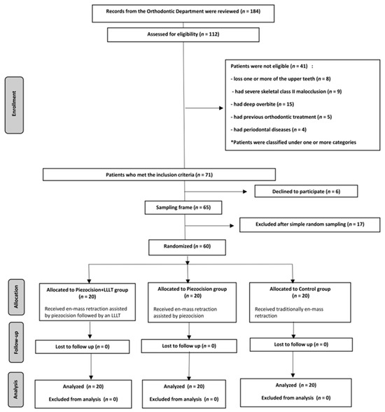
Figure 1.
CONSORT flow diagram of patient recruitment, assignment, follow up, and entry into data analysis. The LLLT included the use of Ga-Al-As diode laser with a wavelength of 808 nm, a power of 1.1 watts, an energy of 1864 joules/point, and an application of 15 s per point.
2.4. Randomization, Allocation Concealment, and Blinding
Simple randomization was carried out to assign the 60 patients into three groups: (1) en masse retraction assisted by flapless corticotomy combined with the later application of LLLT (FC+LLLT), (2) en masse retraction assisted by flapless corticotomy (FC), and (3) conventional en masse retraction (CONT). An academic member who was not involved in this research created a list of random numbers using Minitab® Version 19.1 (Minitab Inc., State College, PA, USA), with an allocation sequence of 1:1:1 ratio. At the end of the leveling and alignment stage, the sequentially numbered, sealed, and opaque envelopes containing the allocation sequence were opened. Blinding was applied only to the outcomes’ assessor, and it did not apply to either the patient or principal researcher (M.M.M).
2.5. Interventions
2.5.1. Leveling and Alignment Phase
The principal investigator (M.M.M) performed the orthodontic treatment under the supervision of one of the coauthors (M.Y.H) at the Orthodontic Department of the Faculty of Dentistry of Damascus University. At the beginning of the treatment, pre-adjusted fixed orthodontic appliances; MBT 0.022-inch slot size (Votion™, OrthoTechnology®, Tampa, FL, USA), were bonded to all patients. Then, self-drilling titanium temporary anchorage devices with 1.6 mm diameter and 8 mm length (3S screw, Hubit®, Seoul, Republic of Korea) were inserted bilaterally at the gingival–mucosal junction approximately 8–10 mm high above the archwires between the maxillary second premolar and the 1st molar, and these teeth were attached to the mini-screw with a ligature wire. Thereafter, patients were referred for extraction of the maxillary 1st premolars. The archwires were applied according to the following sequence: the initial wire 0,014-in Nitinol (NiTi), followed by 0.016-in NiTi, 0.016 × 0.022-in NiTi, 0.017 × 0.025-in NiTi, 0.019 × 0.025-in NiTi, and finally the last wire 0.019 × 0.025-in stainless steel. However, the last wire was kept for two weeks to become neutral before starting the process of en masse retraction. After that, the used wires were replaced with wires of the same size, with soldered hooks placed distally to the lateral incisors.
2.5.2. The Flapless Corticotomy Procedure in the FC+LLLT and FC Groups
The patients received the same aforementioned procedures with the CONT group, and 4 days before the start of the en masse retraction, piezosurgery was performed by the same principal investigator (M.M.M) under the supervision of one of the coauthors (W.A.H) at the Oral and Maxillofacial Surgery Department of the same university. Before the surgical intervention, the patients were asked to use chlorhexidine 0.12% rinse for 60 s. On the buccal aspect, two vertical incisions were made distal to the canine at the site of extraction of the 1st premolars at both sides. Moreover, one vertical gingival incision was made between each of the roots of two adjacent teeth in the anterior region using blade No. (15). However, the same incisions were also performed on the palatal aspect. So, the number of incisions was 18 in total (9 from the buccal side and 9 from the palatal one). These incisions started at a distance of 5 mm apically from the interdental papilla and were 5 mm in length (The purpose of these incisions is to penetrate the periosteum in order to allow the piezosurgery cutting tip to enter and reach the cortical bone), then a piezosurgery micro saw with a BS1 cutting tip (Piezosurgery®, Mectron, Carasco, Italy; Figure 2) was inserted to create alveolar cortical cuts with 3 mm depth (Figure 3). No sutures were applied after performing the incisions. Finally, the area was washed well with physiological serum (sodium chloride) at a concentration of 0.9%. Then, the surgical site was covered using a piece of gauze impregnated with Idoform (BIOGAR Povidone, BIOGAR©, Lattakia, Syria) that was applied for an hour. All patients were strictly asked to adhere to good oral hygiene and avoid nonsteroidal anti-inflammatory drugs using. Paracetamol (acetaminophen, 500 mg) was allowed only when needed, provided the questionnaires had first been answered.
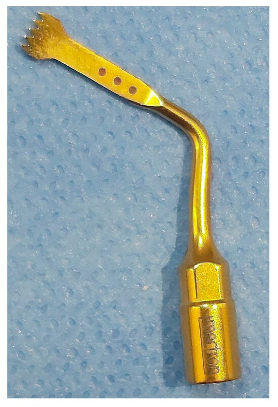
Figure 2.
BS1 cutting tip used for performing the cortical cuts.
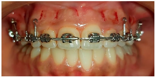
Figure 3.
The minimally invasive piezocision intervention.
2.5.3. The LLLT Procedure in the FC+LLLT Group
Starting from the sixth week after performing piezocision, LLLT was carried out by the same operator (M.M.M). Gallium Aluminum Arsenide (Ga-Al-As) semiconductor diode laser (Klas®-DX Laser, Konftec Corporation, New Taipei City, Taiwan) was used with the following parameters: a wavelength of 808 nm, a power of 1.1 watts, and an energy of 4 joules/point with an application time of 15 s per point. The tip of the biostimulation handpiece was held in contact with the oral mucosa and perpendicular to the root axis during the laser procedure. A total of 32 irradiation points were performed each time, 16 on the buccal aspect and 16 on the palatal one, distributed as follows, with two irradiation points on the apical and cervical root of each of the six upper anterior teeth from both buccal and palatal aspects, so that the total application time was 60 s for one tooth. In addition, there were two irradiation points in the extraction space of the upper 1st premolar on both sides from both buccal and palatal aspects. After the initial application, the laser was applied again on days 3, 7, and 14, and then every 15 days up to 4 sessions. The en masse retraction phase was started 3 days after the piezocision procedure in the FC and FC+LLLT.
2.5.4. En Masse Retraction Phase
The en masse retraction was achieved by using the sliding technique on the base archwires (0.019 3 0.025-in SS) using NiTi closed-coil springs (Ormco Corp, Orange, CA, USA). These springs were suspended from the soldered hooks distally to the lateral incisors to the TADs by applying a force of 250 g per side verified with a force gauge. Every two weeks, the force was checked and adjusted as necessary. The retraction was stopped when a Class I canine relation was achieved and a good incisor relationship was obtained, or spaces lateral to canines were closed.
2.6. Pain, Discomfort, Functional Impairments, and Outcome Measures
The study included two questionnaires distributed to patients within the first month of initiating en masse retraction. The first questionnaire was given to the patients at the following assessment times: 12 h (T1), one day (T2), 3 days (T3), 7 days (T4), and 14 days (T5) after the initiation of the en masse retraction. Additionally, during the third month, the same questionnaire was given at the following time intervals: 12 h (T6), 1 day (T7), 3 days (T8), 7 days (T9), and 14 days (T10) after the activation of coil springs. These questionnaires consisted of four questions regarding the patients’ experiences of pain, discomfort, and their feelings of swelling sensation and difficulty in mastication (Figure 4).
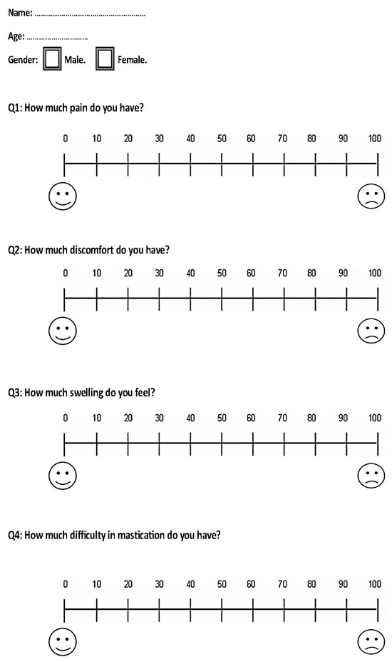
Figure 4.
The questionnaire provided to the included patients at all assessment times except for the last assessment time, i.e., at 14 days after the activation of coil springs in the third month.
Based on the VAS scale, the severity of each studied variable was classified into the following categories: mild (less than 20), mild-to-moderate (from 20 to less than 40), moderate (from 40 to less than 60), moderate-to-severe (from 60 to less than 80), and severe (greater than 80) [25].
The second questionnaire was given to the patients on the 14th day of the third month after retraction initiation (T10). It included the same questions as the first questionnaire, with the addition of two more questions: satisfaction levels assessed on a visual analog scale (VAS) and the possibility of recommending the same treatment to a friend (dichotomous scale) as another measure of satisfaction (Figure 5).
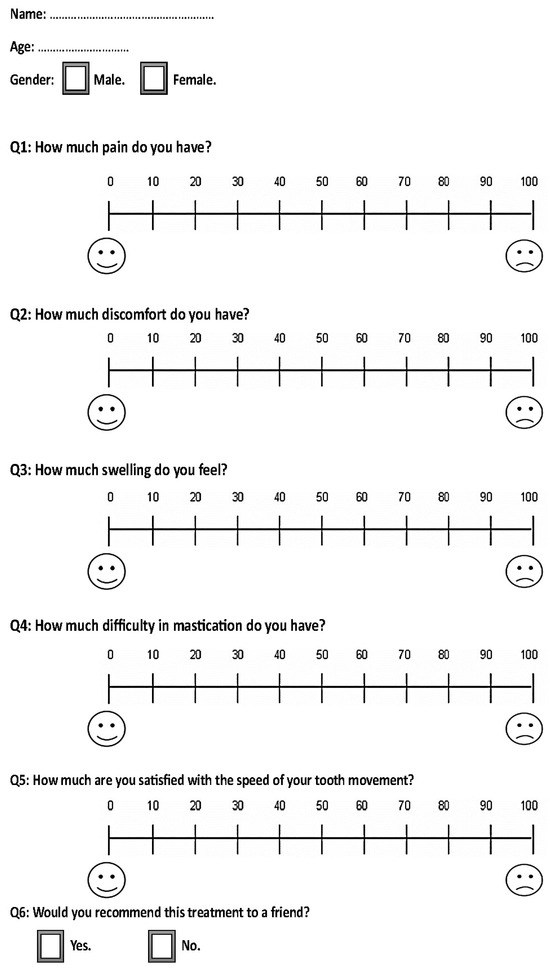
Figure 5.
The questionnaire provided to the included patients at the last assessment time, i.e., at 14 days after the activation of coil springs in the third month.
The questionnaire questions were explained in simple language to ensure patient understanding, and the researcher answered any questions without influencing the patient’s responses. The patients themselves answered the questions, selecting the option that best suited their experience. In cases of severe pain, patients were permitted to take Paracetamol 500 mg, provided that they took the painkiller following the completion of the questionnaire in order not the accuracy of their response.
3. Statistical Analysis
SPSS® Version 20 (SPSS for Windows, version 20, IBM Corporation, New York, NY, USA) was the chosen software for statistical analysis. The Shapiro–Wilk test was used to distinguish the normal distribution of data. One-way ANOVA test or its alternative nonparametric test (i.e., Kruskal–Wallis test) was utilized to make a comparison between the three groups. For pairwise comparisons, the post hoc Bonferroni, LSD, or Games–Howell tests, or the latter’s alternative nonparametric test (i.e., Mann–Whitney test) was applied. Bonferroni’s correction of the significance level was applied due to the multiplicity of pairwise comparisons, and the results of all tests were considered significant at p ≤ 0.017.
4. Results
4.1. Baseline Sample Characteristics
The sample consisted of 60 patients (41 females and 19 males), with a mean age of 21.58 ± 3.19. There were no patient dropouts during the trial, and all patients in the three groups completed their questionnaires at all assessment time points. The baseline sample characteristics regarding age and gender distribution are given in Table 1.

Table 1.
Basic sample characteristics (gender and age).
4.2. Pain and Discomfort Levels
The greatest pain levels were reported 24 h after the initiation of the retraction during the first month in the three groups. The mean pain levels were mild to moderate in the FC+LLLT and FC groups, with 31.55 ± 7.09 mm and 33.7 ± 7.94 mm, respectively, while it was mild in the control group, with a mean pain level of 21.15 ± 7.8.
In the third month, the greatest pain levels were reported after 24 h (T7) in the three groups. The levels were mild in all three groups, with mean values of 9.10 ± 5.64 mm, 22.55 ± 7.39 mm, and 20.8 ± 7.13 mm in the FC+LLLT, FC, and control groups, respectively. In the 1st and 3rd months, the pain levels gradually decreased and became equal or close to zero after 14 days of assessment in all three groups, as seen in Table 2.

Table 2.
Descriptive statistics of pain intensity scores (in mm) at ten assessment times in the three groups using visual analog scales and the results of the significance testing.
Similarly, the discomfort mean values were greatest after 24 h in the first and third months, and gradually decreased thereafter, becoming close to zero after 14 days, as shown in Table 3.

Table 3.
Descriptive statistics discomfort score (in mm) at ten assessment times in the three groups using visual analog scales and the results of the significance testing.
The pain levels were significantly smaller in the CONT group compared to both the FC and FC+LLLT groups at T1, T2, and T3; p < 0.001. However, the pain levels were significantly smaller in the FC+LLLT group compared to the CONT group and FC group at T6, T7, and T8, p < 0.001, as seen in Table 4. The discomfort levels were smaller in the CONT group compared to the FC+LLLT group and FC group at T1, T2, and T3, p < 0.001, as seen in Table 4.

Table 4.
Post hoc tests for pairwise comparisons concerning questions 1 and 2 of the questionnaires.
4.3. Swelling Sensation
The greatest mean levels of swelling were observed after 24 h (T2) in the 1st month of retraction in the CONT, FC+LLLT, and FC groups 16.65 ± 8.41 mm, 39.65 ± 13.03 mm, and 38.3 ± 10.52 mm, respectively, as seen in Table 5. The patients in the CONT group reported significantly smaller levels of perceived swelling compared to the FC+LLLT, and FC groups at T1, T2, and T3 during the first month of the retraction (p ≤ 0.001; Table 5).

Table 5.
Descriptive statistics swelling sensation score (in mm) at ten assessment times in the three groups using visual analog scales and the results of the significance testing.
4.4. Chewing Difficulties
During the first month of assessment, the greatest mean levels of chewing difficulties were recorded at T2 and were generally ‘moderate’ in the FC+LLLT and FC groups; 40.45 ± 9.24, 42.75 ± 10.25, respectively, while they were in the ‘mild to moderate’ category at T2 in the CONT group; 24.8 ± 10.13.
In the third month of assessment, the greatest mean levels of chewing difficulties were at T7 in the three groups, and were classified as being ‘mild’ in FC+LLLT; 9.8 ± 4.22, ‘mild to moderate’ in FC and CONT groups (i.e., 19.6 ± 8.58, 19.1 ± 6.5, respectively). Then, the mean chewing difficulty values gradually decreased over time in the three groups until they approached zero or near zero at T10; Table 6.

Table 6.
Descriptive statistics of questions 4 and 5 score (in mm) at ten assessment times in the three groups using visual analog scales and the results of the significance testing.
The levels of chewing difficulties were significantly smaller in the CONT group at T1, T2, and T3 compared with the FC+LLLT and FC groups (p < 0.001). However, they were significantly smaller in the FC+LLLT group compared to FC and CONT groups at T6, T7, and T8 (p ≤ 0.001; Table 7).

Table 7.
Post hoc tests for pairwise comparisons concerning questions 3, 4, and 5 of the questionnaires.
4.5. Satisfaction with the Orthodontic Tooth Movement (OTM) Speed
The average time required for the en masse retraction phase was 7.85 months in the control group, 5.35 months in the FC group, and 4.5 months in the FC+LLLT group. Notably, the retraction period was significantly shorter in the FC+LLLT group compared to both the FC group (p = 0.009) and the control group (p < 0.001). Furthermore, the retraction period was also significantly shorter in the FC group when compared to the control group (p < 0.001).
High satisfaction levels with the OTM speed were reported in the FC+LLLT and FC groups, whereas levels of satisfaction were classified as being ‘moderate to high’ in the CONT group (i.e., mean VAS value: 84.95 mm, 80.3 and 77.25 mm, respectively, as seen in Table 6). Additionally, satisfaction with the OTM speed was greater in the FC+LLLT group compared with the FC and CONT groups (p < 0.05; Table 7). Furthermore, the majority of patients in the FC+LLLT and FC groups revealed that they would recommend their friends to have the same procedure, 95% and 90%, respectively, and would agree to undergo the same treatment again.
5. Discussion
The current study appears to be the first randomized clinical trial that evaluates the patient-reported outcome measures when using the combined piezocision-LLLT-assisted en masse retraction of upper anterior teeth in comparison with the precision-only-assisted retraction and the traditional method. Various surgical techniques have been developed to expedite dental movement, primarily relying on cortical cutting. These include traditional corticotomy [28], vestibular incision subperiosteal tunnel access [29], and flapless corticotomy using piezosurgery [30]. Among these, piezocision was selected for our study because it is considered the least aggressive among the methods mentioned. Surgical procedures for accelerating orthodontic tooth movement can be adopted in daily orthodontic treatment, provided they do not put an additional burden on patients, such as pain, discomfort, or functional impairments [25].
The VAS was used in this study for pain assessment. Despite its widespread acceptance in clinical research, it is crucial to note that the VAS is inherently subjective. This subjectivity is rooted in the fact that pain is a deeply personal experience. As such, the VAS scores reported in this study reflect the participants’ individual perception of their pain. This could potentially introduce a degree of variability into our results. However, despite this potential limitation, the VAS continues to be a valuable tool in research due to its simplicity and adaptability.
The patient-reported outcome measures were evaluated in the first and third months of retraction. The piezocision was only applied in the first month for both experimental groups, whereas the LLLT was only applied in the FC+LLLT experimental group in the third month.
5.1. The First Month of Assessment
Given that piezocision was applied in both experimental groups before retraction, the comparisons made are confined to the first month only regarding studies that employed piezocision-assisted orthodontic tooth movement. The pain and discomfort levels were in the ‘mild to moderate’ category after 24 h of retraction in the three groups. However, after 72 h of retraction, they were classified as ‘mild to moderate’ in the experimental groups, and ‘mild’ in the control group. Additionally, the levels of pain and discomfort were significantly greater in FC+LLLT and FC groups compared to the control group after 12, 24, and 72 h (p < 0.001). The greater pain levels in both FC+LLLT and FC groups may be attributed to the surgical procedure applied to the patients of these groups compared to the control group.
The current results were inconsistent with those of the study of Hatrom et al. [31], in which the pain levels were mild in the piezocision and control groups during the first 24 h after the initiation of retraction. This discrepancy may be attributed to the fact that in the current trial, incisions were made on both the buccal and palatal sides of the alveolar bone, whereas in Hatrom’s study, the incisions were only placed buccally.
The results of this study disagreed with those reported by Alfawal et al., who evaluated the patient-reported outcome measures associated with canine retraction accelerated by piezosurgery or laser-assisted flapless corticotomy in a compound design RCT. Alfawal et al. found that the levels of pain were moderate and the levels of discomfort were severe in the piezocision group after 24 h of canine retraction in the Alfawal et al. study [32]. This inconsistency may be due to the difference in the onset of retraction force, which was on the same day of the surgical intervention in the study of Alfawal et al. [32], whereas it was four days postoperatively in the current work. The study of Alfawal et al. had a compound design, i.e., two parallel groups with a split-mouth design in each group. This kind of study design may distort patient perception of pain that may originate only from one side of the mouth. Additionally, the assessment of pain on one side of the mouth does not provide a whole picture of the pain that would be encountered when the procedure is performed bilaterally [25].
Patient perception of swelling was ‘mild to moderate’ in the experimental groups and ‘mild’ in the control group after 12, 24, and 72 h of retraction. This can be attributed to edema caused by the surgical procedure in both the FC+LLLT and FC groups. This is consistent with a study by Gibreal et al., which also found increased perception of swelling following piezocision-assisted leveling and alignment [27].
After 24 h of retraction commencement, patients in the experimental groups reported ‘moderate’ levels of chewing difficulty, while those in the control group reported ‘mild to moderate’ levels. Then, after 72 h, the levels of chewing difficulties decreased to the ‘mild to moderate’ category in the experimental groups and were classified as mild in the control group. This can be attributed to the pain and swelling caused by the surgical procedure in the FC+LLLT and FC groups. These findings are consistent with those reported by Alfawal et al. and Gibreal et al. [27,33].
5.2. The Third Month of Assessment
In the third month of retraction, frequent LLLT application was continued in the FC+LLLT group, while the FC and CONT groups did not undergo any further intervention. The primary aim of the laser application was to accelerate orthodontic tooth movement, as previous reports have demonstrated that laser irradiation can accelerate the en masse retraction of upper anterior teeth by 26–54% [34,35,36]. However, in this study, the focus was on patient-centered outcomes during the acceleration of orthodontic tooth movement, with the application of laser from the sixth week following retraction initiation. Therefore, comparisons in the third month were made with other studies that have utilized LLLT-assisted acceleration.
After 12, 24, and 72 h of re-activating the coil springs for the en masse retraction, patients in the FC+LLLT group experienced lower levels of pain and discomfort compared to the FC and CONT groups. After 24 h, the levels were classified as ‘mild to moderate’ in the FC and CONT groups, while they were classified as ‘mild’ in the FC+LLLT group. These lower levels in the FC+LLLT group can be attributed to the dual mechanism of low-level laser therapy, which targets both local and systemic aspects of pain [37], in addition to its role in accelerating orthodontic tooth movement.
The results of this study agreed with those reported by Bhat et al., who found that the LLLT was effective in relieving pain when retracting upper anterior teeth [20]. However, the pain level was assessed only at one time point, which was 7 days after laser application, when most of the pain resulting from orthodontic treatment had already subsided spontaneously [38].
On the other hand, the results of this study disagreed with those of the study of Dalaie et al., who studied the effects of LLLT on canine retraction in a split-mouth-designed RCT. They found that the pain levels were classified as ‘mild to moderate’ after 24 h of retraction in both the experimental and control groups, with no differences between them [39]. This discrepancy can be explained by the differences in laser irradiation parameters. In their study, the retracted canines in the LLLT group were irradiated with an 880 nm Ga-Al-As laser at a dosage of 5 j/cm2 for 10 s at 8 spots, while in the current study, a similar device was used but with a dosage of 4 joules/point for 15 s per point at 32 spots.
The levels of chewing difficulty were classified as ‘mild’ in the FC+LLLT group, whereas they were ‘mild to moderate’ in the FC and CONT groups at 24 h following force application in the third month. This can be explained by the effect of LLLT in the FC+LLLT group, which resulted in reduced levels of pain, and, therefore, reduced chewing difficulties.
The assessment of patient satisfaction in OTM speed with the applied acceleratory procedures revealed a high level of satisfaction in both experimental groups, but the mean scores were greater in the FC+LLLT group than the FC group, and this could be explained by the additional effect of LLLT during the third month of treatment. The finding of high satisfaction was similar to what was observed by Alfawal et al., who reported a mean VAS satisfaction score of 82.94 mm. Additionally, there was an agreement with the study of Khlef et al., which evaluated the patient-reported outcome measures associated with piezocision-assisted en masse retraction of upper anterior teeth, and found that most patients in the piezocision group would recommend the accelerated treatment modality to their friends [40].
6. Limitations
Due to the nature of the study, one of the limitations was the impossibility of blinding the researcher or the patients. Therefore, only the outcome assessor was subject to blinding. Additionally, our study potentially faces the limitation of the inherent subjectivity of the visual analog scale (VAS), which relies on an individual’s personal perception of their pain. This subjectivity could introduce a degree of variability into our findings [41]. Moreover, the absence of daily assessments during the first postoperative week may have limited understanding of the immediate impact of the procedure on pain, discomfort, and functional impairment. It is essential for future studies to investigate the potential long-term complications arising from corticotomy and LLLT interventions, including root resorption, scars, and the vitality of teeth. Furthermore, the evaluation of periodontal indices such as gingival recession and alveolar bone levels after the treatment requires further attention. Lastly, it is recommended to assess patient-centered outcomes when using alternative acceleration methods during orthodontic treatment.
7. Conclusions
The study found that piezocision-accelerated retraction caused mild to moderate discomfort, swelling, and chewing difficulties on the first day, which were significantly higher than in the control group. However, the addition of low-level laser therapy (LLLT) significantly reduced these sensations compared to both the piezocision-only and control groups. Overall, the combination of piezocision and LLLT proved highly effective in reducing pain, discomfort, and chewing difficulties and enhancing patient satisfaction compared to conventional methods. Thus, LLLT is a valuable adjunct to piezocision, improving both patient experience and outcomes.
Author Contributions
Conceptualization, M.Y.H. and A.S.B.; methodology, M.Y.H., A.S.B., O.A. and W.H.A.; software, M.M.M. and W.H.A.; validation, M.M.M. and M.Y.H.; formal analysis, I.A.A. and W.H.A.; investigation, M.M.M.; resources, M.Y.H., K.M.A.D. and O.A.; data curation, M.M.M. and I.A.A.; writing—original draft preparation, M.M.M. and M.A.A.; writing—review and editing, M.Y.H., A.S.B., O.A., K.M.A.D. and M.A.A.; visualization, M.M.M., M.A.A. and I.A.A.; supervision, M.Y.H., A.S.B., O.A. and W.H.A.; project administration, K.M.A.D. and O.A.; funding acquisition, M.M.M. and M.Y.H. All authors have read and agreed to the published version of the manuscript.
Funding
This research was funded by the University of Damascus Postgraduate Research Budget (Reference number: 23654267DEJ).
Institutional Review Board Statement
The study was conducted in accordance with the Declaration of Helsinki and approved by the Local Research Ethics Committee of the University of Damascus (UDDS-632-15012021/SRC-1679).
Informed Consent Statement
Not applicable.
Data Availability Statement
Datasets and spreadsheets detailing the current work are available upon reasonable request from the corresponding authors.
Conflicts of Interest
The authors declare no conflict of interest.
References
- Nimeri, G.; Kau, C.H.; Abou-Kheir, N.S.; Corona, R. Acceleration of tooth movement during orthodontic treatment—A frontier in orthodontics. Prog. Orthod. 2013, 14, 42. [Google Scholar] [CrossRef]
- Uribe, F.; Padala, S.; Allareddy, V.; Nanda, R. Patients’, parents’, and orthodontists’ perceptions of the need for and costs of additional procedures to reduce treatment time. Am. J. Orthod. Dentofac. Orthop. 2014, 145, S65–S73. [Google Scholar] [CrossRef]
- Talic, N.F. Adverse effects of orthodontic treatment: A clinical perspective. Saudi Dent. J. 2011, 23, 55–59. [Google Scholar] [CrossRef]
- Ruškytė, G.; Juozėnaitė, D.; Kubiliūtė, K. Types of root resorptions related to orthodontic treatment. Stomatologija 2019, 21, 22–27. [Google Scholar]
- Mousa, M.M.; Hajeer, M.Y.; Sultan, K.; Almahdi, W.H.; Alhaffar, J.B. Evaluation of the Patient-Reported Outcome Measures (PROMs) With Temporary Skeletal Anchorage Devices in Fixed Orthodontic Treatment: A Systematic Review. Cureus 2023, 15, e36165. [Google Scholar] [CrossRef] [PubMed]
- Baeshen, H.A. The Effect of Partial Corticotomy on the Rate of Maxillary Canine Retraction: Clinical and Radiographic Study. Molecules 2020, 25, 4837. [Google Scholar] [CrossRef]
- Simre, S.S.; Rajanikanth, K.; Bhola, N.; Jadhav, A.; Patil, C.; Mishra, A. Comparative assessment of corticotomy facilitated rapid canine retraction using piezo versus bur: A randomized clinical study. J. Oral Biol. Craniofac. Res. 2022, 12, 182–186. [Google Scholar] [CrossRef] [PubMed]
- Tunçer, N.I.; Arman-Özçirpici, A.; Oduncuoglu, B.F.; Göçmen, J.S.; Kantarci, A. Efficiency of piezosurgery technique in miniscrew supported en-masse retraction: A single-centre, randomized controlled trial. Eur. J. Orthod. 2017, 39, 586–594. [Google Scholar] [CrossRef] [PubMed]
- Yavuz, M.C.; Sunar, O.; Buyuk, S.K.; Kantarcı, A. Comparison of piezocision and discision methods in orthodontic treatment. Prog. Orthod. 2018, 19, 44. [Google Scholar] [CrossRef]
- Alkebsi, A.; Al-Maaitah, E.; Al-Shorman, H.; Abu Alhaija, E. Three-dimensional assessment of the effect of micro-osteoperforations on the rate of tooth movement during canine retraction in adults with Class II malocclusion: A randomized controlled clinical trial. Am. J. Orthod. Dentofac. Orthop. 2018, 153, 771–785. [Google Scholar] [CrossRef]
- Park, Y.G. Corticision: A Flapless Procedure to Accelerate Tooth Movement. Front. Oral Biol. 2016, 18, 109–117. [Google Scholar] [CrossRef] [PubMed]
- Bakr, A.R.; Nadim, M.A.; Sedky, Y.W.; El Kady, A.A. Effects of Flapless Laser Corticotomy in Upper and Lower Canine Retraction: A Split-mouth, Randomized Controlled Trial. Cureus 2023, 15, e37191. [Google Scholar] [CrossRef] [PubMed]
- Carvalho-Lobato, P.; Garcia, V.J.; Kasem, K.; Ustrell-Torrent, J.M.; Tallon-Walton, V.; Manzanares-Cespedes, M.C. Tooth movement in orthodontic treatment with low-level laser therapy: A systematic review of human and animal studies. Photomed. Laser Surg. 2014, 32, 302–309. [Google Scholar] [CrossRef]
- Borzabadi-Farahani, A.; Cronshaw, M. Lasers in Orthodontics. In Lasers in Dentistry—Current Concepts; Coluzzi, D.J., Parker, S.P.A., Eds.; Springer International Publishing: Cham, Switzerland, 2017; pp. 247–271. [Google Scholar] [CrossRef]
- Telatar, B.C.; Gungor, A.Y. Effectiveness of vibrational forces on orthodontic treatment: A randomized, controlled clinical trial. J. Orofac. Orthop. 2021, 82, 288–294. [Google Scholar] [CrossRef] [PubMed]
- Farhadian, N.; Miresmaeili, A.; Borjali, M.; Salehisaheb, H.; Farhadian, M.; Rezaei-Soufi, L.; Alijani, S.; Soheilifar, S.; Farhadifard, H. The effect of intra-oral LED device and low-level laser therapy on orthodontic tooth movement in young adults: A randomized controlled trial. Int. Orthod. 2021, 19, 612–621. [Google Scholar] [CrossRef]
- Showkatbakhsh, R.; Jamilian, A.; Showkatbakhsh, M. The effect of pulsed electromagnetic fields on the acceleration of tooth movement. World J. Orthod. 2010, 11, e52–e56. [Google Scholar]
- Zhi, C.; Guo, Z.; Wang, T.; Liu, D.; Duan, X.; Yu, X.; Zhang, C. Viability of Photobiomodulaton Therapy in Decreasing Orthodontic-Related Pain: A Systematic Review and Meta-Analysis. Photobiomodulation Photomed. Laser Surg. 2021, 39, 504–517. [Google Scholar] [CrossRef]
- Heidari, S.; Torkan, S. Laser applications in orthodontics. J. Lasers Med. Sci. 2013, 4, 151–158. [Google Scholar]
- Bhat, F.; Shetty, N.; Qurashi, N.; Husain, A. Pain Perception during Enmasse Retraction Irradiated By High Energy Low Level Laser Therapy. IOSR J. Dent. Med. Sci. 2020, 19, 71–75. [Google Scholar] [CrossRef]
- Charavet, C.; Lecloux, G.; Bruwier, A.; Rompen, E.; Maes, N.; Limme, M.; Lambert, F. Localized Piezoelectric Alveolar Decortication for Orthodontic Treatment in Adults: A Randomized Controlled Trial. J. Dent. Res. 2016, 95, 1003–1009. [Google Scholar] [CrossRef]
- Farid, K.A.; Eid, A.A.; Kaddah, M.A.; Elsharaby, F.A. The effect of combined corticotomy and low level laser therapy on the rate of orthodontic tooth movement: Split mouth randomized clinical trial. Laser Ther. 2019, 28, 275–283. [Google Scholar] [CrossRef]
- Abdelhameed, A.N.; Refai, W.M.M. Evaluation of the Effect of Combined Low Energy Laser Application and Micro-Osteoperforations versus the Effect of Application of Each Technique Separately On the Rate of Orthodontic Tooth Movement. Open Access Maced. J. Med. Sci. 2018, 6, 2180–2185. [Google Scholar] [CrossRef] [PubMed]
- Moradinejad, M.; Chaharmahali, R.; Shamohammadi, M.; Mir, M.; Rakhshan, V. Low-level laser therapy, piezocision, or their combination vs. conventional treatment for orthodontic tooth movement: A hierarchical 6-arm split-mouth randomized clinical trial. J. Orofac. Orthop. 2022. [Google Scholar] [CrossRef] [PubMed]
- Mousa, M.M.; Hajeer, M.Y.; Burhan, A.S.; Almahdi, W.H. Evaluation of patient-reported outcome measures (PROMs) during surgically-assisted acceleration of orthodontic treatment: A systematic review and meta-analysis. Eur. J. Orthod. 2022, 44, 622–635. [Google Scholar] [CrossRef]
- Turner, L.; Shamseer, L.; Altman, D.G.; Weeks, L.; Peters, J.; Kober, T.; Dias, S.; Schulz, K.F.; Plint, A.C.; Moher, D. Consolidated standards of reporting trials (CONSORT) and the completeness of reporting of randomised controlled trials (RCTs) published in medical journals. Cochrane Database Syst. Rev. 2012, 11, Mr000030. [Google Scholar] [CrossRef] [PubMed]
- Gibreal, O.; Hajeer, M.Y.; Brad, B. Evaluation of the levels of pain and discomfort of piezocision-assisted flapless corticotomy when treating severely crowded lower anterior teeth: A single-center, randomized controlled clinical trial. BMC Oral Health 2019, 19, 57. [Google Scholar] [CrossRef]
- Abbas, N.H.; Sabet, N.E.; Hassan, I.T. Evaluation of corticotomy-facilitated orthodontics and piezocision in rapid canine retraction. Am. J. Orthod. Dentofac. Orthop. 2016, 149, 473–480. [Google Scholar] [CrossRef]
- Zadeh, H.H.; Borzabadi-Farahani, A.; Fotovat, M.; Kim, S.H. Vestibular Incision Subperiosteal Tunnel Access (VISTA) for Surgically Facilitated Orthodontic Therapy (SFOT). Contemp. Clin. Dent. 2019, 10, 548–553. [Google Scholar] [CrossRef]
- Charavet, C.; Lecloux, G.; Jackers, N.; Albert, A.; Lambert, F. Piezocision-assisted orthodontic treatment using CAD/CAM customized orthodontic appliances: A randomized controlled trial in adults. Eur. J. Orthod. 2019, 41, 495–501. [Google Scholar] [CrossRef]
- Hatrom, A.A.; Zawawi, K.H.; Al-Ali, R.M.; Sabban, H.M.; Zahid, T.M.; Al-Turki, G.A.; Hassan, A.H. Effect of piezocision corticotomy on en-masse retraction: A randomized controlled trial. Angle Orthod. 2020, 90, 648–654. [Google Scholar] [CrossRef]
- Alfawal, A.M.H.; Hajeer, M.Y.; Ajaj, M.A.; Hamadah, O.; Brad, B.; Latifeh, Y. Evaluation of patient-centered outcomes associated with the acceleration of canine retraction by using minimally invasive surgical procedures: A randomized clinical controlled trial. Dent. Med. Probl. 2020, 57, 285–293. [Google Scholar] [CrossRef] [PubMed]
- Mousa, M.M.; Al-Sibaie, S.; Hajeer, M.Y. Pain, Discomfort, and Functional Impairments When Retracting Upper Anterior Teeth Using Two-Step Retraction With Transpalatal Arches Versus En-Masse Retraction With Mini-implants: A Randomized Controlled Trial. Cureus 2023, 15, e33524. [Google Scholar] [CrossRef] [PubMed]
- Lalnunpuii, H.; Batra, P.; Sharma, K.; Srivastava, A.; Raghavan, S. Comparison of rate of orthodontic tooth movement in adolescent patients undergoing treatment by first bicuspid extraction and en-mass retraction, associated with low level laser therapy in passive self-ligating and conventional brackets: A randomized controlled trial. Int. Orthod. 2020, 18, 412–423. [Google Scholar] [CrossRef] [PubMed]
- Doshi-Mehta, G.; Bhad-Patil, W.A. Efficacy of low-intensity laser therapy in reducing treatment time and orthodontic pain: A clinical investigation. Am. J. Orthod. Dentofac. Orthop. 2012, 141, 289–297. [Google Scholar] [CrossRef]
- Samara, S.A.; Nahas, A.Z.; Rastegar-Lari, T.A. Velocity of orthodontic active space closure with and without photobiomodulation therapy: A single-center, cluster randomized clinical trial. Lasers Dent. Sci. 2018, 2, 109–118. [Google Scholar] [CrossRef]
- Angelieri, F.; Sousa, M.V.d.S.; Kanashiro, L.K.; Siqueira, D.F.; Maltagliati, L.Á. Efeitos do laser de baixa intensidade na sensibilidade dolorosa durante a movimentação ortodôntica. Dent. Press J. Orthod. 2011, 16, 95–102. [Google Scholar] [CrossRef]
- Turhani, D.; Scheriau, M.; Kapral, D.; Benesch, T.; Jonke, E.; Bantleon, H.P. Pain relief by single low-level laser irradiation in orthodontic patients undergoing fixed appliance therapy. Am. J. Orthod. Dentofac. Orthop. 2006, 130, 371–377. [Google Scholar] [CrossRef]
- Dalaie, K.; Hamedi, R.; Kharazifard, M.J.; Mahdian, M.; Bayat, M. Effect of Low-Level Laser Therapy on Orthodontic Tooth Movement: A Clinical Investigation. J. Dent. 2015, 12, 249–256. [Google Scholar]
- Khlef, H.N.; Mousa, M.M.; Ammar, A.M.; Hajeer, M.Y.; Awawdeh, M.A. Evaluation of Patient-Centered Outcomes Associated With the Acceleration of en-Masse Retraction of Upper Anterior Teeth Assisted by Flapless Corticotomy Compared to Traditional Corticotomy: A Two-Arm Randomized Controlled Trial. Cureus 2023, 15, e42273. [Google Scholar] [CrossRef]
- Correll, D.J. Chapter 22—The Measurement of Pain: Objectifying the Subjective. In Pain Management, 2nd ed.; Waldman, S.D., Ed.; W.B. Saunders: Philadelphia, PA, USA, 2011; pp. 191–201. [Google Scholar] [CrossRef]
Disclaimer/Publisher’s Note: The statements, opinions and data contained in all publications are solely those of the individual author(s) and contributor(s) and not of MDPI and/or the editor(s). MDPI and/or the editor(s) disclaim responsibility for any injury to people or property resulting from any ideas, methods, instructions or products referred to in the content. |
© 2023 by the authors. Licensee MDPI, Basel, Switzerland. This article is an open access article distributed under the terms and conditions of the Creative Commons Attribution (CC BY) license (https://creativecommons.org/licenses/by/4.0/).