Article

A Role for Secondary Metabolites in Desiccation Tolerance in Lichens

by
Nqobile Truelove Ndhlovu
1,
Farida Minibayeva
2 and
Richard Peter Beckett
1,3,*
1
School of Life Sciences, University of KwaZulu-Natal, Private Bag X01, Scottsville 3209, South Africa
2
Kazan Institute of Biochemistry and Biophysics, Federal Research Center “Kazan Scientific Center of RAS”, P.O. Box 261, 420111 Kazan, Russia
3
Open Lab ‘Biomarker’, Kazan (Volga Region) Federal University, Kremlevskaya Str. 18, 420008 Kazan, Russia
*
Author to whom correspondence should be addressed.
Microbiol. Res. 2024, 15(1), 225-235; https://doi.org/10.3390/microbiolres15010016
Submission received: 15 November 2023 / Revised: 16 January 2024 / Accepted: 18 January 2024 / Published: 20 January 2024

Abstract

:
In lichens, secondary metabolites have been shown to protect against biotic stresses such as pathogen attacks and grazing, and abiotic stresses such as ultraviolet (UV) and high photosynthetically active radiation (PAR). Lichen secondary metabolites are known to have strong antioxidant activity, and while theoretically they may have roles in tolerance to other abiotic stresses, these roles remain largely unclear. Here, we used the acetone rinsing method to harmlessly remove most of the secondary metabolites from the thalli of six lichen species. This enabled us to compare the effects of desiccation on thalli with and without the presence of secondary metabolites. Results showed that in general, the presence of lichen substances reduces the effects of desiccation stress. For all species, substances significantly improved the photosystem two (PSII) activity of the photobiont during either desiccation or rehydration. In the mycobiont, in four of the six species, the presence of substances reduced membrane damage, which was assessed by measuring ion leakage during rehydration following desiccation. However, in one species, secondary metabolites had no effect, while in another the presence of substances increased membrane damage. Nevertheless, it seems clear that in addition to their more established roles in protecting lichens against pathogen attacks and grazing, lichen substances can also play a role in aiding desiccation tolerance.

1. Introduction

Lichens can successfully grow in a wide range of habitats, and in some extreme habitats can even dominate [1]. It has been suggested that the ability of lichens to colonize such environments is, at least in part, due to the presence of secondary metabolites that increase their ability to tolerate stress [2]. Good evidence has been presented suggesting that secondary substances can protect lichens against biotic stresses such as pathogen attacks (e.g., viruses, bacteria, and other fungi) and grazing, and abiotic stresses such as UV and high PAR (for review see [3]). The role of secondary metabolites in protecting lichens against other abiotic stresses such as temperature and desiccation remains much less clear. A common effect of abiotic stresses on all organisms is an increase in the formation of reactive oxygen species (ROS) [4]. When isolated from lichens, many secondary metabolites have been demonstrated to display very high antioxidant activity [5,6,7]. Some general correlations between the levels of lichen substances and abiotic stress tolerance have been reported. For example, in their natural habitat, Antarctic lichens are subjected to various stresses such as low temperatures, drought, high UV-B and solar irradiation, and long periods of darkness, especially during winter [8]. At the same time, Antarctic lichens have been shown to contain very high levels of lecanoric acid [8]. While there appear to be few direct demonstrations, in theory, it seems likely that lichen substances may act as a general protectant against ROS formed during abiotic stresses. The majority of lichen secondary metabolites typically occur as hydrophobic crystals on the hyphal cell walls and are also extremely insoluble in water [9]. Furthermore, it has been observed that under certain conditions, rather than acting as antioxidants, they can behave as prooxidants (e.g., [10,11]). In particular, it has been shown that usnic acid treatment greatly increases oxidative stress in the free-living ascomycete Candida albicans [12]. Furthermore, even if beneficial to the mycobiont, it is uncertain how lichen substances on the surface of hyphae could protect the photobiont. Therefore, the role of secondary metabolites in the tolerance of lichens to abiotic stress remains unclear.
The main aim of the present study was to use the “acetone rinsing” method to test for the importance of lichen substances in the desiccation tolerance of a range of lichen species. It has been shown that acetone can be used to harmlessly remove most of these metabolites from the hyphal cell walls, allowing the effects of stress to be compared in thalli with and without substances [13]. To assess the tolerance of the photobiont to desiccation, photosynthesis was measured by testing the maximum potential quantum efficiency of PSII (FV/FM). Furthermore, the effect of substance removal on non-photochemical quenching (NPQ) was also tested. NPQ is an important mechanism used by photosynthetic organisms to harmlessly dissipate excess light energy [14]. The tolerance of the mycobiont to desiccation was assessed by measuring the membrane leakage of electrolytes. Results showed that while secondary metabolites generally appear to reduce the effects of desiccation stress, in some species the substances apparently act as prooxidants and exacerbate stress.

2. Materials and Methods

2.1. Lichen Material

Thalli of Crocodia aurata (Ach.) Link, Parmotrema perlata (Huds.) M. Choisy, Ramalina celastri (Sprengel) Krog & Swinscow, and Usnea undulata Stirt. were collected from a small pocket of the Afromontane Forest in the Fort Nottingham Nature Reserve, KwaZulu Natal, South Africa. Cladonia coniocraea (Flörke) Sprengel was collected from a valley bushveld savanna at Cumberland Nature Reserve, Pietermaritzburg, KwaZulu Natal, South Africa. The lichens used in this study represent the most abundantly occurring Chlorophycean macrolichens in the region in which we work. In addition, Cetraria islandica (L.) Ach. was collected from the understory of a boreal forest on the outskirts of Syktyvkar, Komi Republic, Russia. From the literature, the main lichen substances in these species have been reported to be calycin and pulvinic acid for Crocodia aurata [15]; salizanic and stictic acid for Parmotrema perlata [16]; usnic acid for Ramalina celastri and Usnea undulata [17,18]; and fumarprotocetraric acid for Cladonia coniocraea and Cetraria islandica [19,20].
After collection, thalli were cleared of debris and allowed to air dry at room temperature overnight, and then stored at −24 °C. In general, storage was just for a few days, but in the case of Cetraria islandica it was c. 6 weeks. The day before experimentation, thalli were removed from the freezer, hydrated by being placed on wet filter paper in cool (15 °C), dim (30 µmol m−2 s−1) conditions for 24 h, and then used immediately.

2.2. Acetone Rinsing

To assess the effect of lichen substances on desiccation tolerance, substances were removed using the “acetone rinsing” technique [13,21]. Briefly, before acetone rinsing, thalli were initially left overnight over silica gel to ensure they were completely dry. They were then gently shaken in 100% acetone for 10 min. The acetone was then discarded, and the process repeated twice. After acetone rinsing, the thalli were left at room temperature overnight to allow residual acetone to evaporate. When done in this way, acetone rinsing has only a small effect on the vitality of the thalli. For example, chlorophyll fluorescence measurements indicated that rinsing caused only slight reductions in FV/FM at the start of the experiments.

2.3. Chlorophyll Fluorescence Measurements

To assess the effects of desiccation on the photobiont, the maximum potential quantum efficiency of PSII (FV/FM) was measured through chlorophyll fluorescence using a Hansatech FMS2 (Hansatech Instruments, King’s Lynn, England) fluorometer. After a dark adaptation period of 10 min, material was given a flash of light (8000 μmol m−2 s−1 for 0.8 s), and FV/FM was measured, where
FM = maximum fluorescence, and
FV = variable fluorescence, calculated as FM − Fo, with Fo = minimal fluorescence yield of the dark-adapted state. Very occasionally, thalli with anomalous values of FV/FM at the start of the experiment were discarded. Immediately after measurement of FV/FM the actinic light (25 μmol m−2 s−1) was then switched on, and when the fluorescence signal was stable (typically after 1 min) an additional flash of light was given, enabling estimation of FM’, the maximal fluorescence yield of the light-adapted state. NPQ was then calculated using the formula of [22]:
NPQ = (FM − FM′)/FM′

2.4. Desiccation-Induced Ion Leakage

Ion leakage was used as an index of membrane damage using a modification of the method of [23]. Desiccated lichens were immersed in a beaker containing 100 mL distilled water and stirred with a magnetic stirrer. The electrical conductivity (Cv, µS cm−1) of the water was measured at intervals using a water conductivity meter (Mettler-Toledo AG, Schwerzenbach, Switzerland). To assess the conductivity remaining in the thalli at the end of each experiment (Cf), the material was boiled in 5 mL of water for 30 min, 5 mL added, and conductivity was measured. The conductivity of the mean of several blanks (containing just distilled water) was subtracted from the measured solution conductivities. Ion leakage was calculated as the percentage of the total conductivity lost, calculated as: Cv × 100/(Cf + Cv).

2.5. Experimental Treatments

Apart from the 10 min dark adaption period needed for measuring fluorescence parameters, all experiments were carried out at low laboratory lighting (c. 5 μmol m−2 s−1). For fluorescence measurements, lichen material was desiccated by placing 20 × 2 cm discs or 1 cm thallus segments (10 for the control and 10 with substances removed) in a desiccator over silica gel in the dark. Measurements of FV/FM and NPQ were taken at the start of the experiment and at intervals for 60 min. Material was then stored over silica gel in the dark at 20 °C for 2 weeks, and then hydrated by adding liquid water in the dark. Fluorescence measurements were taken at intervals during the following 2 h. In practice, the requirement for a 10 min dark adaptation period meant that much of the actual desiccation and rehydration was carried out in the dark. For ion leakage measurements, 10 replicates of c. 200 mg dry mass (5 for the control and 5 with substances removed) were placed in a desiccator over silica gel for 2 weeks, as for fluorescence measurements. Material was rapidly hydrated by adding liquid water; conductivity measurements were taken at 2 min intervals for 30 min. To test for any effects of lichen substance removal on drying rates, the water contents of replicate thalli were determined by weighing five replicates of c. 200 mg blotted material that had been hydrated (placed on wet filter paper) for 24 h. Material was then placed over silica gel, and fresh mass was determined while air drying at 10 min intervals for 60 min, and then material was dried overnight in an oven at 80 °C for 48 h to determine dry mass. Water content at each sampling interval was obtained by subtracting dry mass from the fresh mass and expressed as g H2O g−1 dry mass. In all cases, storage for 2 weeks reduced the relative water contents of the material to c. 2%.

2.6. Statistical Analyses

All data were subjected to two-way analysis of variance (ANOVA), with time as one factor and the presence or absence of lichen substances as the second factor, using the data analysis package supplied with Microsoft Excel and after normal tests for normality and homogeneity of variance. In all cases time as a factor was highly significant, and therefore only the significance of the presence or absence of lichen substances is presented in the Section 3.

3. Results

Figure 1 shows the changes in water content that occurred during desiccation in the lichens used in this study. Lichen substance removal had little effect on the maximum water holding capacity of most species, although it was slightly reduced in Crocodia aurata. Rates of drying were similar in thalli with and without substances. While substance removal had a significant effect on drying rates in Cladonia coniocraea, Parmelia perlata, and Usnea undulata (Table 1), the differences in the actual rates of drying were small. The response of photobionts to desiccation was assessed by measuring the maximum potential quantum efficiency of PSII (FV/FM) during a desiccation–rehydration cycle (Figure 2). During desiccation, the FV/FM of four species was significantly more sensitive to desiccation following the removal of lichen substances (Table 1). For R. celastri and Cladonia coniocraea, FV/FM declined at a similar rate in thalli with and without lichen substances. During rehydration following two weeks of desiccation, the initial values of FV/FM and general recovery from desiccation were significantly lower following lichen substance removal in four species, but similar in U. undulata and Crocodia aurata (Table 1). Slow drying increased NPQ in all species, in thalli with or without lichen substances—the only exception being P. perlata with substances removed, where no increase occurred (Figure 3, Table 1). Fully desiccated material of all species had low values of NPQ. During rehydration following desiccation, NPQ initially increased, and then declined. For R. celastri substance removal had no effect, while in U. undulata the removal of lichen substances increased NPQ during rehydration (Figure 3, Table 1). In the other four species, substance removal reduced NPQ during rehydration (Figure 3, Table 1).
Mycobiont responses to desiccation were tested by measuring ion leakage as a proxy for membrane damage from thalli desiccated for two weeks over silica gel (Figure 4). In all species, during rehydration following desiccation, ion leakage was initially rapid, and then gradually slowed. Removal of lichen substances had a variable effect on membrane damage. In Cetraria islandica substance removal had no significant effect on ion leakage (Table 1). In P. perlata ion leakage was significantly greater in thalli with lichen substances present, while in the other four species, ion leakage was significantly greater in thalli with lichen substances removed.

4. Discussion

The results presented here suggest that, in general, lichen substances reduce the effects of desiccation stress in both the photobiont and the mycobiont. For the photobiont, the presence of lichen substances improves PSII activity during either desiccation or rehydration, or both, for all species (Figure 2, Table 1). Interestingly, for the mycobiont, the presence of lichen substances reduces desiccation-induced membrane damage in four of the six species studied, but shows no effect in Cetraria islandica, and actually increases membrane damage in Ramalina celastri (Figure 4, Table 1).

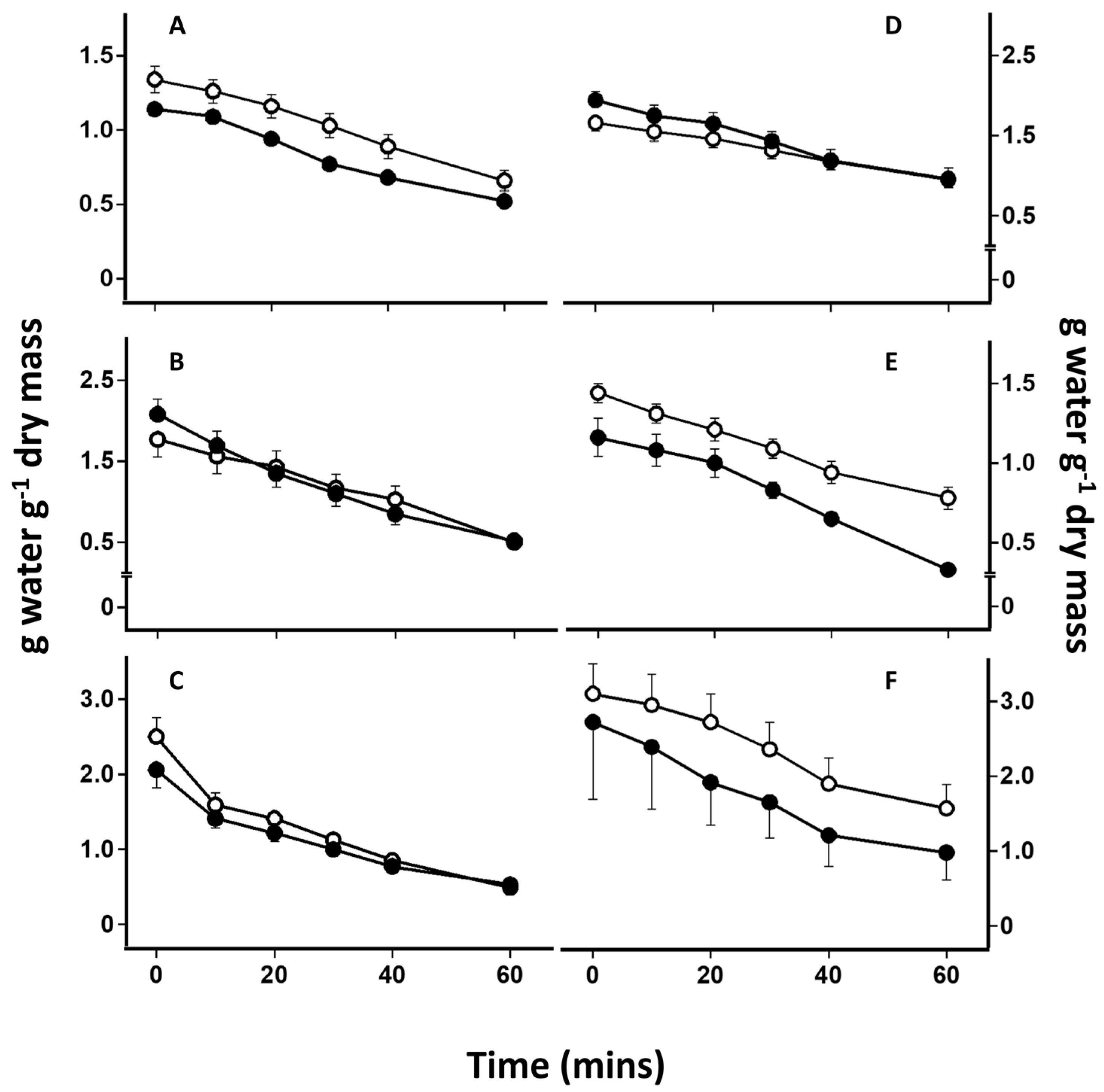
4.1. Effect of Lichen Substances on Rates of Desiccation

It was originally thought that lichen substance removal may increase rates of desiccation, possibly because of the substances in the upper cortex forming a structure analogous to the cuticle in higher plants. It has been proposed that when a thallus is wetted, lichen substances may restrict water entry into thalli, preventing excessive water uptake or “oversaturation” [14]. Oversaturation will have the effect of restricting CO2 diffusion to the photobionts for photosynthesis. Theoretically, therefore, the removal of substances could cause increased total water uptake (on a g H2O g−1 dry mass basis), reducing rates of drying. However, in practice, lichens with and without lichen substances had similar water contents at full turgor, and dried at similar rates (Figure 1). This suggests that the effect of lichen substances on the tolerance of thalli to desiccation is not simply a result of an effect on drying rate.

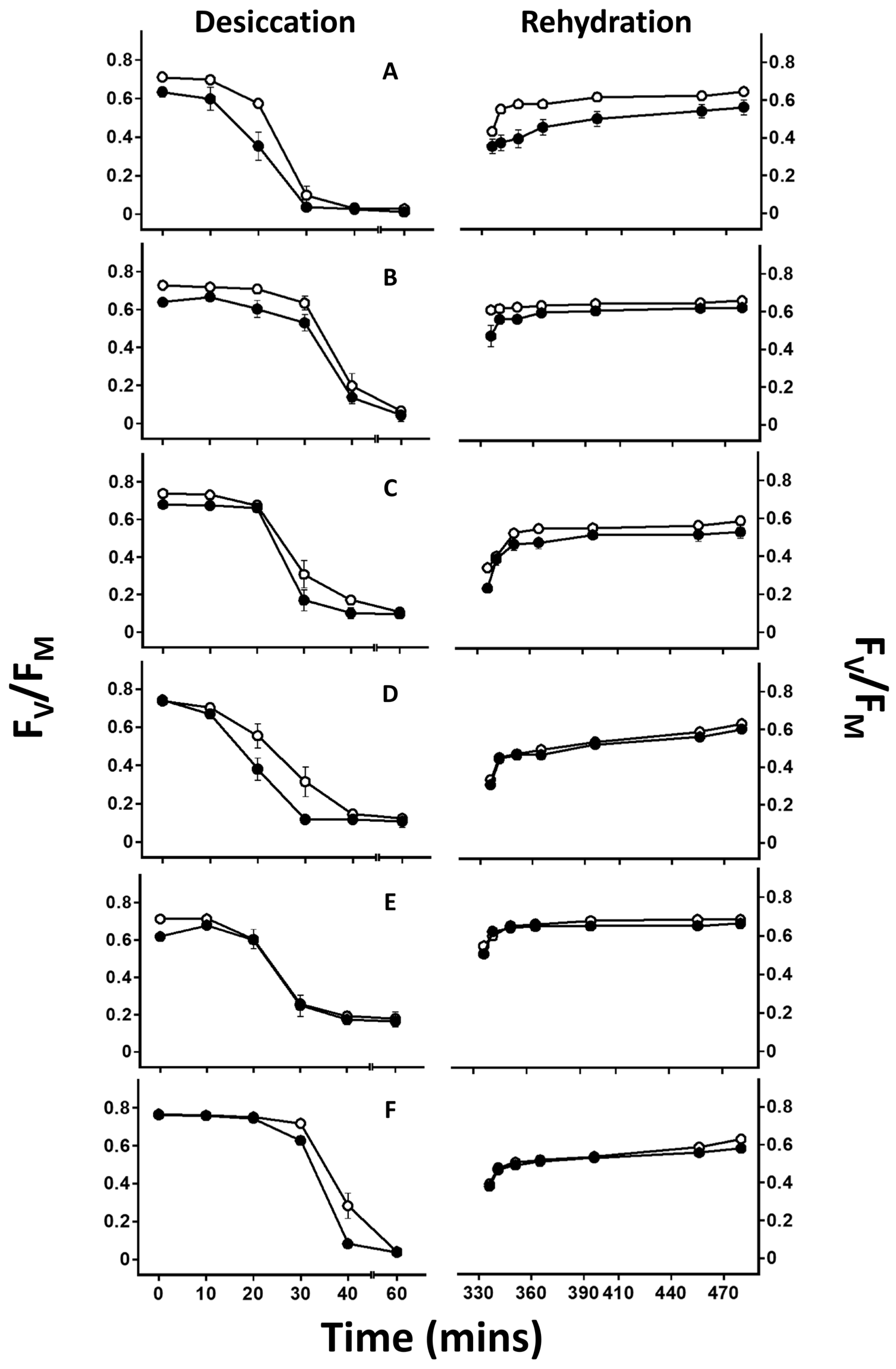
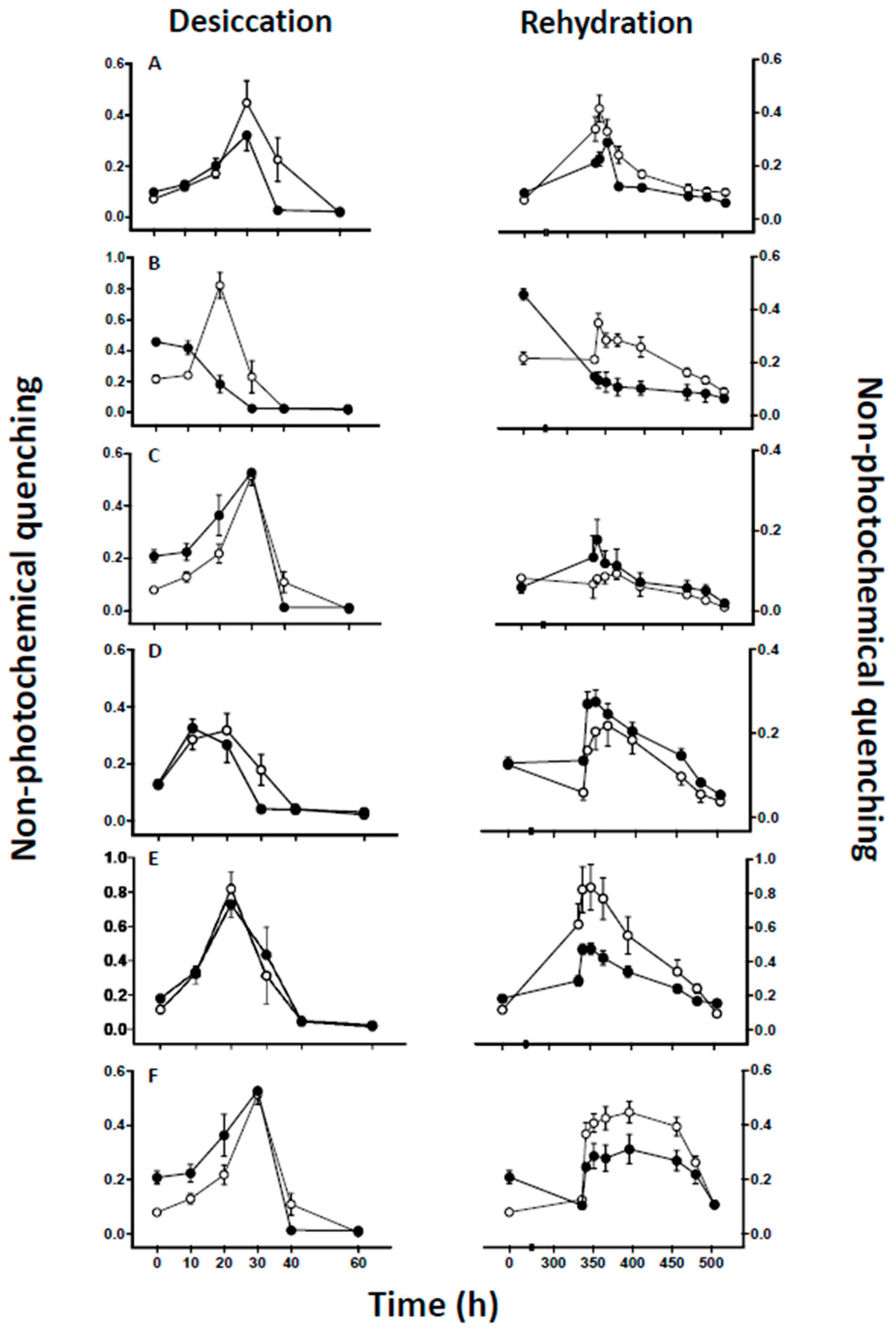
4.2. Effect of Lichen Substance Removal on Photobiont Sensitivity to Desiccation

For all species, the presence of lichen substances improves the PSII activity of the photobiont during either desiccation or rehydration, or in some cases both (Figure 2, Table 1). The most likely explanation for the improvement in PSII activity during desiccation or rehydration is that lichen substances have very high antioxidant activity [5,6,7]. Lichen substances are produced in high concentrations at considerable metabolic expense, suggesting that they play key roles in lichen biology [6]. In addition to their better documented roles in defense against pathogens and herbivores, they may increase desiccation tolerance by scavenging desiccation-induced ROS. While they are rather insoluble and occur as crystals on the surface of the mycobiont hyphae (see Section 1), lichen substances are nevertheless spatially well-positioned to intercept ROS, such as H2O2, on the surface of the surrounding fungal tissues that otherwise may diffuse from the mycobiont to the photobiont during stress. An alternative explanation could be that lichen substances increase thallus reflectance [21,24], potentially reducing any harmful effects of light during desiccation. While light can certainly increase the harmful effects of desiccation on poikilohydric organisms [14,25], here, desiccation and rehydration were deliberately carried out in the dark or under conditions of low laboratory light. Therefore, increased reflection is unlikely to be the explanation for the greater tolerance of thalli with substances present. The efficiency of lichen substances in reducing the effects of desiccation stress appears to differ between species. Lichen substance removal showed the least effect in Cladonia coniocraea (Table 1). This was surprising, because the major lichen substance present in this species has been reported to be fumarprotocetraric acid, which is also present in Cetraria islandica. However, in the present study no attempt was made to quantify the amount of lichen substances in the thalli used, and possibly the C. coniocraea used contained unusually low concentrations of fumarprotocetraric acid. Furthermore, most lichens contain a mixture of secondary substances [2], and it seems likely that the activity of one lichen substance may be modulated by the presence of other substances. While further work is needed to test these possibilities in more detail, the results presented here suggest that lichen substances exert a generally protective effect against desiccation stress in the photobiont.
As indicated above, all experiments were carried out under low laboratory lighting. However, in the field, lichens will often experience wetting and drying cycles under full or partial sunlight. As discussed by, e.g., [14], while desiccation will reduce carbon fixation by photobionts, lichen thalli will continue to absorb similar amounts of PAR. These conditions are conducive to the formation of ROS, which may cause photoinhibition. The most important mechanism used to reduce ROS formation during photosynthesis is probably the ability to “radiationlessly” dissipate excess light energy as heat through a process known as NPQ [14]. This cycle responds dynamically to changing light intensities; the carotenoid violaxanthin is enzymatically converted to zeaxanthin in a pH-regulated process that occurs during increases in light intensity. In the results reported here, consistent with reports in the literature for other lichens [26], NPQ tended to increase during drying (Figure 3). Generally, lichen substance removal had little effect on the increase of NPQ during drying. However, interestingly, in four of the six species substance removal significantly reduced NPQ during rehydration (Figure 3, Table 1). The reasons for the effects of lichen substance removal on NPQ are unclear. It would seem unlikely that the substances, which occur as crystals on the mycobiont, have direct effects on photobiont NPQ. Rather, substance removal probably increases general oxidative stress during recovery from desiccation, which may result in the loss of components of the xanthophyll cycle. In the experiments reported here, any reduction in NPQ during rehydration would have no effect on photobiont health, as they were carried out under low light conditions. However, in the field, if rehydration occurred under sunny conditions, low NPQ would further promote oxidative stress and thereby exacerbate the harmful effects of desiccation on photobiont health. It would be interesting to repeat the present experiments in the presence of light. It seems likely that the negative effect of lichen substance removal on photobionts would be even greater in the presence of light.

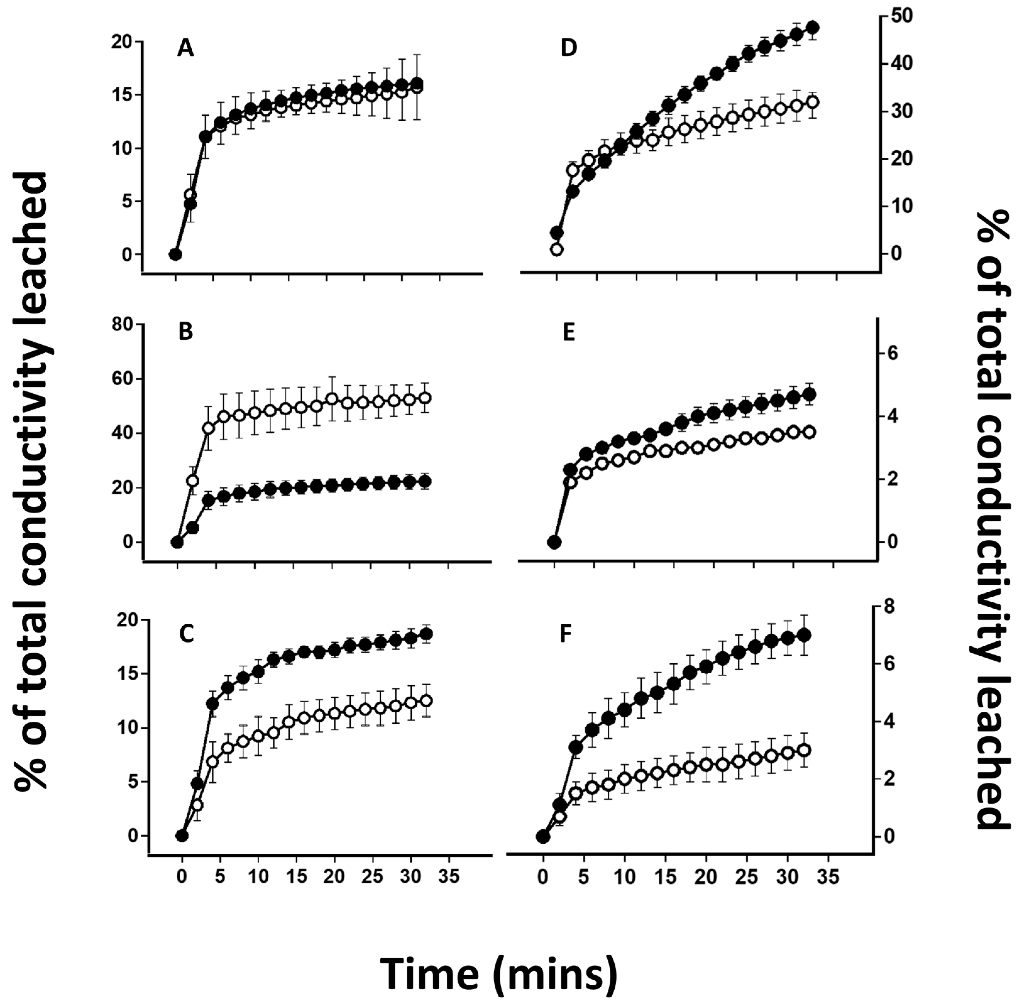
4.3. Effects of Lichen Substance Removal on the Tolerance of the Mycobiont to Desiccation

In four out of the six species tested here, substance removal increased the susceptibility of the lichens to desiccation-induced membrane leakage (Figure 4, Table 1). Membranes are known to be a major site for oxidative damage during abiotic stress [18], and this is particularly true during desiccation stress in lichens [27,28,29]. Although lichen algae can be richer in ions than fungal cells [30], they represent no more than 10% of the total volume of a lichen [31]. Therefore, most ions lost will have originated from the mycobiont. It seems most likely that, as for the photobiont, in the mycobiont the generally protective effects of lichen substances on desiccation-induced membrane damage are likely to be due to their ability to act as antioxidants. However, in Cetraria islandica substance removal had little effect on membrane leakage, and in Parmotrema perlata substance removal actually improved tolerance. As discussed in the Introduction, lichen substances can play many roles in lichen biology, and in some circumstances can act as prooxidants. Thus, for example, while protecting against herbivory or pathogen attacks, the presence of a particular secondary metabolite may increase the susceptibility of a lichen to desiccation stress. Interestingly, the ability of substances to protect the mycobiont is not always correlated with their ability to protect the photobiont. For example, while increasing the sensitivity of the mycobiont of P. perlata (Figure 4), secondary metabolites reduced the sensitivity of the photobiont (Figure 2). Therefore, depending on the species, lichen substance removal may affect the photobiont and the mycobiont in different ways. Species varied considerably in their desiccation tolerance, which was assessed as the proportion of ions that leaked during rehydration following desiccation (Figure 4). Similar variations have been reported in a survey of the amount of K+ lost into the intracellular space following desiccation stress in a range of lichen species [32]. In the present study, sensitivity to desiccation-induced ion leakage was not always consistent with the observed microhabitat of the collection site. For example, the classic shade species Crocodia aurata, which grows in a relatively moist microhabitat, was one of the most resistant. Other studies have shown that in lichens it is difficult to predict the amount of desiccation-induced ion leakage by simple visual inspection of its microhabitat [32]. For example, even the semi-aquatic species Dermatocarpon fluviatile and Lichina pygmaea display only intermediate values of ion leakage [32]. However, within a collection of individual species, ion leakage can provide a sensitive index for the effects of desiccation on membrane integrity, and results presented here suggest that lichen substances generally have a protective effect.

5. Conclusions

In this study we show that although some variation exists between species, removal of lichen substances generally increases the susceptibility of both the photobiont and the mycobiont to desiccation-induced damage. The most likely explanation for these results is that the substances can scavenge potentially harmful ROS formed during desiccation. Future work is needed to study the concentration dependence of any protective effects of lichen substances, which specific substance or combination of substances is most effective, and why, in some cases, lichen substances promote rather than reduce damage. Furthermore, lichens differing in thallus structure will differ in drying rates, and the efficacy of lichen substances at protecting against abiotic stress may depend on drying rates. It would be instructive to take a single species that responds well to substance removal, and then to dry this species at different rates with and without lichen substances. This would provide useful information as to whether the effects of substance removal may differ according to thallus structure. However, it seems clear that in addition to their more established roles in protecting lichens against pathogen attacks and grazing, lichen substances can also play a role in abiotic stress resistance.

Author Contributions

Conceptualization, R.P.B., F.M. and N.T.N.; methodology, N.T.N.; formal analysis, N.T.N.; investigation, N.T.N.; resources, R.P.B.; writing—original draft preparation, R.P.B. and N.T.N.; writing—review and editing, F.M. and R.P.B.; supervision, R.P.B.; project administration, R.P.B.; funding acquisition, R.P.B. and F.M. All authors have read and agreed to the published version of the manuscript.

Funding

R.P.B. thanks the Research Fund of the University of KwaZulu-Natal and the Kazan Federal University Strategic Academic Leadership Program (PRIORITY-2030) for financial support. N.T.N. thanks the Research Fund of the University of KwaZulu-Natal for partial financial support, and the National Research Foundation (NRF) for a postdoctoral bursary. This work was partially performed within the framework of the state assignment of the FRC KazSC RAS. F.M. thanks the Russian Science Foundation (grant no. 23-14-00327, NPQ analyses) for financial support.

Institutional Review Board Statement

Not applicable.

Informed Consent Statement

Not applicable.

Data Availability Statement

No new data were created or analyzed in this study.

Conflicts of Interest

The authors declare no conflicts of interest.

References

  1. Ahmadjian, V. Lichens are more important than you think. Bioscience 1995, 45, 124. [Google Scholar] [CrossRef]
  2. Goga, M.; Elečko, J.; Marcinčinová, M.; Ručová, D.; Bačkorová, M.; Bačkor, M. Lichen metabolites: An overview of some secondary metabolites and their biological potential. In Co-Evolution of Secondary Metabolites. Reference Series in Phytochemistry; Mérillon, J.M., Ramawat, K., Eds.; Springer: New York, NY, USA, 2020; pp. 1–36. [Google Scholar]
  3. Molnár, K.; Farkas, E. Current results on biological activities of lichen secondary metabolites: A review. Z. Naturforsch. C 2010, 65, 157–173. [Google Scholar] [CrossRef] [PubMed]
  4. Gill, S.S.; Tuteja, N. Reactive oxygen species and antioxidant machinery in abiotic stress tolerance in crop plants. Plant Physiol. Biochem 2010, 48, 909–930. [Google Scholar] [CrossRef] [PubMed]
  5. Kosanić, M.; Ranković, B.; Vukojević, J. Antioxidant properties of some lichen species. J. Food Sci. Technol. 2011, 48, 584–590. [Google Scholar] [CrossRef]
  6. Thadhani, V.M.; Choudhary, M.I.; Ali, S.; Omar, I.; Siddique, H.; Karunaratne, V. Antioxidant activity of some lichen metabolites. Nat. Prod. Res. 2011, 25, 1827–1837. [Google Scholar] [CrossRef]
  7. Fernández-Moriano, C.; González-Burgos, E.; Divakar, P.K.; Crespo, A.; Gómez-Serranillos, M.P. Evaluation of the antioxidant capacities and cytotoxic effects of ten Parmeliaceae lichen species. eCAM 2016, 2016, 3169751. [Google Scholar]
  8. Luo, H.; Yamamoto, Y.; Kim, J.A.; Jung, J.S.; Koh, Y.J.; Hur, J.S. Lecanoric acid, a secondary lichen substance with antioxidant properties from Umbilicaria antarctica in maritime Antarctica (King George Island). Pol. Biol. 2009, 32, 1033–1040. [Google Scholar]
  9. Huneck, S.; Yoshimura, I. Identification of Lichen Substances; Springer: New York, NY, USA, 1996. [Google Scholar]
  10. Kohlhardt-Floehr, C.; Boehm, F.; Troppens, S.; Lademann, J.; Truscott, T.G. Prooxidant and antioxidant behaviour of usnic acid from lichens under UVB-light irradiation—Studies on human cells. J. Photochem. Photobiol. B 2010, 101, 97–102. [Google Scholar] [CrossRef]
  11. Šeklić, D.S.; Obradović, A.D.; Stanković, M.S.; Živanović, M.N.; Mitrović, T.L.; Stamenković, S.M.; Marković, S.D. Proapoptotic and antimigratory effects of Pseudevernia furfuracea and Platismatia glauca on colon cancer cell lines. Food Technol. Biotech. 2018, 56, 421–430. [Google Scholar] [CrossRef]
  12. Peralta, M.A.; da Silva, M.A.; Ortega, M.G.; Cabrera, J.L.; Paraje, M.G. Usnic acid activity on oxidative and nitrosative stress of azole-resistant Candida albicans biofilm. Planta Med. 2018, 83, 326–333. [Google Scholar] [CrossRef]
  13. Solhaug, K.A.; Gauslaa, Y. Acetone rinsing-a method for testing ecological and physiological roles of secondary compounds in living lichens. Symbiosis 2001, 30, 301–315. [Google Scholar]
  14. Beckett, R.P.; Minibayeva, F.; Solhaug, K.A.; Roach, T. Photoprotection in lichens: Adaptations of photobionts to high light. Lichenol. 2021, 53, 21–33. [Google Scholar] [CrossRef]
  15. Manojlović, N.T.; Rančić, A.B.; Décor, R.; Vasiljević, P.; Tomović, J. Determination of chemical composition and antimicrobial, antioxidant and cytotoxic activities of lichens Parmelia conspersa and Parmelia perlata. J. Food Meas. Charact. 2021, 15, 686–696. [Google Scholar] [CrossRef]
  16. Fernandes, R.F.; Porto, A.B.; Flores, L.S.; Maia, L.F.; Corrê, C.C.; Spielmann, A.S.; Edwards, H.G.M.; de Oliveira, L.F.C. Nature of light-absorbing pigments from Brazilian lichens identified by Raman spectroscopy. Vib. Spectrosc. 2018, 99, 59–66. [Google Scholar]
  17. Moreira, A.S.N.; Braz-Filho, R.; Mussi-Dias, V.; Vieira, I.J.C. Chemistry and biological activity of Ramalina lichenized fungi. Molecules 2015, 20, 8952–8987. [Google Scholar] [CrossRef] [PubMed]
  18. Popovici, V.; Matei, E.; Cozaru, G.C.; Aschie, M.; Bucur, L.; Rambu, D.; Costache, T.; Cucolea, I.E.; Vochita, G.; Gherghel, D.; et al. Usnic acid and Usnea barbata (L.) F.H. Wigg. dry extracts promote apoptosis and DNA damage in human blood cells through enhancing ROS Levels. Antioxidants 2021, 10, 1171. [Google Scholar] [CrossRef]
  19. Hammer, S. A synopsis of the genus Cladonia in the Northwestern United States. Bryologist 1995, 98, 1–28. [Google Scholar] [CrossRef]
  20. Gudjónsdóttir, G.A.; Ingólfsdóttir, K. Quantitative determination of protolichesterinic- and fumarprotocetraric acids in Cetraria islandica by high-performance liquid chromatography. J. Chromatogr. A 1997, 757, 303–306. [Google Scholar] [CrossRef]
  21. Solhaug, K.A.; Larsson, P.; Gauslaa, Y. Light screening in lichen cortices can be quantified by chlorophyll fluorescence techniques for both reflecting and absorbing pigments. Planta 2010, 231, 1003–1011. [Google Scholar] [CrossRef]
  22. Bilger, W.; Schreiber, U.; Bock, M. Determination of the quantum efficiency of photosystem II and of non-photochemical quenching of chlorophyll fluorescence in the field. Oecologia 1995, 102, 425–432. [Google Scholar] [CrossRef]
  23. Munzi, S.; Pisani, T.; Loppi, S. The integrity of lichen cell membrane as a suitable parameter for monitoring biological effects of acute nitrogen pollution. Ecotox. Environ. Saf. 2009, 72, 2009–2012. [Google Scholar] [CrossRef]
  24. Ndhlovu, N.T.; Minibayeva, F.V.; Beckett, R.P. Unpigmented lichen substances protect lichens against photoinhibition of photosystem II in both the hydrated and desiccated states. Acta Physiol. Plant. 2022, 44, 123. [Google Scholar] [CrossRef]
  25. Challabathula, D.; Zhang, Q.; Bartels, D. Protection of photosynthesis in desiccation-tolerant resurrection plants. J. Plant Physiol. 2018, 227, 84–92. [Google Scholar] [CrossRef]
  26. Calatayud, A.; Deltoro, V.I.; Barreno, E.; del Valle-Tascon, S. Changes in in vivo chlorophyll fluorescence quenching in lichen thalli as a function of water content and suggestion of zeaxanthin-associated photoprotection. Physiol. Plant 1997, 101, 93–102. [Google Scholar] [CrossRef]
  27. Anjum, N.A.; Sofo, A.; Scopa, A.; Roychoudhury, A.; Gill, S.S.; Iqbal, M.; Ahmad, I. Lipids and proteins—Major targets of oxidative modifications in abiotic stressed plants. Environ. Sci. Poll. Res. 2015, 22, 4099–4121. [Google Scholar] [CrossRef]
  28. Kranner, I.; Cram, W.J.; Zorn, M.; Wornik, S.; Yoshimura, I.; Stabentheiner, E.; Pfeifhofer, H.W. Antioxidants and photoprotection in a lichen as compared with its isolated symbiotic partners. Proc. Natl. Acad. Sci. USA 2005, 102, 3141–3146. [Google Scholar] [CrossRef]
  29. Kranner, I.; Beckett, R.; Hochman, A.; Nash, T.H. Desiccation-tolerance in lichens: A review. Bryologist 2008, 111, 576–593. [Google Scholar] [CrossRef]
  30. Asta, J.; Garrec, J.P. Etude de la réparation du calcium, potassium, magnesium et phosphore dans les différentes couches anatomiques de dix lichens par analyse discrete á la microsonde électronique. Crypt. Bryol. Lichen. 1980, 1, 3–20. [Google Scholar]
  31. Collins, C.R.; Farrar, J.F. Structural resistance to mass transfer in the lichen Xanthoria parietina. New Phyt. 1978, 81, 71–83. [Google Scholar] [CrossRef]
  32. Buck, G.W.; Brown, D.H. Effect of desiccation on cation location in lichens. Ann. Bot. 1979, 44, 265–277. [Google Scholar] [CrossRef]
Figure 1. The effect of desiccation on the water content (g water g−1 dry mass) of control (open symbols) and acetone-rinsed (closed symbols) samples of six lichens. (A) Cetraria islandica; (B) Parmotrema perlata; (C) Ramalina celastri; (D) Usnea undulata; (E) Cladonia coniocraea; (F) Crocodia aurata. Thalli were desiccated over silica gel for 60 min. The error bars indicate the standard error of the mean, n = 5.
Microbiolres 15 00016 g001
Figure 2. The effect of desiccation over silica gel for 2 weeks and rehydration in liquid water on the maximum potential quantum efficiency of photosystem two (PSII) (FV/FM) in control (open symbols) and acetone-rinsed (closed symbols) samples of six lichens. (A) Cetraria islandica; (B) Parmotrema perlata; (C) Ramalina celastri; (D) Usnea undulata; (E) Cladonia coniocraea; (F) Crocodia aurata. The error bars indicate the standard error of the mean, n = 10.
Microbiolres 15 00016 g002
Figure 3. The effect of desiccation on nonphotochemical quenching (NPQ) at 25 μmol m−2 s−1 in control (open symbols) and acetone-rinsed (solid symbols) samples of (A) Cetraria islandica; (B) Parmotrema perlata; (C) Ramalina celastri; (D) Usnea undulata; (E) Cladonia coniocraea; and (F) Crocodia aurata following 2 weeks of desiccation followed by rapid rehydration. The error bars indicate the standard error of the mean, n = 10.
Microbiolres 15 00016 g003
Figure 4. The effect of rehydration following desiccation over silica gel for 2 weeks on the percentage of total conductivity lost in control (open symbols) and acetone-rinsed (closed symbols) samples of six lichens. (A) Cetraria islandica; (B) Parmotrema perlata; (C) Ramalina celastri; (D) Usnea undulata; (E) Cladonia coniocraea; (F) Crocodia aurata. The error bars indicate the standard error of the mean, n = 10.
Microbiolres 15 00016 g004
Table 1. Statistical analysis (ANOVA) of the effects of desiccation on photobiont responses (FV/FM), non-photochemical quenching (NPQ), and mycobiont response (ion leakage) in thalli of Cetraria islandica, Parmotrema perlata, Ramalina celastri, Usnea undulata, Cladonia coniocreae, and Crocodia aurata. For all comparisons, the error had 179 degrees of freedom. Significance: * = p < 0.05, ** = p < 0.01, *** = p < 0.001.
SpeciesWaterFV/FMNPQIon
ContentDesiccationRehydrationDesiccationRehydrationLeakage
Cetraria islandica********0.083***0.237
Parmotrema perlata******************
Ramalina celastri0.1900.552***0.6860.132***
Usnea undulata****0.8520.388******
Cladonia coniocraea0.1120.533*0.981******
Crocodia aurata0.204***0.136*******
Disclaimer/Publisher’s Note: The statements, opinions and data contained in all publications are solely those of the individual author(s) and contributor(s) and not of MDPI and/or the editor(s). MDPI and/or the editor(s) disclaim responsibility for any injury to people or property resulting from any ideas, methods, instructions or products referred to in the content.

