
Vaccine-Induced Subacute Thyroiditis (De Quervain’s) after mRNA Vaccine against SARS-CoV-2: A Case Report and Systematic Review

 ,
,  ,
,  , , ,
, , ,
Abstract
:1. Introduction
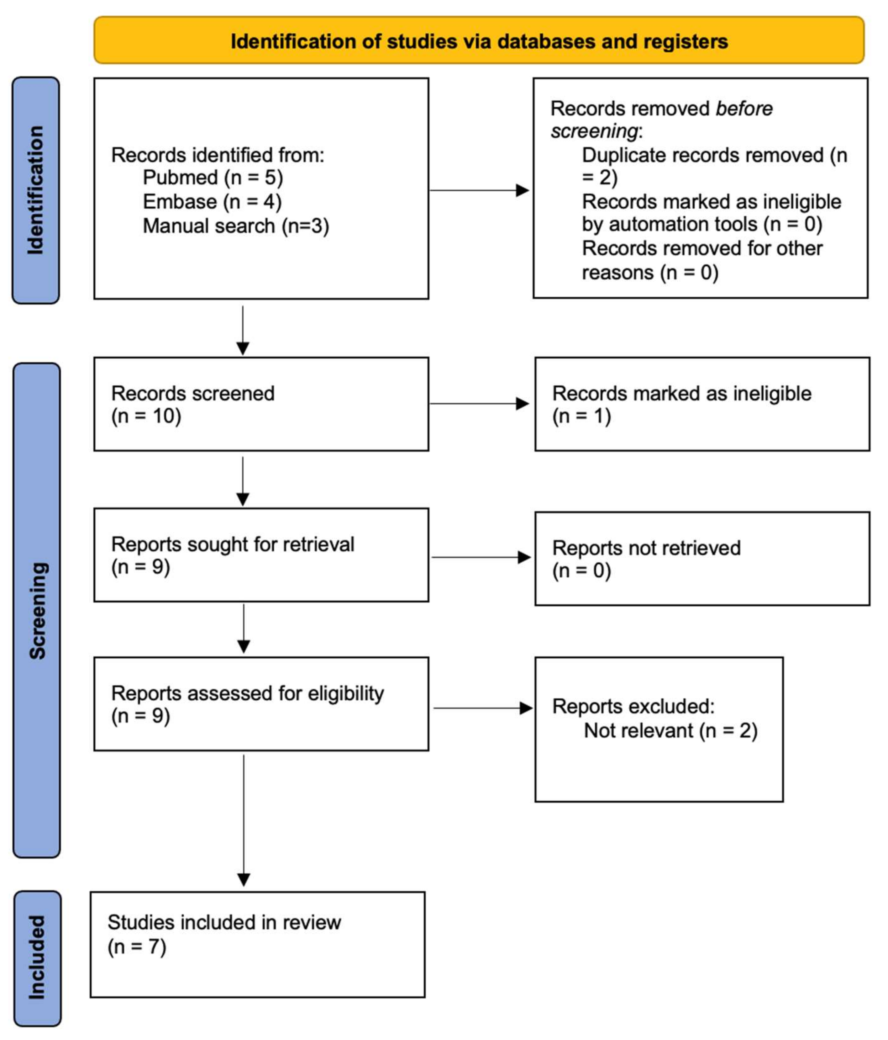
2. Materials and Methods
Systematic Review of the Literature
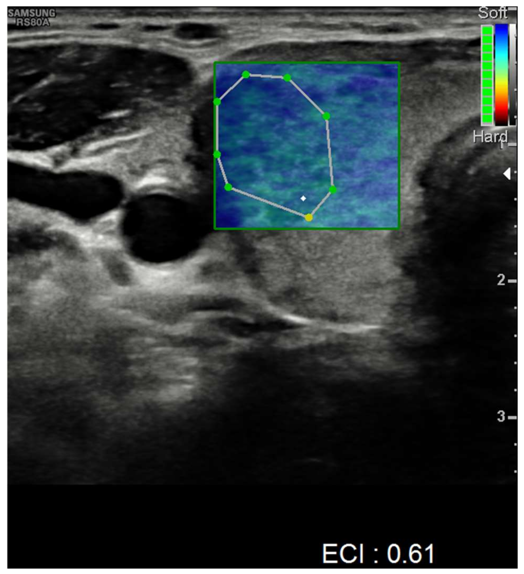
3. Results
4. Discussion
5. Conclusions
Author Contributions
Funding
Institutional Review Board Statement
Informed Consent Statement
Conflicts of Interest
References
- Huang, C.; Huang, L.; Wang, Y.; Li, X.; Ren, L.; Gu, X.; Kang, L.; Guo, L.; Liu, M.; Zhou, X.; et al. 6-month consequences of COVID-19 in patients discharged from hospital: A cohort study. Lancet 2021, 397, 220–232. [Google Scholar] [CrossRef]
- WHO COVID-19 Dashboard. Available online: https://covid19.who.int/ (accessed on 12 August 2021).
- Sohrabpour, S.; Heidari, F.; Karimi, E.; Ansari, R.; Tajdini, A.; Heidari, F. Subacute Thyroiditis in COVID-19 Patients. Eur. Thyroid J. 2020, 9, 321–323. [Google Scholar] [CrossRef] [PubMed]
- Mehmood, M.A.; Bapna, M.; Arshad, M. A Case of Post-COVID-19 Subacute Thyroiditis. Cureus 2020, 12, e12301. [Google Scholar] [CrossRef] [PubMed]
- Sahin, D.; Akpolat, İ. Diagnostic cytological features and differential diagnosis of subacute granulomatous (De Quervain’s) thyroiditis. Diagn. Cytopathol. 2019, 47, 1251–1258. [Google Scholar] [CrossRef]
- Slatosky, J.; Shipton, B.; Wahba, H. Thyroiditis: Differential diagnosis and management. Am. Fam. Physician 2000, 61, 1047–1052, 1054. [Google Scholar]
- Samuels, M.H. Subacute, Silent, and Postpartum Thyroiditis. Med. Clin. N. Am. 2012, 96, 223–233. [Google Scholar] [CrossRef]
- Kojima, M.; Nakamura, S.; Oyama, T.; Sugihara, S.; Sakata, N.; Masawa, N. Cellular Composition of Subacute Thyroiditis. An Immunohistochemical Study of Six Cases. Pathol.—Res. Pract. 2002, 198, 833–837. [Google Scholar] [CrossRef]
- Trivedi, D.P.; Bhagat, R.; Nakanishi, Y.; Wang, A.; Moroz, K.; Falk, N.K. Granulomatous Thyroiditis: A Case Report and Literature Review. Ann. Clin. Lab. Sci. 2017, 47, 620–624. [Google Scholar]
- Kong, M.; La Porte, S. Case Report: De Quervain’s Thyroiditis as a Long-Term Sequelae Complication to SARS-CoV-2 Infection. Case Rep. Acute Med. 2021, 4, 64–70. [Google Scholar] [CrossRef]
- Frates, M.C.; Marqusee, E.; Benson, C.B.; Alexander, E.K. Subacute Granulomatous (de Quervain) Thyroiditis. J. Ultrasound Med. 2013, 32, 505–511. [Google Scholar] [CrossRef]
- Ranganath, R.; Shaha, M.A.; Xu, B.; Migliacci, J.; Ghossein, R.; Shaha, A.R. De Quervain’s thyroiditis: A review of experience with surgery. Am. J. Otolaryngol. 2016, 37, 534–537. [Google Scholar] [CrossRef] [PubMed] [Green Version]
- Wei, L.; Sun, S.; Xu, C.; Zhang, J.; Xu, Y.; Zhu, H.; Peh, S.; Korteweg, C.; McNutt, M.A.; Gu, J. Pathology of the thyroid in severe acute respiratory syndrome. Hum. Pathol. 2007, 38, 95–102. [Google Scholar] [CrossRef] [PubMed]
- Muller, I.; Cannavaro, D.; Dazzi, D.; Covelli, D.; Mantovani, G.; Muscatello, A.; Ferrante, E.; Orsi, E.; Resi, V.; Longari, V.; et al. SARS-CoV-2-related atypical thyroiditis. Lancet Diabetes Endocrinol. 2020, 8, 739–741. [Google Scholar] [CrossRef]
- Rotondi, M.; Coperchini, F.; Ricci, G.; Denegri, M.; Croce, L.; Ngnitejeu, S.T.; Villani, L.; Magri, F.; Latrofa, F.; Chiovato, L. Detection of SARS-CoV-2 receptor ACE-2 mRNA in thyroid cells: A clue for COVID-19-related subacute thyroiditis. J. Endocrinol. Investig. 2021, 44, 1085–1090. [Google Scholar] [CrossRef]
- İremli, B.G.; Şendur, S.N.; Ünlütürk, U. Three Cases of Subacute Thyroiditis Following SARS-CoV-2 Vaccine: Postvaccination ASIA Syndrome. J. Clin. Endocrinol. Metab. 2021, 106, 2600–2605. [Google Scholar] [CrossRef]
- Şahin Tekin, M.; Şaylısoy, S.; Yorulmaz, G. Subacute thyroiditis following COVID-19 vaccination in a 67-year-old male patient: A case report. Hum. Vaccin. Immunother. 2021, 17, 4090–4092. [Google Scholar] [CrossRef]
- Franquemont, S.; Galvez, J. Subacute Thyroiditis After mRNA Vaccine for COVID-19. J. Endocr. Soc. 2021, 5, A956–A957. [Google Scholar] [CrossRef]
- Bornemann, C.; Woyk, K.; Bouter, C. Case Report: Two Cases of Subacute Thyroiditis Following SARS-CoV-2 Vaccination. Front. Med. 2021, 8, 737142. [Google Scholar] [CrossRef]
- Oyibo, S.O. Subacute Thyroiditis After Receiving the Adenovirus-Vectored Vaccine for Coronavirus Disease (COVID-19). Cureus 2021, 13, e16045. [Google Scholar] [CrossRef]
- Patel, K.R.; Cunnane, M.E.; Deschler, D.G. SARS-CoV-2 vaccine-induced subacute thyroiditis. Am. J. Otolaryngol. 2022, 43, 103211. [Google Scholar] [CrossRef]
- Patel, M.; Shahid, M.; Khawaja, A.; Ejike, C.; Vemuri, K. Subacute Thyroiditis Secondary to Moderna COVID-19 Vaccine: A Case Report of a Rare Manifestation. Adv. Clin. Med. Res. Healthc. Deliv. 2021, 1(2), 9. [Google Scholar] [CrossRef]
- Page, M.J.; McKenzie, J.E.; Bossuyt, P.M.; Boutron, I.; Hoffmann, T.C.; Mulrow, C.D.; Shamseer, L.; Tetzlaff, J.M.; Akl, E.A.; Brennan, S.E.; et al. The PRISMA 2020 statement: An updated guideline for reporting systematic reviews. BMJ 2021, 372, n71. [Google Scholar] [CrossRef] [PubMed]
- Aemaz Ur Rehman, M.; Farooq, H.; Ali, M.M.; Ebaad Ur Rehman, M.; Dar, Q.A.; Hussain, A. The Association of Subacute Thyroiditis with COVID-19: A Systematic Review. SN Compr. Clin. Med. 2021, 3, 1515–1527. [Google Scholar] [CrossRef]
- Watad, A.; Quaresma, M.; Brown, S.; Cohen Tervaert, J.W.; Rodríguez-Pint, I.; Cervera, R.; Perricone, C.; Shoenfeld, Y. Autoimmune/inflammatory syndrome induced by adjuvants (Shoenfeld’s syndrome)—An update. Lupus 2017, 26, 675–681. [Google Scholar] [CrossRef]
- Xia, X. Domains and Functions of Spike Protein in SARS-CoV-2 in the Context of Vaccine Design. Viruses 2021, 13, 109. [Google Scholar] [CrossRef] [PubMed]
- Chung, Y.H.; Beiss, V.; Fiering, S.N.; Steinmetz, N.F. COVID-19 Vaccine Frontrunners and Their Nanotechnology Design. ACS Nano 2020, 14, 12522–12537. [Google Scholar] [CrossRef] [PubMed]

| Oyibo | Franquemont and Galvez | Şahin Tekin et al. | İremli et al. | Bornemann et al. | |||||||||||||
|---|---|---|---|---|---|---|---|---|---|---|---|---|---|---|---|---|---|
| At Presentation | At 6 Weeks | At 12 Weeks | At Presentation | At 3 Weeks | At Presentation | At 9 Weeks | 1 at Presentation | 1 at 4 Weeks | 2 at Presentation | 2 at 4 Weeks | 3 at Presentation | 3 at 4 Weeks | 1 at Presentation | 1 At 6 Weeks | 2 at Presentation | 2 at 4 Weeks | |
| Gender | Female | Female | Male | Female | Female | Female | Female | Female | |||||||||
| Age | 55 | 42 | 67 | 35 | 34 | 37 | 26 | 49 | |||||||||
| Vaccine | (I dose) ChAdOx1®, AstraZeneca plc (Cambridge, UK) | (I dose) Comirnaty, Pfizer | (II doses) CoronaVac®, Sinovac Biotech (Beijing, China) | (II doses) CoronaVac®, Sinovac | (I dose-prev. infection) CoronaVac®, Sinovac | (II doses) CoronaVac®, Sinovac | (I dose) ChAdOx1, AstraZeneca® | (I dose) Spikevax®, Moderna inc. (Massachusetts, USA) | |||||||||
| Symptom onset from vaccination (days) | 21 days | 5 days | 19 days (from II dose) | 5 days (from second dose) | 4 days | 7 days (from II dose) | 14 days | 14 days | |||||||||
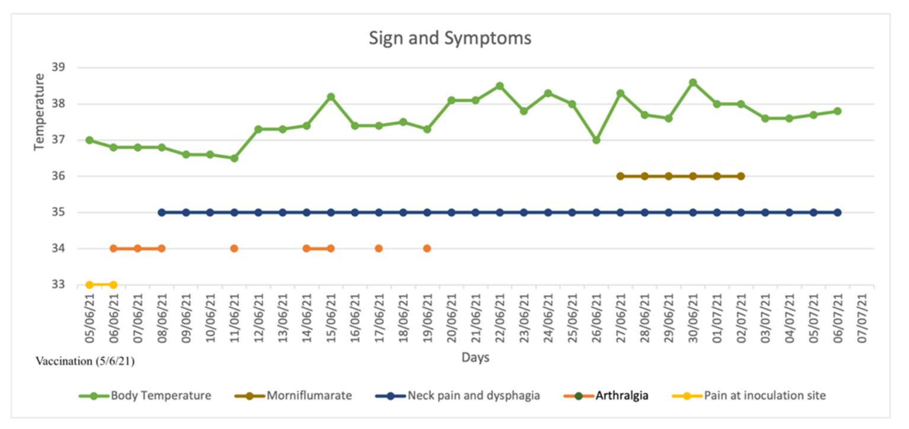
| Signs and symptoms at presentation | Neck pain and swelling, headache, palpitations, sore throat, generalized aches. | Sore throat, palpitations, sinus tachycardia on EKG (>130 bpm) | Fever, hypertension, weight loss (−4 kg in less than 30 days), left ear and anterior neck pain and tenderness at palpation | Severe neck pain, palpitations, fever, fatigue. Thyroid tenderness and enlargement | Anterior neck pain, fatigue, weight loss, fever, palpitations | Anterior neck pain and tenderness. | Cervical pain, fever, chills, thyroid tenderness, cervical lymphadenopathy | Headaches, difficulty concentrating, cervical sore throat, thyroid tenderness | |||||||||
| TSH mU/L (r.v. 0.3–4.2) | 0.09 | 20.3 | 5.35 | <0.01 | <0.01 | <0.005 | 3.15 | 0.47 | 2.27 | 0.01 | <0.015 | 0.9 | 0.018 | 1.75 | 0.83 | 0.05 | 0.01 |
| fT3 ng/mL (r.v. 2.7–4.3) | NA | NA | 4.2 | 7.68 | 8.78 | 8.06 | 2.94 | 4.01 | 3.45 | 7.68 | 5.22 | 3.94 | 4.55 | 3.72 | 2.6 | 3.25 | 3.97 |
| fT4 pg/mL (r.v. 0.93–1.7) | 1.96 | 4.7 | 1.21 | 4.58 | 3.2 | 2.87 | 0.97 | 1.09 | 1.49 | 0.4 | 2 | 1.07 | 2.03 | 0.72 | 0.69 | 0.73 | 1.08 |
| Anti-TPO IU/mL (r.v.) | <10 (r.v. < 34) | NA | NA | <28 | NA | Neg | NA | 1.2 (0–9) | NA | 1.2 | NA | 4.1 | NA | Neg | Neg | Neg | 33 (<6) |
| Anti-TG IU/mL (r.v.) | NA | NA | 15 (r.v. < 3) | NA | Neg | Neg | NA | <0.9 | NA | <0.9 | NA | <0.9 | NA | Neg | Neg | Neg | Neg |
| TRAB IU/L (r.v.) | NA | NA | 1.9 (<2.9) | NA | Neg | Neg | NA | <1.5 | NA | <1.5 | NA | <1.5 | NA | Neg | Neg | Neg | Neg |
| ESR (mm/hr) | 51 | NA | NA | 62 | 26 | 67 | 4 | 53 | 28 | 19 | 16 | 25 | 44 | NA | NA | NA | NA |
| CRP (mg/L) | 87 | NA | NA | NA | Normal | 53.9 | 3.6 | 100.5 | 13.1 | 6 | 5.3 | 2.4 | NA | 29.4 | 1.0 | 21.9 | 22.4 |
| WBC (109/L) | 8.5 | NA | NA | NA | NA | NA | NA | 9.9 | 11.1 | 9.7 | 9.7 | 6.3 | 7.8 | 14.3 | 9.77 | 7.86 | 5.75 |
| PLT (109/L) | 491 | NA | NA | NA | NA | NA | NA | NA | NA | NA | NA | NA | NA | NA | NA | NA | NA |
| HLA-B35 | NA | NA | NA | NA | NA | NA | NA | NA | |||||||||
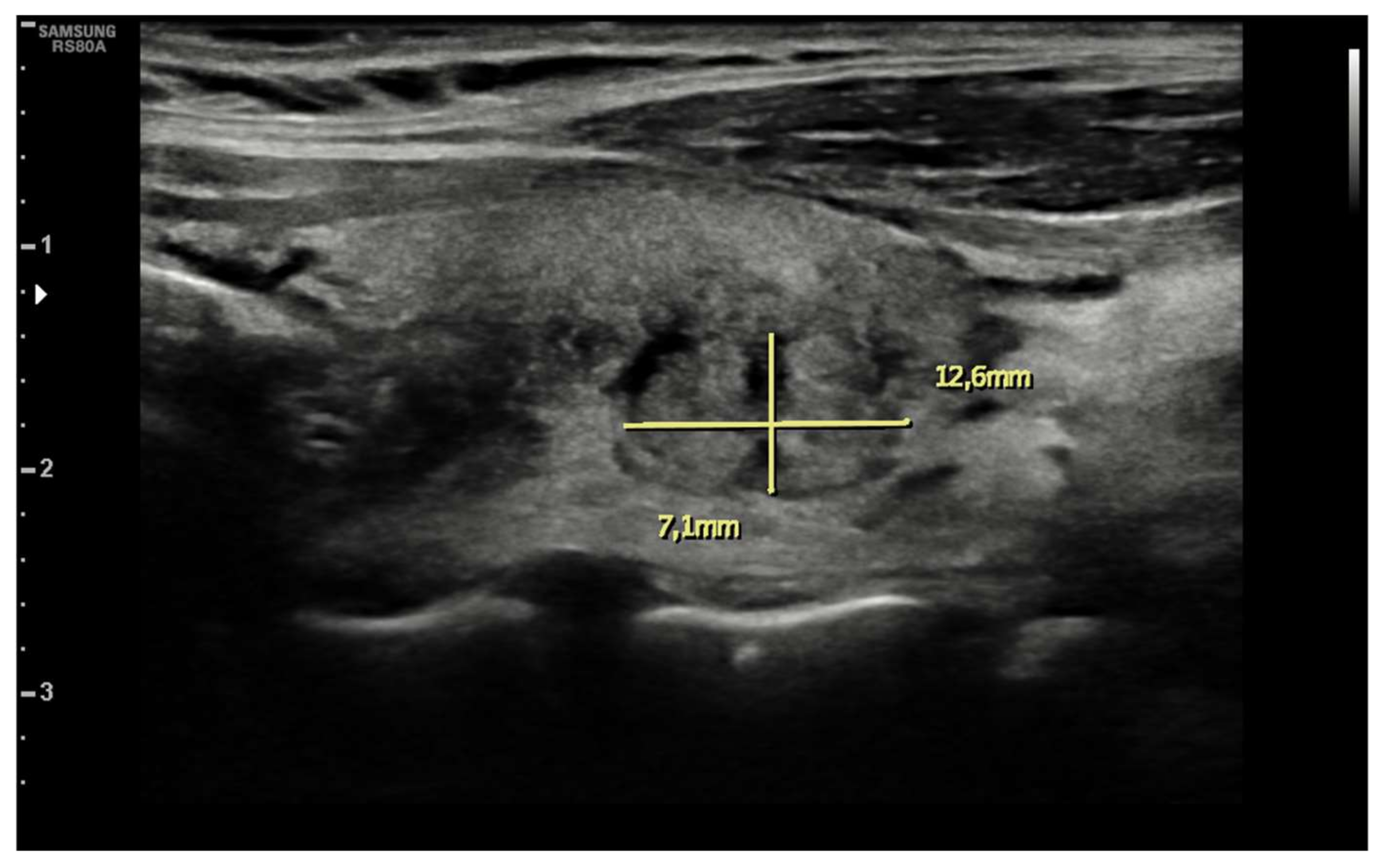
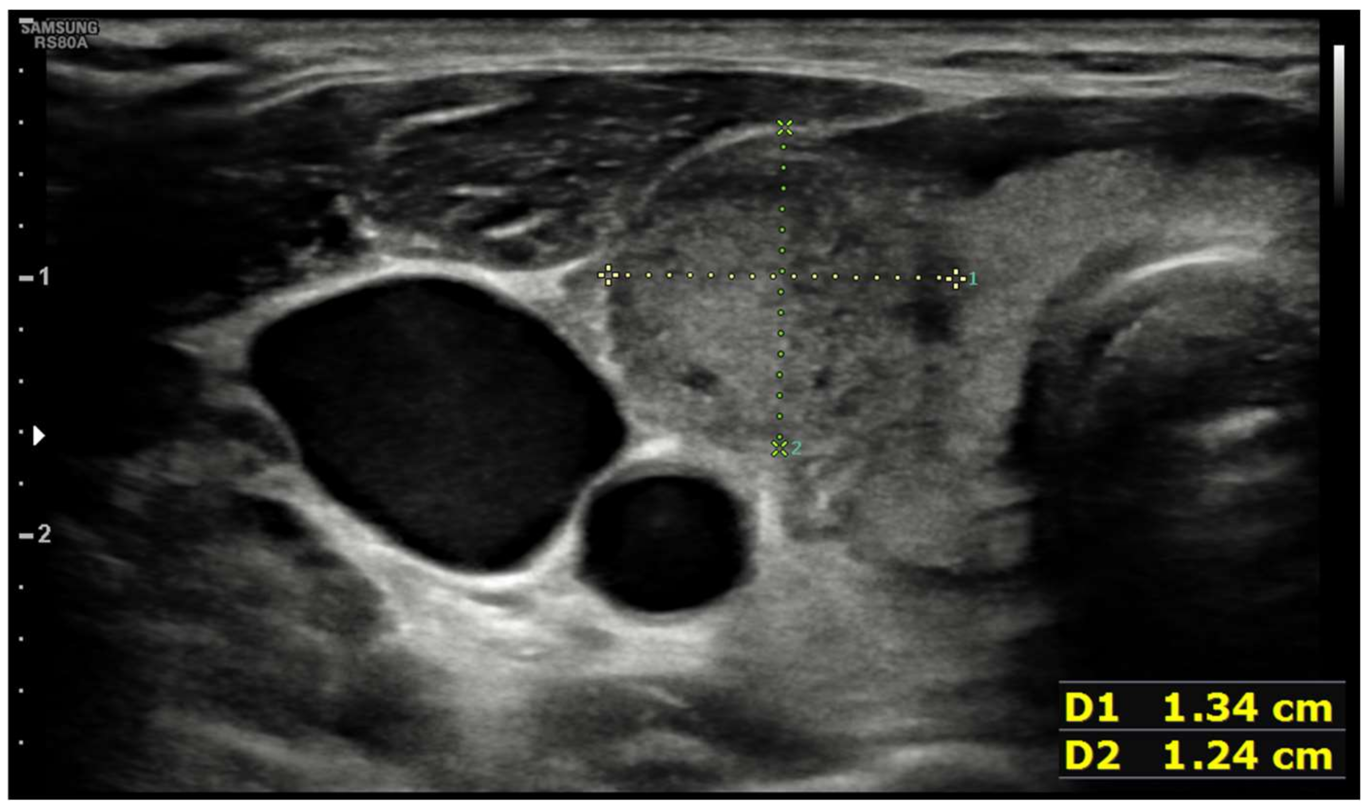
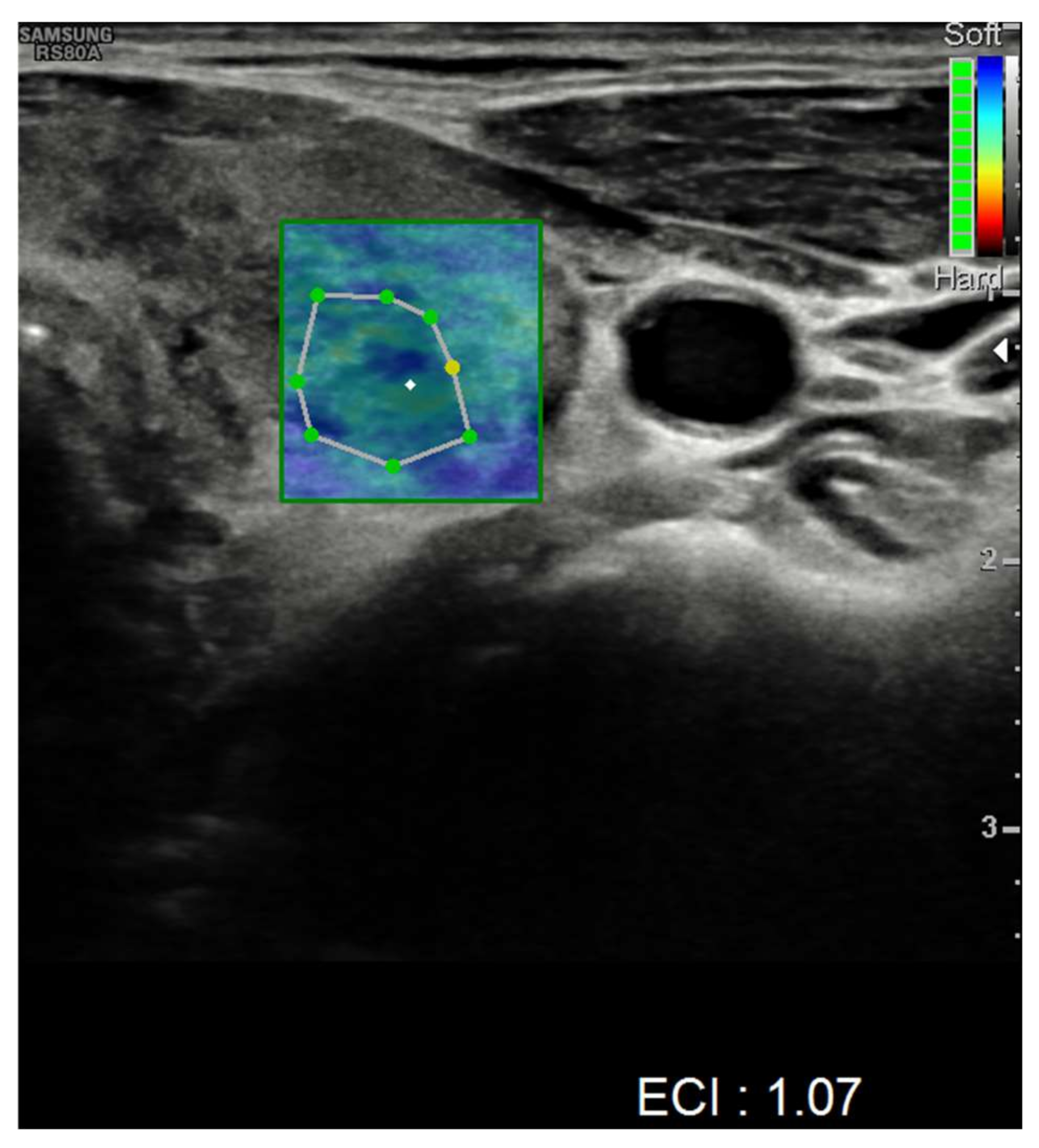
| Ultrasound findings at presentation | Enlarged thyroid gland with heterogeneous echotexture. No nodules no hypervascularity; reduced vascular flow to right lobe | NA | Reduced echogenicity and diffusely heterogeneous texture with pseudonodular areas consistent with thyroiditis | Bilateral focal hypoechoic areas with decreased blood flow on Doppler US | Bilateral focal hypoechoic areas with decreased blood flow on Doppler US | Numerous hypoechoic areas with decreased blood flow on Doppler US | Heterogeneous echogenicity and bilateral hypoechoic areas with decreased blood flow on Doppler US | Hypoechoic confluent areas with decreased vascularity on Doppler US | |||||||||
| Therapy | Propranolol, ibuprofen, paracetamol | Levothyroxine | Levothyroxine | Prednisone, propranolol | NA | Ibuprofen | - | Methylprednisolone, propranolol | methylprednisolone | Methylprednisolone, propranolol | methylprednisolone | No treatment | No treatment | Ibuprofen, prednisolone | - | Diclofenac | Prednisolone |
| Patel M. et al. | Patel KR. et al. | Pipitone et al. | |||||||||||||||
| At presentation | At 4 weeks | At presentation | At <8 weeks | At presentation | At 21 days | ||||||||||||
| Gender | Female | Male | Female | ||||||||||||||
| Age | 46 | 48 | 49 | ||||||||||||||
| Vaccine | (II doses) Spikevax, Moderna® | (II doses) Unspecified SARS-CoV-2 vaccine | (I dose) Comirnaty, Pfizer® | ||||||||||||||
| Symptom onset from vaccination (days) | 32 days from I dose (right following II dose) | 7 days after II dose | 7 days | ||||||||||||||
| Signs and symptoms at presentation | Neck pain, tender, warm, swollen, and firm area on right thyroid. Tachycardia (119 bpm), hypertension (146/76 mmHg). | Right neck swelling, throat discomfort, palpitations, fever, weight loss (−4.5 kg). Tender fullness on right anterior neck. | Anterior cervical pain, anxiety, dysphagia, fatigue, increased bowel movements, insomnia, night sweats, palpitations, tachycardia (115 bpm), fever (38.6 °C), weight loss (−8%) | ||||||||||||||
| TSH mU/L | 0.00 | 0.365 | 0.01 | Suppressed | <0.008 | 0.208 | |||||||||||
| fT3 pg/mL (r.v.) | Elevated | 2.96 | NA | NA | 12.9 (2.3–4.2) | 1.52 | |||||||||||
| fT4 ng/dL (r.v.) | Elevated | 1.02 | 3.6 | decreased | 5.74 | 0.73 | |||||||||||
| Anti-TPO IU/mL (r.v.) | Negative | NA | NA | NA | <28 (<60) | NA | |||||||||||
| Anti-TG IU/mL (r.v.) | Negative | NA | NA | NA | 0.2 (<4.5) | NA | |||||||||||
| TRAB IU/L (r.v.) | NA | NA | NA | NA | <1.8 (<1.8) | NA | |||||||||||
| ESR (mm/hr) | 49 | NA | Elevated | Decreased | 86 | 25 | |||||||||||
| CRP (mg/L) | 86 | NA | Elevated | Decreased | 78 | NA | |||||||||||
| WBC (109/L) | NA | NA | NA | NA | 10,8 | NA | |||||||||||
| PLT (109/L) | NA | NA | NA | NA | 455 | NA | |||||||||||
| HLA-B35 | NA | NA | * 35, * 55 | ||||||||||||||
| Ultrasound findings at presentation | Asymmetric thyroid enlargement with two hypoechoic areas with no internal vascularity on right thyroid lobe | Diffuse thyroid enlargement with hypoechoic areas with heterogeneous echotexture and no evidence of increased vascularity | Diffuse enlargement of thyroid gland with hypoechoic nodules with hyperechoic shoots (micronodular pattern) without increase in vascularity | ||||||||||||||
| Therapy | Analgesics, dexamethasone, propranolol | - | Prednisolone and NSAIDs | - | Prednisolone, propranolol | - | |||||||||||
| Overall (n = 10) | mRNA Vaccines (n = 4) | Inactivated/Vector Vaccines (n = 6) | p-Value | |
|---|---|---|---|---|
| Female | 9 (90%) | 4 (100%) | 5 (83.3%) | 1 |
| SAT after I dose | 6 (60%) | 3 (75%) | 3 (50%) | 0.571 |
| Days between I/II dose and symptoms onset | 10.5 (5–19.5) | 10.5 (5.5–27.5) | 10.5 (4.75–19.5) | 0.762 |
| Age (years) | 44 (34.75–50.50) | 47.5 (43–49) | 36 (32–58) | 0.476 |
| Symptoms | ||||
| Neck pain | 9 (90%) | 3 (75%) | 6 (100%) | 0.400 |
| Sore Throat | 4 (40%) | 3 (75%) | 1 (16.7%) | 0.191 |
| Palpitation/Tachycardia | 6 (60%) | 3 (75%) | 3 (50%) | 0.571 |
| Headache | 3 (30%) | 2 (50%) | 1 (16.7%) | 0.333 |
| Fever | 5 (50%) | 1 (25%) | 4 (66.7%) | 0.262 |
| Fatigue | 3 (30%) | 1 (25%) | 2 (33.3%) | 0.667 |
| Weight loss | 3 (30%) | 1 (25%) | 2 (33.3%) | 1 |
| Hypertension | 3 (30%) | 2 (50%) | 1 (16.7%) | 0.333 |
| Anxiety | 1 (10%) | 1 (25%) | 0 (0%) | 0.400 |
| Night sweats | 1 (10%) | 1 (25%) | 0 (0%) | 0.400 |
| Increase in bowel motility | 1 (10%) | 1 (25) | 0 (0%) | 0.400 |
| TSH at T0 (mU/L) | 0.03 (0.073–0.578) | 0.09 (0.02–0.04) | 0.28 (0.08–1.11) | 0.114 |
| TSH at T4 | 0.287 (0.10–2.49) | 0.109 (0.01–0.33) | 1.55 (0.01–3.70) | 0.257 |
| fT3 at T0 (pg/mL) | 4.01 (3.49–7.87) | 7.68 (3.25-) | 3.98 (2.79–7.78) | 0.548 |
| fT3 at T4 | 3.71 (2.86–4.72) | 3.47 (1.88–7.58) | 3.83 (2.86–4.72) | 1 |
| fT4 at T0 (ng/dL) | 1.09 (0.73–3.73) | 4.58 (0.73-) | 1.08 (0.64–2.19) | 0.262 |
| fT4 at T4 | 1.35 (0.91–2.26) | 1.05 (0.8–2.67) | 1.35 (0.90–2.01) | 0.610 |
| ESR at T0 (mm/h) | 52 (31–65.75) | 62 (49-) | 51 (22–60) | 0.393 |
| ESR at T4 | 25.5 (13–32) | 25.5 (25-) | 22 (7–40) | 1 |
| CRP at T0 (mg/L) | 53.9 (13.95–86.5) | 78 (21.9-) | 41.65 (5.1–90.38) | 0.905 |
| CRP at T4 | 5.3 (2.3–17.75) | 22.4 (22.4–22.4) | 4.45 (1.65–11.15) | 0.400 |
| WBC at T0 (cells/mL) | 9700 (7860–10,800) | 9330 (7860-) | 9700 (7400–12,100) | 1 |
| WBC at T4 | 9700 (6775–10,435) | 5750 (5750–5750) | 9735 (8275–10,770) | 0.400 |
| Pharmacological treatment | ||||
| NSAIDs | 4 (40%) | 1 (25%) | 3 (50%) | 0.571 |
| Steroids | 7 (70%) | 4 (100%) | 3 (50%) | 0.200 |
| Beta-blockers | 5 (50%) | 2 (50%) | 3 (50%) | 0.738 |
| Methimazole | 1 (10%) | 1 (25%) | 0 (0%) | 0.400 |
| Levothyroxine | 1 (10%) | 0 (0%) | 1 (16.7%) | 0.600 |
| Ultrasonographic findings | ||||
| Hypoechoic areas | 8 (80%) | 3 (75%) | 5 (83.3%) | 1 |
| Heterogeneous echotexture | 4 (40%) | 1 (25%) | 3 (50%) | 0.571 |
| Reduced blood flow at Doppler-US | 6 (60%) | 1 (25%) | 5 (83.3%) | 0.119 |
Publisher’s Note: MDPI stays neutral with regard to jurisdictional claims in published maps and institutional affiliations. |
© 2022 by the authors. Licensee MDPI, Basel, Switzerland. This article is an open access article distributed under the terms and conditions of the Creative Commons Attribution (CC BY) license (https://creativecommons.org/licenses/by/4.0/).
Share and Cite
Pipitone, G.; Rindi, L.V.; Petrosillo, N.; Foti, N.A.M.; Caci, G.; Iaria, C.; Donno, D.R.; Boumis, E.; Paviglianiti, G.; Taglietti, F. Vaccine-Induced Subacute Thyroiditis (De Quervain’s) after mRNA Vaccine against SARS-CoV-2: A Case Report and Systematic Review. Infect. Dis. Rep. 2022, 14, 142-154. https://doi.org/10.3390/idr14010018
Pipitone G, Rindi LV, Petrosillo N, Foti NAM, Caci G, Iaria C, Donno DR, Boumis E, Paviglianiti G, Taglietti F. Vaccine-Induced Subacute Thyroiditis (De Quervain’s) after mRNA Vaccine against SARS-CoV-2: A Case Report and Systematic Review. Infectious Disease Reports. 2022; 14(1):142-154. https://doi.org/10.3390/idr14010018
Chicago/Turabian StylePipitone, Giuseppe, Lorenzo Vittorio Rindi, Nicola Petrosillo, Nunzio Adalberto Maria Foti, Grazia Caci, Chiara Iaria, Davide Roberto Donno, Evangelo Boumis, Giuseppe Paviglianiti, and Fabrizio Taglietti. 2022. "Vaccine-Induced Subacute Thyroiditis (De Quervain’s) after mRNA Vaccine against SARS-CoV-2: A Case Report and Systematic Review" Infectious Disease Reports 14, no. 1: 142-154. https://doi.org/10.3390/idr14010018
APA StylePipitone, G., Rindi, L. V., Petrosillo, N., Foti, N. A. M., Caci, G., Iaria, C., Donno, D. R., Boumis, E., Paviglianiti, G., & Taglietti, F. (2022). Vaccine-Induced Subacute Thyroiditis (De Quervain’s) after mRNA Vaccine against SARS-CoV-2: A Case Report and Systematic Review. Infectious Disease Reports, 14(1), 142-154. https://doi.org/10.3390/idr14010018