




Next Issue
Volume 18, August
Previous Issue
Volume 18, June


Neurol. Int., Volume 18, Issue 7 (July 2026) – 19 articles
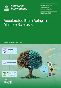
Cover Story (view full-size image): Multiple sclerosis (MS) is linked to accelerated biological brain aging. While the structural MRI-derived ‘brain age gap’ (BAG) is a promising biomarker, its underlying subclinical substrates remain unexplored. This study combines volumetric, spectroscopic, and diffusion imaging to show that this gap extends beyond macroscopic atrophy, capturing subclinical metabolic failure (reduced tNAA/tCr) and microstructural damage (reduced MTR and FA) in normal-appearing tissues. The findings position the BAG as a multi-dimensional proxy for neuroaxonal pathology. The cover image illustrates this concept via a metaphorical brain-tree showing one half healthy and the other withered, alongside diagrams representing machine learning-based brain age prediction and its correlates. View this paper
  • Issues are regarded as officially published after their release is announced to the table of contents alert mailing list.
  • You may sign up for e-mail alerts to receive table of contents of newly released issues.
  • PDF is the official format for papers published in both, html and pdf forms. To view the papers in pdf format, click on the "PDF Full-text" link, and use the free Adobe Reader to open them.
Order results
Result details
Section
Select all
Export citation of selected articles as:
14 pages, 871 KB  
Systematic Review
Influenza Associated Encephalopathy, Encephalitis, and Acute Necrotizing Encephalitis in Adults: A Scoping Review of 83 Cases
by Veljko Rabasovic, Milan Radovanovic, Milan Jovanovic, Vladislav Glusac, Nenad Stojiljkovic, Varun Jain, Natasa Radovanovic, Bojana Milekic, Charles W. Nordstrom and Igor Dumic
Neurol. Int. 2026, 18(7), 140; https://doi.org/10.3390/neurolint18070140 - 22 Jul 2026
Viewed by 455
Abstract
Background: Influenza-associated encephalopathy, encephalitis, and acute necrotizing encephalopathy (ANE) are rare but potentially life-threatening neurological complications of influenza. Adult cases remain poorly characterized because the available literature is largely limited to isolated case reports and small case series. Methods: A PRISMA-guided [...] Read more.
Background: Influenza-associated encephalopathy, encephalitis, and acute necrotizing encephalopathy (ANE) are rare but potentially life-threatening neurological complications of influenza. Adult cases remain poorly characterized because the available literature is largely limited to isolated case reports and small case series. Methods: A PRISMA-guided scoping review of the MEDLINE database was conducted through 31 May 2026. Published adult cases of influenza-associated encephalopathy, encephalitis, and ANE were identified and analyzed for demographic characteristics, clinical presentation, neuroimaging findings, treatment, and outcomes. Results: Eighty-three adult cases reported between 1958 and 2026 were included. The mean age was 45.6 ± 17.8 years, and 57.8% were male. Two-thirds did not have any underlying comorbidities. Neurological symptoms developed a mean of 4.6 days after influenza onset, with influenza A accounting for 80.7% of infections. Fever (91.6%), altered mental status (86.7%), and seizures (36.1%) were the most common manifestations. Encephalitis was the predominant presentation (44.5%), followed by encephalopathy (31.3%) and ANE (24.1%). MRI most frequently demonstrated cerebral hemispheric lesions (53.0%) and bilateral thalamic involvement (36.4%), while EEG abnormalities were reported in 69.6% of patients. Overall mortality was 22.9%, highest among patients with ANE (45.0%). Among survivors, 28.9% experienced persistent neurological sequelae. Conclusions: Influenza-associated encephalopathy, encephalitis, and ANE are rare in adults but are associated with substantial morbidity and mortality. This review represents the largest adult cohort reported to date and provides important insights into the clinical spectrum, neurodiagnostic findings, and outcomes of these uncommon complications. This review highlights significant gaps in knowledge and the need for collaborative multicenter studies to improve the diagnosis, treatment, and outcome of these severe complications of influenza infection. Full article
Show Figures

Figure 1

13 pages, 3300 KB  
Perspective
Protein-First, but Not Protein-Only: Rethinking Neurodegenerative Diseases Through Transgenic Mouse Models
by Chih-Wei Zeng
Neurol. Int. 2026, 18(7), 139; https://doi.org/10.3390/neurolint18070139 - 21 Jul 2026
Viewed by 450
Abstract
Neurodegenerative diseases represent a major and growing global health burden. Although these disorders are often clinically defined by symptoms and affected brain regions, many are mechanistically linked to abnormal protein accumulation, misfolding, impaired proteostasis, RNA dysregulation, mitochondrial dysfunction, and neuroinflammation. In this Perspective [...] Read more.
Neurodegenerative diseases represent a major and growing global health burden. Although these disorders are often clinically defined by symptoms and affected brain regions, many are mechanistically linked to abnormal protein accumulation, misfolding, impaired proteostasis, RNA dysregulation, mitochondrial dysfunction, and neuroinflammation. In this Perspective article, I discuss major neurodegenerative diseases, including Alzheimer’s disease, Parkinson’s disease, dementia with Lewy bodies, multiple system atrophy, amyotrophic lateral sclerosis, frontotemporal dementia, Huntington’s disease, prion diseases, spinocerebellar ataxias, and spinal muscular atrophy, through the lens of disease-associated proteins and experimental modeling. I argue that a protein-centered framework provides a useful approach for understanding disease mechanisms and selecting transgenic mouse models, while recognizing that aging, cellular context, neuroinflammation, mitochondrial dysfunction, vascular dysfunction, and other disease modifiers also shape neurodegeneration. Transgenic and genetically engineered mouse models have been essential for dissecting the pathogenic roles of amyloid-β, tau, α-synuclein, TDP-43, SOD1, FUS, C9ORF72-associated dipeptide repeat proteins, mutant huntingtin, prion protein, ataxins, and SMN deficiency. However, these models have important limitations, including artificial overexpression, familial mutation bias, species differences, and incomplete representation of aging-related sporadic diseases. Rather than seeking a single “best” model, a more productive strategy is to adopt model portfolios tailored to specific biological questions and to integrate mouse studies with human cellular models, postmortem tissue, omics approaches, and biomarker-based validation. Such an approach may improve mechanistic insight, strengthen translational relevance, and enhance the predictive value of preclinical neurodegenerative disease research. Full article
(This article belongs to the Special Issue Advances in Molecular Mechanisms of Neurodegenerative Diseases)
Show Figures

Figure 1

15 pages, 1791 KB  
Article
Cerebrospinal Fluid Transforming Growth Factor β Isoforms and Disease Progression in Alzheimer’s Disease: Longitudinal Evidence from the ADNI Cohort
by Manal Aljuhani, Azhaar Ashraf, Abdullah Alqarni, Mohammed S. Alshuhri, Essam Mohammed Alkhybari, Amani Alharbi, Alanoud Almudayni, Fatmah Jamal Alablani, Azhar Akhmimi and Ahmad A. Alhulail
Neurol. Int. 2026, 18(7), 138; https://doi.org/10.3390/neurolint18070138 - 20 Jul 2026
Viewed by 243
Abstract
Background: The role of cerebrospinal fluid (CSF) transforming growth factor β (TGF-β) isoforms in Alzheimer’s disease (AD) remains unclear. We examined associations of CSF TGF-β1, TGF-β2, and TGF-β3 with AD biomarkers, neurodegeneration, cognition, and clinical progression. Methods: In 294 ADNI participants with baseline [...] Read more.
Background: The role of cerebrospinal fluid (CSF) transforming growth factor β (TGF-β) isoforms in Alzheimer’s disease (AD) remains unclear. We examined associations of CSF TGF-β1, TGF-β2, and TGF-β3 with AD biomarkers, neurodegeneration, cognition, and clinical progression. Methods: In 294 ADNI participants with baseline CSF TGF-β measurements, adjusted regression, and mixed-effect models were used to evaluate associations with CSF biomarkers, neuroimaging, cognitive outcomes, and conversion. False-discovery-rate correction was applied. Results: Higher baseline TGF-β1 was associated with higher CSF total tau (β = 52.68 pg/mL per 1 SD increase; q < 0.001) and p-tau (β = 5.68 pg/mL; q < 0.001). Higher TGF-β2 was associated with faster hippocampal volume loss (β = −45.42 mm3/year; q < 0.001). No isoform was robustly associated with FDG-PET decline, cognitive decline, clinical conversion, or time to conversion. Conclusions: CSF TGF-β1 and TGF-β2 show distinct associations with tau-related pathology and hippocampal neurodegeneration, respectively, but do not appear to be prognostic biomarkers of clinical progression in AD. Full article
(This article belongs to the Section Aging Neuroscience)
Show Figures

Figure 1

11 pages, 7461 KB  
Article
Multidisciplinary Management of Spinal Dural Arteriovenous Fistulas Using an Endovascular-First Treatment Strategy: A Nine-Year Single-Center Experience
by Ivan Vukašinović, Bojana Zivkovic, Zarko Nedeljkovic, Mirko Micovic, Masa Petrovic, Lidija Stanic, Aleksandra Nedeljkovic, Tijana Nastasovic, Mihailo Milićević and Vladimir Bascarevic
Neurol. Int. 2026, 18(7), 137; https://doi.org/10.3390/neurolint18070137 - 16 Jul 2026
Viewed by 310
Abstract
Background/Objectives: Spinal dural arteriovenous fistulas (sDAVFs) are the most common spinal vascular malformation and a treatable cause of progressive myelopathy, yet diagnosis and optimal management remain challenging. This study presents a nine-year institutional experience using a multidisciplinary treatment algorithm that prioritizes endovascular embolization, [...] Read more.
Background/Objectives: Spinal dural arteriovenous fistulas (sDAVFs) are the most common spinal vascular malformation and a treatable cause of progressive myelopathy, yet diagnosis and optimal management remain challenging. This study presents a nine-year institutional experience using a multidisciplinary treatment algorithm that prioritizes endovascular embolization, with surgery reserved for unsuccessful or contraindicated cases. Methods: We retrospectively analyzed 15 patients treated between 2015 and 2023, all diagnosed by MRI and confirmed by digital subtraction angiography. Endovascular embolization was attempted as the initial treatment modality in all patients using contemporary liquid embolic agents. Results: Three patients (20%) subsequently required surgical disconnection following unsuccessful or incomplete embolization. Lesions ranged from Th5 to L5, and most patients presented with varying degrees of motor deficits, gait disturbance, paresthesias, or sphincter dysfunction. Neurological improvement occurred in all but one patient, and no treatment-related complications were observed. Prior embolization attempts aided intraoperative localization in surgically treated cases, facilitating precise fistula identification. Conclusions: These findings demonstrate the feasibility and favorable outcomes of a multidisciplinary, stepwise treatment strategy in this single-center experience. Endovascular embolization served as the initial treatment modality, while surgical disconnection provided an effective complementary option in selected cases where embolization was unsuccessful or incomplete. Full article
Show Figures

Figure 1

20 pages, 2109 KB  
Article
Efficacy and Safety of a Tailored Dosing Strategy with High-Dose IncobotulinumtoxinA at Flexible Injection Intervals for Cervical Dystonia: An Open-Label, Uncontrolled, Single-Arm Study in Japan
by Akira Tamagawa, Masahiro Horiuchi, Takenori Abe, Shinichi Matsumoto, Yohei Mukai, Kunihiko Ikeguchi, Tomoo Mano, Masahito Mihara, Kimiyoshi Arimura, Kotaro Asanuma, Kanako Kurihara, Sonoko Misawa, Ryosuke Miyamoto, Noriko Nishikawa, Yuzuru Sasaki, Shohei Tateishi and Yusaku Nakamura
Neurol. Int. 2026, 18(7), 136; https://doi.org/10.3390/neurolint18070136 - 15 Jul 2026
Viewed by 492
Abstract
Background: This prospective, multicenter, open-label, single-arm study (jRCT2031230690) evaluated the efficacy and safety of a tailored dosing strategy of incobotulinumtoxinA, including high doses (up to 500 U) and flexible injection intervals (as short as 6 weeks), in Japanese patients with cervical dystonia (CD). [...] Read more.
Background: This prospective, multicenter, open-label, single-arm study (jRCT2031230690) evaluated the efficacy and safety of a tailored dosing strategy of incobotulinumtoxinA, including high doses (up to 500 U) and flexible injection intervals (as short as 6 weeks), in Japanese patients with cervical dystonia (CD). Methods: Of 30 enrolled patients, Group A included 27 patients with idiopathic CD for the primary evaluation of efficacy and safety, whereas Group B included 3 patients with tardive dyskinesia (cervical) or tardive CD for exploratory safety assessment. Patients received up to seven injection cycles of incobotulinumtoxinA (120–500 U) over 48 weeks, with minimum 6-week intervals. The primary endpoint, evaluated in Group A, was the change in Toronto Western Spasmodic Torticollis Rating Scale (TWSTRS) total score from baseline to Week 4 after the first injection. Results: Using a mixed model for repeated measures, the least squares mean ± standard error of the change was −11.0 ± 1.77 (95% confidence interval: −14.6, −7.3). The primary efficacy endpoint was achieved in Group A. Due to the small sample size (n = 3), efficacy in Group B was evaluated only for exploratory purposes, although safety findings were broadly consistent with those in Group A. The overall safety profile was consistent with previous studies. Across the study, the most common related adverse events were dysphagia (33.3%) and muscular weakness (22.2%) in Group A and dysphagia (33.3%) in Group B. All cases of dysphagia were mild to moderate in severity and transient, with no apparent dose- or injection interval-related trend observed. Conclusions: IncobotulinumtoxinA was associated with improvements in symptoms and manageable safety profile at high doses and flexible injection intervals in Japanese patients with CD. While these findings suggest a potential treatment option for individualized dose optimization, the absence of a control group and the exploratory nature of the assessment in Group B necessitate cautious interpretation. Full article
(This article belongs to the Special Issue Biomarker Research in Neuromuscular Diseases)
Show Figures

Figure 1

8 pages, 2301 KB  
Case Report
Subependymal Giant Cell Astrocytoma Without Clinical Evidence of Tuberous Sclerosis Complex: Diagnostic and Molecular Insights—Case Report
by José Guilherme Jasper Pickler, Hercílio Fronza Junior, Francis Rossetti Pedack, Luisa Andrade Gabardo, Gabriel Coelho Barros, Suzana Bastos Batista, Bruna Louise Silva, Paulo Henrique Condeixa de França, Rafael Roesler and Karina Munhoz de Paula Alves Coelho
Neurol. Int. 2026, 18(7), 135; https://doi.org/10.3390/neurolint18070135 - 14 Jul 2026
Viewed by 791
Abstract
Introduction: A subependymal giant cell astrocytoma (SEGA) is a benign tumor typically associated with tuberous sclerosis complex (TSC), an autosomal dominant syndrome. Case report: The patient, a 15-year-old male, presented with headaches, nausea, and visual obscurations, consistent with increased intracranial pressure. Neuroimaging identified [...] Read more.
Introduction: A subependymal giant cell astrocytoma (SEGA) is a benign tumor typically associated with tuberous sclerosis complex (TSC), an autosomal dominant syndrome. Case report: The patient, a 15-year-old male, presented with headaches, nausea, and visual obscurations, consistent with increased intracranial pressure. Neuroimaging identified a mass in the anterior left lateral ventricle causing unilateral obstruction at the foramen of Monro. During microsurgery, smears showed a low-grade glial tumor with a biphasic mix of elongated astrocytes and large epithelioid-to-gemistocyte-like cells. Gross total resection was achieved. On permanent sections, a tumor with large polygonal, ganglioid, and gemistocytic-like cells was seen. Nuclear pleomorphism, a feature of SEGA, was present. On immunohistochemistry, the tumor was positive for glial fibrillary acidic protein (GFAP), S100, and CD34, and the cells also displayed nuclear staining for TTF-1. A diagnosis of SEGA in the absence of clinical features of TSC was established; however, definitive classification as sporadic remains limited by the lack of molecular data. Conclusions: This case highlights the importance of evaluating intraventricular masses through the integration of lineage-specific immunohistochemical panels to prevent misclassification of pleomorphic giant cells as high-grade gliomas. Full article
(This article belongs to the Section Brain Tumor and Brain Injury)
Show Figures

Figure 1

13 pages, 4212 KB  
Article
Utility and Wearability of the hitoe® Wearable ECG Monitoring System II for Detecting Covert Paroxysmal Atrial Fibrillation in Patients with Suspected ESUS Across Inpatient and Outpatient Settings: ACROSS-AF in ESUS
by Hisanao Akiyama, Yasutaka Watanabe, Takayuki Fukano, Takahiro Shimizu and Yoshihisa Yamano
Neurol. Int. 2026, 18(7), 134; https://doi.org/10.3390/neurolint18070134 - 13 Jul 2026
Viewed by 324
Abstract
Background/Objectives: Detecting covert paroxysmal atrial fibrillation (AF) in patients with suspected embolic stroke of undetermined source (ESUS) is important for secondary prevention. This study evaluated the feasibility, AF detection, and wearability of the hitoe® wearable electrocardiogram (ECG) monitoring system II, a [...] Read more.
Background/Objectives: Detecting covert paroxysmal atrial fibrillation (AF) in patients with suspected embolic stroke of undetermined source (ESUS) is important for secondary prevention. This study evaluated the feasibility, AF detection, and wearability of the hitoe® wearable electrocardiogram (ECG) monitoring system II, a Holter-type device recording continuously for up to 14 days. Methods: Between March 2022 and October 2023, 31 patients with suspected ESUS were enrolled. After excluding two cases, 29 patients (mean age 74.7 ± 17.3 years; 16 men) underwent ECG monitoring. Clinical outcomes were analyzed in 27 acute-phase patients and wearability in 24 questionnaire respondents. ECG recordings and questionnaire responses were analyzed descriptively, with between-group comparisons. Results: In 29 monitored patients, the mean recording duration was 12.4 ± 3.7 days, the ECG acquisition rate was 64.0 ± 23.4% (median 72.1%), and the mean analyzable duration was 8.1 ± 3.7 days. In the 27 acute-phase patients, covert paroxysmal AF was detected in 2 patients (7.4%), on days 1, 3, and 15 in one patient and on day 7 in the other. In AF-positive patients, the mean ectopic burden was 1.33% for supraventricular and 0.24% for ventricular activity. Wearability was favorable: 77.8% reported no interference with daily activities, none reported sleep disturbance, and 72.7% adapted within 1–4 days. Conclusions: The hitoe® wearable ECG monitoring system II enabled prolonged monitoring across inpatient and outpatient settings, detecting covert paroxysmal AF in 7.4% of acute-phase patients with suspected ESUS. These findings support garment-type wearable ECG monitoring as a non-invasive option for extended rhythm surveillance. Full article
(This article belongs to the Special Issue Cerebrovascular Disease: Update on Diagnosis and Treatment)
Show Figures

Graphical abstract

27 pages, 730 KB  
Review
Therapeutic Advances in Major NBIA Disorders: Current Strategies and Translational Challenges
by Floriana Cascone, Gemma Gasparini, Valeria Tiranti and Ivano Di Meo
Neurol. Int. 2026, 18(7), 133; https://doi.org/10.3390/neurolint18070133 - 10 Jul 2026
Viewed by 473
Abstract
Neurodegeneration with brain iron accumulation (NBIA) comprises a group of rare genetic movement disorders characterized by progressive neurological deterioration, dystonia, parkinsonism, spasticity, and abnormal iron deposition in the basal ganglia. Although iron accumulation is the shared neuroradiological hallmark, most NBIA genes do not [...] Read more.
Neurodegeneration with brain iron accumulation (NBIA) comprises a group of rare genetic movement disorders characterized by progressive neurological deterioration, dystonia, parkinsonism, spasticity, and abnormal iron deposition in the basal ganglia. Although iron accumulation is the shared neuroradiological hallmark, most NBIA genes do not directly regulate iron metabolism. Instead, major NBIA forms arise from disruption of distinct but converging cellular pathways, including coenzyme A (CoA) biosynthesis, lipid metabolism, mitochondrial function, and autophagy. This narrative review aims to examine the pathogenic mechanisms of major NBIA disorders, namely pantothenate kinase-associated neurodegeneration (PKAN), COASY protein-associated neurodegeneration (CoPAN), PLA2G6-associated neurodegeneration (PLAN), mitochondrial membrane protein-associated neurodegeneration (MPAN), and beta-propeller protein-associated neurodegeneration (BPAN), and how these insights are guiding therapeutic development. Preclinical strategies aimed at restoring CoA metabolism, improving mitochondrial function, limiting lipid peroxidation, modulating autophagy, or correcting the underlying genetic defect have shown encouraging results, although none have yet reached robust clinical validation. Clinical translation remains limited by disease rarity, clinical heterogeneity, absence of validated biomarkers, and preclinical models that only partially recapitulate human pathology. Advancing the field will depend on earlier molecular diagnosis, biomarkers capable of tracking disease stage, and trial designs suited to ultra-rare populations. NBIA thus offers a paradigm for how mechanistic classification of a genetically defined disease group can redirect therapeutic strategy away from a shared radiological feature and toward pathway-specific intervention. Full article
(This article belongs to the Special Issue Genetics of Movement Disorders)
Show Figures

Figure 1

14 pages, 820 KB  
Review
Headache as a Sentinel Signal After Cranial Radiotherapy: A Symptom-Driven Approach to Pathophysiology and Management
by Silviu Lunguț, Suzana Turcu and Cristiana Glavce
Neurol. Int. 2026, 18(7), 132; https://doi.org/10.3390/neurolint18070132 - 10 Jul 2026
Viewed by 405
Abstract
Headache is a frequent and clinically relevant symptom in patients undergoing cranial radiotherapy, most often reflecting treatment-induced structural and inflammatory changes such as cerebral edema or radiation-related brain injury. Differentiating secondary headache from primary disorders, particularly migraine, is essential for appropriate management. This [...] Read more.
Headache is a frequent and clinically relevant symptom in patients undergoing cranial radiotherapy, most often reflecting treatment-induced structural and inflammatory changes such as cerebral edema or radiation-related brain injury. Differentiating secondary headache from primary disorders, particularly migraine, is essential for appropriate management. This review aims to examine the pathophysiological mechanisms underlying headache following cranial radiotherapy, evaluate current pharmacological and complementary treatment strategies and highlight key aspects of differential diagnosis with migraine. Unlike existing literature that focuses primarily on radiological findings of radiation injury, this review adopts a symptom-driven approach, reframing headache as a critical clinical gateway for the early detection of structural complications. A structured narrative review of the literature was conducted using PubMed/MEDLINE, Scopus and Google Scholar to identify studies published between 2020 and 2025, focusing on cerebral edema, radiation-related complications, therapeutic approaches and migraine. Relevant clinical trials, systematic reviews and guidelines were included. Cerebral edema consistently emerges as the main mechanism of acute and subacute post-radiotherapy headache, whereas late-onset symptoms are most often linked to radiation necrosis. Corticosteroids remain first-line therapy, while bevacizumab has demonstrated benefit in steroid-refractory cerebral edema and radiation necrosis through inhibition of vascular endothelial growth factor (VEGF), thereby reducing vascular permeability and attenuating peritumoral edema. Its use in the context of cranial radiotherapy requires careful consideration, as the safety of concomitant administration with radiation has not been formally established, and headache itself is among its recognized adverse effects. Evidence for complementary therapies, including Boswellia serrata and plant-based compounds, remains limited. Migraine constitutes a distinct neurovascular disorder requiring careful differentiation from secondary headache in oncological patients. The review emphasizes headache as a clinically relevant indicator of underlying structural complications rather than an isolated symptom. Post-radiotherapy headache should be interpreted as a manifestation of underlying structural pathology. Accurate etiological diagnosis and individualized management are essential. Further research is needed to refine treatment strategies and clarify the role of complementary therapies. Full article
(This article belongs to the Section Pain Research)
Show Figures

Figure 1

17 pages, 2001 KB  
Article
Massage-Related Changes in Cortical Activity and Cerebral Oxygenation in Healthy Term Infants: An Exploratory EEG-fNIRS Study with Sex-Specific Observations
by Rocío Llamas-Ramos, Jorge Juan Alvarado-Omenat, Daniel García-García, Ismael Sanz-Esteban, Juan Luis Sánchez-González, J. Ignacio Serrano and Inés Llamas-Ramos
Neurol. Int. 2026, 18(7), 131; https://doi.org/10.3390/neurolint18070131 - 9 Jul 2026
Viewed by 516
Abstract
Background: Central nervous system development is a rapid and highly plastic process during the first years of life. Tactile stimuli have been shown to induce cortical changes, but potential sex-related differences remain unexplored. This study aimed to investigate sex-specific differences in cortical activity [...] Read more.
Background: Central nervous system development is a rapid and highly plastic process during the first years of life. Tactile stimuli have been shown to induce cortical changes, but potential sex-related differences remain unexplored. This study aimed to investigate sex-specific differences in cortical activity and cerebral oxygenation in response to tactile stimulation via body massage. Methods: Four healthy full-term infants (two females and two males), all aged 11 weeks, were included in this prospective exploratory study. Each infant received a standardized 5 min massage protocol. Cortical activity and cerebral oxygenation were assessed using an 8-channel electroencephalogram (EEG) and functional near-infrared spectroscopy (fNIRS) before, during, and after the intervention, with a 5 min pre-intervention resting period used as the baseline. Results: EEG analysis focused on a single spectral band (4 Hz–30 Hz). This range was selected to capture the main cortical oscillations in infants, including theta, alpha, and beta activity, while delta activity below 4 Hz was partially excluded to reduce movement and physiological artifacts. Standard infant EEG bands were considered when defining this range. Data shows for the female subject an average PSD of −6.726 (± −4.075), and for the male subject, −12.594 (± −10.741). Although babies are of the same gestational age, they exhibited distinct basal cortical activity, which prevented comparisons from being made. Nevertheless, massage induced similar activity patterns in all subjects with increased cortical electrical activity in the left parietal region relative to baseline. fNIRS data showed that comparable HbO concentration patterns between participants were observed only during the second minute of recording. Relative to baseline, pre-intervention HbO responses displayed an opposite distribution, and the effects of the intervention differed by sex. The female participant exhibited a slight reduction in activation in the right hemisphere accompanied by a modest increase in the most ventral region of the left hemisphere. Conversely, the male participant showed an inverse response pattern, characterized by a marked increase in right hemispheric activation and a pronounced decrease in the left hemisphere during the intervention period. Conclusions: These preliminary observations suggest the presence of early variations in cortical processing that warrant further investigation in larger samples, although they cannot be considered conclusive. While baseline response patterns differed between participants, both showed increased left parietal activity during tactile stimulation. The inversion of HbO responses between the pre-intervention and intervention phases points to potential sex-related differences in hemodynamic trajectories. Nevertheless, these results remain preliminary, and larger, well-powered studies are required to determine whether these patterns reflect stable, sex-dependent developmental changes. Full article
Show Figures

Figure 1

14 pages, 622 KB  
Article
Prognostic Factors and Clinical Characteristics of Varicella Zoster Virus Meningitis: Impact of Treatment Delay and Age-Related Differences in a Japanese Tertiary Hospital
by Kenta Tasaki, Makoto Hara and Hideto Nakajima
Neurol. Int. 2026, 18(7), 130; https://doi.org/10.3390/neurolint18070130 - 8 Jul 2026
Viewed by 544
Abstract
Objectives: Varicella zoster virus (VZV) meningitis is a complication of herpes zoster that causes high rates of residual symptoms. However, prognostic factors and optimal management strategies remain unclear. This study investigated factors affecting functional outcomes, age-related differences, and the impact of prior oral [...] Read more.
Objectives: Varicella zoster virus (VZV) meningitis is a complication of herpes zoster that causes high rates of residual symptoms. However, prognostic factors and optimal management strategies remain unclear. This study investigated factors affecting functional outcomes, age-related differences, and the impact of prior oral antiviral therapy in VZV meningitis. Methods: This retrospective observational study enrolled patients admitted for aseptic meningitis between 2013 and 2022. The primary outcome was residual symptoms at discharge, defined as a ≥1-point increase in the modified Rankin Scale (mRS) from baseline. Multiple logistic regression identified independent risk factors. Results: Among 176 patients with aseptic meningitis, 60 (34.1%) had VZV meningitis. Patients with VZV meningitis had higher rates of residual symptoms (43.3% vs. 12.9%, p < 0.001). Independent predictors of residual symptoms included delayed intravenous acyclovir initiation (odds ratio [OR] = 1.303, 95% confidence interval [CI] = 1.060–1.601, p = 0.012), corresponding to a 30.3% increase in the odds of residual symptoms for each additional day before treatment initiation, and pre-onset mRS (OR = 2.352, 95% CI = 1.056–5.237, p = 0.036). Patients ≥ 50 years old displayed lower rates of headache (75.0% vs. 96.9%, p = 0.020), neck stiffness (25.0% vs. 62.5%, p = 0.005), and CSF pleocytosis (56/μL vs. 142/μL, p = 0.023). Prior oral antiviral therapy was not associated with a rate of residual symptoms (p = 0.795). Conclusions: Delayed initiation of intravenous acyclovir was independently associated with residual symptoms at discharge, whereas older patients often presented with atypical clinical features, requiring heightened clinical suspicion. Given the lack of observed benefit associated with prior oral antiviral therapy, prompt initiation of intravenous acyclovir should be considered when VZV meningitis is suspected. Full article
Show Figures

Figure 1

16 pages, 3595 KB  
Article
Resting-State fMRI Functional Connectivity Alterations in Drug-Resistant Epilepsy Compared to Well-Controlled Epilepsy and Healthy Controls
by Petar Vasilev, Ekaterina Viteva, Anna Todeva-Radneva, Antonia Yaneva, Dora Zlatareva, Tina Zdravkova and Sevdalina Kandilarova
Neurol. Int. 2026, 18(7), 129; https://doi.org/10.3390/neurolint18070129 - 7 Jul 2026
Viewed by 366
Abstract
Background/Objectives: Epilepsy is a chronic brain disease characterized by recurrent epileptic seizures. It affects roughly 50 million people worldwide and around one third of the patients have drug-resistant epilepsy (DRE). The current study aimed to find differences in the whole-brain functional connectivity (FC) [...] Read more.
Background/Objectives: Epilepsy is a chronic brain disease characterized by recurrent epileptic seizures. It affects roughly 50 million people worldwide and around one third of the patients have drug-resistant epilepsy (DRE). The current study aimed to find differences in the whole-brain functional connectivity (FC) in patients with DRE compared to patients with well-controlled epilepsy (WCE) and healthy controls (HCs). Methods: This explorative, cross-sectional study included 92 participants (nDRE = 30; nWCE = 30; nHC = 32) who underwent resting-state functional magnetic resonance imaging (fMRI). The CONN Toolbox was used to process and analyze the FC changes among the three groups. Results: There was a statistically significant increase of the FC between the left lateral prefrontal cortex, left inferior temporal gyrus (temporo-occipital), left lobules IV and V of the cerebellum and multiple cortical and subcortical structures in patients with DRE as opposed to WCE and HC. On the other hand, decreased FC was observed between three seeds (the posterior cingulate cortex, precuneus cortex, the right planum polare) and different frontal, temporal and occipital regions. Interestingly, the right nucleus accumbens (r_NAc) showed increased FC with the inferior frontal gyrus in DRE compared to WCE, whereas the r_NAc-left precentral gyrus FC was reduced in DRE as opposed to HC. Conclusions: The acquired information offers valuable insights into the neuronal networks associated with DRE. These data could be used for advancing diagnostic accuracy and future therapeutic strategies. Full article
Show Figures

Figure 1

16 pages, 1242 KB  
Systematic Review
Route-Specific Meningo-Ophthalmic and Orbitomeningeal Communications Relevant to Middle Meningeal Artery Embolization: A Systematic Review and Meta-Analysis
by Alejandro Bruna-Mejias, Loreto Paez-Allendes, Valentina Perez-Lira, Diego Santander-Chavez, Mathis Miranda-Schoen, Juan José Valenzuela-Fuenzalida, María P. Moya, Gustavo Oyanedel-Amaro, Gloria Cifuentes-Suazo, Mathias Orellana-Donoso, Juan J. Cabezas-Salgado, Cristopher Blackwood-Espinoza and Juan Sanchis-Gimeno
Neurol. Int. 2026, 18(7), 128; https://doi.org/10.3390/neurolint18070128 - 6 Jul 2026
Viewed by 314
Abstract
Purpose: Meningo-ophthalmic and orbitomeningeal arterial communications comprise route-specific relationships between the middle meningeal artery (MMA) and the ophthalmic or orbital arterial system. Their recognition is relevant to middle meningeal artery embolization because orbital or ophthalmic collateral pathways may create routes for non-target embolization. [...] Read more.
Purpose: Meningo-ophthalmic and orbitomeningeal arterial communications comprise route-specific relationships between the middle meningeal artery (MMA) and the ophthalmic or orbital arterial system. Their recognition is relevant to middle meningeal artery embolization because orbital or ophthalmic collateral pathways may create routes for non-target embolization. This systematic review aimed to synthesize the prevalence and anatomical patterns of these communications, using quantitative pooling only where the anatomical definition and denominator were sufficiently coherent. Methods: This systematic review and meta-analysis were conducted according to PRISMA 2020 principles and registered in PROSPERO (CRD420261361050). Eligible studies were original human cadaveric anatomical, angiographic, or radiological investigations reporting MMA-ophthalmic or MMA-orbital arterial relationships. After the closure of the full-text retrieval audit, studies and extracted rows were audited by anatomical family, unit of analysis, numerator, denominator, and independence. No global pooled prevalence was calculated across anatomical families. When family-specific pooling was methodologically defensible, proportions were synthesized using logit transformation, restricted maximum likelihood random-effects models, and Hartung-Knapp confidence intervals. Results: Database searches identified 558 records. After removal of 228 duplicates, 330 records were screened, and 285 were excluded by title and abstract. Forty-five reports were sought for retrieval; 10 were not retrieved or were not available as assessable full-text reports after retrieval auditing. Thirty-five full-text reports were assessed; thirteen were excluded for reasons, and three were duplicate reports at the full-text stage. Nineteen studies were included in the qualitative synthesis, and 12 contributed independent data to the final R-ready matrix. MMA arising from the ophthalmic artery was uncommon, with a pooled prevalence of 0.03 (95% CI 0.01 to 0.13; I2 = 75.9%). After excluding the clinically selected chronic subdural hematoma subgroup, the estimate was 0.02 (95% CI 0.01 to 0.06; I2 = 7.7%). The meningolacrimal/lacrimal-MMA route yielded an exploratory pooled proportion of 0.45 (95% CI 0.09 to 0.86; I2 = 95.9%), with substantial anatomical and methodological heterogeneity. Conclusions: The available evidence supports route-specific synthesis rather than a single global prevalence estimate. MMA arising from the ophthalmic artery appears uncommon but procedurally important; however, this estimate should be interpreted as a route-specific estimate across eligible angiographic/anatomical series rather than as a universal anatomical prevalence. Meningolacrimal and lacrimal-MMA routes are frequently described, but their prevalence remains difficult to generalize because detection methods, populations, and denominators differ across studies. Future anatomical and angiographic reports should standardize route definitions, laterality, unit of analysis, and denominator reporting to improve prevalence estimation and procedural safety interpretation. Full article
Show Figures

Figure 1

20 pages, 1235 KB  
Systematic Review
Effects of Transcutaneous Vagus Nerve Stimulation on Gastrointestinal Symptoms and Cardiovascular Autonomic Outcomes: A Systematic Review and Meta-Analysis
by María Pérez-Montalbán, Encarna García-Domínguez, Manuel Pabón-Carrasco and Ángel Oliva-Pascual-Vaca
Neurol. Int. 2026, 18(7), 127; https://doi.org/10.3390/neurolint18070127 - 3 Jul 2026
Viewed by 447
Abstract
Background: Autonomic dysfunction is increasingly recognized as a key mechanism in disorders involving the brain–gut axis and gastrointestinal symptom generation. Transcutaneous vagus nerve stimulation (tVNS) is a noninvasive neuromodulatory technique investigated for its potential effects on autonomic regulation. Methods: A systematic [...] Read more.
Background: Autonomic dysfunction is increasingly recognized as a key mechanism in disorders involving the brain–gut axis and gastrointestinal symptom generation. Transcutaneous vagus nerve stimulation (tVNS) is a noninvasive neuromodulatory technique investigated for its potential effects on autonomic regulation. Methods: A systematic review and meta-analysis were conducted following PRISMA guidelines, with protocol registration in PROSPERO. Randomized controlled trials (RCTs) investigating auricular or cervical tVNS in patients with visceral disorders were included. Continuous outcomes were pooled using mean differences (MDs) or standardized mean differences (SMDs) with 95% confidence intervals (CIs). When multiple publications originated from the same trial population, only independent datasets were considered for quantitative synthesis to avoid double-counting participants. Risk of bias was assessed using the PEDro scale and certainty of evidence with the GRADE approach. Results: Seven RCTs met the inclusion criteria. tVNS demonstrated a small but statistically significant improvement in gastrointestinal symptoms based on change-from-baseline GSRS scores (MD −0.19; 95% CI −0.29 to −0.09; p < 0.001; I2 = 0%). Although statistically significant, the magnitude of this effect was modest, and its clinical relevance remains uncertain. No significant effects were observed on cardiac vagal tone (SMD 0.19; 95% CI −0.53 to 0.90; p = 0.61; I2 = 73%) or systolic blood pressure (MD −1.24 mmHg; 95% CI −6.69 to 4.21; p = 0.66; I2 = 0%). Evidence regarding cardiac autonomic neuropathy (CAN) was limited to a single independent randomized controlled trial, which found no significant differences between tVNS and sham stimulation. Conclusions: tVNS provides modest statistically significant improvements in gastrointestinal symptoms, supporting its role as a symptomatic neuromodulatory intervention. However, the available evidence for this outcome was based on only two studies, and the clinical relevance of the observed effect remains uncertain. No statistically significant pooled effects were observed for the cardiovascular autonomic markers assessed in this review. Evidence regarding CAN was limited to a single independent study. Therefore, the available evidence remains limited and heterogeneous, and further high-quality randomized controlled trials are warranted. Full article
Show Figures

Graphical abstract

16 pages, 1553 KB  
Article
Novel Morphological Classification of Intracranial Aneurysm Wall Irregularity Associates Specific Features with Increased Size and Rupture Risk: A Retrospective Single-Center Cross-Sectional Study
by Kamil Krystkiewicz, Aleksander Kowal, Magdalena Krystkiewicz-Orzechowska, Filip Arczewski, Karol Dziedzic and Marcin Tosik
Neurol. Int. 2026, 18(7), 126; https://doi.org/10.3390/neurolint18070126 - 29 Jun 2026
Viewed by 408
Abstract
Introduction/Objectives: Wall irregularity is a known risk factor in the evaluation of intracranial aneurysms, but the prognostic value of its subtypes remains unclear. Materials and methods: In this retrospective single-center cross-sectional study (2023–2025), we reviewed consecutive adult patients with intracranial aneurysms. Morphology was [...] Read more.
Introduction/Objectives: Wall irregularity is a known risk factor in the evaluation of intracranial aneurysms, but the prognostic value of its subtypes remains unclear. Materials and methods: In this retrospective single-center cross-sectional study (2023–2025), we reviewed consecutive adult patients with intracranial aneurysms. Morphology was classified as daughter sac, multilobulated, or complex irregularity. We compared rupture status and calculated PHASES, ELAPSS, and UIATS scores. Principal Component Analysis (PCA), and logistic and linear regression were applied. Results: A total of 180 patients with 180 index aneurysms were included; mean age was 67.2 ± 12.1 years, and 72.2% were women. Overall, 43.3% of aneurysms were irregular, specifically: daughter sac (25.0%), multilobulated (36.1%), and complex irregularity (11.1%). SAH occurred in 40 patients (22.2%). Ruptured aneurysms had larger maximum diameter, size ratio, and aspect ratio (all p < 0.0001), plus higher 5-year PHASES (p = 0.0091) and ELAPSS growth scores (p < 0.0001). PCA identified three clusters with differing 5-year rupture risks; Cluster 3 had the highest risk (5.71 ± 5.25%) and was characterized by a higher proportion of daughter sac and multilobulated morphology (p = 1.65 × 10⁻⁷ and 8.80 × 10⁻¹⁶). Linear models showed each irregular subtype was associated with significantly larger aneurysm size. Conclusions: Irregular wall patterns were common and associated with larger aneurysm dimensions and higher risk scores. These findings support further investigation of refined morphological descriptors in rupture risk stratification. Full article
(This article belongs to the Special Issue Cerebrovascular Disease: Update on Diagnosis and Treatment)
Show Figures

Graphical abstract

37 pages, 1011 KB  
Review
Recent Insights into the Role of Herpesviridae in Alzheimer’s Disease: A Structured Narrative Review Based on a Systematic Literature Search
by Domenico Plantone, Carlo Manco, Delia Righi, Stefania Lago, Alessio Rocco Sangiorgio, Valentina Schino, Matteo Pardini, Angela Stufano and Guglielmo Lucchese
Neurol. Int. 2026, 18(7), 125; https://doi.org/10.3390/neurolint18070125 - 29 Jun 2026
Viewed by 524
Abstract
Background/Objectives: Herpesviridae have been increasingly investigated as possible contributors to Alzheimer’s disease (AD) because of their neurotropism, lifelong latency, and capacity to modulate inflammatory and amyloid-related pathways Methods: This structured narrative review, based on a systematic literature search, examined original studies [...] Read more.
Background/Objectives: Herpesviridae have been increasingly investigated as possible contributors to Alzheimer’s disease (AD) because of their neurotropism, lifelong latency, and capacity to modulate inflammatory and amyloid-related pathways Methods: This structured narrative review, based on a systematic literature search, examined original studies published between December 2020 and December 2025 on the association between human herpesviridae and AD. Searches were conducted in PubMed, Web of Science, and Scopus using a PRISMA-informed screening framework. Results: The available evidence was heterogeneous across viruses and study designs. HSV-1 emerged as the most consistently implicated virus, supported by epidemiological, biomarker, neuroimaging, and experimental studies, although contradictory findings were also reported. VZV was mainly associated with AD through vascular, inflammatory, and vaccination-related evidence, with several studies suggesting lower dementia risk after zoster vaccination. CMV, EBV, and HHV-6 showed biologically plausible but less consistent associations, whereas HHV-7 and HHV-8 were supported only by limited or indirect evidence. Across studies, the proposed mechanisms included chronic neuroinflammation, vascular injury, amyloid and tau dysregulation, host immune responses, and cumulative infectious burden. Conclusions: Overall, the literature suggests that the relationship between herpesviridae and AD is not uniform and that HSV-1 appears to be the most relevant candidate. However, methodological heterogeneity, possible reverse causation, and unequal study intensity across viruses limit firm conclusions. More robust longitudinal and subtype-specific studies are needed to clarify causal relevance. Full article
(This article belongs to the Section Aging Neuroscience)
Show Figures

Figure 1

16 pages, 4482 KB  
Article
Accelerated Brain Aging in Multiple Sclerosis: Microstructural and Metabolic Correlates of the Brain Age Gap
by Anas Z. Nourelden, Fen Bao, Abigail Biddix, Nidhi Patel, Mawadda Abdelhai, Basil Memon, Vivian Truong, Zaima Liaquat, Carla Santiago-Martinez, Yongsheng Chen and Anza B. Memon
Neurol. Int. 2026, 18(7), 124; https://doi.org/10.3390/neurolint18070124 - 29 Jun 2026
Viewed by 523
Abstract
Background/Objectives: Multiple sclerosis (MS) can cause neurodegeneration leading to accelerated brain atrophy. Brain-predicted age (BA) is an emerging neuroimaging biomarker for neurodegeneration but remains underexplored in MS. This study examines the pathophysiological substrates associated with the brain age gap in MS compared with [...] Read more.
Background/Objectives: Multiple sclerosis (MS) can cause neurodegeneration leading to accelerated brain atrophy. Brain-predicted age (BA) is an emerging neuroimaging biomarker for neurodegeneration but remains underexplored in MS. This study examines the pathophysiological substrates associated with the brain age gap in MS compared with healthy controls (HCs) through a combination of volumetric, spectroscopic, and diffusion imaging. Methods: This retrospective cross-sectional study included 33 HCs and 124 MS patients. Participants underwent 3T MRI including 3D-T1, MR spectroscopy, magnetization transfer, and diffusion imaging. BA and volumes were estimated from T1-weighted scans using brainageR. Metabolic integrity (total N-acetylaspartate to total creatine ratio, tNAA/tCr) and microstructural damage (magnetization transfer ratio [MTR], fractional anisotropy [FA]) were evaluated independently in normal-appearing tissues. Multivariate linear regression assessed MS diagnosis as an independent predictor of BA metrics, controlling for age, sex, and race. Results: MS patients showed significantly higher predicted brain age (53.3 vs. 31.8 years) and a markedly larger age gap (10.2 vs. −0.1 years) compared to HCs. Beyond macroscopic volume loss, accelerated aging paralleled profound subclinical degradation, including lower neuronal integrity (tNAA/tCr: 2.0 vs. 2.4) and widespread microstructural damage, evidenced by reduced MTR and FA across both normal-appearing gray and white matter. Linear regression confirmed MS diagnosis as an independent predictor of both BA and Age Gap (15.09 and 13.50 years) after adjusting for confounders. Conclusions: MS patients exhibit accelerated biological brain aging, characterized by a significant age gap and concurrent tissue volume loss. The brain age gap in MS extends beyond macroscopic atrophy, capturing underlying subclinical metabolic failure and widespread microstructural degradation in normal-appearing tissues. This positions BA as a robust, multi-dimensional proxy for neuroaxonal pathology. Full article
(This article belongs to the Special Issue Advances in Multiple Sclerosis, Third Edition)
Show Figures

Figure 1

27 pages, 9663 KB  
Review
Developmental Neurotoxicity of Alcohol from Neuronal Basis to Behavioural Outcomes: A Comprehensive Review
by Kamal Smimih, Chaima Azzouhri, Bilal El-Mansoury, Ahmed Draoui, Hasna Lahouaoui, Abdelali Bitar, Mohamed Merzouki and Omar El Hiba
Neurol. Int. 2026, 18(7), 123; https://doi.org/10.3390/neurolint18070123 - 25 Jun 2026
Viewed by 606
Abstract
Prenatal alcohol exposure (PAE) is recognized as a major public health concern due to its profound and lasting effects on the central nervous system (CNS) and its ability to induce fetal alcohol spectrum disorders (FASD), which encompass a wide range of cognitive, behavioural, [...] Read more.
Prenatal alcohol exposure (PAE) is recognized as a major public health concern due to its profound and lasting effects on the central nervous system (CNS) and its ability to induce fetal alcohol spectrum disorders (FASD), which encompass a wide range of cognitive, behavioural, and neuropsychiatric disorders that persist throughout life. Experimental and clinical studies have identified several mechanisms underlying ethanol impairing brain development, including apoptosis, oxidative stress, disruption of morphogen and growth factor signalling pathways, impaired neuronal proliferation and migration, neurotransmitter systems’ dysfunction, glial cells damage associated with deficient myelination, vascular and blood–brain barrier (BBB) alterations, and lasting epigenetic reprogramming. However, to date no widely accepted integrative framework explaining how these impairments underline the heterogeneous phenotype observed in FASD is available. The present brings together developmental neurobiology and computational neuroscience to conceptualize PAE as a disorder of emerging neural and functional architecture. Here, we summarize the pharmacokinetics of ethanol in pregnancy, critical windows of vulnerability, and the classical pathways of alcohol teratogenesis affecting neuronal survival, migration, synaptogenesis, myelination, and gene regulation. We have also reviewed MRI, diffusion imaging, and EEG/MEG evidence showing altered brain volumes, white matter microstructure, functional connectivity, and network organization in individuals with PAE. Finally, we propose a systems-level model that conceptualizes PAE as a disorder of emerging neuro-computational architecture, in which ethanol-induced cellular and molecular perturbations collectively alter the building blocks and self-organization rules of brain network assembly. Full article
Show Figures

Figure 1

8 pages, 10095 KB  
Case Report
A Rare EEG Finding of Eye Closure Sensitivity in a Child with Genetic Generalized Epilepsy: A Case Report
by Rayya Ali S. Almarwani, Anas Muslih B. Alkalbi and Juan Toro Perez
Neurol. Int. 2026, 18(7), 122; https://doi.org/10.3390/neurolint18070122 - 24 Jun 2026
Viewed by 580
Abstract
Background: Eye-closure sensitivity (ECS) is a rare reflex epilepsy phenomenon characterized by epileptiform discharges on electroencephalogram (EEG), triggered by eye closure. It has been reported in all genetic generalized epilepsies (GGEs), particularly in adolescents and adults. However, pediatric cases remain uncommon in [...] Read more.
Background: Eye-closure sensitivity (ECS) is a rare reflex epilepsy phenomenon characterized by epileptiform discharges on electroencephalogram (EEG), triggered by eye closure. It has been reported in all genetic generalized epilepsies (GGEs), particularly in adolescents and adults. However, pediatric cases remain uncommon in the literature. Case Presentation: We report a 10-year-old previously healthy girl who presented with recurrent generalized tonic–clonic seizures beginning at age nine. Seizures occurred every few months without identifiable triggers, lasting 1–2 min with complete loss of consciousness, limb stiffening, rhythmic jerking, and upward eye deviation. Her developmental history was unremarkable, with no family history of epilepsy or febrile seizures. Neurological examination was normal. Initial EEG revealed intermittent generalized spike-and-wave and polyspike-and-wave discharges at 3 Hz (range 2–4 Hz), triggered by eye closure, consistent with ECS. These discharges occurred immediately following both spontaneous and instructed eye closure, were more prominent during drowsiness, and resolved upon eye opening. The patient remained alert during these subclinical events. No photosensitivity or hyperventilation response was observed. Brain magnetic resonance imaging was normal. The patient’s electroclinical findings were most consistent with a GGE phenotype with prominent ECS. She was treated with levetiracetam and has remained seizure-free for approximately 1.5 years to date. Conclusions: This case demonstrates that ECS can present in pediatric patients with GGE primarily manifested as generalized tonic–clonic seizures. EEG evaluation should include repeated eye-open/close maneuvers to unmask ECS, particularly in children with suspected generalized seizures. Full article
Show Figures

Figure 1

Previous Issue
Next Issue
Back to TopTop
