
Early Therapeutic Plasma Exchange in Pediatric Transverse Myelitis: A Case Report and Scoping Review

 , and
, and
Abstract
1. Introduction
2. Case Report
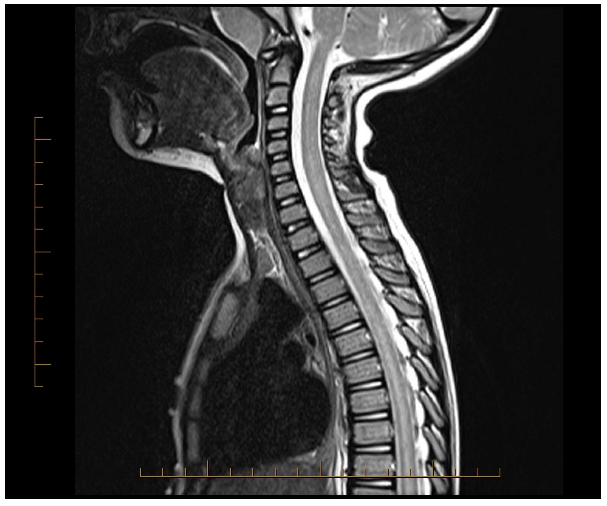
2.1. Patient History and Timeline
2.2. Therapeutic Intervention
2.3. Follow-Up and Outcomes
3. Methods
3.1. Literature Search Strategy
3.2. Inclusion and Exclusion Criteria
- Reported on pediatric patients (age ≤ 18 years) diagnosed with transverse myelitis or longitudinally extensive transverse myelitis.
- Documented the use of TPE as a treatment intervention.
- Provided details on clinical outcomes, including neurological recovery and adverse events.
- Published in English in peer-reviewed journals.
3.3. Data Extraction, Synthesis, and Quality Assessment
4. Results
5. Discussion
6. Conclusions
Author Contributions
Funding
Institutional Review Board Statement
Informed Consent Statement
Data Availability Statement
Conflicts of Interest
References
- Absoud, M.; Greenberg, B.M.; Lim, M.; Lotze, T.; Thomas, T.; Deiva, K. Pediatric transverse myelitis. Neurology 2016, 87 (Suppl. S2), S46–S52. [Google Scholar] [CrossRef] [PubMed]
- Frohman, E.M.; Wingerchuk, D.M. Clinical practice. Transverse myelitis. N. Engl. J. Med. 2010, 363, 564–572. [Google Scholar] [CrossRef] [PubMed]
- Grasso, E.A.; Pozzilli, V.; Tomassini, V. Transverse myelitis in children and adults. In Handbook of Clinical Neurology; Elsevier: Amsterdam, The Netherlands, 2023; Volume 196, pp. 101–117. [Google Scholar] [CrossRef]
- Greenberg, B.M.; Pardo, C.; Recio, A.; Schreiner, T.; Yeh, A.; DeSena, A.; Hopkins, S.; Cutter, G.; Krishnan, C.; McCreary, M. PCORI Final Research Reports. In Does Plasma Exchange Help Improve Physical Function in Children with Transverse Myelitis?—The CAPTURE Study; Patient-Centered Outcomes Research Institute (PCORI): Washington, DC, USA, 2021. [Google Scholar]
- Celik, H.; Aksoy, E.; Oztoprak, U.; Ceylan, N.; Aksoy, A.; Yazici, M.U.; Azapagasi, E.; Eksioglu, A.S.; Yücel, H.; Senel, S.; et al. Longitudinally extensive transverse myelitis in childhood: Clinical features, treatment approaches, and long-term neurological outcomes. Clin. Neurol. Neurosurg. 2021, 207, 106764. [Google Scholar] [CrossRef] [PubMed]
- Suthar, R.; Sankhyan, N.; Sahu, J.K.; Khandelwal, N.K.; Singhi, S.; Singhi, P. Acute transverse myelitis in childhood: A single centre experience from North India. Eur. J. Paediatr. Neurol. 2016, 20, 352–360. [Google Scholar] [CrossRef]
- Chawla, D.; Mishra, D.; Singh, S.; Juneja, M. Longitudinally Extensive Transverse Myelitis. Indian J. Pediatr. 2019, 86, 91–92. [Google Scholar] [CrossRef]
- Ashfaq, M.A.; Javed, I.; Arshad, M.; Yaseen, M.R. Role of Methyl Prednisolone in Longitudinal Extensive Transverse Myelitis (LETM) in Children. Pak. J. Med. Sci. 2020, 36, 451–455. [Google Scholar] [CrossRef]
- Absoud, M.; Brex, P.; Ciccarelli, O.; Diribe, O.; Giovannoni, G.; Hellier, J.; Howe, R.; Holland, R.; Kelly, J.; McCrone, P.; et al. A multicentre randomiSed controlled TRial of IntraVEnous immunoglobulin compared with standard therapy for the treatment of transverse myelitis in adults and children (STRIVE). Health Technol. Assess. 2017, 21, 1–50. [Google Scholar] [CrossRef]
- A Aljezani, M.; Althubaiti, F.; Alhamed, L.; Alharthi, A.; Alamoodi, A.; Bakheet, Y.; Badawi, M.; Hindawi, S. Plasma Exchange in Pediatric Neurology Patients: A Single-Center Experience. Cureus 2024, 16, e52691. [Google Scholar] [CrossRef]
- Fjellbirkeland, O.W.; Szpirt, W.M.; Børresen, M.L. The role of plasmapheresis in severe acute disseminated encephalomyelitis with clinical findings of transverse myelitis. Ther. Apher. Dial. 2024, 28, 119–124. [Google Scholar] [CrossRef]
- Noland, D.K.; Greenberg, B.M. Safety and efficacy of plasma exchange in pediatric transverse myelitis. Neurol. Clin. Pract. 2018, 8, 327–330. [Google Scholar] [CrossRef]
- Savransky, A.; Rubstein, A.; Rios, M.H.; Vergel, S.L.; Velasquez, M.C.; Sierra, S.P.; Marcarian, G.; Alba, R.; Pugliese, A.M.; Tenembaum, S. Prognostic indicators of improvement with therapeutic plasma exchange in pediatric demyelination. Neurology 2019, 93, e2065–e2073. [Google Scholar] [CrossRef] [PubMed]
- Akçay, N.; Menentoğlu, M.E.; Oğur, M.; Tosun, D.; Palabıyık, F.B.; Şevketoğlu, E. COVID-19-associated transverse myelitis treated by therapeutic plasma exchange: A case report. J. Clin. Apher. 2023, 38, 65–68. [Google Scholar] [CrossRef] [PubMed]
- Poyrazoğlu, H.G.; Kırık, S.; Sarı, M.Y.; Esen, I.; Toraman, Z.A.; Eroğlu, Y. Acute demyelinating encephalomyelitis and transverse myelitis in a child with COVID-19. Turk. J. Pediatr. 2022, 64, 133–137. [Google Scholar] [CrossRef] [PubMed]
- Yoo, J.E.; Shin, H.J.; Kang, H.-C.; Lee, J.S.; Kim, H.D.; Lee, H.N. Acute Necrotizing Myelitis Associated with COVID-19. Yonsei Med. J. 2023, 64, 692–695. [Google Scholar] [CrossRef]
- Arabshahi, B.; Pollock, A.N.; Sherry, D.D.; Albert, D.A.; Kreiger, P.A.; Pessler, F. Devic disease in a child with primary Sjögren syndrome. J. Child Neurol. 2006, 21, 285–286. [Google Scholar] [CrossRef]
- Viegas, S.; Weir, A.; Esiri, M.; Kuker, W.; Waters, P.; Leite, M.I.; Vincent, A.; Palace, J. Symptomatic, radiological and pathological involvement of the hypothalamus in neuromyelitis optica. J. Neurol. Neurosurg. Psychiatry 2009, 80, 679–682. [Google Scholar] [CrossRef]
- Lafian, A.; Mahani, T.; Hojjati, M.; Sarlati, T. A Case Report of NMO Transverse Myelitis. Curr. Rheumatol. Rev. 2024, 20, 208–212. [Google Scholar] [CrossRef]
- Tapia-Fonseca, C.V.; Cortés-Enríquez, O.D.; Raya-Garza, L.P.; Gutiérrez-Cuellar, D.M. COVID-19 associated transverse myelitis: Case report. Bol. Med. Hosp. Infant. Mex. 2024, 81, 191–194. [Google Scholar] [CrossRef]
- Khera, D.M.; Didel, S.M.; Panda, S.M.; Tiwari, S.M.; Singh, K.M. Concurrent Longitudinally Extensive Transverse Myelitis and Guillain-Barré Syndrome in a Child Secondary to COVID-19 Infection: A Severe Neuroimmunologic Complication of COVID-19. Pediatr. Infect. Dis. J. 2021, 40, e236–e239. [Google Scholar] [CrossRef]
- Ganelin-Cohen, E.; Konen, O.; Nevo, Y.; Cohen, R.; Halevy, A.; Shuper, A.; Aharoni, S. Prognostic Parameters of Acute Transverse Myelitis in Children. J. Child Neurol. 2020, 35, 999–1003. [Google Scholar] [CrossRef]
- Thabah, M.M.; Sekar, D.; Pranov, R.; Moulitej, M.M.V.; Ramesh, A.; Kadhiravan, T. Neuromyelitis optica spectrum disorder and systemic lupus erythematosus. Lupus 2019, 28, 1722–1726. [Google Scholar] [CrossRef] [PubMed]
- Manguinao, M.; Krysko, K.M.; Maddike, S.; Rutatangwa, A.; Francisco, C.; Hart, J.; Chong, J.; Graves, J.S.; Waubant, E. A retrospective cohort study of plasma exchange in central nervous system demyelinating events in children. Mult. Scler. Relat. Disord. 2019, 35, 50–54. [Google Scholar] [CrossRef] [PubMed]
- Fukuoka, M.; Kuki, I.; Kawawaki, H.; Kim, K.; Hattori, Y.; Tsuji, H.; Horino, A.; Nukui, M.; Okazaki, S. A pediatric patient of hemorrhagic acute transverse myelitis. Brain Dev. 2017, 39, 252–255. [Google Scholar] [CrossRef]
- Fukuoka, M.; Kuki, I.; Horino, A.; Kim, K.; Hattori, Y.; Tsuji, H.; Nukui, M.; Okazaki, S.; Kawawaki, H.; Yoshida, Y.; et al. A child with acute transverse myelitis requiring permanent pacemaker implantation. Brain Dev. 2017, 39, 811–814. [Google Scholar] [CrossRef]
- Hsu, P.-C.; Chen, S.-J. Longitudinal extensive transverse myelitis with an abnormal uFLC ratio in a pediatric patient: Case report and literature review. Medicine 2017, 96, e9389. [Google Scholar] [CrossRef] [PubMed]
- Sarioglu, B.; Kose, S.S.; Saritas, S.; Kose, E.; Kanik, A.; Helvaci, M. Severe acute disseminated encephalomyelitis with clinical findings of transverse myelitis after herpes simplex virus infection. J. Child Neurol. 2014, 29, 1519–1523. [Google Scholar] [CrossRef] [PubMed]
- DeSena, A.; Graves, D.; Morriss, M.C.; Greenberg, B.M. Transverse myelitis plus syndrome and acute disseminated encephalomyelitis plus syndrome: A case series of 5 children. JAMA Neurol. 2014, 71, 624–629. [Google Scholar] [CrossRef]
- Rodrigues, C.E.M.; de Carvalho, J.F. Clinical, radiologic, and therapeutic analysis of 14 patients with transverse myelitis associated with antiphospholipid syndrome: Report of 4 cases and review of the literature. Semin. Arthritis Rheum. 2011, 40, 349–357. [Google Scholar] [CrossRef]
- Csábi, G.; Komáromy, H.; Hollódy, K. Transverse myelitis as a rare, serious complication of Mycoplasma pneumoniae infection. Pediatr. Neurol. 2009, 41, 312–313. [Google Scholar] [CrossRef]
- Pidcock, F.S.; Krishnan, C.; Crawford, T.O.; Salorio, C.F.; Trovato, M.; Kerr, D.A. Acute transverse myelitis in childhood: Center-based analysis of 47 cases. Neurology 2007, 68, 1474–1480. [Google Scholar] [CrossRef]
- Murphy, O.C.; Barreras, P.; Villabona-Rueda, A.; Mealy, M.; Pardo, C.A. Identification of specific causes of myelopathy in a large cohort of patients initially diagnosed with transverse myelitis. J. Neurol. Sci. 2022, 442, 120425. [Google Scholar] [CrossRef] [PubMed]
- Greenberg, B.; Plumb, P.; Cutter, G.; Dean, J.; Desena, A.; Hopkins, S.; Krishnan, C.; Pardo, C.; Recio, A.; Schreiner, T.; et al. Acute flaccid myelitis: Long-term outcomes recorded in the CAPTURE study compared with paediatric transverse myelitis. BMJ Neurol. Open 2021, 3, e000127. [Google Scholar] [CrossRef] [PubMed]
- Wolf, V.L.; Lupo, P.J.; Lotze, T.E. Pediatric acute transverse myelitis overview and differential diagnosis. J. Child Neurol. 2012, 27, 1426–1436. [Google Scholar] [CrossRef] [PubMed]
- Wang, C.; Greenberg, B. Clinical Approach to Pediatric Transverse Myelitis, Neuromyelitis Optica Spectrum Disorder and Acute Flaccid Myelitis. Children 2019, 6, 70. [Google Scholar] [CrossRef] [PubMed]
- Meyer, P.; Leboucq, N.; Molinari, N.; Roubertie, A.; Carneiro, M.; Walther-Louvier, U.; Cuntz-Shadfar, D.; Leydet, J.; Cheminal, R.; Cambonie, G.; et al. Partial acute transverse myelitis is a predictor of multiple sclerosis in children. Mult. Scler. J. 2014, 20, 1485–1493. [Google Scholar] [CrossRef]
- Bigi, S.; Banwell, B.; Yeh, E.A. Outcomes after early administration of plasma exchange in pediatric central nervous system inflammatory demyelination. J. Child Neurol. 2015, 30, 874–880. [Google Scholar] [CrossRef]
- Osman, C.; Jennings, R.; El-Ghariani, K.; Pinto, A. Plasma exchange in neurological disease. Pract. Neurol. 2020, 20, 92–99. [Google Scholar] [CrossRef]
- Schwartz, J.; Padmanabhan, A.; Aqui, N.; Balogun, R.A.; Connelly-Smith, L.; Delaney, M.; Dunbar, N.M.; Witt, V.; Wu, Y.; Shaz, B.H. Guidelines on the Use of Therapeutic Apheresis in Clinical Practice-Evidence-Based Approach from the Writing Committee of the American Society for Apheresis: The Seventh Special Issue. J. Clin. Apher. 2016, 31, 149–338. [Google Scholar] [CrossRef]
- Strasser, E. Principles of Therapeutic Apheresis in Neurological Disease. Transfus. Med. Hemotherapy 2023, 50, 88–97. [Google Scholar] [CrossRef]
- Weinstein, R. Therapeutic apheresis in neurological disorders: A survey of the evidence in support of current category I and II indications for therapeutic plasma exchange. J. Clin. Apher. 2008, 23, 196–201. [Google Scholar] [CrossRef]
- Magaña, S.M.; Keegan, B.M.; Weinshenker, B.G.; Erickson, B.J.; Pittock, S.J.; Lennon, V.A.; Rodriguez, M.; Thomsen, K.; Weigand, S.; Mandrekar, J.; et al. Beneficial plasma exchange response in central nervous system inflammatory demyelination. Arch. Neurol. 2011, 68, 870–878. [Google Scholar] [CrossRef] [PubMed]
- Ipe, T.S.; Meyer, E.K.; Sanford, K.W.; Joshi, S.K.; Wong, E.C.C.; Raval, J.S. Use of therapeutic plasma exchange for pediatric neurological diseases. J. Clin. Apher. 2021, 36, 161–176. [Google Scholar] [CrossRef] [PubMed]
- Michon, B.; Moghrabi, A.; Winikoff, R.; Barrette, S.; Bernstein, M.L.; Champagne, J.; David, M.; Duval, M.; Hume, H.A.; Robitaille, N.; et al. Complications of apheresis in children. Transfusion 2007, 47, 1837–1842. [Google Scholar] [CrossRef]
- Vitaliti, G.; Tabatabaie, O.; Matin, N.; Ledda, C.; Pavone, P.; Lubrano, R.; Serra, A.; Di Mauro, P.; Cocuzza, S.; Falsaperla, R. The usefulness of immunotherapy in pediatric neurodegenerative disorders: A systematic review of literature data. Hum. Vaccines Immunother. 2015, 11, 2749–2763. [Google Scholar] [CrossRef] [PubMed]


| Etiology Category | Specific Causes | Notes |
|---|---|---|
| CNS Demyelinating Disorders |
| TM can be an initial event in MS or appear as a feature in NMOSD, MOGAD, and ADEM, each with characteristic manifestations (e.g., longitudinally extensive lesions in NMOSD, and monophasic symptoms in ADEM). |
| Systemic Autoimmune Disorders |
| TM may result from these systemic inflammatory diseases, with varying mechanisms of central nervous system involvement. |
| Less Common Systemic Autoimmune Disorders |
| These conditions are less frequently associated with TM but can present neurologic involvement affecting the spinal cord. |
| Infections |
| Infectious agents are rare causes of TM; enteroviruses are linked to acute flaccid myelitis (AFM), which mimics polio-like symptoms and occurs in outbreaks. |
| Paraneoplastic Syndromes |
| TM related to paraneoplastic syndromes may present with other nervous system symptoms and is often linked to specific autoantibodies associated with malignancies like SCLC. |
| Vaccinations |
| TM has been temporally associated with vaccines, but causation is unproven. Studies have found no significant correlation between TM and recent vaccination. |
| Idiopathic |
| Idiopathic TM is diagnosed when no specific cause is found despite thorough evaluation. Many cases report an antecedent illness (respiratory, GI, or systemic) but lack definitive evidence of causation. |
| Year | First Author | Type of Study | Location | No. of Subjects | Age (Years) | Sex | Case History | Treatment | Outcome |
|---|---|---|---|---|---|---|---|---|---|
| 2006 | Arabshahi [17] | CR | Philadelphia, Pennsylvania USA | 1 | 11 | F | NMOSD as initial presentation of primary Sjögren’s syndrome with optic neuritis and TM | MP CMP TPE | Remission with treatment led to remission, suggests potential overlap between NMOSD and other autoimmune conditions |
| 2007 | Pidcock [32] | CS | Baltimore, Maryland, USA | 47 | 0–17 | 24 M 23 F | Acute TM, median follow-up 3.2 years. Evaluated functional outcomes and prognostic factors affecting recovery. | Variable | Long-term, 43% of children were unable to walk 30 feet, 68% experienced urinary urgency, and 75% had ongoing numbness. Age < 3 years associated with worse functional outcomes. |
| 2009 | Csábi [31] | CR | Pécs, Hungary | 1 | 14 | M | TM associated with mycoplasma pneumoniae presented with paraplegia + bowel/bladder dysfunction | MP TPE Roxithromycin | No recovery for patient, potential for persistent disability in mycoplasma-associated TM |
| 2011 | Rodrigues [30] | CS | São Paulo, Brazil | 14 | 8 to 83 | F | TM associated with APS, majority of pts experiencing severe symptoms like sphincter dysfunction and thoracic spinal cord involvement | MP CMP TPE | 64% had complete recovery |
| 2014 | Meyer [37] | CS | Montpellier, France | 30 | 3–15 (median 11) | 14 M 16 F | Not specified | 87% MP 50% IVIG 37% both | 60% probability of relapse at 5 years, 80% of patients able to walk independently within 1 month, Children with acute TM and brain MRI abnormalities may later develop MS. |
| 2014 | DeSena [29] | CS | Dallas Texas, USA | 5 | 1–13 2–14 3–9 4–8 5–29 | 1-F 2-M 3-F 4-M 5-F | TM and ADEM pts with atypical peripheral nerve involvement-leading to worse prognoses than typical TM/ADEM cases | MP TPE | 1. Unable to ambulate with unilateral support; urinary retention resolved. 2. Urinary retention improved; urgency and occasional incontinence persist. 3. Fully recovered at 3-month follow-up. 4. Unable to walk; requires bilateral support to stand. 5. Deteriorated post-steroid taper; new brain MRI lesions required re-escalation of steroids |
| 2014 | Sarioglu [28] | CR | Izmir, Turkey | 1 | 25 months | M | Developed TM after HSV infection presented with urinary retention, respiratory distress, and progressive weakness; neuroimaging showed CNS lesions consistent with ADEM | MP TPE | Significant residual motor impairment |
| 2016 | Suthar [6] | CS | North India | 36 | All patients < 12 years old. Median age: 7.5 years (Only children under 12 are admitted to this hospital) | 21 M 15 F | Symmetrical weakness at onset, progressing over 2 days. Thoracic cord most often affected. Median follow-up 35 months: 15 non-ambulatory or needed assistance. | MP (all). 3 children received acyclovir 4 children received azithromycin. | Worse outcomes linked to severe weakness (MRC ≤ 1), spinal shock, respiratory muscle weakness, mechanical ventilation, or delayed diagnosis/treatment. |
| 2017 | Hsu [27] | CR | Taipei, Taiwan | 1 | 12 | M | Sudden onset low back pain followed by quadriplegia, hyperalgesia, flaccid bladder, and altered consciousness. MRI showed diffuse T2 hyperintensity from cervical cord to conus medullaris. | MP TPE | After TPE, limb function improved, bladder/rectal issues resolved. At 6 months: independent eating, supported ambulation, EDSS 5 |
| 2017 | Absoud [9] | RCT | United Kingdom | 2 | Child aged 10–15 years, adult aged 60–65 | F child Adult M | Despite recruiting (26 across 15 centers in the U.K.) only two patients randomized-- due to strict inclusion criteria. | MP IVIG | Impact of the use of IVIG in addition to standard therapy children/adults with TM/NMO could not be determined due to low enrollment. |
| 2017 | Fukuoka [25] | CR | Osaka, Japan | 1 | 3 | F | TM presented with limb weakness, urinary retention, and respiratory issues | MP IVIG TPE | Rehabilitation improved motor function--but became bedridden and upper limb-predominant paralysis remained 2 years after onset, experienced two cardiac arrests requiring pacemaker |
| 2017 | Fukuoka [26] | CR | Osaka, Japan | 1 | 11 | M | Idiopathic TM: developed progressive hypesthesis, urinary dysfunction, and significant motor impairment | MP IVIG TPE | Hemorrhagic spinal lesions, complete recovery after 4 months |
| 2018 | Noland [12] | CS | Dallas, Texas | 19 | 7 months –17 | 11 F 8 M | Review of patients receiving TPE for TM. | TPE | 79% had major improvements in symptoms after 4–7 TPE Majority required further inpatient or outpatient physical therapy 4 patients returned to baseline, 75% regained ability to ambulate 4 adverse events noted over 114 treatments |
| 2019 | Chawla [7] | CR | Delhi, India | 1 | 5 | Not reported | Presented with fever, rapid paraparesis, upper limb weakness, urinary retention, and constipation. Had hypertonic lower limbs, extensor plantar reflexes, and absent abdominal reflexes MRI had signal abnormalities in the spinal cord | MP | Gradual improvement of power, complete recovery by 5th week. |
| 2019 | Manguinao [24] | CS | San Francisco, California, USA | 26 | 2–17 | 16 F 10 M | Acute events: MS (n = 15), NMOSD (n = 7), MOG (n = 2), TM (n = 1), ADEM (n = 1). Diagnoses included MS, NMOSD, TM, ADEM, or MOG-antibody demyelination with disease onset and TPE treatment before age 18 | MP TPE | 14/24 patients improved after PS; 13 improved further with TPE, and 8/10 non-responders to PS improved with TPE. Median EDSS: pre-TPE 4.0, post-TPE 3.75. Adverse events included hypotension, thrombocytopenia, spinal hemorrhage, and GI discomfort. |
| 2019 | Savransky [13] | CS | Buenos Aires, Argentina | 65 | Median age at TPE: 10.5 (1.9–18) | 36 M 29 F | Mixed Optic Neuritis and LETM, 20/42 pts had seropositivity immunoglobin G/myelin oligo. glycoprotein | TPE | 72% showed neurologic improvement; at 6 months, 88.5% improved. Benefits included lower EDSS scores, better visual outcomes, and gait scales. Adverse events occurred in 5.9% of procedures, with 4 severe cases out of 524, TPE effectively rescued severe steroid-unresponsive cases, improving function with low adverse event frequency |
| 2019 | Thabah [23] | CR | India | 1 | 18 | F | Presented with LETM and diagnosed with NMOSD (indicated by high AQP4-IgG antibody titers) | MP TPE maintenance with prednisolone, azathioprine, and hydroxychloroquine | pt responded well to initial treatment and TPE, relapse free at 1 year |
| 2020 | Ashfaq [8] | CS | Pakistan | 34 | 1–13 | 20 M 14 F | 73.5% with acute paraplegia, 26.47% with quadriplegia; 71% had prior febrile illness | MP | 41.2% achieved complete recovery, 58.8% partial recovery, with outcomes linked to gender, spinal cord involvement, muscle power, and autonomic dysfunction. |
| 2020 | Ganelin-Cohen [22] | CS | Israel | 23 | 0.5–17 | 11 M 12 F | Pediatric TM patients (<18 years) with non-recurrent demyelinating events presented with limb paresis, paresthesia, sensory disturbance, sphincter dysfunction, and back pain. CSF showed pleocytosis; mean follow-up: 6 years, 10 months | Variable | Factors associated with prognosis: CSF pleocytosis, no quadriparesis, prolonged time to nadir; 70% good outcome, 30% poor |
| 2021 | Celik [5] | CS | Turkey | 15 | 1–15 | 7 M 8 F | Symptoms: inability to walk (12), incontinence (9), back pain (4), abdominal pain (2), arm weakness (2). Barthel index: 8 independent, 3 moderately dependent, 2 slightly dependent. | MP | 1-month: improved muscle strength. 6-month: 2 with stable monoparesis, 2 with paraparesis. Paraparesis reduced from 80% to 13.3%. 66.6% fully recovered during follow-up, results support MP and early TPE if steroids ineffective |
| 2021 | PCORI Greenberg [4] | CS | Washington, DC, USA | 90 | 0–18 | 54 M 36 F | Children diagnosed with TM or AFM within 6 months; MS and NMOSD excluded. | MP IVIG TPE | TPE First-Line: 3/3 (100%) achieved ≥ 22-point WeeFIM increase; no significant risk difference vs. non-TPE (p = 0.24). IVIG First-Line: 4/8 (50%) achieved ≥ 22-point WeeFIM increase. |
| 2021 | Khera [21] | CR | Jodhpur, India | 1 | 11 | F | Developed LETM and GBS post-COVID-19 with febrile illness and severe paralysis needing ventilatory support | MP TPE | improved after 2 weeks of hospitalization, extubated, able to walk at 6 weeks |
| 2022 | Poyrazoğlu [15] | CR | Elazığ, Turkey | 1 | 10 | M | Presented with ADEM and TM after COVID-19 infection: severe weakness and inability to walk | MP IVIG TPE | patient regained partial function after treatments |
| 2023 | Fjellbirkeland [11] | CR | CopenhagenDenmark | 1 | 16 | F | 16 y F with ADEM: 2-week history of headaches, nausea, neck pain. CSF: monolytic pleocytosis, low CSF glucose, elevated protein, increased lactate | MP TPE | 1 month: normal motor function, fatigue, headaches. 3 months: back to school, intermittent pain, incontinence resolved in a year. |
| 2023 | Yoo [16] | CR | Seoul, Korea | 1 | 10 | F | COVID-19 positive with rapid motor and sensory deficits: neck pain, leg weakness, urinary dysfunction | MP IVIG TPE | 6 months: motor function returned, significant neurological deficits remained |
| 2023 | Akçay [14] | CR | Istanbul, Turkey | 1 | 9 | F | TM following COVID-19 with severe symptoms and rapid worsening | MP IVIG TPE | TPE significantly improved neurological symptoms |
| 2024 | Aljezani [10] | CS | Jeddah, Saudi Arabia | 15 | 2–18 | 12 M 3 F | 2 patients with TM | MP TPE | TPE improved outcomes |
| 2024 | Tapia-Fonseca [20] | CR | Monterrey, Mexico | 1 | 15 | M | Presented with cervical-dorsal medullary syndrome: ascending symmetric paraparesis rapidly progressing to paraplegia (T3 sensory level). COVID-19 positive. | MP TPE | Slow recovery following treatment |
| 2024 | Lafian [19] | CR | California, USA | 1 | 18 | F | Presented with intractable hiccups and paraparesis--MRI showing T2 hyperintensity from the medulla to the conus | MP TPE Rituximab | Complete recovery, serology for west nile virus positive |
| Neurological Disorder | Pathophysiology | Role of TPE | Clinical Use |
|---|---|---|---|
| Myasthenia Gravis (MG) | Autoantibodies target acetylcholine receptors, impairing neuromuscular transmission. | Removes autoantibodies, improving neuromuscular function, especially during crises. | Acute exacerbations or preoperative stabilization. |
| Guillain-Barre Syndrome (GBS) | Immune-mediated attack on peripheral nerve myelin or axons, often triggered by infection. | Clears anti-ganglioside antibodies, halting progression and accelerating recovery. | Most effective within two weeks of onset, reduces ventilatory dependence. |
| Chronic Inflammatory Demyelinating Polyneuropathy (CIDP) | Chronic immune-mediated demyelination of peripheral nerves. | Reduces immune factors causing nerve damage and improves function. | Long-term management alongside immunosuppressants. |
| Neuromyelitis Optica Spectrum Disorders (NMOSD) | Autoantibodies (e.g., anti-aquaporin-4) target astrocytes in the CNS, causing severe demyelination. | Removes anti-aquaporin-4 antibodies, mitigating acute relapses. | Acute relapses, combined with corticosteroids. |
| Autoimmune Encephalitis | Autoantibodies target neuronal or synaptic proteins, leading to neuroinflammation. | Clears pathogenic antibodies, reducing inflammation and preventing long-term damage. | During acute episodes, in conjunction with immunotherapy. |
| Multiple Sclerosis (MS) | Autoimmune-mediated demyelination of CNS neurons, causing acute relapses. | Removes autoantibodies exacerbating demyelination, effective in steroid-refractory relapses. | Second-line treatment for severe relapses not responding to steroids. |
Disclaimer/Publisher’s Note: The statements, opinions and data contained in all publications are solely those of the individual author(s) and contributor(s) and not of MDPI and/or the editor(s). MDPI and/or the editor(s) disclaim responsibility for any injury to people or property resulting from any ideas, methods, instructions or products referred to in the content. |
© 2024 by the authors. Licensee MDPI, Basel, Switzerland. This article is an open access article distributed under the terms and conditions of the Creative Commons Attribution (CC BY) license (https://creativecommons.org/licenses/by/4.0/).
Share and Cite
Khan, A.; Peña, J.; Briceño, G.; Gronquist, J.M.; Khan, K.; Reddy, R.; Yadav, V.; Singh, A. Early Therapeutic Plasma Exchange in Pediatric Transverse Myelitis: A Case Report and Scoping Review. Neurol. Int. 2024, 16, 1674-1690. https://doi.org/10.3390/neurolint16060122
Khan A, Peña J, Briceño G, Gronquist JM, Khan K, Reddy R, Yadav V, Singh A. Early Therapeutic Plasma Exchange in Pediatric Transverse Myelitis: A Case Report and Scoping Review. Neurology International. 2024; 16(6):1674-1690. https://doi.org/10.3390/neurolint16060122
Chicago/Turabian StyleKhan, Akram, José Peña, Genesis Briceño, Juliann M. Gronquist, Khurram Khan, Raju Reddy, Vijayshree Yadav, and Asha Singh. 2024. "Early Therapeutic Plasma Exchange in Pediatric Transverse Myelitis: A Case Report and Scoping Review" Neurology International 16, no. 6: 1674-1690. https://doi.org/10.3390/neurolint16060122
APA StyleKhan, A., Peña, J., Briceño, G., Gronquist, J. M., Khan, K., Reddy, R., Yadav, V., & Singh, A. (2024). Early Therapeutic Plasma Exchange in Pediatric Transverse Myelitis: A Case Report and Scoping Review. Neurology International, 16(6), 1674-1690. https://doi.org/10.3390/neurolint16060122