Review

Mayaro Virus: The State-of-the-Art for Antiviral Drug Development

by
Ana Paula Andreolla
1,2,
Alessandra Abel Borges
3,
Juliano Bordignon
1,*,† and
Claudia Nunes Duarte dos Santos
1,*
1
Laboratório de Virologia Molecular, Instituto Carlos Chagas, ICC/Fiocruz, Curitiba 81350-010, PR, Brazil
2
Departamento de Biologia Celular e Molecular, Universidade Federal do Paraná, Curitiba 81530-900, PR, Brazil
3
Laboratório de Pesquisas em Virologia e Imunologia, Universidade Federal de Alagoas, Maceió 57072-900, AL, Brazil
*
Authors to whom correspondence should be addressed.
Current address: Instituto Carlos Chagas, ICC/Fiocruz, Rua Prof. Algacyr Munhoz Mader 3775, Cidade Industrial de Curitiba, Curitiba 81310-020, PR, Brazil.
Viruses 2022, 14(8), 1787; https://doi.org/10.3390/v14081787
Submission received: 3 August 2022 / Revised: 10 August 2022 / Accepted: 11 August 2022 / Published: 16 August 2022
(This article belongs to the Special Issue Chikungunya Virus and Emerging Alphaviruses)

Abstract

:
Mayaro virus is an emerging arbovirus that causes nonspecific febrile illness or arthralgia syndromes similar to the Chikungunya virus, a virus closely related from the Togaviridae family. MAYV outbreaks occur more frequently in the northern and central-western states of Brazil; however, in recent years, virus circulation has been spreading to other regions. Due to the undifferentiated initial clinical symptoms between MAYV and other endemic pathogenic arboviruses with geographic overlapping, identification of patients infected by MAYV might be underreported. Additionally, the lack of specific prophylactic approaches or antiviral drugs limits the pharmacological management of patients to treat symptoms like pain and inflammation, as is the case with most pathogenic alphaviruses. In this context, this review aims to present the state-of-the-art regarding the screening and development of compounds/molecules which may present anti-MAYV activity and infection inhibition.

1. Introduction

Arboviruses are viral diseases transmitted by arthropod vectors (arthropod-borne virus) to vertebrate hosts during blood feeding [1,2]. Arboviruses have a wide geographic distribution, predominantly in countries with tropical and subtropical climates, such as Brazil [3]. This class of viruses is leading to a growing concern of viral dispersion and new transmission cycles due to its ability to adapt to new vectors and environmental conditions [4].
There are more than 100 species of arboviruses that cause disease in humans, classified into five families: Peribunyaviridae, Flaviviridae, Reoviridae, Rhabdoviridae, and Togaviridae [1,5]. The arboviruses that currently pose the major threats to public health in Brazil are Dengue (DENV), Zika (ZIKV), and Yellow Fever (YFV) of the family Flaviviridae, and the Chikungunya virus (CHIKV) of the family Togaviridae [3]. ZIKV and CHIKV were considered neglected arboviruses until the recent reemergence of both, leading to explosive epidemics and causing severe clinical symptoms [6,7]. Additionally, among neglected arboviruses, the Mayaro virus (MAYV), is endemic in some regions of Central America, the Caribbean, and South America [8]. Although MAYV is frequently detected in the northern and central-western regions of Brazil, it has been spreading to non-endemic regions in recent years, posing a risk of causing epidemics in large areas of the country [9,10,11,12,13,14]. Because most of the populations in these areas are immunologically naïve to MAYV, and considering the high potential rate of vector infestation, the scenario for large-scale epidemics seems plausible [3,15,16,17]. It is worth noting that some degree of cross-protection against MAYV by humoral response induced by a previous CHIKV infection has been observed due to the close phylogenetic and antigenic relationships between CHIKV and MAYV [18,19,20].
In general, MAYV infections in humans cause focal outbreaks characterized by nonspecific febrile illness or arthralgia syndromes [9,21,22,23,24,25]. The main symptoms include chills, fever, gastrointestinal manifestations, dizziness, itching, eye pain, myalgia, and arthralgia; they can last for months to years, thus making MAYV more debilitating than that caused by other arboviruses, whose symptoms might last approximately three to four weeks [23,26]. Severe complications like myocarditis and neurological and hemorrhagic manifestations can also occur due to MAYV infection [15,24,25,26,27,28,29].
Epidemiological studies demonstrate a growing number of confirmed cases of MAYV in endemic and non-endemic regions, both in Brazil and the Americas, and the occurrence of imported human cases in other continents like Europe [8,21,22,23,26,30,31,32,33,34]. However, due to clinical manifestations often confused with other arboviruses, such as DENV, there is a large number of underreported MAYV cases, as shown by retrospective studies, especially during other arboviruses epidemics [9,11,32,35]. Several urban vectors potentially able to transmit MAYV, like Aedes aegypti, might also contribute to viral spread and human infection [36]. Recent surveillance studies have demonstrated the circulation and dispersion of MAYV to previously unreported areas, as well as the detection of human infections in urban settings, evidencing the potential risk of epidemics [11,37]. If epidemics caused by MAYV eventually occur, there are no vaccines or other prophylactic methods or effective antivirals approved for human use to prevent or treat the disease [38]. The clinical approach with anti-inflammatory drugs, antipyretics, and analgesics is usually used to treat secondary symptoms, and, in risk areas, arthropod vector control is implemented [39]. Thus, there is an urgent need to search for new substances/molecules that specifically target MAYV infections.

2. Viral Structure and Replication Cycle
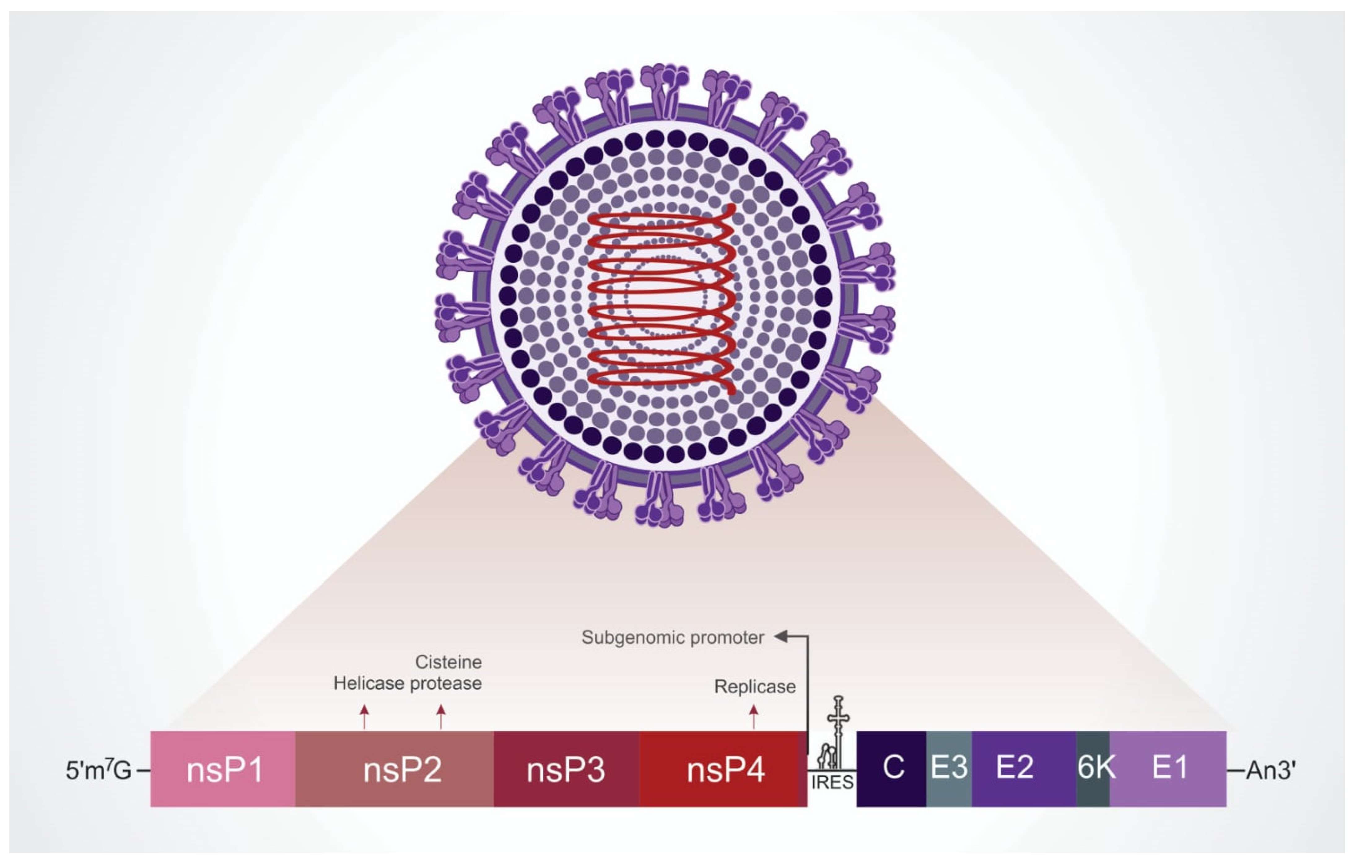
2.1. Viral Particle

Mayaro virus (MAYV) is a member of the family Togaviridae, genus Alphavirus, belonging to the Semliki forest serocomplex. The viral particle has an icosahedral capsid of about 70 nm in diameter and a lipid envelope derived from the host cell in which heterodimers of the transmembrane glycoproteins E1 and E2 are embedded [40]. The heterodimers are ordered in trimers on the virion surface [41]. The genetic material is composed of a single strand of positive-sense RNA of ~12 kb with two open reading frames (a 7 kb and a 4 kb) that each encodes a polyprotein, separated by a short non-coding sequence (Figure 1). From the cleavage of the non-structural polyprotein, the following proteins are found: nsP1, involved in mRNA capping; nsP2, with helicase/protease activities; nsP3, which has three recognized domains (the macrodomain, the alphavirus unique domain (AUD) and the hypervariable region) all of which are required for replication; nsP4, which is solely responsible for the RNA synthetic properties of the viral replicase complex because it contains the core RdRp domain and motifs. After processing of the structural polyprotein, there are associated proteins that result in the viral particle: C, capsid; E1, envelope glycoproteins of higher molecular mass 1; E2, envelope glycoproteins of higher molecular mass 2; E3, lower mass glycoprotein; 6K, protein that associates E1 and E2; TF, transframe protein involved in viral assembly/budding and also virulence factor [42] (Figure 1) [21,41,43,44,45,46,47,48]. Notably, the E3 protein is absent in the virion structure at 4.4 Ǻ resolution from cryo-electron microscopy analysis (cryo-EM) [41].

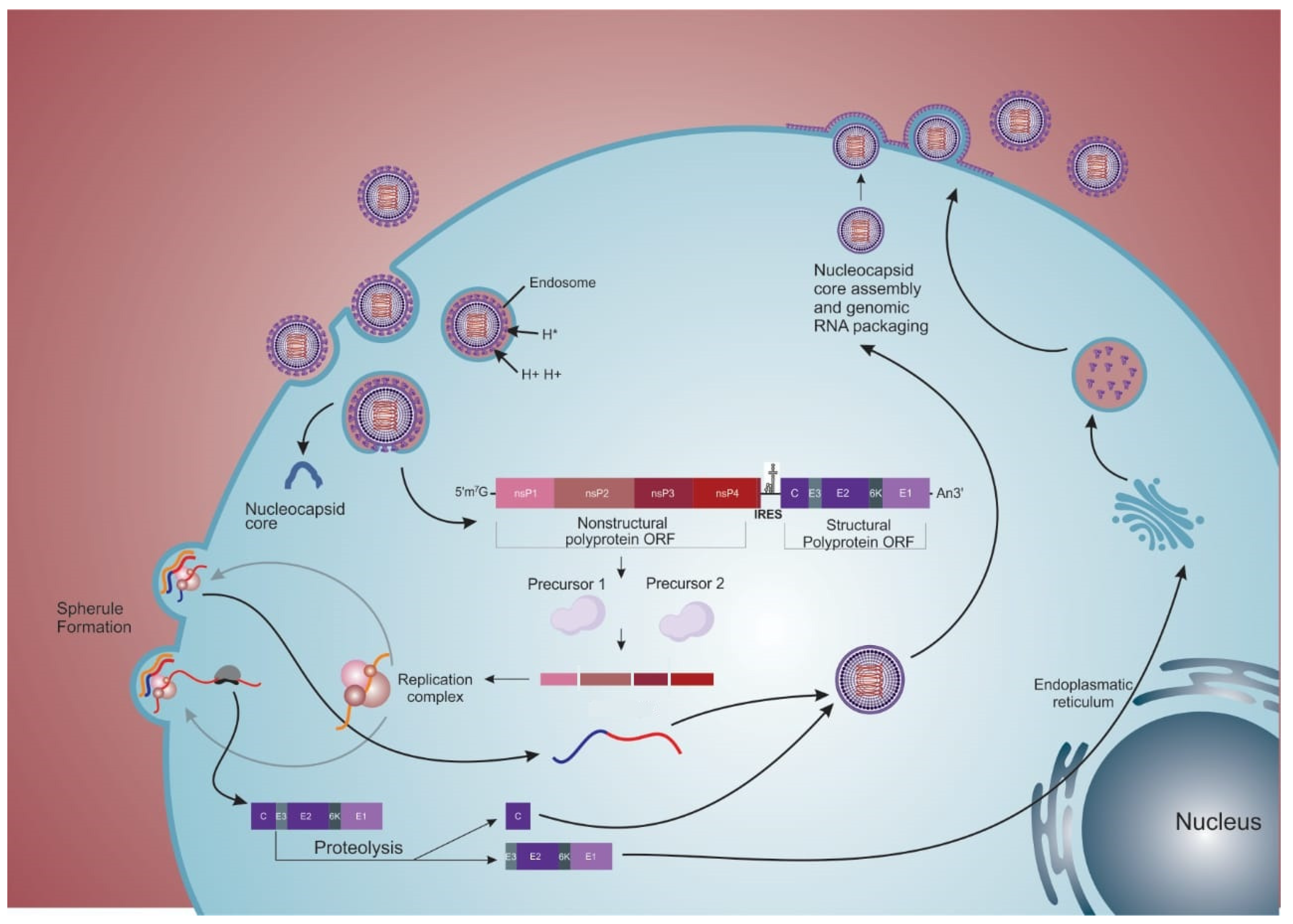
2.2. Internalization

The MAYV replication cycle and the precise function of each viral protein produced are not completely understood (Figure 2). The putative protein functions are inferred from other alphaviruses. The binding of the virions to the cell receptor is primarily mediated by the E2 protein [49]. The target cell receptors that mediate the entry of the virus are also not fully known; however, the adhesion molecule Mxra8 (also known as DICAM, ASP3, or limitrin, a member of the immunoglobulin superfamily) was recently described as a receptor for MAYV and other arthritogenic alphaviruses, like CHIKV, Ross River, O’nyong nyong, and Barmah Forest virus [46,50]. However, for encephalitic alphaviruses, such as VEEV and EEV, this receptor, when blocked, does not seem to affect the infection [50]. The uptake of the viral particle is closely linked to the lipid rafts of cell membranes, where cholesterol is the main component [49]. Endocytosis occurs dependent and/or independent of clathrin (caveolar via), the former pathway most frequently used by MAYV [49]. Once endocytosed, the clathrin layer (when present) is quickly removed from the vesicle, and the virus is delivered to the initial compartment of the endosome. The presence of ATP-dependent proton pumps is the critical characteristic of the endosome, which causes acidification of the vesicle during the maturation stages. This pH change is fundamental to viral RNA delivery to the cell cytoplasm, as it will be the initial event of intermembrane fusion (vesicle and viral envelope) [49,51].
The events of alphavirus membrane fusion are comprised of three steps: (1) dissociation of E1 from E2; (2) E1 insertion into the target membrane; and (3) the formation of E1 homotrimers [51,52]. Intermembrane fusion starts from the acidification of the endosome, which will promote the dissociation of the two envelope proteins (E1 and E2). These proteins are associated in metastable conformation in the form of heterodimers, and upon dissociation, the E1 fusion loop is exposed and inserted into the target membrane of the vesicle. In a pH-independent interaction, the stem region of the E1 complex folds, distorting the target membrane through the cooperative action of several trimers, providing the necessary energy to mediate membrane fusion. Fusion proceeds through a hemifusion step where the two outer leaflets fuse. The final stable post-fusion E1 homotrimer is formed, located on the same side as the fusion loops, and transmembrane domains are anchored. Concomitantly, the E1 homotrimer refolds, resulting in complete fusion and thus forming the fusion pore, which will allow the exit of the viral nucleocapsid (NC) from the endosome to the cytoplasm of the cell [26,44,49,51].

2.3. Replication

Once the NC is in the cytoplasm of the cell, its disassembly is facilitated by interactions with free ribosomes [53,54,55]. Cellular ribosomes recognize the CAP region and the 5′-AUG initiator codon of the viral RNA in the cytoplasm, leading to the translation of 49S (genomic) RNA and its processing [53,54,56]. This entire process occurs within bulb-like structures present in the plasma membrane, known as spherules (Figure 2), and the structure of these membranes as a type I cytopathic vacuole (CPV-I) [57,58,59]. After recognition of the CAP region, the non-structural genes are recognized and translated, giving rise to polyproteins P123 (precursor of nsP1, nsP2, and nsP3) in higher amounts and P1234 (P123 and nsP4) in lower amounts. This phenomenon is due to the presence of a stop codon at the junction of nsP3 and nsP4 [60,61]. In both situations, the P123 polyprotein is processed sequentially, individualizing each protein into mature species. Through its proteolytic function, mature nsP2 will catalyze cleavages on P1234 releasing RNA-dependent RNA polymerase (nsP4), which forms the replication complex (P123+nsP4) when associated with P123 [62], involved in the synthesis of complementary negative (−) RNA (Figure 2) [62,63,64,65,66]. In addition, nsP2 will promote the shutdown of transcription mediated by RNA polymerase II of the host. This process was evidenced through the interaction of nsP2 with subunit A of RNA polymerase II (Rpb1) and subunit 2 of transcription initiation factor IIE (TFIIE2), suggesting a shutdown of cellular transcription [67]. In addition to the pivotal role of nsP2, the release of nsP1 is also reported as essential for creating a replication complex by associating with nsP4 (nsP1 + P23 + nsP4), which will use the (−) RNA as a template to synthesize new positive strands of genomic RNA [61,62,64]. However, the recent study by Gorchakov et al. (2008) [68] with the SINV indicated that the P123/nsP4 complex was able to synthesize (+) RNAs without the need for cleaving nsP1 from the P123 protein [69]. This process is still not fully understood with regard to MAYV. In addition to the transcription of (+) RNA, a subgenomic mRNA (26S transcript) is also transcribed, which will be translated into a structural polyprotein and give rise to structural proteins after cleavage (Figure 2) [70].
From the structural polyprotein precursor, the protein C is the first structural protein generated through its processing by the action of the autoproteolytic serine protease to encapsulate the newly synthesized RNA molecules, originating new NCs [48,51,71,72]. Concomitantly, the remaining polyprotein comprising the glycoprotein precursor (E3-E2-6k-E1) is routed to the endoplasmic reticulum (ER). The N-terminal portion of the E3 glycoprotein contains a signal peptide which interacts with the ER membrane, translocating the polyprotein to the ER lumen. Once in the ER, the host signalase recognize both the N and C-terminal portions of the 6k, creating the products E3E2, 6k, and E1, which will be anchored in the organelle membrane. Next, E3E2 and E1 associate to form dimers that move with 6K through vesicles to the plasma membrane. Finally, E3E2 is cleaved by furin proteases present in a post-Golgi compartment, generating the glycoproteins E3 and E2 (Figure 2) [40,44,71,72].
Notably, the 6k protein is palmitoylated, enabling it to anchor to the membranes. This protein is involved in the transport of E1 and E2 to the plasma membrane and is a viroporin that enhances membrane permeability in infected cells by forming ion channels [42]. The 6K protein presents two transmembrane helices, classified as a Class II subclass A of viroporin types, connected by a short cytoplasmic loop [42,73]. In addition, the 6k gene undergoes a frameshift event and starts to produce the TF protein [42]. Within the gene encoding 6K, a heptanucleotide slip site (UUUUUUA) is highly conserved among the alphaviruses that causes a -1 ribosomal shift. This frameshift produces the TF protein in place of 6K and E1 with a frequency ranging from 5% to 40%, depending on the alphavirus species [74,75]. The 6K and TF proteins have an identical N-terminal portion (including the first transmembrane domain with ion channel activity) but differ in the C-terminal portion. The TF protein contains a C-terminal extension of the 6K protein and a longer cytoplasmic domain than 6K [74]. Functionally, TF seems essential for viral assembly, presenting a virulence role as antagonist of the host type I interferon responses [76].

2.4. Exit

The complete intracellular cycle of viral replication takes place four hours after the virus enters the cell. Virus release occurs after the anchoring of the viral capsid in the cell membrane; the viral lipid envelope is then formed through the budding mechanism (Figure 2) [72,77,78].
NC interacts with E2 proteins, initiating the process of assembly and the budding of viral particles [79,80,81,82]. This interaction occurs in a pocket (hydrophobic on the bottom and hydrophilic on the top) in the C-terminal domain of protein C, to which the E2 protein is non-covalently linked through its C-terminal domain [41]. In this interface, a consensus motif containing threonine, proline, and tyrosine (TPY) is present, which is conserved within the genus Alphavirus [83]. Previous results have suggested the co-transport of C and E2 to the plasma membrane, as both proteins colocalize in mobile vesicles in the cell cytoplasm [79].
The viral envelope is enriched with cholesterol and sphingolipids precisely because, in mammals, it sprouts from host cell membrane domains rich in these components [49,84]. These sites are present both in the cell body and in intercellular extensions induced by the virus itself (a phenomenon called cell-to-cell transmission) that facilitates the infection of neighboring cells [85]. The alphavirus exit from cells is determined by host factors like actin cytoskeleton remodeling, which promotes the transport of glycoproteins to the plasma membrane. Microscopic studies have revealed actin rearrangements and the accumulation of actin clusters in the cytoplasm at the final steps of viral infection with the co-localization of E2 in these foci and along the filaments [86]. The precise mechanism(s) of cleavage of the budding particles is still undetermined. It is unknown whether only the NC’s interaction with the envelope is strong enough to detach itself; nevertheless, other viral proteins, like 6K and TF, are involved in the process [42,87]. Studies with mutants in the 6K and TF proteins have shown that they play an essential role in the viral assembly and budding, although the exact mechanism is not completely understood [42]. According to Ribeiro-Filho et al. (2021) [41] the cryo-EM structure suggests that MAYV capsid assembly is based on electrostatic contacts between the C proteins. In addition, data suggest a probable action of nsP1 by inhibiting tetherin, an interferon-induced membrane protein, which has the action of restricting the exit of enveloped viruses [88,89,90]. Additional studies are needed to define the precise budding mechanism of alphaviruses from infected cells.

2.5. “Cell-to-Cell” Transmission

Alphavirus infection induces drastic remodeling of the cell cytoskeleton. This remodeling promotes the production of two types of cell extensions, the short- and long-filopodia-like structures [85,91] with length, contacts, and components at their main differences. The short ones are from 2 to 7 µm in length and comprise only F-actin filaments. Its induction requires the E2-C interaction [84,92] and is involved with nsP1, as assays performed using only structural proteins did not trigger such formations, and specific viral protein labeling demonstrated the co-localization of E2-C and nsP1. However, it is unclear whether nsP1 can mediate cell-to-cell transmission or act to protect viral particles from neutralizing antibodies [79,91].
The long intercellular extensions have more than 10 µm, can reach up to 60 µm, and are composed of F-actin and tubulin [79,91,93]. They are usually identified when there is a production of active structural proteins, with E2, E1, and C detected throughout the length of the filament. These extensions emerge from an infected cell towards to an uninfected cell through retrograde actin transport, and during this process viral particles moved and budded from the infected cell through these extensions. The contact tip of the filopodium-like structure is usually flat and does not fuse with the other cell, hence the name “virological synapse” [94]. Interestingly, there is a loss of stress fibers in infected cells in the late periods of infection, correlating with the appearance of extensions [79,91].
The “cell-to-cell” transmission requires the budding and release of active viral particles. Thus, transmission is not mediated by viral RNA transfer, replication complexes, or type II cytopathic vacuoles (CPVIIs) [86]. This is in agreement with the observed lack of cytoplasmic or membrane continuity between the infected cells and the target [93]. Intercellular transmission prevents the action of neutralizing antibodies on the virus, suggesting that nascent particles are somehow protected from the extracellular environment [85,95,96]. The data also strongly suggest that infection of target cells occurs by endocytosis and fusion triggered by low pH, as does initial infection. Furthermore, the host determinants for such a phenomenon to occur are unknown, and such structures are species and/or cell-line dependent.

3. Antivirals

Presently, there are no specific antiviral treatments or vaccines against MAYV infections. In urban environments of endemic areas, vector control through insecticide spraying is the only available measure to prevent infection. The clinical management of infected patients is limited to drug administration such as analgesics, non-steroidal anti-inflammatory drugs, and antipyretics to relieve symptoms.
Several natural and synthetic compounds have been evaluated to identify those with low cytotoxicity to host cells and the compounds’ ability to inhibit viral infection [97]. The parameters most commonly used for this purpose are the cytotoxic concentration for 50% of the cells (CC50), inhibitory concentration for 50% of the infected cells (IC50), and the selectivity index (SI) [98,99]. The SI is a factor calculated using the two previous parameters (CC50 and IC50), facilitating the comparison of compounds in terms of cytotoxicity and antiviral potency [100]. Substances that present antiviral activity are able to inhibit critical steps of viral replication, like events related to virus entry into host cells, activity of enzymes involved in the replication complex (proteases, viral helicase, and RNA-dependent RNA polymerase), or in late stages during particles assembly and release to the extracellular medium [38].
In addition to substances that directly affect the activity of virus proteins or enzymes, some compounds can modulate the host response and affect virus replication. Such substances can present the advantage of broad-spectrum use against other related alphaviruses. However, these compounds must be carefully studied, as the side effects generated by their use can cause disorders compared to compounds that act directly on virus proteins or enzymes [101]. In this review, we describe the state-of-the-art on promising substances/molecules with anti-MAYV activity, which are demonstrated in Figure 3.

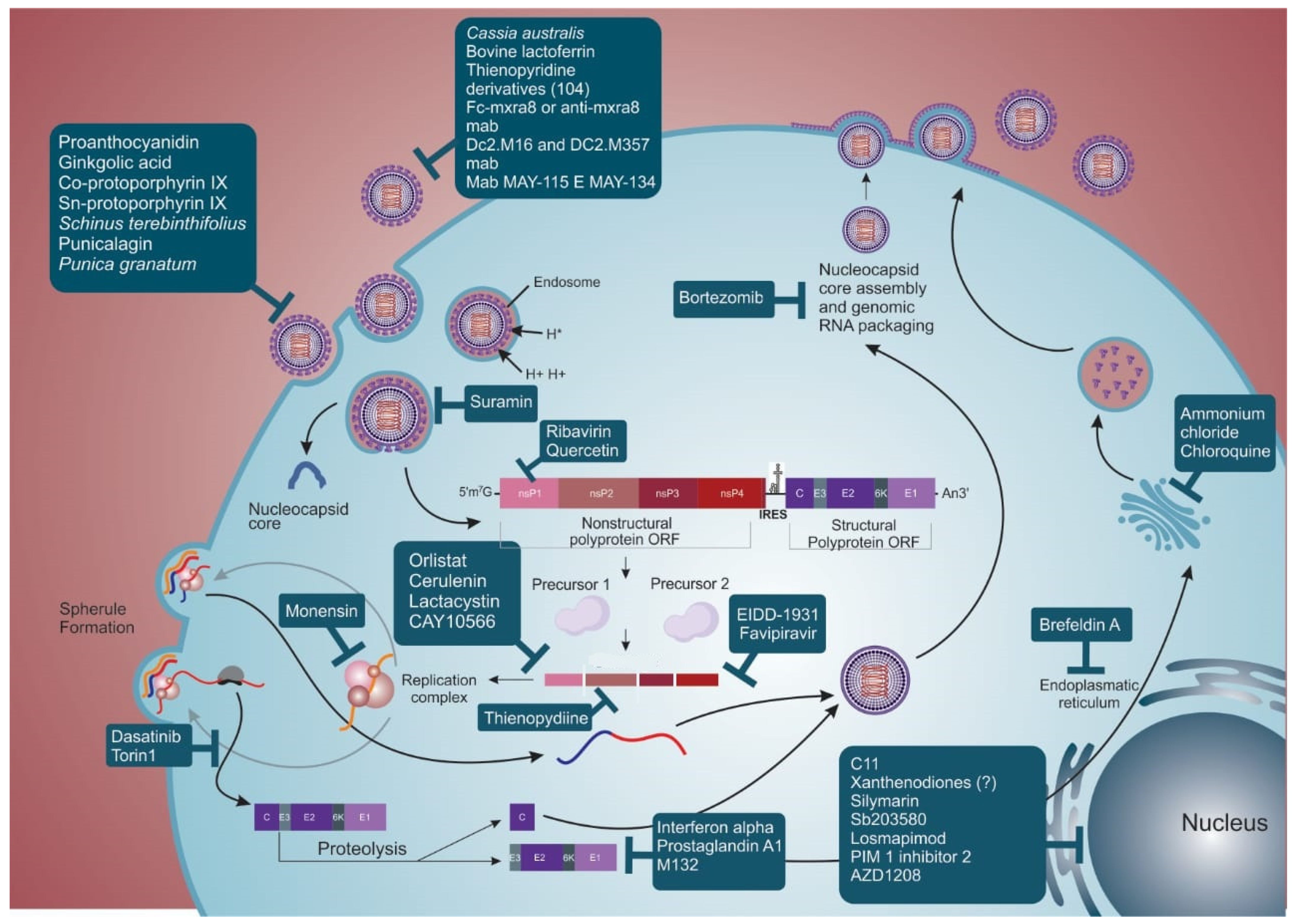
3.1. Virucidal Compounds

Compounds or plant extracts are considered virucidal when they interact directly with the viral particle and, consequently, prevent infection onset. Several reports indicate that the flavonoid proanthocyanidin ([(-) epicatechin-(4β -> 8)-(-)-4′-methylepigallocatechin]; PAC) isolated from methanol extraction of Maytenus imbricata (Celastraceae) roots showed a virucidal effect on MAYV. This compound acted directly in MAYV particles and not in the host cells, with an EC50 (effective concentration for 50% of infected cells) of 37.9 ± 2.4 µM and a SI above 43 [102]. Ginkgolic acid, a substance isolated from the Ginkgo biloba plant, has a similar effect on MAYV infectivity [103].
Hydrophobic molecules like Co-protoporphyrin IX and Sn-protoporphyrin IX also have virucidal activity. By elucidating the mechanism of action of these compounds, it was found that they act on the conformation of envelope proteins, deforming the viral particle and preventing adsorption of receptors on the cell plasma membrane in an in vitro model [104].
Compounds present in the extract of Schinus terebinthifolius fruit, which is a condiment widely used in cooking, were evaluated against MAYV infection and proved to be virucidal in an in vitro model. Four substances (two bioflavonoids and two ethyl acetate) were isolated, and three of them had SI (60, 12, 98, 6, respectively) greater than that of ribavirin (used as antiviral control; SI = 8) [105]. The virucidal mechanism of these substances remains unknown. Another isolated compound is punicalagin from the extract of pomegranate (Punica granatum). Both extract and isolate proved virucidal for MAYV, with SI = 14 and 48, respectively [106]. The mechanisms of action for MAYV inhibition are still unclear. Studies using SARS-CoV2 demonstrated that its action is through the reversible and non-competitive binding to the viral surface glycoproteins [107].

3.2. Compounds That Interfere with Adsorption and Internalization of the Virus Particle

Some substances/molecules can prevent the anchorage, entry, and internalization of MAYV to the cell by inhibiting fundamental pathways for the initiation of viral replication. For example, it was found that extracts from Cassia australis leaves have an anti-MAYV effect, especially those containing condensed tannins (SI = 33; relative potency—RP = 16.5). This substance seems responsible for the antiviral activity since, characteristically, tannins have the ability to produce complex lipo and glycoproteins. The viral envelope is mostly composed of glycoproteins, so the compound possibly inhibits viral adsorption and internalization [108].
Bovine lactoferrin, a multifunctional globular protein isolated on a large scale from bovine milk and easily found commercially, acts by blocking the entry of MAYV into the host cell. The block seems to depend on the presence of sulfated glycosaminoglycans on the cell membrane surface, preventing viral anchorage [109].
Compounds already employed in the clinic for the treatment of other conditions or diseases, such as suramin, used to treat African sleeping sickness and onchocerciasis, also showed anti-MAYV efficacy. After treating cells with this drug, the inhibition of MAYV entry was evidenced, possibly because it causes conformational changes in the viral envelope proteins, impairing fusion with the host cell membrane [110]. A study analyzing the in vitro infection with CHIKV suggested that suramin interacts with the E2 protein, inhibiting the adsorption and/or conformational changes necessary for membrane fusion [111].
Another class of drugs is that based on antibodies. Only a few studies characterizing the cell receptors involved in MAYV entry into host cells are available [46,51,112,113,114]. As mentioned previously, it has been recently shown that the Mxra8 receptor is the putative entry molecule for MAYV and other arthritogenic alphaviruses and, thus, a potential therapeutic target. Using the Fc-Mxra8 fusion protein or anti-Mxra8 monoclonal antibody as treatment, in vitro infection by MAYV, CHIKV, Ross River virus, O’nyong nyong virus (ONNV), and Barmah Forest virus was reduced. Additionally, mice (C57BL/6) infected with CHIKV and ONNV and treated with Fc-Mxra8 fusion protein or anti-Mxra8 blocking antibodies had reduced infection and disease signals (foot swelling) [50].
Specific anti-MAYV neutralizing antibodies are also available, representing potential therapeutic targets to interfere with viral adsorption and internalization. Of the 151 hybridomas generated in the study by Earnest, J.T. et al. (2019) [115], 11 presented neutralization activity against MAYV. Neutralizing antibodies bind to E2 and E1 protein from D and L genotypes, with EC50 lower than 10 ng/mL. Additionally, the anti-MAYV neutralizing antibodies presented cross-reactivity and also inhibited the in vitro infection by other alphaviruses, like UNAV and CHIKV [115]. The neutralization of MAYV in vitro infection occurred mainly through monoclonal antibody recognition of the E2 protein, as previously demonstrated for antibodies produced against CHIKV [116] and Eastern Equine Encephalitis virus (EEEV) [117]. It was also demonstrated that an antibody cocktail that recognizes E1 and E2 had a greater effect in in vivo infection of C57BL/6J mice [115]. There are reports of humanized monoclonal antibodies that recognize different alphaviruses, like MAYV and CHIKV. These monoclonal antibodies were developed through expression of the E3-E2-E1 protein, using a more conserved region among the arthritogenic alphaviruses. Two monoclonal antibodies, DC2.M16 and DC2.M357, were generated and recognized the B domain of the E2 protein; they promoted neutralization, possibly through phagocytosis performed by monocytes (in vitro test) [20].

3.3. Compounds That Interfere with Replication, Morphogenesis, and Viral Exit

There are substances/molecules with inhibitory activity beyond viral entry, affecting steps such as viral replication, translation and the processing of viral proteins, morphogenesis/maturation, and viral sprouting, which are critical events for the formation of viral progeny [26].
Belonging to the class of immunomodulatory proteins, interferon alpha (IFN-α) is one of the viral inhibitors more widely used as an in vitro molecule to treat MAYV infection in cell lines [118,119,120,121]. Analysis by SDS-PAGE and protein densitometry showed that the inhibition of MAYV protein synthesis is heterogeneous after treatment with IFN-α, acting more significantly on glycoproteins E1 (99%) and E2 (79%) and their precursors (84% and 77%, respectively) compared to protein C (28%) [120]. This phenomenon can be explained by the increase in intracellular pH caused by treatment with IFN, compromising the traffic of proteins in the endoplasmic reticulum and Golgi complex, as previously shown in in vitro infections by vesicular stomatitis virus [122], herpes simplex viruses [119], CHIKV, ZIKV, and SARS-CoV-2 [118] and in the human treatment of hepatitis B [123].
Ribavirin (RIBV), another compound used in the clinic to treat viral infections [124,125,126,127,128] and widely used to control MAYV infection, has well-known pharmacological properties, such as bioavailability and toxicity. The putative mechanism of action of this compound is through (1) inhibiting enzymes related to the capping reaction of the viral RNA, since it is a guanosine analog; (2) suppressing the viral RNA synthesis by inhibiting the enzyme inosine-5′-monophosphate dehydrogenase; or (3) when phosphorylated, it can directly interfere with viral RNA synthesis during transcription [126,129,130]. RIBV has already been shown to be effective against CHIKV in mouse models, mainly when associated with doxycycline [131].
Furthermore, it has been demonstrated that compounds fractioned from plant extracts also impair MAYV replication. An example is quercetin isolated from Bauhinia longifolia (Bong.), which has a robust anti-MAYV activity (SI = 94; relative potency = 5) compared to ribavirin (SI = 8), and is used as a control compound [132]. Although the exact mechanism of action of quercetin during MAYV infection remains unclear, it has been suggested that it may be due to inhibition of viral RNA metabolism as observed for DENV-2 [132,133].
Epicatechin extracted from Salacia crassifolia is another compound that showed an anti-MAYV effect (SI = 7; RP = 2). There is strong evidence of the interaction of epicatechin with some viral components, possibly blocking important stages of the replication cycle and formation of new viral progenies without interfering with host cell metabolism. However, the precise mechanism of action of the compound action remains unknown [102].
In addition to natural compounds, synthetic molecules like thienopyridine derivatives also stand out as antivirals. Candidate compound 104 (SI = 125), for example, has low toxicity risks and severely influences viral protein synthesis, probably by inhibiting nsP2 transcription. There is a deficiency mainly in the assembly of the MAYV particle, favoring the production of non-infectious particles [134]. This compound showed promising action in inhibiting MAYV replication, acting at different points in the viral cycle. This conclusion was supported because (a) the viral protein synthesis was highly impaired; and (b) transmission electron microscopy after treatment with the candidate compound indicated action on the viral envelope [134].
Additionally, drugs already employed to treat other pathologies have shown anti-MAYV activity, such as ammonium chloride and chloroquine. Their action is demonstrated by a large amount of virus within intracytoplasmic vacuoles, in addition to free precursors and proteins (possibly capsid or nucleocapsid) in the cell cytoplasm, which are rarely seen during untreated infection. This effect possibly occurs because these substances raise the pH of the organelles of the Golgi complex, thus inhibiting the transport of glycoproteins to the membrane. In addition, this pH change can impair the function of some enzymes involved in the processing and maturation of viral glycoproteins, also affecting sprouting and viral infectivity. Consequently, these drugs seem to force the virus to follow an alternative morphogenesis route, leading to premature intracellular viral maturation [135].
The drugs EIDD-1931 and favipiravir have also been efficient in inhibiting MAYV infection. In the clinic, they are used to treat influenza A and B and resistant pandemic influenza viruses. Both drugs acted on MAYV viral replication in in vitro models by inhibiting polymerase and causing lethal mutagenesis during viral replication [110], similar to what has been reported for CHIKV [136,137] and Venezuelan Equine Encephalitis virus (VEEV) [138]. In a C57BL/6 mouse model, favipiravir pre-treatment reduced infection, RNA copies, and viral particle production in different tissues (e.g., brain, liver, heart, and spleen) during infection by MAYV [139], CHIKV [140] and Western equine encephalitis virus [141]. However, when the treatment of this compound is post-infection, of all tissues tested (e.g., brain, liver, heart and kidney) only the right quadriceps muscles showed a statistical decrease in MAYV infection [139]. A reduction in footpad swelling was also found in an animal model for the evaluation of arthritis and arthralgia triggered by MAYV [139]. In addition, in a retrospective study with pregnant women who used favipiravir, no major teratogenic effects were observed [142].
Inhibitors of the membrane-associated kinase family, such as Dasatinib and Torin 1, appear to act more specifically in viral translation without interfering with host mRNA translation [143,144]. It has been demonstrated that Dasatinib and Torin 1 inhibit the translation of subgenomic mRNA, resulting in a decrease in viral infection not only for MAYV but also for other alphaviruses [145].
In addition to drugs used as antivirals, some antibiotics have also been evaluated as potential anti-MAYV molecules. Monensin is an example of a natural antibiotic used as an additive in ruminant feed to increase feed efficiency. Its action, which has been previously described for other alphaviruses, such as the Semliki Forest and Sindbis viruses, has also been evaluated for MAYV. Treatment of both vertebrate and invertebrate cells with Monesin showed severe inhibition of MAYV replication, although viral morphogenesis was not affected [146].

3.4. Drugs That Modulate Host Response to Viral Infection

As previously mentioned, substances that may act on specific host factors are also promising alternatives for the development of broad-spectrum antiviral drugs. Nevertheless, there are still gaps in understanding the MAYV infection cycle in the host cells that would make the development and testing of this class of compounds a challenge.
After a high-throughput screening of 52,000 compounds, a small molecule named C11 that activates the signaling of the innate immune response was identified. C11 acts as an agonist of the adaptor protein STING and activates antiviral genes through phosphorylation of IRF3, mainly inducing the IFN type I response. Since alphaviruses are sensitive to the IFN response, including MAYV, this substance decreased the viral titer in an in vitro model [147].
Brefeldin A, a fungal metabolite reported to block newly synthesized endoplasmic reticulum protein transport, can also inhibit MAYV replication. By analyzing its effect during infection, a drastic reduction in the synthesis of viral proteins in mammalian cells was found which may or may not affect their glycosylation [148].
Prostaglandin A1 (PGA1) is another compound that has been shown to be effective against MAYV. This widely studied compound plays a role in several physiological and pathological processes, in addition to influencing the replication of several viruses like SINV [149], vaccinia virus [150] and Sendai virus [151]. After treating Vero cells with PGA1 and challenging them with MAYV, inhibition of E1 and E2 glycoprotein synthesis was found to be associated with triggering the synthesis of 70 kDa heat shock cell protein (HSP) [152].
The cyclic ketones, which are classified into different groups based on their chemical structure, have also been evaluated for their activity against different microorganisms and diseases. Among them, the xanthenodiones stand out as anti-bactericidal, anti-leishmania, antifungal, antitumor, anti-trypanosome, and antiviral substances [153,154]. Several compounds of this group were produced from the mixture of 1,3-diketone, aldehyde, and ZrOCl28H2O and evaluated during in vitro infection by MAYV [155]. Only one of them, compound 9, remained in the test, as it obtained the best SI (15.8) [156]. However, when evaluating its anti-MAYV action, it was found to be effective only in pre-treatment or in high concentrations (338.8 µmol/L) [156], unlike what was verified for ZIKV, in which it interacts with viral envelope proteins preventing adsorption [155]. The likely mechanism of action is unclear, and it is believed that it may act on intracellular events. This hypothesis is based on some antiviral assays where there was no virucidal action or activity in the early stages of entry, as there is no decrease in infection compared to the positive control [134,156].
Drugs that act to inhibit the synthesis of new long-chain fatty acids, like orlistat (anti-obesity FDA approved) and cerulenin (antibiotic in test phase), which impair the activity of fatty acid synthase, as well as the substance CAY10566 that acts on the activity of stearoyl-CoA desaturase, have been shown to reduce in vitro MAYV replication. These enzymes seem important in the MAYV replication cycle and other arthritogenic alphaviruses, as they interfere with the plasma membrane anchoring of the nsP1 protein present in the replication complex [157]. Furthermore, cerulenin, considered an inhibitor of lipid synthesis, has been shown to reduce MAYV replication, probably preventing the incorporation of [3H] glycerol into lipids at any time after infection. Activity analysis at different time points during infection showed that the inhibition was more pronounced at the beginning of the infection, as it interferes with the synthesis of viral proteins [158].
Proteasome inhibitors are another class of virus inhibitory substances that can affect different stages of the replication cycle of CHIKV, rotavirus, and vaccinia virus [159,160,161]. Many viruses have evolved to hijack the proteasome machinery to make their replication more efficient, as this pathway is essential for cellular processes such as the cell cycle, immunity, and autophagy [162,163,164]. These drugs are described in the literature as acting at different stages of the viral replication cycle, both in protein expression [165], maturation [166] and budding [167,168]. For MAYV infection, the compounds MG132 and lactacystin have been shown to modulate the synthesis of E1 and nsP1, constituting critical steps during the replicative cycle. This drug activity reduces the amount of infectious viral particles produced in in vitro assays [77]. Another study with EEEV and VEEV compared the effectiveness of MG132 with another proteasome inhibitor, Bortezomib, which is FDA-approved to treat some forms of myeloma and lymphoma. In an in vitro model, there was a decrease in viral genomic copies, possibly due to interference by the ubiquitination of the viral capsid, preventing viral RNA from going into the cell’s cytoplasm [169]. Furthermore, MG132 also showed activity in the inhibition of additional targets, like certain lysosomal cysteine proteases, calpains, and cathepsins [170].
Another example is a plant-derived compound, silymarin, obtained from Silybum marianum. This substance acts on MAYV replication with a SI = 29.6, but the mechanism of action remains to be elucidated. One hypothesis that explains the anti-MAYV activity of silymarin in vitro is the modulation of cellular oxidative stress triggered by viral infection, decreasing the formation of ROS and, consequently, the levels of MDA (malondialdehyde) and protein carbonyl [171]. The efficacy of silymarin was assayed in a non-lethal model of disease in BALB/C mice [172,173]. In addition to the drastic decrease of viral infection in various organs (e.g., liver, spleen, brain) and serum, there was a control of the oxidant activity caused by the virus (carbonyl protein and MDA) and an increase of antioxidants (catalase, glutathione, and SOD activity). Thus, protection of the liver (ALT and AST activity) and other organs and the controlling of pro-inflammatory factors like IFN-b, TNF, IL-6, and IL-1B were also observed [28,173,174,175]. The in vivo (mouse model) as well as in vitro cell assays findings support the hypothesis that the modulation of MAYV-induced oxidative stress increases disease severity [176].
Other cellular factors that appear to be necessary for efficient replication of MAYV, at least in a cell model, are the mitogen-activated protein kinases (MAPKs) [177]. These proteins are involved in several cell cycle stages, like proliferation, growth, cytokine production, and stress response, controlling and regulating transduction signaling pathways [178]. It has been shown that the main pathway of MAPKs involved in MAYV replication is p38 since the addition of the p38 inhibitor (SB203580) leads to a severe reduction in infection and the production of viable particles [177]. Studies carried out with enteroviruses [179] and SARS-CoV-2 [144] found that this pathway interferes directly with the synthesis of viral proteins. When treating MAYV infection with two compounds that act on this pathway, NR-7h (which blocks several isoforms of p38) and losmapimod (a compound used in Phase 2 clinical study for the treatment of individuals with facioscapulohumeral muscular dystrophy, which blocks p38) there is a reduction in the expression of E1 [177].
Another family of protein kinases, the proviral integration sites of Moloney murine leukemia virus (PIMs), was evaluated during MAYV infection [180]. PIMs act in different cellular pathways, including migration, metabolism, and proliferation, and are closely linked to the prognosis of some cancers, such as colon, pancreatic and prostatic [181,182,183]. Studies using an in vitro model have shown that PIM 1 inhibitor 2 (isoform 1 only) and AZD1208 (pan-PIM kinase inhibitor for all PIM isoforms) show decreased rates of MAYV infection, viral progeny and protein synthesis of E1 and nsP1 [184]. This activity was verified during ZIKV and UNAV infections but not in CHIKV [185,186].

4. Conclusions

In this review, we describe compounds that are potentially effective against MAYV infection. They include synthetic molecules extracted from natural products as well as approved medicines already used to treat other diseases. Drug repurposing seems to be a promising strategy since the toxicity parameters have already been determined for use in humans, making the final approval by regulatory agencies faster than with other drugs. Still, non-commercial compounds (synthetic or extracted from plants) do not have safety test data, as most are in the preclinical development stages. Despite this, chemical engineering can benefit and improve molecules that have promising inhibitory results, both the synthetic ones and those isolated from natural extracts. Chemical engineering of molecules could lead to reduced toxicity, as well as high antiviral activity in drugs.
Considering that MAYV infection causes an acute and debilitating disease and the recent spread of this virus to broader regions in Brazil and South America [10,14,187,188,189], there is an urgent need for effective anti-MAYV compounds. Additionally, the lack of knowledge on the precise mechanism of MAYV replication and pathogenesis makes this neglected emergent virus a potential threat to all tropical and sub-tropical areas of the globe. Finally, further studies on MAYV replication and pathogenesis, including animal models, are essential for the future development of new, effective, and safe anti-MAYV drugs.

Funding

C.N.D.d.S. (307176-2018-5) and J.B. (312671/2020-2 and 205096/2018-2) are CNPq fellows. A.P.A. is a Ph.D. fellow from CAPES, Brazil.

Acknowledgments

Wagner Nagib for the graphic design of the figures.

Conflicts of Interest

The authors declare that they have no conflict of interest.

References

  1. Calisher, C.H.; Karabatsos, N. Arbovirus Serogroups: Definition and Distribution. In The Arboviruses: Epidemiology and Ecology; Monath, T.P., Ed.; CRC Press: New York, NY, USA, 1988; pp. 19–57. [Google Scholar]
  2. Knipe, D.M.; Howley, P.M. Fields Virology, 6th ed.; Lippincott Williams & Wilkins: Philadelphia, PA, USA, 2013; ISBN 9780849340871. [Google Scholar]
  3. da Saúde, M. Boletim de Vigilância Laboratorial Dos Arbovírus. Bol. Epidemiológico 2021, 52, 7–18. [Google Scholar]
  4. Coffey, L.L.; Forrester, N.; Tsetsarkin, K.; Vasilakis, N.; Weaver, S.C. Factors Shaping the Adaptive Landscape for Arboviruses: Implications for the Emergence of Disease. Future Microbiol. 2013, 8, 155–176. [Google Scholar] [CrossRef] [PubMed]
  5. Travassos da Rosa, A.; Shope, R.; Pinheiro, F.; Travassos da Rosa, J.; Vasconcelos, P.; Herve, J.; Degallier, N. Arbovirus Research in the Brazilian Amazon. In Proceedings of the Fifth Symposium on Arbovírus Research in Australia, Brisbane, Australia, 28 August–1 September 1989; pp. 4–8. [Google Scholar]
  6. Zanluca, C.; De Melo, V.C.A.; Mosimann, A.L.P.; Dos Santos, G.I.V.; dos Santos, C.N.D.; Luz, K. First Report of Autochthonous Transmission of Zika Virus in Brazil. Mem. Inst. Oswaldo Cruz 2015, 110, 569–572. [Google Scholar] [CrossRef] [PubMed]
  7. Albuquerque, I.G.C.D.; Marandino, R.; Mendonça, A.P.; Nogueira, R.M.R.; Vasconcelos, P.F.D.C.; Guerra, L.R.; Brandão, B.C.; Mendonça, A.P.P.; Aguiar, G.R.; Bacco, P.A.M.D. Chikungunya Virus Infection: Report of the First Case Diagnosed in Rio de Janeiro, Brazil. Rev. Soc. Bras. Med. Trop. 2012, 45, 128–129. [Google Scholar] [CrossRef]
  8. Pezzi, L.; Rodriguez-Morales, A.J.; Reusken, C.B.; Ribeiro, G.S.; LaBeaud, A.D.; Lourenço-de-Oliveira, R.; Brasil, P.; Lecuit, M.; Failloux, A.B.; Gallian, P.; et al. GloPID-R Report on Chikungunya, o’nyong-Nyong and Mayaro Virus, Part 3: Epidemiological Distribution of Mayaro Virus. Antivir. Res. 2019, 172, 104610. [Google Scholar] [CrossRef]
  9. Lopes, N.; Nozawa, C.; Linhares, R.E.C. Características Gerais e Epidemiologia Dos Arbovírus Emergentes No Brasil. Rev. Pan-Amazônica Saúde 2014, 5, 55–64. [Google Scholar] [CrossRef]
  10. Saatkamp, C.J.; Rodrigues, L.R.R.; Pereira, A.M.N.; Coelho, J.A.; Marques, R.G.B.; Souza, V.C.D.; Nascimento, V.A.D.; Saatkamp, J.G.D.S.; Naveca, F.G.; Figueiredo, R.M.P.D. Mayaro Virus Detection in the Western Region of Pará State, Brazil. Rev. Soc. Bras. Med. Trop. 2021, 54, 1–3. [Google Scholar] [CrossRef]
  11. Romeiro, M.F.; Fumagalli, M.J.; dos Anjos, A.B.; Figueiredo, L.T.M. Serological Evidence of Mayaro Virus Infection in Blood Donors from São Carlos, São Paulo, Brazil. Trans. R. Soc. Trop. Med. Hyg. 2020, 114, 693–696. [Google Scholar] [CrossRef]
  12. Brunini, S.; França, D.D.S.; Silva, J.B.; Silva, L.N.; Silva, F.P.A.; Spadoni, M.; Rezza, G. High Frequency of Mayaro Virus IgM among Febrile Patients, Central Brazil. Emerg. Infect. Dis. 2017, 23, 1025–1026. [Google Scholar] [CrossRef]
  13. Coimbra, T.L.M.; Santos, C.L.S.; Suzuki, A.; Petrella, S.M.C.; Bisordi, I.; Nagamori, A.H.; Marti, A.T.; Santos, R.N.; Fialho, D.M.; Lavigne, S.; et al. Mayaro Virus: Imported Cases of Human Infection in São Paulo State, Brazil. Rev. Inst. Med. Trop. Sao Paulo 2007, 49, 221–224. [Google Scholar] [CrossRef]
  14. de Paula Silveira-Lacerda, E.; Laschuk Herlinger, A.; Tanuri, A.; Rezza, G.; Anunciação, C.E.; Ribeiro, J.P.; Tannous, I.P.; Abrantes, G.R.; da Silva, E.G.; Arruda, K.F.; et al. Molecular Epidemiological Investigation of Mayaro Virus in Febrile Patients from Goiania City, 2017–2018. Infect. Genet. Evol. 2021, 95, 104981. [Google Scholar] [CrossRef] [PubMed]
  15. Esposito, D.L.A.; Fonseca, B.A.L.d. Will Mayaro Virus Be Responsible for the next Outbreak of an Arthropod-Borne Virus in Brazil? Braz. J. Infect. Dis. 2017, 21, 540–544. [Google Scholar] [CrossRef] [PubMed]
  16. Azevedo, R.S.S.; Chiang, J.O.; Martins, L.C.; Viana, G.M.R. Monitoramento de Casos de Febre Do Mayaro e Febre Do Oropouche Até a Semana Epidemiológica 35, 2019. Bol. Epidemiológico 2019, 50, 14–16. [Google Scholar]
  17. Lorenz, C.; Ribeiro, A.F.; Chiaravalloti-neto, F. Mayaro Virus Distribution in South America. Acta Trop. 2019, 198, 105093. [Google Scholar] [CrossRef]
  18. Webb, E.M.; Azar, S.R.; Haller, S.L.; Langsjoen, R.M.; Cuthbert, C.E.; Ramjag, A.T.; Luo, H.; Plante, K.; Wang, T.; Simmons, G.; et al. Effects of Chikungunya Virus Immunity on Mayaro Virus Disease and Epidemic Potential. Sci. Rep. 2019, 9, 20399. [Google Scholar] [CrossRef]
  19. Fumagalli, M.J.; de Souza, W.M.; de Castro-Jorge, L.A.; de Carvalho, R.V.H.; Castro, Í.A.; de Almeida, L.G.N.; Consonni, S.R.; Zamboni, D.S.; Figueiredo, L.T.M. Chikungunya Virus Exposure Partially Cross-Protects against Mayaro Virus Infection in Mice. J. Virol. 2021, 95, e01122-21. [Google Scholar] [CrossRef]
  20. Malonis, R.J.; Earnest, J.T.; Kim, A.S.; Angeliadis, M.; Holtsberg, F.W.; Javad Aman, M.; Jangra, R.K.; Chandran, K.; Daily, J.P.; Diamond, M.S.; et al. Near-Germline Human Monoclonal Antibodies Neutralize and Protect against Multiple Arthritogenic Alphaviruses. Proc. Natl. Acad. Sci. USA 2021, 118, e2100104118. [Google Scholar] [CrossRef]
  21. de Figueiredo, M.L.G.; Figueiredo, L.T.M. Emerging Alphaviruses in the Americas: Chikungunya and Mayaro. Rev. Soc. Bras. Med. Trop. 2014, 47, 677–683. [Google Scholar] [CrossRef]
  22. Lima, W.G.; Pereira, R.S.; da Cruz Nizer, W.S.; Brito, J.C.M.; Godói, I.P.; Cardoso, V.N.; Fernandes, S.O.A.; Ferreira, J.M.S. Rate of Exposure to Mayaro Virus (MAYV) in Brazil between 1955 and 2018: A Systematic Review and Meta-Analysis. Arch. Virol. 2021, 166, 347–361. [Google Scholar] [CrossRef]
  23. da Costa, V.G.; de Rezende Féres, V.C.; Saivish, M.V.; de Lima Gimaque, J.B.; Moreli, M.L. Silent Emergence of Mayaro and Oropouche Viruses in Humans in Central Brazil. Int. J. Infect. Dis. 2017, 62, 84–85. [Google Scholar] [CrossRef]
  24. Filho, P.; de Paula, F. Epidemias Simultâneas de Mayaro e Febre Amarela Em Belterra, Pará. Bol. Epidemiológico 1978, 10, 146–152. [Google Scholar]
  25. Anderson, C.R.; Downs, W.G.; Wattley, G.H.; Ahin, N.W.; Reese, A.A. Mayaro Virus: A New Human Disease Agent: II. Isolation from Blood of Patients in Trinidad, B.W.I. Am. J. Trop. Med. Hyg. 1957, 6, 1012–1016. [Google Scholar] [CrossRef] [PubMed]
  26. Diagne, C.T.; Bengue, M.; Choumet, V.; Hamel, R.; Pompon, J.; Missé, D. Mayaro Virus Pathogenesis and Transmission Mechanisms. Pathogens 2020, 9, 738. [Google Scholar] [CrossRef] [PubMed]
  27. McGill, P.E. Viral Infections: α-Viral Arthropathy. Bailliere’s Clin. Rheumatol. 1995, 9, 145–150. [Google Scholar] [CrossRef]
  28. Pinheiro, F.P.; Freitas, R.B.; Travassos da Rosa, J.F.; Gabbay, Y.B.; Mello, W.A.; LeDuc, J.W. An Outbreak of Mayaro Virus Disease in Belterra, Brazil. I. Clinical and Virological Findings. Am. J. Trop. Med. Hyg. 1981, 30, 674–681. [Google Scholar] [CrossRef]
  29. Theilacker, C.; Held, J.; Allering, L.; Emmerich, P.; Schmidt-Chanasit, J.; Kern, W.V.; Panning, M. Prolonged Polyarthralgia in a German Traveller with Mayaro Virus Infection without Inflammatory Correlates. BMC Infect. Dis. 2013, 13, 2011–2014. [Google Scholar] [CrossRef]
  30. do Rosário Casseb, A.; Casseb LM, N.; da Silva, S.P.; da Costa Vasconcelos, P.F. Arbovírus: Importante Zoonose Na Amazônia Brasileira. Vet. Zootec. 2013, 20, 9–21. [Google Scholar]
  31. Lednicky, J.; Beau De Rochars, V.M.; Elbadry, M.; Loeb, J.; Telisma, T.; Chavannes, S.; Anilis, G.; Cella, E.; Ciccozzi, M.; Okech, B.; et al. Mayaro Virus in Child with Acute Febrile Illness, Haiti, 2015. Emerg. Infect. Dis. 2016, 22, 2000–2002. [Google Scholar] [CrossRef]
  32. Aguilar-Luis, M.A.; del Valle-Mendoza, J.; Sandoval, I.; Silva-Caso, W.; Mazulis, F.; Carrillo-Ng, H.; Tarazona-Castro, Y.; Martins-Luna, J.; Aquino-Ortega, R.; Peña-Tuesta, I.; et al. A Silent Public Health Threat: Emergence of Mayaro Virus and Co-Infection with Dengue in Peru. BMC Res. Notes 2021, 14, 29. [Google Scholar] [CrossRef]
  33. Slegers, C.A.D.; Keuter, M.; Günther, S.; Schmidt-Chanasit, J.; van der Ven, A.J.; de Mast, Q. Persisting Arthralgia Due to Mayaro Virus Infection in a Traveler from Brazil: Is There a Risk for Attendants to the 2014 FIFA World Cup? J. Clin. Virol. 2014, 60, 317–319. [Google Scholar] [CrossRef]
  34. Llagonne-Barets, M.; Icard, V.; Leparc-Goffart, I.; Prat, C.; Perpoint, T.; André, P.; Ramière, C. A Case of Mayaro Virus Infection Imported from French Guiana. J. Clin. Virol. 2016, 77, 66–68. [Google Scholar] [CrossRef]
  35. Vieira, C.J.d.S.P.; da Silva, D.J.F.; Barreto, E.S.; Siqueira, C.E.H.; Colombo, T.E.; Ozanic, K.; Schmidt, D.J.; Drumond, B.P.; Mondini, A.; Nogueira, M.L.; et al. Detection of Mayaro Virus Infections during a Dengue Outbreak in Mato Grosso, Brazil. Acta Trop. 2015, 147, 12–16. [Google Scholar] [CrossRef]
  36. Pereira, T.N.; Carvalho, F.D.; De Mendonça, S.F.; Rocha, M.N.; Moreira, L.A. Vector Competence of Aedes Aegypti, Aedes Albopictus, and Culex Quinquefasciatus Mosquitoes for Mayaro Virus. PLoS Negl. Trop. Dis. 2020, 14, e0007518. [Google Scholar] [CrossRef] [PubMed]
  37. PAHO; WHO. Epidemiological Alert: Mayaro Fever; PAHO/WHO: Washington, DC, USA, 2019; pp. 1–5. [Google Scholar]
  38. Mello, M.V.P.; Domingos, T.F.S.; Ferreira, D.F.; Ribeiro, M.M.J.; Ribeiro, T.P.; Rodrigues, C.R.; Souza, A.M.T. Antiviral Drug Discovery and Development for Mayaro Fever—What Do We Have so Far? Mini-Rev. Med. Chem. 2020, 20, 921–928. [Google Scholar] [CrossRef] [PubMed]
  39. Lima-Camara, T.N. Arboviroses Emergentes e Novos Desafios Para a Saúde Pública No Brasil. Rev. Saude Publica 2016, 50, 1–7. [Google Scholar] [CrossRef]
  40. Mezencio, J.M.; de Souza, W.; Fonseca, M.E.; Rebello, M.A. Replication of Mayaro Virus in Aedes Albopictus Cells: An Electron Microscopic Study. Arch. Virol. 1989, 104, 299–308. [Google Scholar] [CrossRef]
  41. Ribeiro-Filho, H.V.; Coimbra, L.D.; Cassago, A.; Rocha, R.P.F.; Guerra, J.V.d.S.; de Felicio, R.; Carnieli, C.M.; Leme, L.; Padilha, A.C.; Paes Leme, A.F.; et al. Cryo-EM Structure of the Mature and Infective Mayaro Virus at 4.4 Å Resolution Reveals Features of Arthritogenic Alphaviruses. Nat. Commun. 2021, 12, 3038. [Google Scholar] [CrossRef]
  42. Ramsey, J.; Mukhopadhyay, S. Disentangling the Frames, the State of Research on the Alphavirus 6K and TF Proteins. Viruses 2017, 9, 228. [Google Scholar] [CrossRef]
  43. Espósito, D.L.A.; da Fonseca, B.A.L. Complete Genome Sequence of Mayaro Virus (Togaviridae, Alphavirus) Strain BeAr 20290 from Brazil. Genome Announc. 2015, 3, 141660. [Google Scholar] [CrossRef]
  44. Jose, J.; Snyder, J.E.; Kuhn, R.J. A Structural and Functional Perspective of Alphavirus Replication and Assembly. Future Microbiol. 2009, 4, 837–856. [Google Scholar] [CrossRef]
  45. Mezencio, J.M.; Rebello, M.A. Mayaro Virus Proteins. Mem. Inst. Oswaldo Cruz 1992, 88, 299–304. [Google Scholar] [CrossRef] [PubMed]
  46. Strauss, J.H.; Strauss, E.G. The Alphaviruses: Gene Expression, Replication, and Evolution. Microbiol. Rev. 1994, 58, 491–562. [Google Scholar] [CrossRef] [PubMed]
  47. Lavergne, A.; Thoisy, D.; Lacoste, V.; Tolou, H.; Dussart, P.; Morvan, J.; Talarmin, A.; Kazanji, M. Mayaro Virus: Complete Nucleotide Sequence and Phylogenetic Relationships with Other Alphaviruses. Virus Res. 2006, 117, 283–290. [Google Scholar] [CrossRef] [PubMed]
  48. Bakar, F.A.; Ng, L.F.P. Nonstructural Proteins of Alphavirus–Potential Targets for Drug Development. Viruses 2018, 10, 71. [Google Scholar] [CrossRef]
  49. Carvalho, C.A.M.; Silva, J.L.; Oliveira, A.C.; Gomes, A.M.O. On the Entry of an Emerging Arbovirus into Host Cells: Mayaro Virus Takes the Highway to the Cytoplasm through Fusion with Early Endosomes and Caveolae-Derived Vesicles. PeerJ 2017, 5, e3245. [Google Scholar] [CrossRef] [PubMed]
  50. Zhang, R.; Kim, A.S.; Fox, J.M.; Nair, S.; Basore, K.; Klimstra, W.B.; Rimkunas, R.; Fong, R.H.; Lin, H.; Poddar, S.; et al. Mxra8 Is a Receptor for Multiple Arthritogenic Alphaviruses. Nature 2018, 557, 570–574. [Google Scholar] [CrossRef] [PubMed]
  51. Kielian, M.; Chanel-vos, C.; Liao, M. Alphavirus Entry and Membrane Fusion. Viruses 2010, 2, 796–825. [Google Scholar] [CrossRef]
  52. Kononchik, J.P., Jr.; Hernandez, R.; Brown, D.T. An Alternative Pathway for Alphavirus Entry. Virol. J. 2011, 8, 304. [Google Scholar] [CrossRef]
  53. Singh, I.; Helenius, A. Role of Ribosomes in Semliki Forest Virus Nucleocapsid Uncoating. J. Virol. 1992, 66, 7049–7058. [Google Scholar] [CrossRef]
  54. Wengler, G.; Wengler, G. Identification of a Transfer of Viral Core Protein to Cellular Ribosomes during the Early Stages of Alphavirus Infection. Virology 1984, 134, 435–442. [Google Scholar] [CrossRef]
  55. Wengler, G.; Würkner, D.; Wengler, G. Identification of a Sequence Element in the Alphavirus Core Protein Which Mediates Interaction of Cores with Ribosomes and the Disassembly of Cores. Virology 1992, 191, 880–888. [Google Scholar] [CrossRef]
  56. Glanville, N.; Ranki, M.; Morser, J. Initiation of Translocation Directed by 42S and 26S RNAs from Semliki Forest Virus in Vitro. Proc. Natl. Acad. Sci. USA 1976, 73, 3059–3063. [Google Scholar] [CrossRef] [PubMed]
  57. Pietilä, M.K.; Hellström, K.; Ahola, T. Alphavirus Polymerase and RNA Replication. Virus Res. 2017, 234, 44–57. [Google Scholar] [CrossRef]
  58. Grimley, P.M.; Berezesky, I.K.; Friedman, R.M. Cytoplasmic Structures Associated with an Arbovirus Infection: Loci of Viral Ribonucleic Acid Synthesis. J. Virol. 1968, 2, 1326–1338. [Google Scholar] [CrossRef] [PubMed]
  59. Friedman, R.M.; Levin, J.G.; Grimley, P.M.; Berezesky, I.K. Membrane-Associated Replication Complex in Arbovirus Infection. J. Virol. 1972, 10, 504–515. [Google Scholar] [CrossRef] [PubMed]
  60. Li, G.; Rice, C.M. The Signal for Translational Readthrough of a UGA Codon in Sindbis Virus RNA Involves a Single Cytidine Residue Immediately Downstream of the Termination Codon. J. Virol. 1993, 67, 5062–5067. [Google Scholar] [CrossRef]
  61. Kallio, K.; Hellström, K.; Jokitalo, E.; Ahola, T. RNA Replication and Membrane Modification Require the Same Functions of Alphavirus Nonstructural Proteins. J. Virol. 2016, 90, 1687–1692. [Google Scholar] [CrossRef]
  62. Shirako, Y.; Strauss, J.H. Cleavage between NsP1 and NsP2 Initiates the Processing Pathway of Sindbis Virus Nonstructural Polyprotein P123. Virology 1990, 177, 54–64. [Google Scholar] [CrossRef]
  63. Lemm, J.A.; Rice, C.M. Assembly of Functional Sindbis Virus RNA Replication Complexes: Requirement for Coexpression of P123 and P34. J. Virol. 1993, 67, 1905–1915. [Google Scholar] [CrossRef]
  64. Lemm, J.A.; Rümenapf, T.; Strauss, E.G.; Strauss, J.H.; Mrice, C. Polypeptide Requirements for Assembly of Functional Sindbis Virus Replication Complexes: A Model for the Temporal Regulation of Minus- and plus-Strand RNA Synthesis. EMBO J. 1994, 13, 2925–2934. [Google Scholar] [CrossRef]
  65. Sawicki, D.L.; Sawicki, S.G. Short-Lived Minus-Strand Polymerase for Semliki Forest Virus. J. Virol. 1980, 34, 108–118. [Google Scholar] [CrossRef] [PubMed]
  66. Sawicki, D.L.; Sawicki, S.G. Alphavirus Positive and Negative Strand RNA Synthesis and the Role of Polyproteins in Formation of Viral Replication Complexes. Arch. Virol. Suppl. 1994, 9, 393–405. [Google Scholar] [CrossRef] [PubMed]
  67. Ishida, R.; Cole, J.; Lopez-Orozco, J.; Fayad, N.; Felix-Lopez, A.; Elaish, M.; Luo, S.Y.; Julien, O.; Kumar, A.; Hobman, T.C. Mayaro Virus Non-Structural Protein 2 Circumvents the Induction of Interferon in Part by Depleting Host Transcription Initiation Factor IIE Subunit 2. Cells 2021, 10, 3510. [Google Scholar] [CrossRef]
  68. Rupp, J.C.; Sokoloski, K.J.; Gebhart, N.N.; Hardy, R.W. Alphavirus RNA Synthesis and Non-Structural Protein Functions. J. Gen. Virol. 2015, 96, 2483–2500. [Google Scholar] [CrossRef] [PubMed]
  69. Adkins, S.; Stawicki, S.S.; Faurote, G.; Siegel, R.W.; Kao, C.C. Mechanistic Analysis of RNA Synthesis by RNA-Dependent RNA Polymerase from Two Promoters Reveals Similarities to DNA-Dependent RNA Polymerase. RNA 1998, 4, 455–470. [Google Scholar] [PubMed]
  70. Fros, J.J.; Pijlman, G.P. Alphavirus Infection: Host Cell Shut-Off and Inhibition of Antiviral Responses. Viruses 2016, 8, 166. [Google Scholar] [CrossRef]
  71. Mezencio, J.M.; de Souza, W.; Fonseca, M.E.; Rebello, M.A. Ultrastructural Study of Mayaro Virus Replication in BHK-21 Cells. Arch. Virol. 1990, 114, 229–235. [Google Scholar] [CrossRef]
  72. Nieva, J.L.; Madan, V.; Carrasco, L. Viroporins: Structure and Biological Functions. Nat. Rev. Microbiol. 2012, 10, 563–574. [Google Scholar] [CrossRef]
  73. Snyder, J.E.; Kulcsar, K.A.; Schultz, K.L.W.; Riley, C.P.; Neary, J.T.; Marr, S.; Jose, J.; Griffin, D.E.; Kuhn, R.J. Functional Characterization of the Alphavirus TF Protein. J. Virol. 2013, 87, 8511–8523. [Google Scholar] [CrossRef]
  74. Firth, A.E.; Chung, B.Y.W.; Fleeton, M.N.; Atkins, J.F. Discovery of Frameshifting in Alphavirus 6K Resolves a 20-Year Enigma. Virol. J. 2008, 5, 108. [Google Scholar] [CrossRef]
  75. Rogers, K.J.; Jones-Burrage, S.; Maury, W.; Mukhopadhyay, S. TF Protein of Sindbis Virus Antagonizes Host Type I Interferon Responses in a Palmitoylation-Dependent Manner. Virology 2020, 542, 63–70. [Google Scholar] [CrossRef] [PubMed]
  76. Llamas-González, Y.Y.; Campos, D.; Pascale, J.M.; Arbiza, J.; González-Santamaría, J. A Functional Ubiquitin-Proteasome System Is Required for Efficient Replication of New World Mayaro and Una Alphaviruses. Viruses 2019, 11, 370. [Google Scholar] [CrossRef] [PubMed]
  77. Mota, M.T.D.O.; Ribeiro, M.R.; Vedovello, D.; Nogueira, M.L. Mayaro Virus: A Neglected Arbovirus of the Americas. Future Virol. 2015, 10, 1109–1122. [Google Scholar] [CrossRef]
  78. Brown, R.S.; Wan, J.J.; Kielian, M. The Alphavirus Exit Pathway: What We Know and What We Wish We Knew. Viruses 2018, 10, 89. [Google Scholar] [CrossRef] [PubMed]
  79. Liu, N.; Brown, D.T. Phosphorylation and Dephosphorylation Events Play Critical Roles in Sindbis Virus Maturation. Virology 1993, 196, 703–711. [Google Scholar] [CrossRef] [PubMed]
  80. Strauss, J.H.; Strauss, E.G.; Kuhn, R.J. Budding of Alphaviruses. Trends Microbiol. 1995, 3, 346–350. [Google Scholar] [CrossRef]
  81. Zhao, H.; Garoff, H. Role of Cell Surface Spikes in Alphavirus Budding. J. Virol. 1992, 66, 7089–7095. [Google Scholar] [CrossRef]
  82. West, J.; Hernandez, R.; Ferreira, D.; Brown, D.T. Mutations in the Endodomain of Sindbis Virus Glycoprotein E2 Define Sequences Critical for Virus Assembly. J. Virol. 2006, 80, 4458–4468. [Google Scholar] [CrossRef]
  83. Jose, J.; Taylor, A.B.; Kuhn, R.J. Spatial and Temporal Analysis of Alphavirus Replication and Assembly in Mammalian and Mosquito Cells. MBio 2017, 8, e02294-16. [Google Scholar] [CrossRef]
  84. Hahon, N.; Zimmerman, W.D. Chikungunya Virus Infection of Cell Monolayers by Cell-to-Cell and Extracellular Transmission. Appl. Microbiol. 1970, 19, 389–391. [Google Scholar] [CrossRef]
  85. Radoshitzky, S.R.; Pegoraro, G.; Chī, X.; Dǒng, L.; Chiang, C.Y.; Jozwick, L.; Clester, J.C.; Cooper, C.L.; Courier, D.; Langan, D.P.; et al. SiRNA Screen Identifies Trafficking Host Factors That Modulate Alphavirus Infection. PLoS Pathog. 2016, 12, e1005466. [Google Scholar] [CrossRef] [PubMed]
  86. Rao, S.; Taylor, A. Arthritogenic Alphavirus Capsid Protein. Life 2021, 11, 230. [Google Scholar] [CrossRef] [PubMed]
  87. Mahauad-Fernandez, W.D.; Jones, P.H.; Okeoma, C.M. Critical Role for Bone Marrow Stromal Antigen 2 in Acute Chikungunya Virus Infection. J. Gen. Virol. 2014, 95, 2450–2461. [Google Scholar] [CrossRef]
  88. Ooi, Y.S.; Dubé, M.; Kielian, M. BST2/Tetherin Inhibition of Alphavirus Exit. Viruses 2015, 7, 2147–2167. [Google Scholar] [CrossRef]
  89. Jones, P.H.; Maric, M.; Madison, M.N.; Maury, W.; Roller, R.J.; Okeoma, C.M. BST-2/Tetherin-Mediated Restriction of Chikungunya (CHIKV) VLP Budding Is Counteracted by CHIKV Non-Structural Protein 1 (NsP1). Virology 2013, 438, 37–49. [Google Scholar] [CrossRef]
  90. Martinez, M.G.; Snapp, E.-L.; Perumal, G.S.; Macaluso, F.P.; Kielian, M. Imaging the Alphavirus Exit Pathway. J. Virol. 2014, 88, 6922–6933. [Google Scholar] [CrossRef]
  91. Jose, J.; Przybyla, L.; Edwards, T.J.; Perera, R.; Burgner, J.W.; Kuhn, R.J. Interactions of the Cytoplasmic Domain of Sindbis Virus E2 with Nucleocapsid Cores Promote Alphavirus Budding. J. Virol. 2012, 86, 2585–2599. [Google Scholar] [CrossRef] [PubMed]
  92. Martinez, M.G.; Kielian, M. Intercellular Extensions Are Induced by the Alphavirus Structural Proteins and Mediate Virus Transmission. PLoS Pathog. 2016, 12, e1006061. [Google Scholar] [CrossRef] [PubMed]
  93. Lee, C.Y.; Kam, Y.W.; Fric, J.; Malleret, B.; Koh, E.G.L.; Prakash, C.; Huang, W.; Lee, W.W.L.; Lin, C.; Lin, R.T.P.; et al. Chikungunya Virus Neutralization Antigens and Direct Cell-to-Cell Transmission Are Revealed by Human Antibody-Escape Mutants. PLoS Pathog. 2011, 7, e1002390. [Google Scholar] [CrossRef]
  94. Labadie, K.; Larcher, T.; Joubert, C.; Mannioui, A.; Delache, B.; Brochard, P.; Guigand, L.; Dubreil, L.; Lebon, P.; Verrier, B.; et al. Chikungunya Disease in Nonhuman Primates Involves Long-Term Viral Persistence in Macrophages. J. Clin. Investig. 2010, 120, 894–906. [Google Scholar] [CrossRef]
  95. Hoarau, J.-J.; Jaffar Bandjee, M.-C.; Krejbich Trotot, P.; Das, T.; Li-Pat-Yuen, G.; Dassa, B.; Denizot, M.; Guichard, E.; Ribera, A.; Henni, T.; et al. Persistent Chronic Inflammation and Infection by Chikungunya Arthritogenic Alphavirus in Spite of a Robust Host Immune Response. J. Immunol. 2010, 184, 5914–5927. [Google Scholar] [CrossRef] [PubMed]
  96. Fung, M.; Thornton, A.; Mybeck, K.; wu, J.H.H.; Hornbuckle, K.; Muniz, E. Evaluation of the Characteristics of Safety Withdrawal of Prescription Drugs from Worldwide Pharmaceutical Markets-1960 to 1999. Ther. Innov. Regul. Sci. 2001, 35, 293–317. [Google Scholar] [CrossRef]
  97. Martin, H.L.; Adams, M.; Higgins, J.; Bond, J.; Morrison, E.E.; Bell, S.M.; Warriner, S.; Nelson, A.; Tomlinson, D.C. High-Content, High-Throughput Screening for the Identification of Cytotoxic Compounds Based on Cell Morphology and Cell Proliferation Markers. PLoS ONE 2014, 9, e88338. [Google Scholar] [CrossRef] [PubMed]
  98. Cruz, D.J.M.; Koishi, A.C.; Taniguchi, J.B.; Li, X.; Milan Bonotto, R.; No, J.H.; Kim, K.H.; Baek, S.; Kim, H.Y.; Windisch, M.P.; et al. High Content Screening of a Kinase-Focused Library Reveals Compounds Broadly-Active against Dengue Viruses. PLoS Negl. Trop. Dis. 2013, 7, e2073. [Google Scholar] [CrossRef] [PubMed]
  99. Aguiar, A.C.C.; Murce, E.; Cortopassi, W.A.; Pimentel, A.S.; Almeida, M.M.F.S.; Barros, D.C.S.; Guedes, J.S.; Meneghetti, M.R.; Krettli, A.U. Chloroquine Analogs as Antimalarial Candidates with Potent in Vitro and in Vivo Activity. Int. J. Parasitol. Drugs Drug Resist. 2018, 8, 459–464. [Google Scholar] [CrossRef]
  100. Quartuccio, L.; Zabotti, A.; Del Zotto, S.; Zanier, L.; De Vita, S.; Valent, F. Risk of Serious Infection among Patients Receiving Biologics for Chronic Inflammatory Diseases: Usefulness of Administrative Data. J. Adv. Res. 2019, 15, 87–93. [Google Scholar] [CrossRef]
  101. Ferreira, P.G.; Ferraz, A.C.; Figueiredo, J.E.; Lima, C.F.; Rodrigues, V.G.; Taranto, A.G.; Ferreira, J.M.S.; Brandão, G.C.; Vieira-Filho, S.A.; Duarte, L.P.; et al. Detection of the Antiviral Activity of Epicatechin Isolated from Salacia Crassifolia (Celastraceae) against Mayaro Virus Based on Protein C Homology Modelling and Virtual Screening. Arch. Virol. 2018, 163, 1567–1576. [Google Scholar] [CrossRef]
  102. Campos, D.; Navarro, S.; Llamas-González, Y.Y.; Sugasti, M.; González-Santamaría, J. Broad Antiviral Activity of Ginkgolic Acid against Chikungunya, Mayaro, Una, and Zika Viruses. Viruses 2020, 12, 449. [Google Scholar] [CrossRef]
  103. Neris, R.L.S.; Figueiredo, C.M.; Higa, L.M.; Araujo, D.F.; Carvalho, C.A.M.; Verçoza, B.R.F.; Silva, M.O.L.; Carneiro, F.A.; Tanuri, A.; Gomes, A.M.O.; et al. Co-Protoporphyrin IX and Sn-Protoporphyrin IX Inactivate Zika, Chikungunya and Other Arboviruses by Targeting the Viral Envelope. Sci. Rep. 2018, 8, 9805. [Google Scholar] [CrossRef]
  104. Salles, T.S.; Meneses, M.D.F.; Yamamoto, K.A.; Sá-Guimarães, T.E.; Caldas, L.A.; Silva, J.H.S.; da Silva Ferreira, P.; Amaral, A.C.F.; Ventura, J.A.; Azevedo, R.C.; et al. Chemical Composition and Anti-Mayaro Virus Activity of Schinus Terebinthifolius Fruits. VirusDisease 2021, 32, 526–534. [Google Scholar] [CrossRef]
  105. Salles, T.S.; Meneses, M.D.F.; Caldas, L.A.; Sá-Guimarães, T.E.; de Oliveira, D.M.; Ventura, J.A.; Azevedo, R.C.; Kuster, R.M.; Soares, M.R.; Ferreira, D.F. Virucidal and Antiviral Activities of Pomegranate (Punica Granatum) Extract against the Mosquito-Borne Mayaro Virus. Parasites Vectors 2021, 14, 443. [Google Scholar] [CrossRef] [PubMed]
  106. Du, R.; Cooper, L.; Chen, Z.; Lee, H.; Rong, L.; Cui, Q. Discovery of Chebulagic Acid and Punicalagin as Novel Allosteric Inhibitors of SARS-CoV-2 3CLpro. Antivir. Res. 2021, 190, 105075. [Google Scholar] [CrossRef] [PubMed]
  107. Spindola, K.C.W.; Simas, N.K.; Salles, T.S.; De Meneses, M.D.F.; Sato, A.; Ferreira, D.; Romão, W.; Kuster, R.M. Anti-Mayaro Virus Activity of Cassia Australis Extracts (Fabaceae, Leguminosae). Parasites Vectors 2014, 7, 537. [Google Scholar] [CrossRef] [PubMed]
  108. Carvalho, C.A.M.; Sousa, I.P.; Silva, J.L.; Oliveira, A.C.; Gonçalves, R.B.; Gomes, A.M.O. Inhibition of Mayaro Virus Infection by Bovine Lactoferrin. Virology 2014, 452–453, 297–302. [Google Scholar] [CrossRef]
  109. Langendries, L.; Abdelnabi, R.; Neyts, J.; Delang, L. Repurposing Drugs for Mayaro Virus: Identification of Eidd-1931, Favipiravir and Suramin as Mayaro Virus Inhibitors. Microorganisms 2021, 9, 734. [Google Scholar] [CrossRef]
  110. Albulescu, I.C.; White-Scholten, L.; Tas, A.; Hoornweg, T.E.; Ferla, S.; Kovacikova, K.; Smit, J.M.; Brancale, A.; Snijder, E.J.; van Hemert, M.J. Suramin Inhibits Chikungunya Virus Replication by Interacting with Virions and Blocking the Early Steps of Infection. Viruses 2020, 12, 314. [Google Scholar] [CrossRef] [PubMed]
  111. Klimstra, W.B.; Ryman, K.D.; Johnston, R.E. Adaptation of Sindbis Virus to BHK Cells Selects for Use of Heparan Sulfate as an Attachment Receptor. J. Virol. 1998, 72, 7357–7366. [Google Scholar] [CrossRef] [PubMed]
  112. Klimstra, W.B.; Nangle, E.M.; Smith, M.S.; Yurochko, A.D.; Ryman, K.D. DC-SIGN and L-SIGN Can Act as Attachment Receptors for Alphaviruses and Distinguish between Mosquito Cell- and Mammalian Cell-Derived Viruses. J. Virol. 2004, 78, 7862. [Google Scholar] [CrossRef]
  113. La Linn, M.; Eble, J.A.; Lübken, C.; Slade, R.W.; Heino, J.; Davies, J.; Suhrbier, A. An Arthritogenic Alphavirus Uses the A1β1 Integrin Collagen Receptor. Virology 2005, 336, 229–239. [Google Scholar] [CrossRef]
  114. Earnest, J.T.; Basore, K.; Roy, V.; Bailey, A.L.; Wang, D.; Alter, G.; Fremont, D.H.; Diamond, M.S. Neutralizing Antibodies against Mayaro Virus Require Fc Effector Functions for Protective Activity. J. Exp. Med. 2019, 2282–2301. [Google Scholar] [CrossRef]
  115. Fox, J.M.; Long, F.; Edeling, M.A.; Lin, H.; Van Duijl-Richter, M.K.S.; Fong, R.H.; Kahle, K.M.; Smit, J.M.; Jin, J.; Simmons, G.; et al. Broadly Neutralizing Alphavirus Antibodies Bind an Epitope on E2 and Inhibit Entry and Egress. Cell 2015, 163, 1095–1107. [Google Scholar] [CrossRef] [PubMed]
  116. Kim, A.S.; Austin, S.K.; Gardner, C.L.; Zuiani, A.; Reed, D.S.; Trobaugh, D.W.; Sun, C.; Basore, K.; Williamson, L.E.; Crowe, J.E.; et al. Protective Antibodies against Eastern Equine Encephalitis Virus Bind to Epitopes in Domains A and B of the E2 Glycoprotein. Nat. Microbiol. 2019, 4, 187–197. [Google Scholar] [CrossRef] [PubMed]
  117. Grabarz, F.; Paulo, A.; Lopes, Y.; Barazzone, G.C.; Santos, J.C.; Botosso, V.F.; Attie, S.; Jorge, C.; Lucia, A.; Oller, T.; et al. Strategies for the Production of Soluble Interferon-Alpha Consensus and Potential Application in Arboviruses and SARS-CoV-2. Life 2021, 11, 460. [Google Scholar] [CrossRef] [PubMed]
  118. Maheshwari, R.K.; Sidhu, G.S.; Singh, A.K.; Sivaram, S.S.; Kinchington, P.R.; Hay, J.; Friedman, R.M. Defective Transport of Herpes Simplex Virus Glycoprotein in Interferon-Treated Cells: Role of Intracellular PH. J. Interferon Res. 1994, 14, 319–324. [Google Scholar] [CrossRef] [PubMed]
  119. Ferreira, D.F.; Rebello, M.C.S. Interferon Selectively Inhibits the Synthesis of Mayaro Virus Glycoproteins. Rev. Microbiol. 1998, 29, 219–221. [Google Scholar] [CrossRef]
  120. Rebello, M.C.; Fonseca, M.E.; Marinho, J.O.; Rebello, M.A. Studies on the Replication of Mayaro Virus Grown in Interferon Treated Cells. Mem. Inst. Oswaldo Cruz 1994, 89, 619–623. [Google Scholar] [CrossRef]
  121. Maheshwari, R.K.; Sidhu, G.S.; Bhartiya, D.; Friedman, R.M. Primary Amines Enhance the Antiviral Activity of Interferon against a Membrane Virus: Role of Intracellular PH. J. Gen. Virol. 1991, 72, 2143–2152. [Google Scholar] [CrossRef]
  122. Li, J.; Kemper, T.; Broering, R.; Chen, J.; Yuan, Z.; Wang, X.; Lu, M. Interferon Alpha Induces Cellular Autophagy and Modulates Hepatitis B Virus Replication. Front. Cell. Infect. Microbiol. 2022, 12, 27. [Google Scholar] [CrossRef]
  123. Murgue, B.; Domart, Y.; Coudrier, D.; Rollin, P.E.; Darchis, J.P. Efficacy of Interferon Alpha-2b and Ribavirin against West Nile Virus In Vitro. Emerg. Infect. Dis. 2002, 8, 107–108. [Google Scholar]
  124. Livonesi, M.C.; De Moro Sousa, R.L.; Badra, S.J.; Figueiredo, L.T.M. In Vitro and in Vivo Studies of Ribavirin Action on Brazilian Orthobunyavirus. Am. J. Trop. Med. Hyg. 2006, 75, 1011–1016. [Google Scholar] [CrossRef]
  125. Graci, J.D.; Cameron, C.E. Mechanisms of Action of Ribavirin against Distinct Viruses. Rev. Med. Virol. 2006, 16, 37–48. [Google Scholar] [CrossRef] [PubMed]
  126. Cummings, K.J.; Lee, S.M.; West, E.S.; Cid-Ruzafa, J.; Fein, S.G.; Aoki, Y.; Sulkowski, M.S.; Goodman, S.N. Interferon and Ribavirin vs Interferon Alone in the Re-Treatment of Chronic Hepatitis C Previosly Nonresponsive to Interferon a Meta-Analysis of Randomized Trials. J. Am. Med. Assoc. 2001, 285, 193–199. [Google Scholar] [CrossRef]
  127. Ravichandran, R.; Manian, M. Ribavirin Therapy for Chikungunya Arthritis. J. Infect. Dev. Ctries. 2008, 2, 140–142. [Google Scholar] [CrossRef] [PubMed]
  128. Andrei, G.; De Clercq, E. Molecular Approaches for the Treatment of Hemorrhagic Fever Virus Infections. Antivir. Res. 1993, 22, 45–75. [Google Scholar] [CrossRef]
  129. Briolant, S.; Garin, D.; Scaramozzino, N.; Jouan, A.; Crance, J.M. In Vitro Inhibition of Chikungunya and Semliki Forest Viruses Replication by Antiviral Compounds: Synergistic Effect of Interferon-α and Ribavirin Combination. Antivir. Res. 2004, 61, 111–117. [Google Scholar] [CrossRef] [PubMed]
  130. Rothan, H.A.; Bahrani, H.; Mohamed, Z.; Teoh, T.C.; Shankar, E.M.; Rahman, N.A.; Yusof, R. A Combination of Doxycycline and Ribavirin Alleviated Chikungunya Infection. PLoS ONE 2015, 10, e0126360. [Google Scholar] [CrossRef]
  131. Dos Santos, A.E.; Kuster, R.M.; Yamamoto, K.A.; Salles, T.S.; Campos, R.; De Meneses, M.D.F.; Soares, M.R.; Ferreira, D. Quercetin and Quercetin 3-O-Glycosides from Bauhinia Longifolia (Bong.) Steud. Show Anti-Mayaro Virus Activity. Parasites Vectors 2014, 7, 130. [Google Scholar] [CrossRef]
  132. Zandi, K.; Teoh, B.T.; Sam, S.S.; Wong, P.F.; Mustafa, M.; Abubakar, S. Antiviral Activity of Four Types of Bioflavonoid against Dengue Virus Type-2. Virol. J. 2011, 8, 560. [Google Scholar] [CrossRef]
  133. Amorim, R.; de Meneses, M.D.F.; Borges, J.C.; da Silva Pinheiro, L.C.; Caldas, L.A.; Cirne-Santos, C.C.; de Mello, M.V.P.; de Souza, A.M.T.; Castro, H.C.; de Palmer Paixão, I.C.N.; et al. Thieno[2,3-b]Pyridine Derivatives: A New Class of Antiviral Drugs against Mayaro Virus. Arch. Virol. 2017, 162, 1577–1587. [Google Scholar] [CrossRef]
  134. Ferreira, D.F.; Santo, M.P.E.; Rebello, M.A.; Rebello, M.C.S. Weak Bases Affect Late Stages of Mayaro Virus Replication Cycle in Vertebrate Cells. J. Med. Microbiol. 2000, 49, 313–318. [Google Scholar] [CrossRef]
  135. Ehteshami, M.; Tao, S.; Zandi, K.; Hsiao, H.-M.; Jiang, Y.; Hammond, E.; Amblard, F.; Russell, O.O.; Merits, A.; Schinazi, R.F. Characterization of B-D-N4-Hydroxycytidine as a Novel Inhibitor of Chikungunya Virus. Antimicrob. Agents Chemother. 2017, 61, e02395-16. [Google Scholar] [CrossRef] [PubMed]
  136. Delang, L.; Guerrero, N.S.; Tas, A.; Quérat, G.; Pastorino, B.; Froeyen, M.; Dallmeier, K.; Jochmans, D.; Herdewijn, P.; Bello, F.; et al. Mutations in the Chikungunya Virus Non-Structural Proteins Cause Resistance to Favipiravir (T-705), a Broad-Spectrum Antiviral. J. Antimicrob. Chemother. 2014, 69, 2770–2784. [Google Scholar] [CrossRef] [PubMed]
  137. Urakova, N.; Kuznetsova, V.; Crossman, D.K.; Sokratian, A.; Guthrie, D.B.; Kolykhalov, A.A.; Lockwood, M.A.; Natchus, M.G.; Crowley, M.R.; Painter, G.R.; et al. β-d-N4-Hydroxycytidine Is a Potent Anti-Alphavirus Compound That Induces a High Level of Mutations in the Viral Genome. J. Virol. 2018, 92, e01965-17. [Google Scholar] [CrossRef] [PubMed]
  138. Bengue, M.; Pintong, A.R.; Liegeois, F.; Nougairède, A.; Hamel, R.; Pompon, J.; de Lamballerie, X.; Roques, P.; Choumet, V.; Missé, D. Favipiravir Inhibits Mayaro Virus Infection in Mice. Viruses 2021, 13, 2213. [Google Scholar] [CrossRef]
  139. Abdelnabi, R.; Jochmans, D.; Verbeken, E.; Neyts, J.; Delang, L. Antiviral Treatment Efficiently Inhibits Chikungunya Virus Infection in the Joints of Mice during the Acute but Not during the Chronic Phase of the Infection. Antivir. Res. 2018, 149, 113–117. [Google Scholar] [CrossRef]
  140. Julander, J.G.; Smee, D.F.; Morrey, J.D.; Furuta, Y. Effect of T-705 Treatment on Western Equine Encephalitis in a Mouse Model. Antivir. Res. 2009, 82, 169–171. [Google Scholar] [CrossRef]
  141. Ertem, O.; Guner, O.; Incir, C.; Kalkan, S.; Gelal, A. The Outcomes of Favipiravir Exposure in Pregnancy: A Case Series. Arch. Gynecol. Obstet. 2022. [Google Scholar] [CrossRef]
  142. Ingley, E. Src Family Kinases: Regulation of Their Activities, Levels and Identification of New Pathways. Biochim. Biophys. Acta-Proteins Proteom. 2008, 1784, 56–65. [Google Scholar] [CrossRef]
  143. Bouhaddou, M.; Memon, D.; Meyer, B.; White, K.M.; Rezelj, V.V.; Correa Marrero, M.; Polacco, B.J.; Melnyk, J.E.; Ulferts, S.; Kaake, R.M.; et al. The Global Phosphorylation Landscape of SARS-CoV-2 Infection. Cell 2020, 182, 685–712.e19. [Google Scholar] [CrossRef]
  144. Broeckel, R.; Sarkar, S.; May, N.A.; Totonchy, J.; Kreklywich, C.N.; Smith, P.; Graves, L.; DeFilippis, V.R.; Heise, M.T.; Morrison, T.E.; et al. Src Family Kinase Inhibitors Block Translation of Alphavirus Subgenomic MRNAs. Antimicrob. Agents Chemother. 2019, 63, e02325-18. [Google Scholar] [CrossRef]
  145. De Campos, R.M.; Ferreira, D.F.; Da Veiga, V.F.; Rebello, M.A.; Rebello, M.C.S. Effect of Monensin on Mayaro Virus Replication in Monkey Kidney and Aedes Albopictus Cells. Acta Virol. 2003, 47, 113–119. [Google Scholar] [PubMed]
  146. Gall, B.; Pryke, K.; Abraham, J.; Mizuno, N.; Botto, S.; Sali, T.M.; Broeckel, R.; Haese, N.; Nilsen, A.; Placzek, A.; et al. Emerging Alphaviruses Are Sensitive to Cellular States Induced by a Novel Small-Molecule Agonist of the STING Pathway. J. Virol. 2018, 92, e01913-17. [Google Scholar] [CrossRef] [PubMed]
  147. Da Costa, L.J.; Rebello, M.A. Effect of Brefeldin A on Mayaro Virus Replication in Aedes Albopictus and Vero Cells. Acta Virol. 1999, 43, 357–360. [Google Scholar] [PubMed]
  148. Burlandy, F.M.; Meneses, M.D.F. De Prostaglandin A 1 Inhibits the Replication of Sindbis Virus in Monkey Kidney and Mosquito Cells. Rev. Ciênc. Médicas E Biológicas 2008, 7, 169–174. [Google Scholar]
  149. Santoro, M.G.; Jaffe, B.M.; Garaci, E.; Esteban, M. Antiviral Effect of Prostaglandins of the A Series: Inhibition of Vaccinia Virus Replication in Cultured Cells. J. Gen. Virol. 1982, 63 2, 435–440. [Google Scholar] [CrossRef]
  150. Santoro, M.G.; Fukushima, M.; Benedetto, A.; Amici, C. PGJ2, a New Antiviral Prostaglandin: Inhibition of Sendai Virus Replication and Alteration of Virus Protein Synthesis. J. Gen. Virol. 1987, 68 Pt 4, 1153–1158. [Google Scholar] [CrossRef]
  151. Burlandy, F.M.; Rebello, M.A. Inhibition of Mayaro Virus Replication by Prostaglandin A1 in Vero Cells. Intervirology 2001, 44, 344–349. [Google Scholar] [CrossRef]
  152. Sivropoulou, A.; Nikolaou, C.; Papanikolaou, E.; Kokkini, S.; Lanaras, T.; Arsenakis, M. Antimicrobial, Cytotoxic, and Antiviral Activities of Salvia Fructicosa Essential Oil. J. Agric. Food Chem. 1997, 45, 3197–3201. [Google Scholar] [CrossRef]
  153. Shagufta; Ahmad, I. Recent Insight into the Biological Activities of Synthetic Xanthone Derivatives. Eur. J. Med. Chem. 2016, 116, 267–280. [Google Scholar] [CrossRef]
  154. da Silva, Í.E.P.; Lopes da Silva, M.; Dias, R.S.; Santos, E.G.; Brangioni de Paula, M.C.; Silva de Oliveira, A.; Costa da Silveira Oliveira, A.F.; Marques de Oliveira, F.; Canedo da Silva, C.; Teixeira, R.R.; et al. Xanthenedione (and Intermediates Involved in Their Synthesis) Inhibit Zika Virus Migration to the Central Nervous System in Murine Neonatal Models. Microbes Infect. 2020, 22, 489–499. [Google Scholar] [CrossRef]
  155. Fernandes, L.S.; da Silva, M.L.; Dias, R.S.; Lucindo, M.S.d.S.; da Silva, Í.E.P.; Silva, C.C.; Teixeira, R.R.; de Paula, S.O. Evaluation of Antiviral Activity of Cyclic Ketones against Mayaro Virus. Viruses 2021, 13, 2123. [Google Scholar] [CrossRef] [PubMed]
  156. Bakhache, W.; Neyret, A.; McKellar, J.; Clop, C.; Bernard, E.; Weger-Lucarelli, J.; Briant, L. Fatty Acid Synthase and Stearoyl-CoA Desaturase-1 Are Conserved Druggable Cofactors of Old World Alphavirus Genome Replication. Antivir. Res. 2019, 172, 104642. [Google Scholar] [CrossRef]
  157. Pereira, H.S.; Rebello, M.A. Inhibition of Mayaro Virus Replication by Cerulenin in Aedes Albopictus Cells. Acta Virol. 1998, 42, 383–388. [Google Scholar] [PubMed]
  158. Karpe, Y.A.; Pingale, K.D.; Kanade, G.D. Activities of Proteasome and M-Calpain Are Essential for Chikungunya Virus Replication. Virus Genes 2016, 52, 716–721. [Google Scholar] [CrossRef]
  159. Contin, R.; Arnoldi, F.; Mano, M.; Burrone, O.R. Rotavirus Replication Requires a Functional Proteasome for Effective Assembly of Viroplasms. J. Virol. 2011, 85, 2781–2792. [Google Scholar] [CrossRef] [PubMed]
  160. Satheshkumar, P.S.; Anton, L.C.; Sanz, P.; Moss, B. Inhibition of the Ubiquitin-Proteasome System Prevents Vaccinia Virus DNA Replication and Expression of Intermediate and Late Genes. J. Virol. 2009, 83, 2469–2479. [Google Scholar] [CrossRef] [PubMed]
  161. Isaacson, M.K.; Ploegh, H.L. Ubiquitination, Ubiquitin-like Modifiers, and Deubiquitination in Viral Infection. Cell Host Microbe 2009, 5, 559–570. [Google Scholar] [CrossRef] [PubMed]
  162. Swatek, K.N.; Komander, D. Ubiquitin Modifications. Cell Res. 2016, 26, 399–422. [Google Scholar] [CrossRef]
  163. Viswanathan, K.; Früh, K.; DeFilippis, V. Viral Hijacking of the Host Ubiquitin System to Evade Interferon Responses. Curr. Opin. Microbiol. 2010, 13, 517–523. [Google Scholar] [CrossRef]
  164. La Frazla, S.; Amici, C.; Santoro, M.G. Antiviral Activity of Proteasome Inhibitors in Herpes Simplex Virus-1 Infection: Role of Nuclear Factor-ΚB. Antivir. Ther. 2006, 11, 995–1004. [Google Scholar] [CrossRef]
  165. Watanabe, H.; Tanaka, Y.; Shimazu, Y.; Sugahara, F.; Kuwayama, M.; Hiramatsu, A.; Kiyotani, K.; Yoshida, T.; Sakaguchi, T. Cell-Specific Inhibition of Paramyxovirus Maturation by Proteasome Inhibitors. Microbiol. Immunol. 2005, 49, 835–844. [Google Scholar] [CrossRef] [PubMed]
  166. Harty, R.N.; Brown, M.E.; McGettigan, J.P.; Wang, G.; Jayakar, H.R.; Huibregtse, J.M.; Whitt, M.A.; Schnell, M.J. Rhabdoviruses and the Cellular Ubiquitin-Proteasome System: A Budding Interaction. J. Virol. 2001, 75, 10623–10629. [Google Scholar] [CrossRef] [PubMed]
  167. Schubert, U.; Ott, D.E.; Chertova, E.N.; Welker, R.; Tessmer, U.; Princiotta, M.F.; Bennink, J.R.; Kräusslich, H.G.; Yewdell, J.W. Proteasome Inhibition Interferes with Gag Polyprotein Processing, Release, and Maturation of HIV-1 and HIV-2. Proc. Natl. Acad. Sci. USA 2000, 97, 13057–13062. [Google Scholar] [CrossRef] [PubMed]
  168. Amaya, M.; Keck, F.; Lindquist, M.; Voss, K.; Scavone, L.; Kehn-Hall, K.; Roberts, B.; Bailey, C.; Schmaljohn, C.; Narayanan, A. The Ubiquitin Proteasome System Plays a Role in Venezuelan Equine Encephalitis Virus Infection. PLoS ONE 2015, 10, e0124792. [Google Scholar] [CrossRef]
  169. Kisselev, A.F.; Goldberg, A.L. Proteasome Inhibitors: From Research Tools to Drug Candidates. Chem. Biol. 2001, 8, 739–758. [Google Scholar] [CrossRef]
  170. Camini, F.C.; da Silva, T.F.; da Silva Caetano, C.C.; Almeida, L.T.; Ferraz, A.C.; Alves Vitoreti, V.M.; de Mello Silva, B.; de Queiroz Silva, S.; de Magalhães, J.C.; de Brito Magalhães, C.L. Antiviral Activity of Silymarin against Mayaro Virus and Protective Effect in Virus-Induced Oxidative Stress. Antivir. Res. 2018, 158, 8–12. [Google Scholar] [CrossRef]
  171. Caetano, C.C.d.S.; Camini, F.C.; Almeida, L.T.; Ferraz, A.C.; da Silva, T.F.; Lima, R.L.S.; de Freitas Carvalho, M.M.; de Freitas Castro, T.; Carneiro, C.M.; de Mello Silva, B.; et al. Mayaro Virus Induction of Oxidative Stress Is Associated With Liver Pathology in a Non-Lethal Mouse Model. Sci. Rep. 2019, 9, 15289. [Google Scholar] [CrossRef]
  172. Ferraz, A.C.; Almeida, L.T.; da Silva Caetano, C.C.; da Silva Menegatto, M.B.; Souza Lima, R.L.; de Senna, J.P.N.; de Oliveira Cardoso, J.M.; Perucci, L.O.; Talvani, A.; Geraldo de Lima, W.; et al. Hepatoprotective, Antioxidant, Anti-Inflammatory, and Antiviral Activities of Silymarin against Mayaro Virus Infection. Antivir. Res. 2021, 194, 105168. [Google Scholar] [CrossRef]
  173. Tappe, D.; Pérez-Girón, J.V.; Just-Nübling, G.; Schuster, G.; Gómez-Medina, S.; Günther, S.; Muñoz-Fontela, C.; Schmidt-Chanasit, J. Sustained Elevated Cytokine Levels during Recovery Phase of Mayaro Virus Infection. Emerg. Infect. Dis. 2016, 22, 750–752. [Google Scholar] [CrossRef]
  174. Santiago, F.W.; Halsey, E.S.; Siles, C.; Vilcarromero, S.; Guevara, C.; Silvas, J.A.; Ramal, C.; Ampuero, J.S.; Aguilar, P.V. Long-Term Arthralgia after Mayaro Virus Infection Correlates with Sustained Pro-Inflammatory Cytokine Response. PLoS Negl. Trop. Dis. 2015, 9, e0004104. [Google Scholar] [CrossRef]
  175. Camini, F.C.; da Silva Caetano, C.C.; Almeida, L.T.; da Costa Guerra, J.F.; de Mello Silva, B.; de Queiroz Silva, S.; de Magalhães, J.C.; de Brito Magalhães, C.L. Oxidative Stress in Mayaro Virus Infection. Virus Res. 2017, 236, 1–8. [Google Scholar] [CrossRef] [PubMed]
  176. Sugasti-Salazar, M.; Llamas-González, Y.Y.; Campos, D.; González-Santamaría, J. Inhibition of P38 Mitogen-Activated Protein Kinase Impairs Mayaro Virus Replication in Human Dermal Fibroblasts and Hela Cells. Viruses 2021, 13, 1156. [Google Scholar] [CrossRef] [PubMed]
  177. Plotnikov, A.; Zehorai, E.; Procaccia, S.; Seger, R. The MAPK Cascades: Signaling Components, Nuclear Roles and Mechanisms of Nuclear Translocation. Biochim. Biophys. Acta-Mol. Cell Res. 2011, 1813, 1619–1633. [Google Scholar] [CrossRef] [PubMed]
  178. Peng, H.; Shi, M.; Zhang, L.; Li, Y.; Sun, J.; Zhang, L.; Wang, X.; Xu, X.; Zhang, X.; Mao, Y.; et al. Activation of JNK1/2 and P38 MAPK Signaling Pathways Promotes Enterovirus 71 Infection in Immature Dendritic Cells. BMC Microbiol. 2014, 14, 147. [Google Scholar] [CrossRef] [PubMed]
  179. Santio, N.M.; Koskinen, P.J. PIM Kinases: From Survival Factors to Regulators of Cell Motility. Int. J. Biochem. Cell Biol. 2017, 93, 74–85. [Google Scholar] [CrossRef]
  180. Eerola, S.K.; Kohvakka, A.; Tammela, T.L.J.; Koskinen, P.J.; Latonen, L.; Visakorpi, T. Expression and ERG Regulation of PIM Kinases in Prostate Cancer. Cancer Med. 2021, 10, 3427–3436. [Google Scholar] [CrossRef]
  181. Wu, J.; Chu, E.; Kang, Y. Pim Kinases in Multiple Myeloma. Cancers 2021, 13, 4304. [Google Scholar] [CrossRef]
  182. Zhang, X.; Song, M.; Kundu, J.K.; Lee, M.-H.; Liu, Z.-Z. PIM Kinase as an Executional Target in Cancer. J. Cancer Prev. 2018, 23, 109–116. [Google Scholar] [CrossRef]
  183. Keeton, E.K.; McEachern, K.; Dillman, K.S.; Palakurthi, S.; Cao, Y.; Grondine, M.R.; Kaur, S.; Wang, S.; Chen, Y.; Wu, A.; et al. AZD1208, a Potent and Selective Pan-Pim Kinase Inhibitor, Demonstrates Efficacy in Preclinical Models of Acute Myeloid Leukemia. Blood 2014, 123, 905–913. [Google Scholar] [CrossRef]
  184. Sugasti-Salazar, M.; Campos, D.; Valdés-Torres, P.; González-Santamaría, J. Targeting Host PIM Protein Kinases Reduces MayaroVirus Replication. Viruses 2022, 14, 422. [Google Scholar] [CrossRef]
  185. Zhou, F.; Wan, Q.; Chen, Y.; Chen, S.; He, M. liang PIM1 Kinase Facilitates Zika Virus Replication by Suppressing Host Cells’ Natural Immunity. Signal Transduct. Target. Ther. 2021, 6, 3–5. [Google Scholar] [CrossRef] [PubMed]
  186. Mutricy, R.; Matheus, S.; Mosnier, É.; Martinez-Lorenzi, E.; De Laval, F.; Nacher, M.; Niemetzky, F.; Naudion, P.; Djossou, F.; Rousset, D.; et al. Mayaro Virus Infection in French Guiana, a Cross Sectional Study 2003–2019. Infect. Genet. Evol. 2022, 99, 105243. [Google Scholar] [CrossRef] [PubMed]
  187. de Souza Costa, M.C.; Siqueira Maia, L.M.; Costa de Souza, V.; Gonzaga, A.M.; Correa de Azevedo, V.; Ramos Martins, L.; Chavez Pavoni, J.H.; Gomes Naveca, F.; Dezengrini Slhessarenko, R. Arbovirus Investigation in Patients from Mato Grosso during Zika and Chikungunya Virus Introdution in Brazil, 2015–2016. Acta Trop. 2019, 190, 395–402. [Google Scholar] [CrossRef] [PubMed]
  188. Salgado, B.B.; de Jesus Maués, F.C.; Pereira, R.L.; Chiang, J.O.; de Oliveira Freitas, M.N.; Ferreira, M.S.; Martins, L.C.; da Costa Vasconcelos, P.F.; Ganoza, C.; Lalwani, P. Prevalence of Arbovirus Antibodies in Young Healthy Adult Population in Brazil. Parasites Vectors 2021, 14, 403. [Google Scholar] [CrossRef] [PubMed]
  189. Caicedo, E.Y.; Charniga, K.; Rueda, A.; Dorigatti, I.; Mendez, Y.; Hamlet, A.; Carrera, J.P.; Cucunubá, Z.M. The Epidemiology of Mayaro Virus in the Americas: A Systematic Review and Key Parameter Estimates for Outbreak Modelling. PLoS Negl. Trop. Dis. 2021, 15, e0009418. [Google Scholar] [CrossRef]
Figure 1. Schematic representation of MAYV particle and genomic organization. Electron microscopy data [41] shows that the viral particle is about 70 nm in diameter, constituted of structural proteins (in purple scale: C—capsid; E1—envelope glycoproteins 1; E2—envelope glycoproteins 2; E3—envelope glycoproteins 3; 6K—protein that associates E1 and E2). In addition to these, non-structural proteins 1 to 4 (in red scale: nsP 1–4) are synthesized in the early phase of the replication process.
Viruses 14 01787 g001
Figure 2. The replicative cycle of MAYV. Cell entry of MAYV is mediated by endocytosis and occurs via a clathrin-coated vesicle or, alternatively, caveolin. Internalization begins by binding the E2 virus with host cell receptors, possibly Mxra8. After endosome formation, acidification of its interior occurs, leading to structural changes in the viral envelope, exposing the E1 protein and promoting cell membrane and virus fusion. As a result, the nucleocapsid and viral genome are released into the cell’s cytoplasm. The genome is recognized by ribosomes, and a pair of nsP precursors is synthesized from the generated viral mRNA. The replication complex (RC) produced from the interaction of nsP is among the precursors. The RC catalyzes the synthesis of a negative-strand RNA that will, in turn, be the template for generating genomic (49S) and subgenomic (26S) RNA. The 26S structural polyprotein is generated, and the capsid protein is released and surrounds the 49S RNA through the action of the autoproteolytic serine protease, assembling the nucleocapsid. The remainder of the polyprotein is directed to the processing and maturation of E2 glycoproteins (through the E2 precursor protein; pE2) and E1 glycoproteins in the endoplasmic reticulum and Golgi complex. The mature glycoproteins will associate with and be transported to the cell membrane. The presence of the nucleocapsid in the membrane allows the recruitment of E1, initiating the process of viral assembly and the release of new MAYV and other alphavirus progenies through the cell membrane.
Viruses 14 01787 g002
Figure 3. MAYV replication cycle and anti-MAYV compounds. Compounds with anti-MAYV activity are described at different time points of viral replication. Proanthocyanidin, ginkgolic acid, Co and Sn-protoporphyrin IX, schinus terebinthifolius, and punicalagin from Punica granatum have virucidal action. The compounds of Cassia australis, bovine lactoferrin, thienopyridine derivatives (no. 104), and the antibodies Fc-Mxra8 and anti-Mxra8, DC2M16 and DC2M357, MAY-115 and MAY-134 prevent adsorption of the viral particle. Suramin acts by blocking fusion during the viral internalization step. Ribavirin appears to interact with viral RNA before initiating replication, and quercetin acts to inhibit viral RNA metabolism. Some compounds interact with non-structural proteins, such as orlistat, cerulenin, CAY10566, and lactacystin that interact with nsP1; thienopyridine interacts with protease (nsP2); EIDD-1931 and favipiravir interact with nsP4. Monensin and epicatechin act during viral replication. Dasatinib and torin-1 inhibit the translation of subgenomic mRNA. M132 interferes during the synthesis of E1 and nsP1, possibly by compromising the action of lysosomal cysteine proteases. Interferon alpha inhibits the protein synthesis of envelope glycoproteins, possibly by increasing intracellular pH, compromising protein traffic. Prostaglandin A1 also interferes with the synthesis of envelope glycoproteins by acting on the synthesis of HSP70. Brefeldin A blocks the transport of envelope proteins in the endoplasmic reticulum. Ammonium chloride and chloroquine act on the Golgi complex, shifting the pH of the organelle and preventing the transport of glycoproteins to the cell membrane. Bortezomib interferes with the ubiquitination of the viral capsid, preventing the packaging of replicated viral RNA. Some compounds also target host factors like C11 (STING antagonist and IRF3 phosphorylation), xanthenodiones (intracellular event not determined), silymarin (oxidative stress modulation), SB203580 and losmapimod (p38 protein suppressors), PIM1 inhibitor 2 and AZD1208 (which interferes with the negative regulation of interferon-stimulated, like PML, OASL, and TRIM5 genes).
Viruses 14 01787 g003
Publisher’s Note: MDPI stays neutral with regard to jurisdictional claims in published maps and institutional affiliations.

