Article

Detection of Feline Coronavirus Variants in Cats without Feline Infectious Peritonitis

1
Clinic of Small Animal Medicine, Centre for Clinical Veterinary Medicine, LMU Munich, Veterinärstraße 13, 80539 Munich, Germany
2
Section of Clinical and Comparative Neuropathology, Institute of Veterinary Pathology, Centre for Clinical Veterinary Medicine, LMU Munich, Veterinärstraße 13, 80539 Munich, Germany
3
Clinical Laboratory, Department of Clinical Diagnostics and Services, Center for Clinical Studies, Vetsuisse Faculty, University of Zurich, Winterthurerstrasse 260, 8057 Zurich, Switzerland
*
Author to whom correspondence should be addressed.
Viruses 2022, 14(8), 1671; https://doi.org/10.3390/v14081671
Submission received: 30 April 2022 / Revised: 25 July 2022 / Accepted: 27 July 2022 / Published: 29 July 2022
(This article belongs to the Special Issue Feline Viruses and Viral Diseases 2.0)

Abstract

:
(1) Background: This study aimed to detect feline coronavirus (FCoV) and characterize spike (S) gene mutation profiles in cats suffering from diseases other than feline infectious peritonitis (FIP) using commercial real-time reverse transcription polymerase chain reaction (RT-qPCR) and reevaluating results by sequencing. (2) Methods: In 87 cats in which FIP was excluded by histopathology and immunohistochemistry, FCoV 7b gene and S gene mutation RT-qPCR was performed prospectively on incisional biopsies and fine-needle aspirates of different organs, body fluids, and feces. Samples positive for S gene mutations or mixed FCoV underwent sequencing. (3) Results: In 21/87 cats, FCoV RNA was detectable. S gene mutations were detected by commercial RT-qPCR (and a diagnostic algorithm that was used at the time of sample submission) in at least one sample in 14/21 cats (66.7%), with only mutated FCoV in 2/21, only mixed in 1/21, and different results in 11/21 cats; in the remaining 7/21 cats, RNA load was too low to differentiate. However, sequencing of 8 tissue samples and 8 fecal samples of 9 cats did not confirm mutated FCoV in any of the FCoV RNA-positive cats without FIP. (4) Conclusions: Sequencing results did not confirm results of the commercial S gene mutation RT-qPCR.

1. Introduction

Feline coronavirus (FCoV) is the etiologic agent of feline infectious peritonitis (FIP). Worldwide, up to 90% of cats are infected with FCoV, especially those living in multi-cat environments, but only a small percentage of FCoV-infected cats (7–14% in multi-cat environments) develop FIP [1]. Cats with FCoV infection without FIP also can have viremia, and FCoV has been detected in organs of healthy cats [2,3,4,5].
There are two serotypes of FCoV: type I and type II [6,7,8]. Serotype I is considered to be of only feline origin, while serotype II originates from a recombination of canine coronavirus and feline coronavirus [8,9]. Both, type I and type II FCoV can cause FIP. FCoV infection is generally asymptomatic or leads to mild transient enteritis, while FIP is fatal if untreated. A key event towards the development of FIP is the development of mutations of the FCoV genome that are responsible for a change in viral cell tropism from enterocytes to monocytes/macrophages. FIP-associated FCoV have the ability to effectively replicate within monocytes/macrophages [10]. It is not exactly known which mutations are responsible for the switch in pathogenicity from less virulent FCoV to FIP-associated FCoV. None of the mutations that have been identified to date appear to consistently correlate with the FIP-associated pathotype [11,12,13,14]. However, as the FCoV spike (S) protein is considered responsible for receptor binding and viral cell entry [15], mutations in the FCoV S gene have been suggested to play an important role in the switch in cell tropism and pathogenicity [11,16,17,18]. By sequencing S genes of a large number of FCoV strains from feces of healthy cats and ascites or tissues of cats with FIP, two single nucleotide polymorphisms (SNP) in close proximity were found at nucleotide position 23531 and 23537 of the S gene only in FCoV of cats with FIP but not in FCoV of healthy cats. These SNPs at nucleotide position 23531 and 23537 lead to amino acid substitutions at position 1058 (M1058L) and 1060 (S1060A), respectively, within the spike protein. In one study, one of the SNPs was present in 96% of FCoV S genes in cats with FIP [11]. However, another research group [19] suggested that the SNPs in nucleotide 23531, resulting in amino acid substitution M1058L, would be more likely associated with any systemic spread of FCoV rather than with the FIP phenotype, since this substitution could also be detected in tissues from cats without FIP [19]. Therefore, the role of these S gene mutations in FIP pathogenesis is still unclear.
Diagnosis of FIP in practice is a challenge. The only confirmative method is immunohistochemical (IHC) staining of FCoV antigen in macrophages within tissues with characteristic changes [20,21,22], but this requires invasive tissue collection. Reverse transcription polymerase chain reaction (RT-PCR) is frequently applied to detect FCoV RNA in different samples, but FCoV RNA can also be detected in cats without FIP [15,19,20,21,22], although cats without FIP generally have significantly lower FCoV RNA loads measured by quantitative RT-PCR than cats with FIP [15,19,23,24,25,26]. Thus, quantification of FCoV RNA loads is an option to narrow the diagnosis of FIP, since the presence of high levels of FCoV RNA in tissue samples is highly suggestive of FIP (ABCD Diagnostic tree) [27].
Since a few years, a real-time RT-PCR (RT-qPCR) assay, aiming not only to detect all FCoV but specifically those with S gene mutations, is commercially available. A number of studies have investigated the value of this commercial assay in the diagnosis of FIP [25,28,29,30,31,32], but contrasting results were obtained and occasionally, FCoV with S gene mutations were detected using this assay in cats without FIP [25].
The aim of the present study was to evaluate how frequently different FCoV variants (those with the described S gene mutations and those without these mutations) occur in cats without FIP (definitively excluded by IHC) by analyzing many different tissues, body fluids, and feces, and thus, to characterize the S gene mutation profile of the FCoV found in cats without FIP. First, all samples were analyzed by a commercially available RT-qPCR detecting the presence of FCoV RNA (FCoV 7b gene RT-qPCR) followed in positive samples by an additional RT-qPCR for detection of presence or absence of S gene mutations (FCoV S gene mutation RT-qPCR). Second, the presence or absence of S gene mutations was reevaluated by sequencing of the S gene region.

2. Materials and Methods

2.1. Study Population and Exclusion of FIP

In total, 87 cats were prospectively included from July 2018 to August 2019. Inclusion criteria were (1) the definitive exclusion of FIP by necropsy, histopathology, and negative IHC of mesenteric and popliteal lymph nodes, liver, spleen, lung, omentum, kidney, intestine (duodenum, jejunum, ileum, colon), and brain, and (2) a definitive diagnosis of a disease other than FIP established by necropsy including histopathology (Supplementary Material Table S1).
For histopathology and IHC, specimens of all tissues were fixed by immersion in 10% buffered formalin, embedded in paraffin, and processed for histopathology and IHC. For histopathology, sections were stained by hematoxylin and eosin.
IHC was performed using an FIPV3-70 monoclonal antibody (Linaris Medizinische Produkte GmbH, Dossenheim, Germany) on formalin-fixed, paraffin-embedded tissue sections as described previously [33]. For signal detection, diaminobenzidine (DAB) staining was implemented (DAB Substrate Kit; Vector Laboratories, Burlingame, CA, USA). Negative controls were included in every immunohistochemical staining process. These were brain tissue samples from a cat with confirmed FIP affecting the central nervous system in which the antibody was substituted by phosphate buffered saline (PBS). Additionally, to ensure adequate performance of the antibody, a positive tissue control (tissue from a cat with confirmed FIP) was included in every IHC run. Samples were considered positive for FIP in IHC if typical histopathological lesions were present (e.g., pyogranulomatous and fibrinonecrotic tissue lesions at predilection sites with exclusion of other pathogens) and FCoV antigen was detected within macrophages in those lesions. Samples were considered negative for FIP in IHC if no histopathological lesions suggestive of FIP were detected and FCoV antigen was absent in all tissue samples including lymph nodes.

2.2. Sample Collection

Different samples (tissues, body fluids, and feces) (Table 2) were investigated for the presence of FCoV RNA. Tissue samples included fine-needle aspirates (FNA) as well as incisional biopsies of mesenteric and popliteal lymph nodes, liver, kidney, spleen, and lungs, and only incisional biopsies of omentum, intestines (duodenum, jejunum, ileum, colon), and brain.
For FNA, a 21-G needle and a 2 mL syringe were used. Cellular material was layered on two slides without staining. Incisional biopsies of all tissues were stored in Eppendorf tubes with 0.9% saline. Whole blood (ethylenediamine tetra acetic acid (EDTA) blood) was collected antemortem if possible; otherwise, blood was drawn postmortem by cardiac puncture. Additionally, body cavity fluid samples (effusion or peritoneal lavage samples), aqueous humor, and cerebrospinal fluid (CSF) were collected into Eppendorf tubes. In cats without effusion, peritoneal lavage was performed by instilling 20 mL/kg saline (0.9%) into the peritoneal cavity, massaging the abdomen, and withdrawing fluid by paracentesis. All samples were immediately stored at 4 °C until processing. Samples were sent to the commercial laboratory within 24 h of collection. Samples were mostly analyzed on the day of arrival at the commercial laboratory. If this was not possible, they were stored at 4 °C until analysis, but time between sampling and examination never exceeded 72 h.

2.3. FCoV 7b Gene RT-qPCR

First, FCoV 7b gene RT-qPCR was performed on all samples at a commercial laboratory (IDEXX Laboratories, Ludwigsburg, Germany). The RT-qPCR targeting the FCoV 7b gene was performed to detect the presence of FCoV RNA. Briefly, total nucleic acid was extracted from samples applying the “MagVetTM Universal Kit” (ThermoFisher, Darmstadt, Germany) on a KingFisher Flex Purification System platform (ThermoFisher), and 7b gene RT-qPCR was performed using primers and probes as previously described [34].

2.4. FCoV S Gene Mutation RT-qPCR

In a second step, in all samples positive for FCoV RNA in the 7b gene RT-qPCR, two additional RT-qPCR assays targeting the S gene SNPs at nucleotide positions 23531 and 23537 (corresponding to the M1058L and S1060A substitutions within the fusion peptide of the S protein, respectively) were performed at the commercial laboratory (FCoV S gene mutation RT-qPCR; IDEXX Laboratories, Ludwigsburg, Germany). Briefly, highly specific hydrolysis probes were used to detect mutated sequences within the S gene. These probes were fluorophore-labeled (6-FAM and VIC, respectively). Results were analyzed to determine the 6-FAM:VIC (mutated FCoV: non-mutated FCoV) fluorescence ratio emitted by the hydrolysis probes. All sample types, including feces, were evaluated by the FCoV S gene mutation RT-qPCR. However, it should be mentioned that the commercial FCoV S gene mutation RT-qPCR has not been validated for use with feces, and fecal specimens are not an accepted sample type for this test by the commercial laboratory.
Interpretation of the results from FCoV S gene mutation RT-qPCR was performed by an algorithm. This algorithm (termed “original algorithm” in the following) used for interpretation was updated later (approximately September 2019) between the time the samples were collected (July 2018 to August 2019) and the evaluation of the data and preparation of the manuscript (“new algorithm” in the following). Originally, the “original algorithm” employed a subjective evaluation of the cycling curve for mutated FCoV to non-mutated FCoV fluorescence ratios that were <2. The presence of a strong curve was an indication for the presence of “mixed FCoV”.
Interpretation of the results using the current commercial algorithm (“new algorithm”) were as follows: (1) “negative” if no FCoV at all was detected by FCoV 7b gene RT-qPCR and no further S gene mutation RT-qPCR was performed, (2) “low” if FCoV load in the FCoV 7b gene RT-qPCR was below a cut-off of 1.5 million RNA equivalents per mL, and due to the low load, no further differentiation of the FCoV strains via FCoV S gene mutation RT-qPCR was possible, (3) “non-mutated FCoV” if FCoV was detected by FCoV 7b gene RT-qPCR but the mutated to non-mutated fluorescence ratio was <1.5, (4) “borderline” if a specific PCR product (amplicon) could neither be detected with certainty nor its presence could be ruled out, (5) “mutated FCoV” if the FCoV 7b gene RT-qPCR was positive and the mutated to non-mutated fluorescence ratio was >2 (either mutation in nucleotide 23531 or 23537), or (6) “mixed FCoV”, if the FCoV 7b gene RT-qPCR was positive and the fluorescence ratios of mutated to non-mutated fell in between “mutated FCoV” and “non-mutated FCoV” (1.5–2).
RT-qPCR was run with six quality controls, including: (1) RT-qPCR-positive controls (using synthetic DNA covering the real-time PCR target region); (2) RT-qPCR-negative controls (PCR-grade nuclease-free water); (3) negative extraction controls (extraction positions filled with lysis solution and PCR-grade nuclease free water only); (4) RNA pre-analytical quality control targeting feline ssr rRNA (18S rRNA) gene complex; (5) a swab-based environmental contamination monitoring control, and (6) an internal positive control spiked into the lysis solution to monitor the nucleic acid extraction efficiency, and presence or absence of inhibitory substances (using lambda phage DNA). These controls assessed the functionality of the PCR test protocols (1), for the absence of contamination in the reagents (2) and laboratory (5), absence of cross-contamination during the extraction process (3), quality and integrity of the RNA as a measure of sample quality (4), and absence of PCR inhibitory substances as a carry-over from the sample matrix (6).

2.5. Sanger Sequencing

Sanger sequencing was performed on all tissue and fluid samples (except feces) that were positive for a mutation either in nucleotide 23531 or 23537 in the FCoV S gene mutation RT-qPCR according to the original algorithm and on all fecal samples that were defined as “mixed FCoV” or “mutated FCoV” in the FCoV S gene mutation RT-qPCR according to the original algorithm.
All samples had been stored at −20 °C until processing. Retained samples were transferred to the Clinical Laboratory, Vetsuisse Faculty Zurich for RNA isolation and sequencing in April 2021. Viral RNA was isolated with the RNeasy Mini Kit (Qiagen, Hombrechtikon, Switzerland). Presence of viral RNA was re-assessed by quantitative 7b gene RT-PCR as described previously [34]. FCoV 7b gene RT-qPCR-positive samples underwent conventional RT-PCR amplifying part of the FCoV S gene containing nucleotides 23531 and 23537 [11,35]. RT-PCR amplicons were purified and sequenced either directly or after cloning into the pCR®II-TOPO plasmid with the TOPO TA Cloning Kit for Sequencing (Invitrogen, Thermo Scientific, Cleveland, OH, USA) as previously described [35,36]. For the amplification of the S gene targeting the single nucleotide polymorphisms leading to amino acid substitutions M1058L and S1060A, a modified previously published nested RT-PCR assay was used [11]. The FCoV-UCD1-S.3022 (5′-CAA TAT TAC AAT GGC ATA ATG G-3′) forward and FCoV-UCD1-S.3636 (5′-CCC TCG AGT CCC GCA GAA ACC ATA CCT A-3′) reverse primer were used for a first amplification (amplicon 615 bp) using the one-step RT-PCR kit (SuperScript III RT/Platinum Taq Mix, Invitrogen). The reaction had the same composition as above. Cycling conditions were 55 °C for 30 min, 94 °C for 2 min; 40 cycles of 94 °C for 15 s, 47 °C for 30 s, and 68 °C for 2 min; 5 min at 68 °C and then cooling to 10 °C. For the second nested PCR step, if needed, two newly designed primer pairs amplifying the region of interest (amplicon 134 bp), FCoV-UCD1-S.3027f (5’-AAT GGT GCT TCC TGG GGT TG-3’) and FCoV-UCD1-S.3160r (5’-GCA CCT GCA TAG CAA AAG GC-3’), and the Phusion Hot Start II High-Fidelity DNA Polymerase (Thermo Fisher Scientific, Waltham, MA, USA) were used. Five μL of one-step RT-PCR product were added to 20 μL of Mastermix (composed of 5 μL 5× HF Buffer, 0.5 μL d’NTPs (10 mM), 0.625 μL of each primer (20 μM), 0.5 μL Phusion High-Fidelity Polymerase (2 U/μL), and 12.75 μL RNase-free water). Cycling conditions were 98 °C for 3 min; 40 cycles of 98 °C for 10 s, 59 °C for 30 s, and 72 °C for 2 min; 10 min at 72 °C and then cooling to 10 °C. Sanger sequencing was performed by a commercial laboratory (Microsynth AG, Balgach, Switzerland) either using the RT-PCR primers or M13 forward and M13 reverse primers on 800 ng plasmid DNA as described previously [35]. Furthermore, this method had been applied on effusion and blood samples originating from histopathologically confirmed cases of FIP, where the mutated leucine at position 1058 was described, as reported by Meli et al. [36].

3. Results

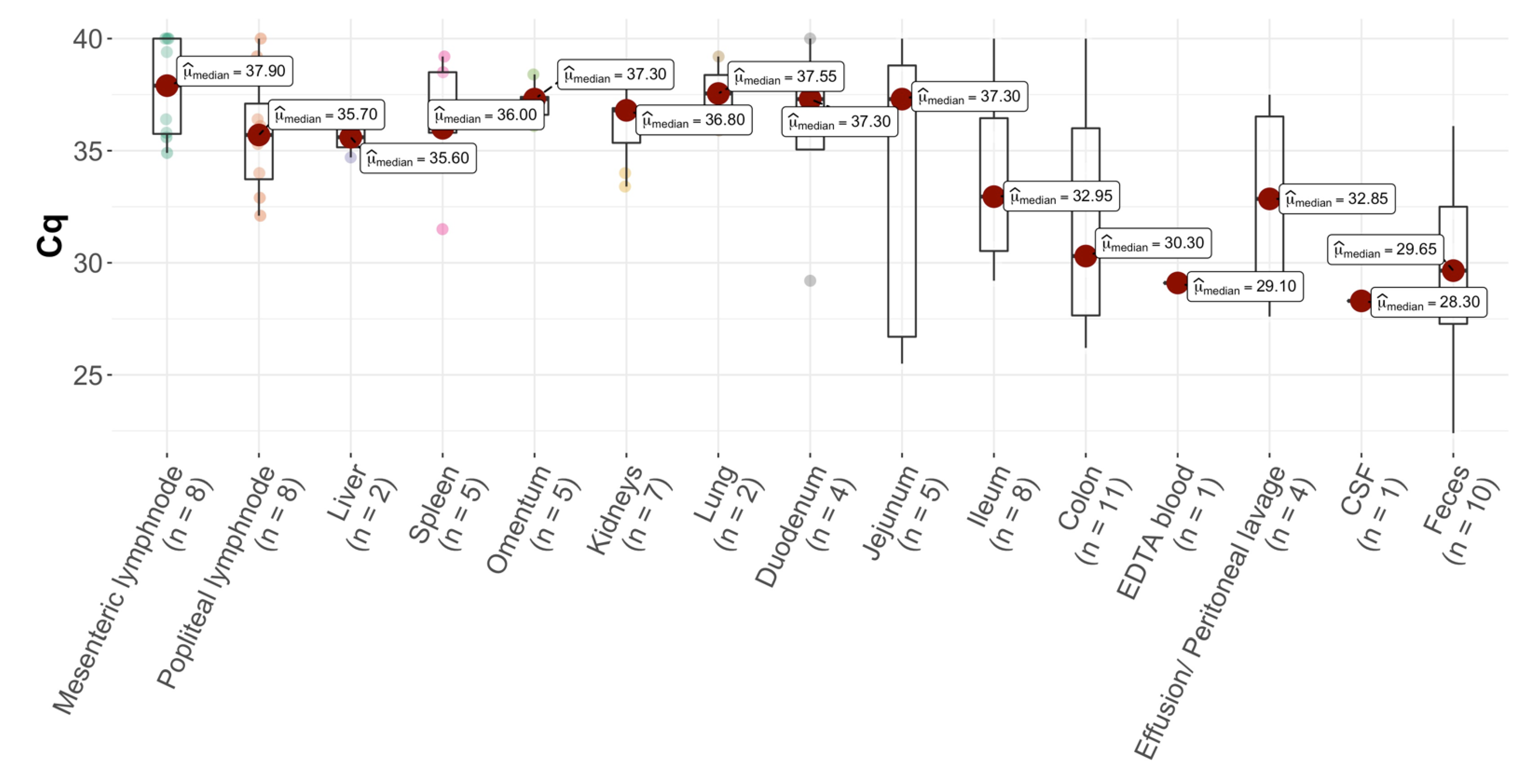
3.1. Presence of FCoV RNA in Cats without FIP

A total of 1861 samples of 87 cats were analyzed by FCoV 7b gene RT-qPCR for presence of FCoV RNA. FCoV RNA in at least one sample was detected in 21 of the 87 cats (24.1%). A total of 78 samples of these 21 cats were FCoV 7b gene RT-qPCR-positive (Table 1 and Table 2).

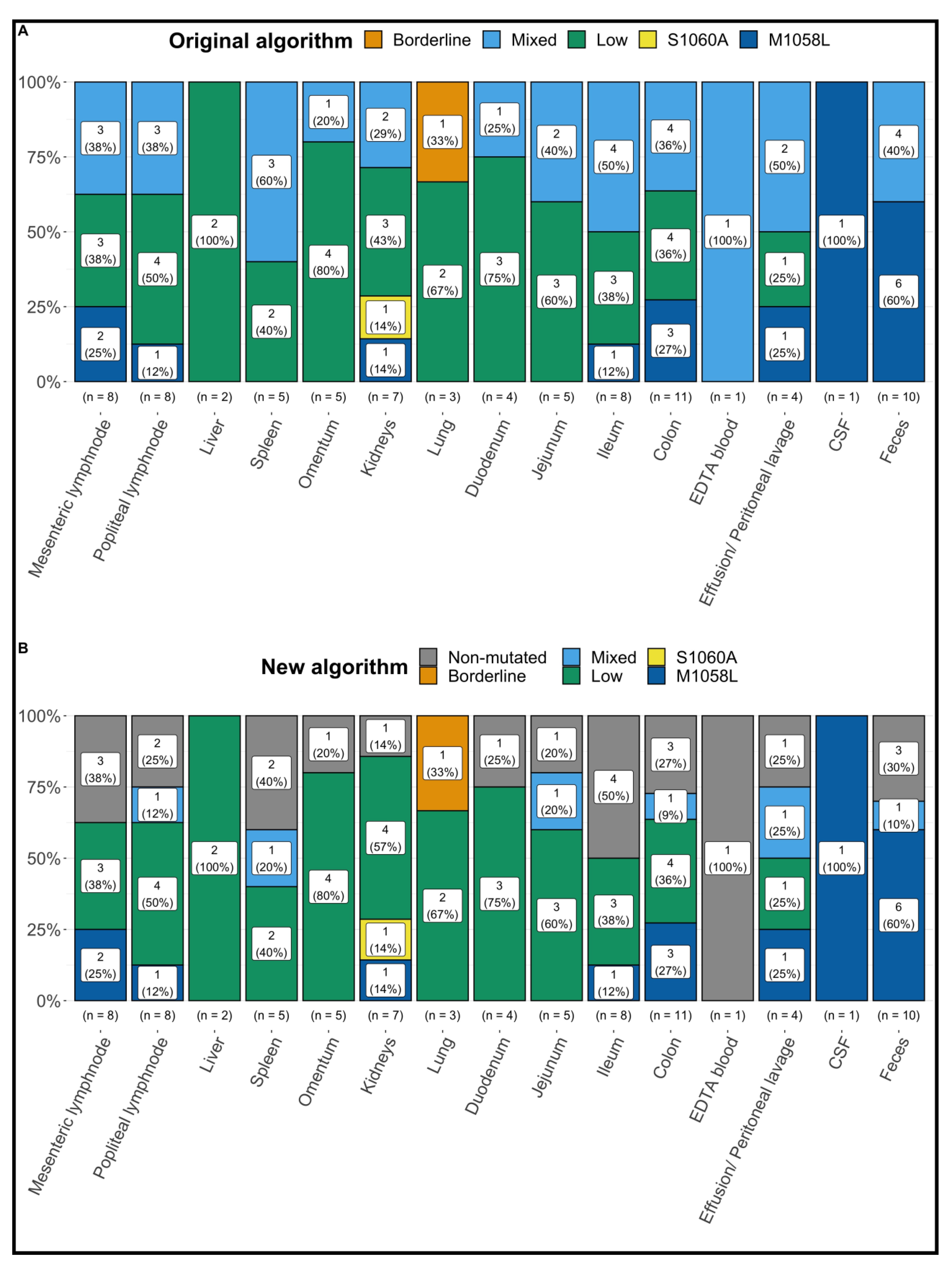
3.2. Results of FCoV S Gene Mutation RT-qPCR following the Original Algorithm

FCoV S gene mutation RT-qPCR detected “mutated FCoV” (with a fluorescence ratio of mutated to non-mutated FCoV > 2) and “mixed FCoV” in at least one sample in 14 of the 21 FCoV 7b gene RT-qPCR-positive cats (66.7%; only “mutated FCoV” were detected in 2/21 (9.5%), only “mixed FCoV” in 1/21 (4.8%), and different results with at least one sample positive for “mixed FCoV” or “mutated FCoV” in different tissues were found in 11/21 (52.4%) cats). Based on the original algorithm, none of the positive samples were interpreted as “non-mutated FCoV”. The remaining 7/21 (33.3%) cats were FCoV 7b gene RT-qPCR-positive but RNA load was too low in all tested samples to differentiate between mutated and non-mutated FCoV (“low”) (Table 1 and Table 2, Figure 1). The crossing point (cq) values of the positive samples are shown in Figure 2.

3.3. Results of FCoV S Gene Mutation RT-qPCR following the New Algorithm

FCoV S gene mutation RT-qPCR detected “mutated FCoV” (with a fluorescence ratio of mutated to non-mutated FCoV > 2) and “mixed FCoV” in at least one sample in 10 of the 21 FCoV 7b gene RT-qPCR-positive cats (47.6%; only “mutated FCoV” were detected in 2/21 (9.5%), only “mixed FCoV” in 0/21 (0%), and different results with at least one sample positive for “mixed FCoV” or “mutated FCoV” in different tissues were found in 8/21 (38.1%) cats). Different results with samples positive for “non-mutated FCoV” and “low” were detected in 4/21 cats (19.0%). The remaining 7/21 (33.3%) cats were FCoV 7b gene RT-qPCR-positive but RNA load was too low in all tested samples to differentiate between mutated and non-mutated FCoV (“low”) (Table 1 and Table 2, Figure 1).

3.4. Results of Sanger Sequencing

In total, 24 samples (14 body fluid and tissue samples and 10 fecal samples) of the 87 samples identified by the original algorithm as “FCoV with S gene mutations” or as “mixed FCoV” were selected for sequencing. Sequencing was possible in 18/24 samples (9/14 body fluid and tissue samples and 9/10 fecal samples) with 16/18 successfully sequenced samples (8 tissue and 8 fecal samples). This sample set included 3 fecal specimens and 1 sample from the ileum that were subsequently recategorized as “non-mutated FCoV” based upon the new algorithm. Only non-mutated FCoV was detected in all 16 samples (Table 3) by direct sequencing of the RT-PCR amplicons.
Of these 16 samples, 4 tissue samples and 4 fecal samples were selected for further characterization by cloning of the RT-PCR amplicons and sequencing of 10 clones each to detect possible underrepresented sequence variants. In all sequenced clones, only non-mutated FCoV was detected.

4. Discussion

This is the first study that included a large number of different tissue, body fluid, and fecal samples from a defined population of cats in which FIP was definitively ruled out by gold standard methods. The results of the commercially available FCoV S gene mutation RT-qPCR were quite extraordinary and unexpected because mutated FCoV was found in many tissues of these non-FIP cats and even in fecal samples. Therefore, reevaluation by Sanger sequencing was performed. This also allowed discussion about specificity of the commercially available FCoV S gene mutation RT-qPCR in the diagnosis of FIP.
The commercially available FCoV S gene mutation RT-qPCR used in the present study was developed to detect mutations considered specific for FIP and therefore, to have a higher diagnostic specificity to diagnose FIP than the detection of all FCoV by FCoV 7b gene RT-qPCR. Interestingly, the commercial assay diagnosed positive FCoV S gene mutation RT-qPCR results in several tissue, fluid, and even fecal samples of these cats in which FIP was definitively ruled out. However, it should be noted that fecal samples are not validated for use with this FCoV S gene mutation RT-qPCR by the commercial laboratory. Still, the fact that mutated FCoV was also found in feces using the FCoV S gene mutation RT-qPCR and that according to the original algorithm, non-mutated FCoV was not at all found in any sample of any of the tested cats, not even in feces, was very unexpected. The fluorescence from both, the mutated and non-mutated probes was detected in the FCoV S gene mutation RT-qPCR; however, using the original algorithm based on the ratio and the cycling curve, none of the results were interpreted as “non-mutated”. To further clarify the presence of S gene mutations in cats without FIP, samples positive by FCoV S gene mutation RT-qPCR were subsequently analyzed by Sanger sequencing of the S gene region of interest. Strikingly, sequencing did not confirm the results of the FCoV S gene mutation RT-qPCR in any of the sequenced samples; only non-mutated FCoV could be detected by sequencing. These contrasting results were highly surprising. Possible explanations include methodological errors of one of the diagnostic methods. Erroneous results of the Sanger sequencing seem highly unlikely, since the validity of the sequencing assay used in the present study has been confirmed previously [36] using 15 samples of 15 cats with confirmed FIP (and positive FCoV S gene mutation RT-qPCR): in 6 cats, the S gene mutation leading to amino acid substitution M1058L could be confirmed by sequencing; in 2 cats, the S gene mutation leading to amino acid substitution S1060A was present, and in 1 cat, a S gene mutation leading to amino acid substitution M1058F was present; in 4 cats, sequencing could not be performed because of low viral load [36]. Furthermore, to exclude the presence of a small proportion of mutated together with non-mutated FCoV (mixed FCoV) in the present study, the RT-PCR amplicon was cloned into a plasmid in selected samples, and 10 clones were sequenced from each amplicon. Even with this approach, no mutated FCoV could be detected in any of the analyzed samples. While these results do not completely rule out a very low number of mutated FCoV besides non-mutated FCoV, they do call the diagnostic value of the FCoV S gene mutation RT-qPCR into question.
So far, most previous studies evaluating the diagnostic specificity of the commercial FCoV S gene mutation RT-qPCR were hampered by the fact that they often did not include control cats without FIP that were positive for FCoV RNA by FCoV 7b gene RT-qPCR. Thus, a clear statement about the diagnostic specificity of the FCoV S gene mutation RT-qPCR could not be made [30,31]. Additionally, previous studies only included a small number of cats without FIP [25,28,30]. Still, one study also reported a positive FCoV S gene mutation RT-qPCR result in one cat in which FIP was definitely excluded [25]; mutated FCoV (amino acid substitution M1058L) was found in an effusion sample in this cat that suffered from chronic kidney disease. In those previous studies, it was discussed that positive results of the commercial FCoV S gene mutation RT-qPCR in cats without FIP could potentially be explained by the fact that cats with positive results could have suffered from early-stage FIP, although there were no histopathological changes of FIP. Furthermore, mutations in the S gene’s nucleotides 23531 and 23537 leading to amino acid substitutions M1058L and S1060A have been discussed as being markers of systemic spread of FCoV rather than being markers of the FIP phenotype [19]. Finally, a true false positive result because of a methodological error was possible. The fact that in the present study, sequencing (whenever it was possible) only detected FCoV with S genes without mutations in all the cats without FIP makes misdiagnosis and early-stage FIP as an explanation very unlikely.
Additionally, these findings indicate that the S gene mutations, if detected by sequencing, could indeed be specific for FIP, which was suggested by earlier studies [11,37], and that sequencing can be used to distinguish between cats with FIP and cats without FIP. This is in contrast to studies, in which presence of M1058L or S1060A substitutions in the FCoV S protein were supposed to be a hallmark of systemic spread of FCoV rather than a definitive marker of FIP [19,23]. In these studies, mutated FCoV was detected by sequencing in cats without FIP in tissue and effusion samples [23]. However, the results of the present study, in which mutated FCoV could not be detected in tissues from cats without FIP, together with the results of the study by Meli et al. [36] in which mutated FCoV was indeed detected by sequencing in various tissues and body fluids of cats with FIP, support the hypothesis that the mutations are indeed a marker for FIP and not only for systemic spread of FCoV [19].
As determined by Sanger sequencing, non-mutated FCoV was found in all 9 cats without FIP of which samples could successfully be sequenced. Three of the 9 cats had only a single sample sequenced and that sample was recategorized by the new algorithm to be non-mutated. In these cats, non-mutated FCoV could not only be detected in the feces, but also in various tissues. The presence of non-mutated FCoV in feces and in the colon and ileum was to be expected, as enteric infection and viral persistence has been described in otherwise clinically healthy cats and in cats suffering from diseases other than FIP [2,3,38]. Interestingly, however, non-mutated FCoV was also detected in other tissues, such as mesenteric and popliteal lymph nodes and kidneys. This has been shown before [35] and underlines the fact that non-mutated FCoV can spread systemically not only in cats with, but also in cats without FIP, and that the detection of FCoV by FCoV 7b gene RT-qPCR per se is not specific for FIP. Thus, a positive FCoV 7b gene RT-qPCR result, particularly if without determination of the viral load, cannot be used as a single diagnostic test to diagnose FIP.
FIP is a fatal disease if left untreated, and thus, specificity of confirmative tests is essential to avoid euthanasia or potentially harmful and expensive treatments in cats misdiagnosed with FIP. The results of the present study indicate that the commercially available FCoV S gene mutation RT-qPCR evaluated in the present study (at least in its current form) was not able to correctly identify the S gene mutations previously described to be specific for FIP [11]. In view of these results, this assay in its current form cannot be recommended for the diagnosis of FIP. On the other hand, sequencing of the regions of interest within the FCoV S gene could potentially be more suitable for the detection of S gene mutations, since no mutated FCoV sequences were detected by Sanger sequencing.
One limitation of this study was the fact that collection of the samples occurred postmortem. Therefore, it is possible that the amount of viable viral RNA was degraded. Another limitation was that the material analyzed by RT-qPCR was not exactly the same as the material that was sequenced; the examined material originated from the same biopsies, but the RNA was extracted independently. In addition, the commercial RT-qPCR assays were performed from July 2018 to August 2019 and sequencing was done in April 2021; however, all samples had been stored at −20 °C until processing. Finally, another limitation of the study was that sequencing was not performed on all samples. Sequencing was performed to specifically verify the correctness of results of samples classified as “mutated FCoV” in FCoV S gene mutation RT-qPCR.

5. Conclusions

FCoV RNA can be detected in cats without FIP, not only in feces but also systemically in various tissues and body fluids, but none of the detected FCoV contained S gene mutations as confirmed by Sanger sequencing of the S gene region. The applied commercially available FCoV S gene mutation RT-qPCR, however, classified FCoV as “mutated FCoV” or “mixed FCoV” in several samples. The results of the present study thus indicate that the accuracy of the herein used commercial FCoV S gene mutation RT-qPCR is limited, and other methods, such as quantification of FCoV RNA loads, should be used for the diagnosis of FIP. Alternatively, or additionally, sequencing of the regions of interest within the S gene is recommended for detection of S gene mutations which can increase diagnostic specificity when compared to detection of FCoV by 7b gene RT-qPCR alone.

Supplementary Materials

The following supplementary materials are available online at https://www.mdpi.com/article/10.3390/v14081671/s1, Table S1: Cats without feline infectious peritonitis (FIP) included in the study.

Author Contributions

Conceptualization, K.H., M.B. and S.F.; methodology, S.J., K.E., M.B., M.L.M., R.H.-L., K.M. and S.F.; formal analysis, S.J.; investigation, S.J., M.L.M. and K.E.; resources, K.H., R.H.-L. and K.M.; data curation, S.J.; writing—original draft preparation, S.J., K.H. and S.F.; writing—review and editing, K.H., S.F., M.L.M., R.H.-L. and K.M.; visualization, S.J.; supervision, K.H., M.B. and R.H.-L.; project administration, K.H., M.B., K.M. and R.H.-L.; funding acquisition, K.H., K.M. and R.H.-L. All authors have read and agreed to the published version of the manuscript.

Funding

FCoV 7b gene and FCoV S gene mutation RT-qPCR were kindly provided for free by IDEXX Laboratories. IDEXX played no role in the interpretation of data or in the decision to submit the manuscript for publication. Other than receiving the RT-qPCR results for free, this research received no external funding.

Institutional Review Board Statement

The prospective study in cats fulfilled the German guidelines for prospective studies and was approved by the ethical committee (reference number 120-08-04-2018) of the Centre for Clinical Veterinary Medicine of the LMU Munich.

Informed Consent Statement

Not applicable.

Data Availability Statement

The authors confirm that the datasets analyzed during the study are available from the corresponding author upon reasonable request.

Acknowledgments

We thank IDEXX for providing the RT-qPCR assays for free. The authors also thank Theres Meili from the University of Zurich for her excellent technical assistance. The RT-PCR amplification for sequencing was performed using the logistics of the Centre for Clinical Studies at the Vetsuisse Faculty of the University of Zurich.

Conflicts of Interest

The authors declare no potential conflict of interest with respect to the research, authorship, and/or publication of this article. Although the RT-qPCR assays were provided for free by IDEXX, IDEXX had no role in the design of the study, in the collection and interpretation of data, in the writing of the manuscript, or in the decision to submit the manuscript for publication. There is no commercial conflict of interest as the information generated here is solely for scientific dissemination. The authors declare that they have no competing interests.

References

  1. Addie, D.D.; Jarrett, O. A Study of Naturally Occurring Feline Coronavirus Infections in Kittens. Vet. Rec. 1992, 130, 133–137. [Google Scholar] [CrossRef] [PubMed]
  2. Kipar, A.; Meli, M.L.; Baptiste, K.E.; Bowker, L.J.; Lutz, H. Sites of Feline Coronavirus Persistence in Healthy Cats. J. Gen. Virol. 2010, 91, 1698–1707. [Google Scholar] [CrossRef] [PubMed] [Green Version]
  3. Desmarets, L.M.B.; Vermeulen, B.L.; Theuns, S.; Conceicąõ-Neto, N.; Zeller, M.; Roukaerts, I.D.M.; Acar, D.D.; Olyslaegers, D.A.J.; van Ranst, M.; Matthijnssens, J.; et al. Experimental Feline Enteric Coronavirus Infection Reveals an Aberrant Infection Pattern and Shedding of Mutants with Impaired Infectivity in Enterocyte Cultures. Sci. Rep. 2016, 6, 20022. [Google Scholar] [CrossRef] [PubMed]
  4. Meli, M.; Kipar, A.; Müller, C.; Jenal, K.; Gönczi, E.; Borel, N.; Gunn-Moore, D.; Chalmers, S.; Lin, F.; Reinacher, M.; et al. High Viral Loads despite Absence of Clinical and Pathological Findings in Cats Experimentally Infected with Feline Coronavirus (FCoV) Type I and in Naturally FCoV-Infected Cats. J. Feline Med. Surg. 2004, 6, 69–81. [Google Scholar] [CrossRef] [Green Version]
  5. Kipar, A.; Meli, M.L.; Failing, K.; Euler, T.; Gomes-Keller, M.A.; Schwartz, D.; Lutz, H.; Reinacher, M. Natural Feline Coronavirus Infection: Differences in Cytokine Patterns in Association with the Outcome of Infection. Vet. Immunol. Immunopathol. 2006, 112, 141–155. [Google Scholar] [CrossRef] [PubMed]
  6. Malbon, A.J.; Meli, M.L.; Barker, E.N.; Davidson, A.D.; Tasker, S.; Kipar, A. Inflammatory Mediators in the Mesenteric Lymph Nodes, Site of a Possible Intermediate Phase in the Immune Response to Feline Coronavirus and the Pathogenesis of Feline Infectious Peritonitis? J. Comp. Pathol. 2019, 166, 69–86. [Google Scholar] [CrossRef] [PubMed]
  7. Hu, C.M.J.; Chang, W.S.; Fang, Z.S.; Chen, Y.T.; Wang, W.L.; Tsai, H.H.; Chueh, L.L.; Takano, T.; Hohdatsu, T.; Chen, H.W. Nanoparticulate Vacuolar ATPase Blocker Exhibits Potent Host-Targeted Antiviral Activity against Feline Coronavirus. Sci. Rep. 2017, 7, 13043. [Google Scholar] [CrossRef] [PubMed] [Green Version]
  8. Herrewegh, A.A.P.M.; Smeenk, I.; Horzinek, M.C.; Rottier, P.J.M.; de Groot, R.J. Feline Coronavirus Type II Strains 79-1683 and 79-1146 Originate from a Double Recombination between Feline Coronavirus Type I and Canine Coronavirus. J. Virol. 1998, 72, 4508–4514. [Google Scholar] [CrossRef] [Green Version]
  9. Xia, H.; Li, X.; Zhao, W.; Jia, S.; Zhang, X.; Irwin, D.M.; Zhang, S. Adaptive Evolution of Feline Coronavirus Genes Based on Selection Analysis. BioMed Res. Int. 2020, 2020, 9089768. [Google Scholar] [CrossRef] [PubMed]
  10. Vennema, H.; Poland, A.; Foley, J.; Pedersen, N.C. Feline Infectious Peritonitis Viruses Arise by Mutation from Endemic Feline Enteric Coronaviruses. Virology 1998, 243, 150–157. [Google Scholar] [CrossRef] [PubMed] [Green Version]
  11. Chang, H.W.; Egberink, H.F.; Halpin, R.; Spiro, D.J.; Rottie, P.J.M. Spike Protein Fusion Peptide and Feline Coronavirus Virulence. Emerg. Infect. Dis. 2012, 18, 1089–1095. [Google Scholar] [CrossRef]
  12. Pedersen, N.C.; Liu, H.; Scarlett, J.; Leutenegger, C.M.; Golovko, L.; Kennedy, H.; Kamal, F.M. Feline Infectious Peritonitis: Role of the Feline Coronavirus 3c Gene in Intestinal Tropism and Pathogenicity Based upon Isolates from Resident and Adopted Shelter Cats. Virus. Res. 2012, 165, 17–28. [Google Scholar] [CrossRef] [PubMed]
  13. Bank-Wolf, B.R.; Stallkamp, I.; Wiese, S.; Moritz, A.; Tekes, G.; Thiel, H.J. Mutations of 3c and Spike Protein Genes Correlate with the Occurrence of Feline Infectious Peritonitis. Vet. Microbiol. 2014, 173, 177–188. [Google Scholar] [CrossRef]
  14. Borschensky, C.M.; Reinacher, M. Mutations in the 3c and 7b Genes of Feline Coronavirus in Spontaneously Affected FIP Cats. Res. Vet. Sci. 2014, 97, 333–340. [Google Scholar] [CrossRef] [PubMed]
  15. Bosch, B.J.; van der Zee, R.; de Haan, C.A.M.; Rottier, P.J.M. The Coronavirus Spike Protein Is a Class I Virus Fusion Protein: Structural and Functional Characterization of the Fusion Core Complex. J. Virol. 2003, 77, 8801–8811. [Google Scholar] [CrossRef] [PubMed] [Green Version]
  16. Lewis, C.S.; Porter, E.; Matthews, D.; Kipar, A.; Tasker, S.; Helps, C.R.; Siddell, S.G. Genotyping Coronaviruses Associated with Feline Infectious Peritonitis. J. Gen. Virol. 2015, 96, 1358–1368. [Google Scholar] [CrossRef] [PubMed] [Green Version]
  17. Licitra, B.N.; Millet, J.K.; Regan, A.D.; Hamilton, B.S.; Rinaldi, V.D.; Duhamel, G.E.; Whittaker, G.R. Mutation in Spike Protein Cleavage Site and Pathogenesis of Feline Coronavirus. Emerg. Infect. Dis. 2013, 19, 1066–1073. [Google Scholar] [CrossRef] [PubMed]
  18. Shirato, K.; Chang, H.W.; Rottier, P.J.M. Differential Susceptibility of Macrophages to Serotype II Feline Coronaviruses Correlates with Differences in the Viral Spike Protein. Virus Res. 2018, 255, 14–23. [Google Scholar] [CrossRef]
  19. Porter, E.; Tasker, S.; Day, M.J.; Harley, R.; Kipar, A.; Siddell, S.G.; Helps, C.R. Amino Acid Changes in the Spike Protein of Feline Coronavirus Correlate with Systemic Spread of Virus from the Intestine and Not with Feline Infectious Peritonitis. Vet. Res. 2014, 45, 49–59. [Google Scholar] [CrossRef] [PubMed] [Green Version]
  20. Hartmann, K.; Binder, C.; Hirschberger, J.; Cole, D.; Reinacher, M.; Schroo, S.; Frost, J.; Egberink, H.; Lutz, H.; Hermanns, W. Comparison of Different Tests to Diagnose Feline Infectious Peritonitis. J. Vet. Intern. Med. 2003, 17, 781–790. [Google Scholar] [CrossRef] [PubMed]
  21. Kipar, A.; Bellmann, S.; Kremendahl, J.; Köhler, K.; Reinacher, M. Cellular Composition, Coronavirus Antigen Expression and Production of Specific Antibodies in Lesions in Feline Infectious Peritonitis. Vet. Immunol. Immunopathol. 1998, 65, 243–257. [Google Scholar] [CrossRef]
  22. Sharif, S.; Arshad, S.S.; Hair-Bejo, M.; Omar, A.R.; Zeenathul, N.A.; Alazawy, A. Diagnostic Methods for Feline Coronavirus: A Review. Vet. Med. Int. 2010, 2010, 809480. [Google Scholar] [CrossRef] [PubMed] [Green Version]
  23. Barker, E.N.; Stranieri, A.; Helps, C.R.; Porter, E.L.; Davidson, A.D.; Day, M.J.; Knowles, T.; Kipar, A.; Tasker, S. Limitations of Using Feline Coronavirus Spike Protein Gene Mutations to Diagnose Feline Infectious Peritonitis. Vet. Res. 2017, 48, 60. [Google Scholar] [CrossRef] [PubMed] [Green Version]
  24. Dunbar, D.; Kwok, W.; Graham, E.; Armitage, A.; Irvine, R.; Johnston, P.; McDonald, M.; Montgomery, D.; Nicolson, L.; Robertson, E.; et al. Diagnosis of Non-Effusive Feline Infectious Peritonitis by Reverse Transcriptase Quantitative PCR from Mesenteric Lymph Node Fine-Needle Aspirates. J. Feline Med. Surg. 2019, 21, 910–921. [Google Scholar] [CrossRef] [Green Version]
  25. Felten, S.; Leutenegger, C.M.; Balzer, H.J.; Pantchev, N.; Matiasek, K.; Wess, G.; Egberink, H.; Hartmann, K. Sensitivity and Specificity of a Real-Time Reverse Transcriptase Polymerase Chain Reaction Detecting Feline Coronavirus Mutations in Effusion and Serum/Plasma of Cats to Diagnose Feline Infectious Peritonitis. BMC Vet. Res. 2017, 13, 228. [Google Scholar] [CrossRef] [PubMed]
  26. Longstaff, L.; Porter, E.; Crossley, V.J.; Hayhow, S.E.; Helps, C.R.; Tasker, S. Feline Coronavirus Quantitative Reverse Transcriptase Polymerase Chain Reaction on Effusion Samples in Cats with and without Feline Infectious Peritonitis. J. Feline Med. Surg. 2017, 19, 240–245. [Google Scholar] [CrossRef] [PubMed] [Green Version]
  27. Addie, D.; Belák, S.; Boucraut-Baralon, C.; Egberink, H.; Frymus, T.; Gruffydd-Jones, T.; Hartmann, K.; Hosie, M.J.; Lloret, A.; Lutz, H.; et al. Feline Infectious Peritonitis ABCD Guidelines on Prevention and Management. J. Feline Med. Surg. 2009, 11, 594–604. [Google Scholar] [CrossRef] [PubMed]
  28. Sangl, L.; Matiasek, K.; Felten, S.; Gründl, S.; Bergmann, M.; Balzer, H.J.; Pantchev, N.; Leutenegger, C.M.; Hartmann, K. Detection of Feline Coronavirus Mutations in Paraffin-Embedded Tissues in Cats with Feline Infectious Peritonitis and Controls. J. Feline Med. Surg. 2019, 21, 133–142. [Google Scholar] [CrossRef] [Green Version]
  29. Emmler, L.; Felten, S.; Matiasek, K.; Balzer, H.J.; Pantchev, N.; Leutenegger, C.; Hartmann, K. Feline Coronavirus with and without Spike Gene Mutations Detected by Real-Time RT-PCRs in Cats with Feline Infectious Peritonitis. J. Feline Med. Surg. 2020, 22, 791–799. [Google Scholar] [CrossRef] [PubMed]
  30. Felten, S.; Matiasek, K.; Leutenegger, C.M.; Sangl, L.; Herre, S.; Dörfelt, S.; Fischer, A.; Hartmann, K. Diagnostic Value of Detecting Feline Coronavirus Rna and Spike Gene Mutations in Cerebrospinal Fluid to Confirm Feline Infectious Peritonitis. Viruses 2021, 13, 186. [Google Scholar] [CrossRef]
  31. Sangl, L.; Felten, S.; Matiasek, K.; Dörfelt, S.; Bergmann, M.; Balzer, H.J.; Pantchev, N.; Leutenegger, C.; Hartmann, K. Detection of Feline Coronavirus RNA, Spike Gene Mutations, and Feline Coronavirus Antigen in Macrophages in Aqueous Humor of Cats in the Diagnosis of Feline Infectious Peritonitis. J. Vet. Diagn. 2020, 32, 527–534. [Google Scholar] [CrossRef] [PubMed]
  32. Felten, S.; Hartmann, K. Diagnosis of Feline Infectious Peritonitis: A Review of the Current Literature. Viruses 2019, 11, 1068. [Google Scholar] [CrossRef] [Green Version]
  33. Felten, S.; Matiasek, K.; Gruendl, S.; Sangl, L.; Wess, G.; Hartmann, K. Investigation into the Utility of an Immunocytochemical Assay in Body Cavity Effusions for Diagnosis of Feline Infectious Peritonitis. J. Feline Med. Surg. 2017, 19, 410–418. [Google Scholar] [CrossRef]
  34. Gut, M.; Leutenegger, C.M.; Huder, J.B.; Pedersen, N.C.; Lutz, H. One-Tube Fluorogenic Reverse Transcription-Polymerase Chain Reaction for the Quantitation of Feline Coronaviruses. J. Virol. Methods 1999, 77, 37–46. [Google Scholar] [CrossRef]
  35. Lutz, M.; Steiner, A.R.; Cattori, V.; Hofmann-Lehmann, R.; Lutz, H.; Kipar, A.; Meli, M.L. Fcov Viral Sequences of Systemically Infected Healthy Cats Lack Gene Mutations Previously Linked to the Development of Fip. Pathogens 2020, 9, 603. [Google Scholar] [CrossRef] [PubMed]
  36. Meli, M.L.; Spiri, A.M.; Zwicklbauer, K.; Krentz, D.; Felten, S.; Bergmann, M.; Dorsch, R.; Matiasek, K.; Alberer, M.; Kolberg, L.; et al. Fecal Feline Coronavirus RNA Shedding and Spike Gene Mutations in Cats with Feline Infectious Peritonitis Treated with GS-441524. Viruses 2022, 14, 1069. [Google Scholar] [CrossRef] [PubMed]
  37. Barker, E.N.; Tasker, S.; Gruffydd-Jones, T.J.; Tuplin, C.K.; Burton, K.; Porter, E.; Day, M.J.; Harley, R.; Fews, D.; Helps, C.R.; et al. Phylogenetic Analysis of Feline Coronavirus Strains in an Epizootic Outbreak of Feline Infectious Peritonitis. J. Vet. Intern. Med. 2013, 27, 445–450. [Google Scholar] [CrossRef] [PubMed] [Green Version]
  38. Vogel, L.; van der Lubben, M.; te Lintelo, E.G.; Bekker, C.P.J.; Geerts, T.; Schuijff, L.S.; Grinwis, G.C.M.; Egberink, H.F.; Rottier, P.J.M. Pathogenic Characteristics of Persistent Feline Enteric Coronavirus Infection in Cats. Vet. Res. 2010, 41, 71–82. [Google Scholar] [CrossRef] [PubMed] [Green Version]
Figure 1. Summary of the results of the commercial real-time feline coronavirus (FCoV) 7b gene and spike (S) gene mutation reverse transcription polymerase chain reactions (RT-qPCR) following following the original algorithm (A) and the new algorithm (B) in all tissue, fluid, and fecal samples from the 21 cats positive for FCoV RNA in at least one sample. M1058L = positive FCoV S gene mutation RT-qPCR detecting mutation in nucleotide 23,531, corresponding to amino acid substitution M1058L; S1060A = positive FCoV S gene mutation RT-qPCR detecting mutation in nucleotide 23537, corresponding to amino acid substitution S1060A; Low = positive FCoV 7b gene RT-qPCR with viral load below cut-off (therefore no further differentiation possible by FCoV S gene mutation RT-qPCR); Mixed = positive FCoV 7b gene RT-qPCR and presence of FCoV with and without S gene mutations; Borderline = a specific PCR product (amplicon) could neither be detected with certainty nor could its presence be ruled out; EDTA = ethylenediamine tetra acetic acid; CSF = cerebrospinal fluid.
Viruses 14 01671 g001
Figure 2. Box-whisker-plot comparing Cq values in tissue, fluid, and fecal samples from the 21 cats positive for feline coronavirus (FCoV) RNA in at least one sample. EDTA = ethylenediamine tetra acetic acid; CSF = cerebrospinal fluid.
Viruses 14 01671 g002
Table 1. Results of the commercial real-time feline coronavirus (FCoV) 7b gene reverse transcription polymerase chain reaction (RT-qPCR) and spike (S) gene mutation RT-qPCR following the original algorithm, and the new algorithm for interpretation of the results in the 87 cats.
Original AlgorithmNew Algorithm
Total number of cats8787
FCoV 7b Gene RT-qPCR FCoV 7b gene RT-qPCR-
negative in all samples
6666
FCoV 7b gene RT-qPCR-
positive in at least 1 sample
2121
FCoV S Gene Mutation RT-qPCR0Same Results in All Samples1Only Non-mutated00
2Only Mutated (M1058L *)22
3Only Mutated (S1060A )00
4Only Mixed §10
Only Low 77
Different Results in Different SamplesMixed § and Mutated (M1058L *)10
Mixed § and Low 40
Borderline Ω, Mixed § and Low 10
Mutated (M1058L *) and Low 33
Mutated (both M1058L * and S1060A ) and Low 11
Mixed § and Mutated (M1058L *) and Low 10
Non-mutated and Low 04
Non-mutated and M1058L *01
Non-mutated and Mixed §01
Non-mutated, M1058L *, Mixed § and Low 01
Non-mutated, Borderline Ω, Mixed § and Low 01
M1058L * = positive FCoV S gene mutation RT-qPCR detecting mutation in nucleotide 23531, corresponding to amino acid substitution M1058L; S1060A = positive FCoV S gene mutation RT-qPCR detecting mutation in nucleotide 23537, corresponding to amino acid substitution S1060A; Low = positive FCoV 7b gene RT-qPCR with viral load below cut-off (therefore no further differentiation possible by FCoV S gene mutation RT-qPCR); Mixed FCoV § = positive FCoV 7b gene RT-qPCR and presence of FCoV with and without S gene mutations; Borderline Ω = a specific PCR product (amplicon) could neither be detected with certainty nor its presence could be ruled out; 0 FCoV S gene mutation RT-qPCR was only performed on the 21 FCoV 7b gene RT-qPCR-positive samples; 1 Only Non-mutated FCoV = non-mutated FCoV in each positive sample; 2 Only M1058L * = M1058L * in each positive sample; 3 Only S1060A = S1060A in each positive sample; 4 Only Mixed FCoV § = mixed FCoV § in each positive sample; Bold indicates samples positive for Mixed §, M1058L * or S1060A .
Table 2. Results of the commercial real-time feline coronavirus (FCoV) 7b gene and spike (S) gene mutation reverse transcription polymerase chain reactions (RT-qPCR) following the original algorithm and the new algorithm in all tissue, fluid, and fecal samples from the 21 cats positive for FCoV RNA in at least one sample.
Cat Mesenteric
Lymph Node
Popliteal
Lymph Node
LiverSpleenOmentumKidneysLungDuodenumJejunumIleumColonEDTA
Blood
Effusion/Peritoneal LavageCSFAqueous HumorFeces
1Cq39.4 >40.0 39.0
Origi-nalLow ------Low -Low ------
NewLow ------Low -Low ------
2Cq 31.5 39.2
Origi-nal---Mixed§--Low ---------
New Non-mutated Low
3Cq 34.935.334.738.5 34.0
Origi-nalMixed§Low Low Low -Low ----------
NewNon-mutatedLow Low Low Low
4Cq 38.3
Origi-nal-----Low ----------
New Low
5Cq35.634.036.535.836.136.735.929.226.729.226.229.127.6 27.5
Origi-nalMixed §Mixed §Low Mixed §Mixed §Mixed § Mixed §Mixed §Mixed §Mixed §Mixed §Mixed §--Mixed §
NewNon-mutatedNon-mutatedLow Non-mutatedNon-mutatedNon-mutatedLow Non-mutatedNon-mutatedNon-mutatedNon-mutatedNon-mutatedNon-mutated Non-mutated
6Cq >40.0>40.0
Origi-nalLow Low --------------
NewLow Low
7Cq >40.0
Origi-nal---------Low ------
New Low
8Cq >40.0
Origi-nal----------Low -----
New Low
9Cq 38.4
Origi-nal----Low -----------
New Low
10Cq 29.528.3
Origi-nal------------M1058L *M1058L *--
New M1058L *M1058L *
11Cq Cq 35.835.2 (FNA)
32.1
37.336.8 >40.030.728.1 36.2
Origi-nalM1058L *M1058L *--Low M1058L *--Low M1058L *M1058L *-Low ---
NewM1058L *M1058L * Low M1058L * Low M1058L *M1058L * Low
12Cq 38.8
Origi-nal--------Low -------
New Low
13Cq>40.036.4 39.236.636.8No Cq value37.025.530.026.3 23.2
Origi-nalMixed §Mixed §-Mixed §Low Mixed §Borderline ΩLow Mixed §Mixed §Mixed §----Mixed §
NewNon-mutatedNon-mutated Mixed §Low Low Borderline ΩLow Mixed §Non-mutatedMixed § Mixed §
14Cq 36.439.2 36.037.433.4 35.627.2 22.4
Origi-nalM1058L *Low -Low Low S1060A ---Low M1058L *----M1058L *
NewM1058L *Low Low Low S1060A Low M1058L * M1058L *
15Cq>40.0 37.8 27.2
Origi-nalLow --------- Low ---M1058L *
NewLow Low M1058L *
16Cq 32.9 37.0 37.637.333.129.6 32.2
Origi-nal-Mixed §---Low -Low Low Mixed §M1058L *----M1058L *
New Mixed § Low Low Low Non-mutatedM1058L * M1058L *
17Cq 32.834.2 30.6
Origi-nal---------Mixed §Low ----Mixed §
New Non-mutatedLow Non-mutated
18Cq 33.8 32.6
Origi-nal----------Low ----M1058L *
New Low M1058L *
19Cq 38.7 36.1
Origi-nal----------Mixed §----M1058L *
New Non-mutated M1058L *
20Cq 36.1 30.3 37.5 28.7
Origi-nal-Low --------Mixed §-Mixed §--Mixed §
New Low Non-mutated Mixed § Non-mutated
21Cq value 34.2
Origi-nal---------------M1058L *
New M1058L *
Original = Original algorithm for FCoV S gene mutation RT-qPCR; New = new algorithm for FCoV S gene mutation RT-qPCR; Cq = crossing point value of FCoV 7b gene RT-qPCR; M1058L* = positive FCoV S gene mutation RT-qPCR detecting mutation in nucleotide 23531, corresponding to amino acid substitution M1058L; S1060A = positive FCoV S gene mutation RT-qPCR detecting mutation in nucleotide 23537, corresponding to amino acid substitution S1060A; - = negative FCoV 7b gene RT-qPCR; Low = positive FCoV 7b gene RT-qPCR with viral load below cut-off (therefore no further differentiation possible by FCoV S gene mutation RT-qPCR); Mixed § = positive FCoV 7b gene RT-qPCR and presence of FCoV with and without S gene mutations; Borderline Ω = a specific PCR product (amplicon) could neither be detected with certainty nor could its presence be ruled out; EDTA = ethylenediamine tetra acetic acid; CSF = cerebrospinal fluid; Bold indicates samples positive for Mixed §, M1058L *or S1060A.
Table 3. Results of the commercial real-time feline coronavirus (FCoV) spike (S) gene mutation reverse transcription polymerase chain reaction (RT-qPCR) and corresponding Sanger sequencing and cloning in the 16 samples (8 tissue and 8 fecal samples) of 9 nine cats that could be successfully sequenced.
CatMaterial Commercial FCoV S Gene Mutation RT-qPCR According to the Original AlgorithmCommercial FCoV S Gene Mutation RT-qPCR According to the New AlgorithmSanger SequencingSequencing after Cloning
5FecesMixed §Non-mutatedNon-mutatedNon-mutated
11Popliteal lymph nodeM1058L *M1058L *Non-mutatedNon-mutated
11KidneyM1058L *M1058L *Non-mutated
11IleumM1058L *M1058L *Non-mutatedNon-mutated
11Mesenteric lymph nodeM1058L * M1058L * Non-mutated
13FecesMixed §Mixed §Non-mutatedNon-mutated
14KidneyM1058L *M1058L *Non-mutatedNon-mutated
14ColonM1058L *M1058L *Non-mutatedNon-mutated
14FecesM1058L *M1058L *Non-mutatedNon-mutated
16IleumMixed §Non-mutated Non-mutated
16ColonM1058L *M1058L *Non-mutated
16FecesM1058L *M1058L *Non-mutatedNon-mutated
17FecesMixed §Non-mutatedNon-mutated
18FecesM1058L *M1058L *Non-mutated
19FecesM1058L *M1058L *Non-mutated
20FecesMixed §Non-mutatedNon-mutated
M1058L * positive FCoV S gene mutation RT-qPCR detecting mutation in nucleotide 23531, corresponding to amino acid substitution M1058L; Mixed § = presence of both FCoV with and without S gene mutations.
Publisher’s Note: MDPI stays neutral with regard to jurisdictional claims in published maps and institutional affiliations.

