Article

Repurposing an In Vitro Measles Virus Dissemination Assay for Screening of Antiviral Compounds

1
Department of Viroscience, Erasmus MC, 3015 GD Rotterdam, The Netherlands
2
Gilead Sciences, Foster City, CA 94404, USA
3
Robert Frederick Smith School of Chemical and Biomolecular Engineering, Cornell University, Ithaca, NY 14850, USA
4
Department of Pediatrics, Columbia University Medical Center, New York, NY 10032, USA
5
Center for Host–Pathogen Interaction, Columbia University Irving Medical Center, New York, NY 10032, USA
6
Department of Physiology and Cellular Biophysics, Columbia University Irving Medical Center, New York, NY 10032, USA
7
Department of Microbiology and Immunology, Columbia University Irving Medical Center, New York, NY 10032, USA
8
Department of Experimental Medicine, University of Campania “Luigi Vanvitelli”, 81100 Caserta, Italy
*
Author to whom correspondence should be addressed.
Current address: Division of Veterinary Medicine, Paul-Ehrlich-Institut, 63225 Langen, Germany.
These authors contributed equally to this work.
§
As part of: Master Program Integrated Immunology, FAU Erlangen-Nürnberg and Elite Network of Bavaria.
Viruses 2022, 14(6), 1186; https://doi.org/10.3390/v14061186
Submission received: 27 March 2022 / Revised: 25 May 2022 / Accepted: 25 May 2022 / Published: 29 May 2022
(This article belongs to the Special Issue Measles)

Abstract

:
Measles virus (MV) is a highly contagious respiratory virus responsible for outbreaks associated with significant morbidity and mortality among children and young adults. Although safe and effective measles vaccines are available, the COVID-19 pandemic has resulted in vaccination coverage gaps that may lead to the resurgence of measles when restrictions are lifted. This puts individuals who cannot be vaccinated, such as young infants and immunocompromised individuals, at risk. Therapeutic interventions are complicated by the long incubation time of measles, resulting in a narrow treatment window. At present, the only available WHO-advised option is treatment with intravenous immunoglobulins, although this is not approved as standard of care. Antivirals against measles may contribute to intervention strategies to limit the impact of future outbreaks. Here, we review previously described antivirals and antiviral assays, evaluate the antiviral efficacy of a number of compounds to inhibit MV dissemination in vitro, and discuss potential application in specific target populations. We conclude that broadly reactive antivirals could strengthen existing intervention strategies to limit the impact of measles outbreaks.

1. Introduction

Measles virus (MV) is a highly contagious respiratory virus that causes outbreaks associated with significant morbidity and mortality. Multiple initiatives, including the Measles and Rubella Initiative, set out to eliminate measles [1]. Safe and effective live-attenuated measles vaccines are available, but vaccination hesitancy and vaccination coverage gaps have decreased MV-specific herd immunity, leading to global resurgences of measles. Yearly, measles accounts for more than 100,000 deaths, mostly in children under the age of five in Africa and Asia [2]. In recent years, increasing numbers of measles cases and associated deaths were recorded. In 2019, measles cases hit their highest number in 23 years and were responsible for more than 207,000 deaths [3]. Case numbers drastically declined in 2020 and 2021 due to social distancing measures and travel restrictions, but a further measles surge is expected in the upcoming years as measles immunization programs have been affected by the COVID-19 pandemic [4,5].

1.1. Measles Virus

MV is a member of the genus Morbillivirus of the family Paramyxoviridae and has a negative-sense unsegmented RNA genome [6]. MV is a monotypic virus. Although eight different clades and 24 genotypes have been identified, most of these no longer circulate, and genotype B3 and D8 are currently the most prevalent worldwide, while genotype H1 still circulates in China [7,8]. Despite harboring an RNA-dependent RNA-polymerase (RdRp) that lacks proofreading capacity, the mutation rate of MV is low. Most likely, the virus has a narrow fitness peak and is limited in the mutations it can tolerate. Therefore, vaccines developed in the 1960s still offer clinical protection from measles. The MV genome encodes six structural and two non-structural proteins [9]: The nucleoprotein (N) encapsidates the viral genome. The two viral transmembrane glycoproteins, the hemagglutinin (H) and the fusion protein (F), are responsible for cell attachment and entry [10,11]. Entry is initiated by the H protein, binding to the cellular receptors CD150 (SLAM) or nectin-4 [12,13,14] and triggering confirmational changes in the F protein. These conformational changes facilitate the fusion of the viral and cell membrane. Subsequently, the genome content is released into the cell, transcribed by the viral RdRp (large protein, L) and its cofactor phosphoprotein (P), and by orchestration of the matrix (M) protein, all viral proteins are ultimately assembled at the cell membrane before being released [15,16]. Detailed characterization of the two non-structural proteins V and C, which are transcribed from the P gene, remains to be performed, but both appear to be virulence factors involved in antagonizing innate immune responses [17].

1.2. Pathogenesis

Measles virus is highly contagious and has a basic reproductive number (R0) of 12 – 18 [18]. It enters via the respiratory tract, infects CD11c+ myeloid cells, probably alveolar macrophages and DCs, which disseminate the virus to the local lymphoid tissues [19,20]. In the draining lymph nodes, MV infects CD150+ B- and T-lymphocytes, which effectively amplify and distribute the virus throughout the body [21]. Ultimately, a major fraction of CD150+ white blood cells, including lymphocytes and DCs in the epithelial submucosa, are infected. Those infected cells can transfer the virus to nectin-4-positive respiratory epithelial cells from where MV is released via the upper respiratory tract and transmitted by aerosols or small droplets [22,23].
The MV incubation phase is between 9 and 19 days, followed by a prodromal phase, which is characterized by fever, conjunctivitis, malaise, cough and coryza. The preferential massive infection and depletion of CD150-positive memory lymphocytes results in transient immune amnesia [24], which may last more than two years after measles. This leaves measles patients susceptible to opportunistic infections and leads to increased mortality due to infectious complications [25]. Paradoxically, a robust MV-specific immune response is formed, which clears the infection and lasts for life [26]. The typical measles rash following the prodromal phase with a two-to-four-day delay is a result of MV-specific T-cell invasion into the skin to clear infected keratinocytes [27].
In rare cases, measles may also lead to neurological complications such as acute post measles encephalitis (APME), measles inclusion body encephalitis (MIBE) or subacute sclerosing panencephalitis (SSPE). While APME is associated with an auto-immune reaction against oligodendrocytes, fatal in 20% of the cases, MIBE and SSPE occur after MV infection of the central nervous system (CNS) in immunocompromised or immunocompetent patients, respectively. Both MIBE and SSPE are almost always fatal and can only be treated symptomatically (reviewed in [28]).

1.3. Treatment Challenges

Given its high transmissibility, herd immunity levels of 95% are required for sufficient measles control [29]. Decreasing vaccination coverage results in rising case numbers and more complications associated with measles. Therefore, there is a significant risk for individuals who cannot be vaccinated, such as young infants (<6 months) and immunocompromised individuals but also for unvaccinated healthcare workers and their vulnerable patients. Antivirals could be used for measles prophylaxis in an insufficiently protected population or serve as treatment during acute infection or measles-associated long-term complications. However, to date, no specific antiviral treatment for MV is approved. The pathogenesis of measles is complex, making antiviral treatment challenging. Measles has a long incubation time, and initial symptoms are often attributed to other infectious diseases, partly because general practitioners are rarely confronted with measles. Rather than staying confined to the respiratory tract, MV rapidly spreads systemically, even before the onset of symptoms. Once in the dissemination phase, it is likely that treatment will have limited impact on disease outcome or immune suppression, leaving an extremely narrow treatment window in the extended incubation phase to prevent lymphopenia.
On the other hand, measles outbreaks usually occur in geographically clustered communities. Rapid identification of index cases allows for contact tracing of unvaccinated individuals and early post-exposure treatment. Post-exposure vaccination or passive immunization with intra-venous immunoglobulin (IVIg) is recommended by the World Health Organization (WHO) and has been used with diverse levels of success [30,31,32]. Effective measles prevention and treatment requires optimal infrastructure and resources, which are not always present in countries where measles is most prevalent.

1.4. In Vitro Testing

Antiviral compounds investigated for in vitro activity against MV include nucleoside analogs, non-nucleoside polymerase inhibitors, fusion and entry inhibitors, host-directed compounds, plant extracts, disinfectants, anti-microbial agents, and interferons (Table 1).
Nucleoside analogs commonly target viral replication, particularly the viral DNA or RNA polymerase. They contain non-canonical bases that are integrated into the viral genome after intracellular phosphorylation [89,90]. The purine-analog ribavirin has demonstrated broad spectrum antiviral activity [91] and has been approved to treat several viruses, including respiratory syncytial virus (RSV) [49]. Multiple mechanisms of actions are being discussed, including the inhibition of viral polymerase and viral RNA capping [92]. Ribavirin base-pairs with equal efficiency with cytosine and uracil [93], resulting in hypermutations of the newly synthesized strand, which block virus replication via error catastrophe [94]. However, resembling adenosine and guanosine also carries the disadvantage of increased interaction with the host cell machinery, leading to poor selectivity and toxicity, which may result in adverse events such as anemia [92].
Remdesivir (GS-5734) is a nucleoside analog prodrug with broad-spectrum in vitro antiviral activity against RNA-viruses from many virus families [41]. As an adenosine-analog, remdesivir interferes with the viral RNA synthesis [95] and leads to delayed RNA-chain termination three to five nucleotides after its incorporation [92,96]. Remdesivir has demonstrated efficacy in the treatment of both hospitalized and non-hospitalized COVID-19 patients in multiple clinical trials and was initially approved by the U.S. Food and Drug administration (FDA) in 2020 [97].
The RdRp presents a promising target for broad-spectrum non-nucleoside antiviral drugs, as it is highly conserved across several RNA viruses such as influenza viruses, coronaviruses and paramyxoviruses, including MV [98]. Different non-nucleoside inhibitors such as ERDRP-0519 or AS-136a show efficacy in in vitro and in vivo studies [48,51]. Targeting the L subunit of the RdRp, ERDRP-0519 inhibits the formation of phosphodiester bonds during initiation of RNA synthesis and RNA elongation [51].
Next to the viral polymerase, an important protein in the MV lifecycle is the fusion protein. The fusion of enveloped viruses with the host cell is a key step in their infectivity, and interference with this process can lead to highly effective antivirals. Fusion inhibitors for class I viral fusion proteins are often peptides or small molecules and can have multiple mechanisms of action. Fusion inhibitory peptide (FIP) was first used as an acronym for Z-D- Phe-L-Phe-L-Gly, a synthesized oligopeptides with an amino acid sequence that is similar to the N-terminal region of F1 [54]. FIP-analogs block membrane fusion by stabilizing the prefusion state of the viral F protein. In contrast, peptides mimicking one of the two heptad repeat (HR) domains capture the viral F protein in a post-triggering state and freeze the fusion process at the beginning.
Overall, a dozen different antiviral assays with minor modifications have been performed to evaluate the antiviral potency of numerous antiviral drugs against MV (Table 2).
Given the pathogenesis of measles, highly predictive in vitro models mimicking the in vivo situation are needed. Considering the earliest realistic timepoint of treatment and the early events during MV infection, an in vitro assay resembling the dissemination phase of MV in immune cells is anticipated to provide translational value. Moreover, wild-type-based viruses in comparison to vaccine-based viruses seem superior when evaluating the antiviral potential of a drug to clinically relevant MV. Therefore, we repurposed a previously established MV-dissemination assay in lymphoid cells for screening of antivirals [100]. In this assay, we evaluated the antiviral efficacy of IVIg, remdesivir and a selection of fusion inhibitory lipopeptides designed to inhibit MV fusion in comparison to ribavirin. Moreover, we screened 153 antiviral candidates from the Medicines for Malaria Venture Pandemic Response Box for their potential to inhibit MV dissemination.

2. Materials and Methods

2.1. Cells and Viruses

All experiments were performed in human Epstein–Barr-virus-transformed (EBV) B-lymphoblastic cell lines (B-LCL). B-LCLs were maintained in RPMI-1640 medium (Lonza) supplemented with penicillin (1000 U/mL), streptomycin (100 µg/mL), 2 mM L-glutamine and 10% fetal bovine serum (FBS). Virus stocks were grown in B-LCL, MRC-5 cells or Vero-human-SLAM (VHS) cells. MRC-5 and VHS were maintained in DMEM (Lonza) supplemented with penicillin, streptomycin, L-glutamine and 10% FBS. The recombinant MV expressing the reporter protein Venus at position three of the genome (rMVKSVenus(3)) was based on a clinical isolate from Khartoum, Sudan [101]; rMVKSVenus(3) was grown in B-LCL. The recombinant MV expressing the reporter protein EGFP at position three of the genome (rMVEZEGFP(3)) was based on the Edmonston-Zagreb vaccine virus [102]; rMVEZEGFP(3) was grown in MRC-5 cells. All clinical isolates (MVi/Khartoum.SUD/34.97/2 − genotype B3, MVi/Bilthoven. NLD/1991 − genotype C2, MVi/Amsterdam.NLD/19.11 − genotype D4, MVi/ Dodewaaard.NLD/29.13 − genotype D8, MVi/Amsterdam.NLD/49.97 − genotype G2, MVi/Amsterdam/NLD/27.97 − genotype H1) used in this study were isolated in B-LCLs, and stocks were grown in VHS.

2.2. Antiviral Compounds

IVIg (Privigen) was diluted to 100 mg/mL and used at final concentrations of 2 mg/mL to 0.2 ng/mL (10-fold dilutions). Remdesivir was obtained from Gilead Sciences, Inc. (Foster City, CA, USA) as a 20 mM solution dissolved in DMSO. In all assays, remdesivir was used at final concentrations of 10 µM to 0.1 nM (5-fold dilution series). Ribavirin was diluted in DMSO to a concentration of 25 mM and used at final concentrations of 250 µM to 2 µM (2-fold dilution series). All fusion inhibitory peptides were received as a 5 mM solution and used at final concentrations of 10 µM to 0.1 nM (5-fold dilution series). The Pandemic Response Box was designed and kindly supplied by Medicines for Malaria Venture (MMV). Compounds classified as ‘antiviral’ were diluted to 2 mM in DMSO and aliquoted until further use. For primary screenings, antivirals were tested at 20 µM and 4 µM. Potential hits were defined as compounds that resulted in less than 50% MV infection at a concentration of 20 μM and/or less than 90% MV infection at a concentration of 4 μM, independent of toxicity and time point of evaluation (48 or 72 h post co-culture, hpc). Those hits were re-screened at 4 µM, 800 nM, 160 nM and 32 nM.

2.3. In Vitro MV Dissemination Assay

A previously established MV-dissemination assay was used to test the efficacy of the selected antiviral compounds (Figure 1a) [100]. B-LCLs were infected with MV (clinical isolate or recombinant MV) for 48 to 72 h. Infection percentage in the live cell fraction was determined using flow cytometry (BD FACS Lyric), either by measuring expression of the green fluorescent reporter protein or by staining for MV nuclear protein (NP). In the assay, 150 MV-infected cells were added to 60,000 uninfected B-LCLs. Antiviral compounds were added to those cultures simultaneously, six hours before or six hours after co-culture. As control, co-cultures were performed without antiviral (in the presence of equimolar concentrations of DMSO). MV dissemination and compound or vehicle toxicity were analyzed 48 and 72 h post co-culture. For toxicity, cell viability was evaluated based on FSC/SSC and for infection percentage FITC+ events were recorded. In assays performed with clinical isolates, cells were washed in PBS, fixed and permeabilized (Cytofix-Cytoperm, BD) according to the manufacturer’s instructions and stained with anti-MV-NP/FITC (KK2, Millipore). Data shown were collected 48 h post co-culture unless differently stated. At this point, infection levels in control co-cultures were >90%. Remdesivir, ribavirin and fusion-inhibitory peptides were tested in duplicate in three or more independent assays.

2.4. Data Analysis

The half-maximal inhibitory concentration (IC50) was determined using a four-parameter non-linear regression on normalized (to DMSO-treated co-cultures) and transformed infection percentages. All analyses were performed in GraphPad Prism 9.

3. Results

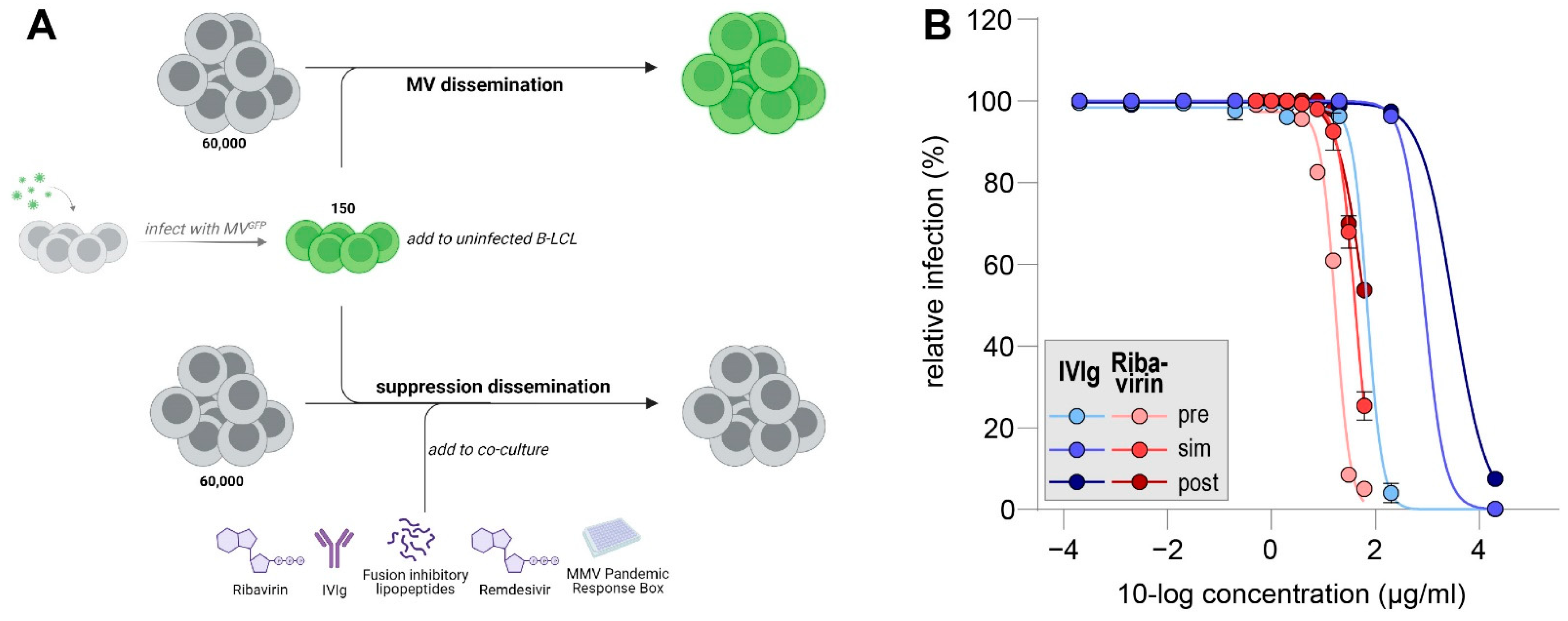
3.1. IVIg Inhibitis MV-Dissemination at Physiological Concentrations

We designed an MV-dissemination assay to evaluate the efficacy of viral inhibitors in the dissemination phase. The assay assesses the cell-to-cell spread of MV in the presence or absence of antiviral compounds. As MV replication in B-LCLs requires approximately 24 h, a window of post-exposure treatment is given to address multiple treatment conditions (Figure 1A).
We validated our assay using multiple compounds which do not inhibit MV and compared those to ribavirin, often shown to inhibit MV in vitro (data not shown).
After successful validation, we were aiming to evaluate the potency of IVIg to inhibit wild-type MV dissemination and compare it to ribavirin. To this end, we used a recombinant MV based on a clinical isolate from Khartoum, Sudan (Genotype B3), which expresses a fluorescent protein at position three of its genome (rMVKSVenus(3)). Serial dilutions of IVIg or ribavirin were added either before, simultaneously with or after the addition of MV-infected cells to uninfected cultures and incubated for 48 h. Next, Venus-expressing cells were detected by flow cytometry as a measure of MV infection (Figure 1B).
In all treatment conditions, the highest tested concentration of IVIg (2 mg/mL) inhibited MV dissemination almost completely. MV dissemination was also suppressed when cells were incubated with 0.2 mg/mL of IVIg prior to the addition of MV-infected B-LCLs. The further dilution of IVIg led to MV infection of almost all uninfected cells in all treatment conditions. The half-maximal inhibitory concentrations (IC50) were 0.07 mg/mL, 0.87 mg/mL and 3.2 mg/mL for pre-incubation, simultaneous incubation or post-exposure incubation, respectively. Ribavirin only effectively inhibited MV-dissemination when pre-incubated with uninfected cells. If ribavirin was added simultaneously or six hours post-co-culture, infection percentages at 61 µg/mL were 25% or 54%, respectively. IC50 values for ribavirin pre-incubation were 17 µg/mL, for ribavirin incubation at the start of co-culture 40 µg/mL and for ribavirin post-exposure treatment 62 µg/mL. Our data support an inhibitory effect of IVIg and ribavirin on the dissemination of MV. Importantly, this effect is highly time-dependent, leading to reduced antiviral efficacy when inhibitors are added post-exposure.

3.2. Remdesivir Efficiently Inhibitis MV-Dissemination at Sub-Micromolar Concentrations

Next, we evaluated the effect of remdesivir on rMVKSVenus(3)-dissemination in pre-, simultaneous or post-co-culture treatment regimens and compared this to ribavirin (Figure 2A). Remdesivir inhibited its dissemination in all tested conditions. The pre-exposure of uninfected cultures with remdesivir resulted in almost complete inhibition of dissemination at 80 nM or higher. If remdesivir was added to the co-culture simultaneously or six hours post co-culture, 10% infection or less was measured at 400 nM and 10 µM, respectively. Mean IC50 values of four or more experiments were 54 nM for pre-addition, 175 nM for simultaneous addition and 304 nM for post-addition conditions. Similar differences as those related to the timing of treatment were observed for ribavirin, as shown in Figure 1B. The mean IC50 values for ribavirin obtained in three or more experiments were 38 µM, 72 µM and 119 µM for pre-, simultaneous or post-addition, respectively. Notably, IC50 values for post co-culture addition varied greatly between assays, and no upper limit of a 95% confidence interval could be determined.
Next, we assessed the influence of simultaneous addition of remdesivir and ribavirin on a vaccine strain-based (rMVEZEGFP(3)) or wildtype-based (rMVKSVenus(3)) recombinant MV (Figure 2B). rMVEZEGFP(3) dissemination was slightly better inhibited by remdesivir and ribavirin when compared to the rMVKSVenus(3). The corresponding mean IC50s were 34 nM and 156 nM (4.5-fold decrease) for remdesivir and 28 µM and 63 µM for ribavirin, respectively.
Finally, we tested the efficacy of remdesivir and ribavirin added simultaneously against low-passage clinical MV isolates belonging to different genotypes (Figure 2C). A total of six different virus isolates were evaluated and no inter-genotype differences were detected. IC50 values for remdesivir varied between 27 nM and 87 nM and for ribavirin between 39 µM and 77 µM. Generally, IC50 values for all wild-type viruses appeared slightly lower when compared to the recombinant wild-type-based virus. This could be possibly attributed to the method of infection detection as recombinant wild-type-based virus infections were evaluated based on EGFP or Venus expression, while the N protein was stained for all wild-type viruses.
In conclusion, the nucleoside-analog remdesivir inhibited MV dissemination at sub-micromolar concentrations.

3.3. Fusion Inhibitory Lipopeptides Prevent MV Dissemination at Nanomolar Concentrations

We evaluated three classes of previously described fusion inhibitory lipopeptides in our MV dissemination assay (Figure 3A). Peptides mimicking different sequences of the C-terminal heptad repeat domain (HRC) of the fusion protein (green) [60,61] inhibited rMVKSVenus(3) dissemination completely at 80 nM or higher. FIP ((Z)-d-Phe-l-Phe-Gly)-modified lipopeptides [54,64] (blue) performed less well, and FIP-HRC hybrid lipopeptides [64] (red) had an intermediate effect on viral dissemination when compared to HRC- or FIP-based peptides. IC50 values ranged from 22 nM for HRC-based peptides, to 47–68 nM for hybrid peptides and more than 400 nM for FIP-based peptides.
Peptide cytotoxicity was determined using flow cytometry. With the exception of the [FIP-PEG4]2-chol peptide (light blue), no lipopeptide was cytotoxic at the highest tested concentration (Figure 3B). Cellular viability was inversely proportional to infection percentages (r2 = 0.76) suggesting that cytotoxicity was induced by viral infection, rather than drug-related (Figure 3C).
Overall, these data demonstrate that fusion inhibitory peptides can efficiently inhibit MV dissemination at low concentrations with neglectable cytotoxicity. Importantly, the different classes of fusion inhibitors varied in efficacy, with HRC-based lipopeptides performing best in the dissemination assay.

3.4. MMV Pandemic Response Box Screening

The Pandemic Response Box (PRB) contains 400 diverse molecules of antiviral, antibacterial or antifungal properties. Selected compounds are at various stages of research and development and have been characterized into the three categories based on literature. Initially, we screened all 153 compounds categorized as antivirals, 5 antibacterials and 2 antifungals for their inhibitory properties against rMVKSVenus(3) at two concentrations in our dissemination assay. Hits were defined as compounds that resulted in less than 50% MV infection at a concentration of 20 µM and/or less than 90% MV infection at a concentration of 4 µM, independent of toxicity and the time point of evaluation (48 or 72 h post-co-culture (hpc)). Importantly, using those cut-off criteria, we overestimated the number of compounds with possible anti-MV effect. At 48 hpc, 14 compounds showed less than 50% MV infection at a concentration of 20 μM, while this was measured for 15 compounds at 72 hpc. Using compounds at a concentration of 4 μM, 14 and 16 compounds exhibited less than 90% MV infection 48 or 72 hpc, respectively. In total, 21 compounds reduced the percentage of MV-infected cells and qualified for further testing (Figure 4A, enlarged, colored symbols, one symbol per compound).
We used a five-fold dilution series starting from 4 µM to test the 21 compounds, namely, MMV394033, Emricasan, MMV1782101, MMV1782115, Ozanimod, MMV690621, MMV642550, Topotecan, Panobinostat, Selinexor, URMC-099-C, Fludarabine Triapine, DNDI1417411, MMV1782214, MMV218827, Rubitecan, ML324, Verdinexor, MMV019724 and 1,1-dioxide 1-Thioflavone, with remdesivir as a control. Five compounds (Topotecan, Fludarabine, Panobinostat, Verdinexor and 1,1-dioxide 1-Thioflavone) inhibited MV dissemination by more than 50% at 4 µM. (Figure 4B, dotted line). However, all five compounds proved to be cytotoxic with a relative viability of less than 20% in co-cultures infected and treated with the compounds (Figure 4C). Therefore, we concluded that no antiviral compound of the PRB was able to inhibit MV dissemination efficiently but that the induced cytotoxicity led to decreased infection.

4. Discussion

We have repurposed an MV dissemination assay to test the efficacy of various antiviral compounds. Originally, a low number of infected EBV-transformed human B-lymphoblastic cells (B-LCL) were co-cultured with a large number of uninfected cells and MV-specific, HLA-matched T-cell clones (TCC), which suppressed the dissemination of MV. The repurposed assay uses B-LCLs as target cells to mimic the early dissemination of MV in lymphocytes. B-LCL express two MV receptors, CD150 and CD46. While CD46 is no natural MV receptor and can only be used by MV vaccine strains or cell culture adapted strains, CD150 is expressed on myeloid and lymphoid cells and used by MV vaccine strains and wild-type strains. However, non-human primate studies have shown that intra-muscular injected MV vaccine strains preferentially target CD150-expressing DCs and macrophages instead of CD46-expressing myocytes and therefore resemble the tropism of wild-type MV viruses [102]. Studies with wild-type-based MV in NHPs have shown that MV amplification starts in bronchus-associated lymphoid tissues [19] or local draining lymph nodes [20], which is represented by a small amount of infected B-LCLs in a large number of uninfected cells in our antiviral assay. Local amplification leads to dissemination throughout the entire body. In humans, peripheral blood mononuclear cells (PBMC) are infected as early as five days before the onset of rash, and the peak of the cell-associated viremia is measured before the onset of rash [103]. In NHPs, viremia peaks at nine days post-inoculation and also precedes the onset of rash [24], suggesting that the NHP model closely resembles human disease. Simultaneously, the lowest white blood cell count can also be measured during or just after peak viremia, which is associated with follicular depletion in lymph nodes [24]. This indicates progressive lymphodepletion, both in peripheral blood and in lymphoid tissues. In our in vitro antiviral assay, we achieved best treatment outcomes if antivirals were added prior to or simultaneously with the start of MV dissemination. Treatment of measles patients should therefore ideally be initiated before the start of viremia, i.e., more than five days before the onset of rash, as this marks the highest chance of a mostly local MV infection
IVIg and measles vaccination are the only currently approved post-exposure intervention methods. Usually, IVIg is administered within six days after exposure and is often limited to immunocompromised contacts, pregnant women and children below the age of 6 months. Dependent on the country, dosages vary between 150 mg/kg bodyweight and 400 mg/kg bodyweight leading to maximum concentrations in blood of 3–8 mg/mL (assuming approx. 50 mL serum per kg bodyweight) [104]. In our assay, IC50 values, but not complete inhibition, for six-hour post-exposure treatment with IVIg were 3.2 mg/mL, which is similar to what is achieved in vivo and what possibly explains the variable efficacy in patients. Importantly, in our assay, IVIg effectivity increased with earlier treatment, supporting the importance of early treatment in vivo [31,32,105].
We have repeatedly shown that remdesivir efficiently inhibits MV dissemination at half-maximal inhibitory concentrations below 180 nM, regardless if wild-type-based viruses or vaccine-strain-based viruses were used. In line with this, Lo et al. estimated the IC50 of remdesivir against MV infection in HeLa cells at 37 nM, in VeroE6 cells at 5 µM, in NCl-H358 cells at 25 nM and in HSAEC1-KT cells at 63 nM, using an rMVEZEGFP(3) reporter-based assay [41,42]. Our assay resembles the early dissemination phase and mimics in vivo cell-to-cell viral spread instead of cell-free virus production. Similar to the time-dependent effect seen for IVIg, remdesivir inhibited viral dissemination best when administered early. However, almost complete inhibition could also be observed when co-cultures were treated 6 h post-initiation of co-culture and likely all uninfected cells were already exposed to MV. It is unclear how those in vitro results will translate in vivo and whether remdesivir can be established as an effective post-exposure prophylaxis or treatment for measles. Crucial considerations include the emergence of resistance mutations which remain to be investigated, the timing of treatment initiation and the translation of the in vitro determined effective dose to an in vivo administered dose, reaching high enough systemic concentrations to inhibit MV in all target organs.
Remdesivir’s intravenous administration is feasible for the treatment of severe measles cases or neurological complications which require hospitalization, but makes it less attractive for early post-exposure prophylaxis. Therefore, advances to design an orally available compound that generates the same bioactive metabolite as remdesivir need to be made. 1-O-octadecyl-2-O-benzyl-sn-glycerylester (ODBG) lipid-modified monophosphate prodrug (ODBG-P-RVn) is an orally available derivative of Nuc, remdesivir’s parent nucleotide, and proved to be stable in plasma and highly active against MV (82–160 nM) [42], potentially making it more widely available and easier accessible.
Intranasal administration of fusion inhibitory peptides has been suggested as prophylaxis for MV and SARS-CoV-2, preventing virus infection in the respiratory tract [61,106]. However, treatment of measles, as a primarily systemic disease, may need to be looked at from the perspective of other systemic infections such as HIV. For treatment-experienced HIV patients, enfuvirtide (T20), the only marketed fusion inhibitor, can be used but needs to be administered twice daily by subcutaneous injection, possibly challenging patient compliance [107,108]. Moreover, drug resistance to enfuvirtide has been numerously reported in patients [109]. For MV, resistant mutants to FIP and related fusion inhibitors have been detected or were induced by selective pressure in in vitro experiments [56,110]. Fortunately, a fusion inhibitory peptide with an unrelated working mechanism still conferred protection in CD150 transgenic suckling mice infected with FIP-resistant MV [63].
Mechanistically, fusion-inhibitory peptides based on one HR-domain directed against viruses containing a class I fusion protein such as MV, SARS-CoV-2 or HIV work similarly. Peptides mimic one of the two HR-domains, which usually facilitate fusion by forming a six-helix bundle (6-HB) with the complementary HR. Increasing concentrations of peptides outcompete the complementation of both HRs, thus inhibiting the transition of the F protein into its post-fusion state. In our MV antiviral assay, we showed the highest antiviral activity for the three peptides derived from HR domains. [HRC450-485-PEG4]2-chol, also referred to as HRC4, has previously been shown to prevent infection in vitro and to reduce viral replication in hippocampal brain slices of neonatal SLAM transgenic mice ex vivo [60,61]. To this end, the investigators had dimerized the HRC peptide and conjugated it to a cholesterol group, as this had been shown previously to enhance antiviral potency [111,112].
Next to peptides preventing the transition to post-fusion confirmation, fusion inhibitors stabilizing the prefusion state have been described as early as 1980. FIP, (Z)-d-Phe-l-Phe-Gly, has been investigated for paramyxoviruses and myxoviruses [54]. Lipid-conjugation and dimerization of FIP have once more been shown to increase peptide potency [64]. Importantly, similarly to what has been observed by us, a conjugation of the FIP domain to the HRC domain (hybrid peptide) resulted in a more than 10-fold more potent inhibitor. The authors also report a 10-fold improvement when compared to HRC-based peptides, which is in contrast to our results, in which HRC-based peptides have performed best. While Bovier et al. not only reported a synergistic effect between both peptides, they also speculated that resistance emergence might be less likely considering the dual action mechanism. In our hands, both HRC and hybrid peptides have IC50 values below 100 nM and should be further evaluated in post-exposure animal models.
Few antivirals made it to pre-clinical evaluation in MV animal models. The non-human primate model is still considered the gold standard in measles research, as NHPs are the only animal species naturally susceptible to MV infection. Moreover, biodistribution, pharmacokinetics, pharmacodynamics as well as off-target effects of antiviral drugs can be inferred from data obtained in NHPs. However, ethical concerns and high costs led researchers to re-evaluate the use of NHPs as experimental animal model. Rodents are widely being used to determine drug characteristics in vivo. Unfortunately, natural measles cannot be mimicked in rodents, and treatment effects can therefore not be assessed. Alternatively, ferrets can be infected with the closely related morbillivirus canine distemper virus (CDV) and are being used as a surrogate model. In contrast to MV, CDV causes high mortality in ferrets and therefore only mimics a high-susceptibility-with-severe-outcome scenario, rather than a typical MV infection.
Over the last 10 years the non-nucleoside polymerase inhibitor ERDRP-0519 has been investigated for its anti-MV effect and one of the only antivirals, which has been tested in more than one animal model. Krumm et al. explored the potential of ERDRP-0519 to inhibit CDV in ferrets [50]. They have chosen for a CDV strain which was passaged in ferrets and proved to be highly virulent [113]. All ferrets were treated by gastric gavage with 50 mg/kg for 14 days, starting either 24 h pre-infection or at the onset of viremia (3 days post-infection). Pre-treatment prolonged the survival of ferrets when compared to untreated controls, but ultimately did not prevent fatal outcome. Surprisingly, all ferrets in the post-exposure treatment group survived and only experienced mild lymphopenia. Next, ERDRP-0159 was also tested for its potential as an MV inhibitor in squirrel monkeys infected with a clinical MV isolate from Germany [52]. As measles is usually not lethal in monkeys, the investigators used rash, viral titers in PBMC, viral titers in nose swabs and leukopenia as a measure of efficacy. Next to a pre-exposure-treatment group and an onset-of-viremia-treatment group (3 dpi), they also chose for an onset-of-clinical-signs-treatment group (7 dpi). In all groups, clinical benefits or decreased viral shedding were detected, suggesting a positive influence on the viral epidemiology. However, a clear disease benefit was only seen in the former two groups as treatment 7 dpi did not affect the height of viral load in blood and did also not protect from leukopenia.
In comparison to ERDRP-0159, multiple fusion inhibitors have been investigated in non-natural hosts for morbilliviruses. The HRC4 peptide and the FIP–HRC hybrid peptide (also used in this study) were evaluated in the cotton rat model, a non-lethal model mostly used to investigate respiratory tract involvement [114]. In all studies, the investigators could show that no virus could be isolated from lungs of intranasally, prophylactically treated and MV-challenged cotton rats [61,62,64]. Moreover, the HRC4 peptide could also be traced in serum and in the brain of cotton rats, suggesting a potential treatment effect after systemic spread.
Characterization of fusion-inhibitory lipopeptides has often been performed in the context of CNS disease, especially SSPE: a late-onset rare but fatal complication of measles. As there is no reliable CNS disease model in a morbillivirus-natural host, most studies have been performed in CD150-transgenic mice, CD150-transgenic suckling mice or CD150 x IFNAR-knock out mice [60,61,62]. Overall, subcutaneous and intranasal application of HRC4 or similar lipopeptides were evaluated in pre-MV-exposure-treatment regimens, showing promising results on survival. Similar to the cotton rat model, the lipopeptides could be detected in the circulation, the lungs as well as the brain with variable concentrations, endorsing some peptides as a potent treatment method, while others could be better used preventative. In other studies, intracranial inoculation of nude mice was used as a model system for MV-induced CNS disease [115]. Watanabe et al. then used an MV-HRC-based peptide as treatment and achieved an increase in survival rate from 0% to 67% [59].
CNS disease, specifically SSPE, has further been studied in the hamster model. Studies performed in the 1990s suggested a beneficial effect of ribavirin when administered intracranially at least 36 h post inoculation [116]. In this case, half (5/10) of all treated hamsters survived the lethal infection; full survival was achieved when treatment was initiated as early as 12 h post inoculation with 10 mg/kg/day for 10 days, which correlates to an intra-brain concentration of more than 50 µg/g [117]. The 50% effective dose was calculated to be 1.4 mg/kg/day [116]. Experimental intravenous treatment of two SSPE patients with ribavirin led to neurological improvements [118]. However, both experienced neurological deterioration a few months after the therapy was stopped. Serum concentrations of ribavirin in both patients were between 10 and 20 µg/mL, and CSF concentrations between 7 and 17 µg/mL. As rationalized by the previous hamster studies, prolonged patient improvement could be observed when ribavirin was administered intraventricularly and CSF concentrations were higher than 100 µg/mL [119]. While some studies report in vitro IC50 values of ribavirin for MV or the SSPE-strain Yamagata of less than 10 µg/mL [33,34,36,43], we and others determined IC50 values of higher than 35 µg/mL [33,73], especially when ribavirin was used as post-exposure treatment.
Next to efficacy, many other drug characteristics are validated in animal models. A perfect antiviral should have a low toxicity profile and should be usable by the patient independently of a health care professional (e.g., orally available). In the case of MV, antivirals require systemic biodistribution to reach all lymphoid tissues and epithelia. Furthermore, antivirals should have low production costs and could ideally be synthesized at multiple locations. A long shelf stability would be advantageous to guarantee sufficient supply in case of outbreaks.
When discussing MV treatment options, we should not forget that measles is a difficult disease to treat. To date, most licensed antivirals are used in chronic disease such as HIV or HCV, which have their own challenges but which give ample opportunities to initiate and monitor treatment. An acute disease with a long asymptomatic phase and an early initiation of long-term immunosuppression such as measles leaves a narrow treatment window. Moreover, despite rising measles cases in the Western world, antivirals would mostly make an impact on children in measles-endemic countries. However, clinical trials in a pediatric patient population have major challenges, and as vaccination is yet the best standard of care, acceptance of clinical trials in a population not refusing vaccination will be difficult. This leaves researchers with a restricted population for clinical studies. Once marketed, target groups in the Western world include non-vaccinated healthcare workers and immunosuppressed patients, household or school contacts and patients suffering from measles-related neurological symptoms. Given the limited market, the research and development of an MV-specific antiviral may not be cost-effective, urging us to invest in broad-spectrum antivirals and a combination of targeted therapies. The latter will additionally be of increasing interest to reduce individual drug doses and related adverse events, as well as the emergence of drug-resistant viruses. It remains to be investigated if combination therapy of remdesivir or polymerase inhibitors with fusion inhibitory peptides will be a potent post-exposure prophylaxis for measles.

Author Contributions

Conceptualization, K.S.S., R.D.d.V. and R.L.d.S.; methodology, K.S.S., R.D.d.V. and R.L.d.S.; validation K.S.S. and M.V.L.; formal analysis K.S.S., M.V.L. and L.G.; investigation K.S.S., M.V.L. and L.G.; data curation K.S.S.; writing—original draft preparation K.S.S., K.H.; writing—review and editing D.P.P., C.A.A., A.M., M.P., R.D.d.V. and R.L.d.S.; visualization K.S.S.; supervision R.D.d.V. and R.L.d.S. All authors have read and agreed to the published version of the manuscript.

Funding

This research was partly funded by Gilead Sciences. Gilead Sciences had no role in the design of the study; in the collection, analyses, or interpretation of data; or in the decision to publish the results. This work was partly supported by funding from ZonMw (Infectious Diseases, 10150511910052) to KSS, RDdV and RLdS and by funding from the National Institutes of Health (AI159085, NS105699 and NS091263) to M.P.

Acknowledgments

We thank Medicines for Malaria Venture for support, designing and supplying the Pandemic Response Box.

Conflicts of Interest

C.A.A., A.M. and M.P. are listed as inventors on a patent for the FIP–HRC and the HRC fusion inhibitors. The other authors declare no conflict of interest.

References

  1. Measles & Rubella Initiative. Measles & Rubella Strategic Framework 2021—2030. Available online: https://measlesrubellainitiative.org/measles-rubella-strategic-framework-2021-2030/ (accessed on 20 February 2022).
  2. WHO. Measles Fact Sheet. Available online: https://www.who.int/news-room/fact-sheets/detail/measles (accessed on 20 February 2022).
  3. WHO. Worldwide Measles Deaths Climb 50% from 2016 to 2019 Claiming over 207,500 Lives in 2019. Available online: https://www.who.int/news/item/12-11-2020-worldwide-measles-deaths-climb-50-from-2016-to-2019-claiming-over-207-500-lives-in-2019 (accessed on 20 February 2022).
  4. Dixon, M.G.; Ferrari, M.; Antoni, S.; Li, X.; Portnoy, A.; Lambert, B.; Hauryski, S.; Hatcher, C.; Nedelec, Y.; Patel, M.; et al. Progress toward Regional Measles Elimination—Worldwide, 2000–2020. Morb. Mortal Wkly. Rep. 2021, 70, 1563–1569. [Google Scholar] [CrossRef] [PubMed]
  5. Torjesen, I. Measles outbreaks likely as covid pandemic leaves millions of world’s children unvaccinated, WHO warns. BMJ 2021, 375, n2755. [Google Scholar] [CrossRef] [PubMed]
  6. Rima, B.; Balkema-Buschmann, A.; Dundon, W.G.; Duprex, P.; Easton, A.; Fouchier, R.; Kurath, G.; Lamb, R.; Lee, B.; Rota, P.; et al. ICTV Virus Taxonomy Profile: Paramyxoviridae. J. Gen. Virol. 2019, 100, 1593–1594. [Google Scholar] [CrossRef] [PubMed]
  7. WHO. Genetic diversity of wild-type measles viruses and the global measles nucleotide surveillance database (MeaNS). Wkly. Epidemiol. Rec. 2015, 90, 373–380. [Google Scholar]
  8. Rota, P.A.; Brown, K.; Mankertz, A.; Santibanez, S.; Shulga, S.; Muller, C.P.; Hübschen, J.M.; Siqueira, M.; Beirnes, J.; Ahmed, H.; et al. Global Distribution of Measles Genotypes and Measles Molecular Epidemiology. J. Infect. Dis. 2011, 204, S514–S523. [Google Scholar] [CrossRef]
  9. Dowling, P.C.; Blumberg, B.M.; Menonna, J.; Adamus, J.E.; Cook, P.; Crowley, J.C.; Kolakofsky, D.; Cook, S.D. Transcriptional map of the measles virus genome. J. Gen. Virol. 1986, 67 Pt 9, 1987–1992. [Google Scholar] [CrossRef]
  10. Bankamp, B.; Horikami, S.M.; Thompson, P.D.; Huber, M.; Billeter, M.; Moyer, S.A. Domains of the Measles Virus N Protein Required for Binding to P Protein and Self-Assembly. Virology 1996, 216, 272–277. [Google Scholar] [CrossRef] [Green Version]
  11. Plattet, P.; Alves, L.; Herren, M.; Aguilar, H.C. Measles Virus Fusion Protein: Structure, Function and Inhibition. Viruses 2016, 8, 112. [Google Scholar] [CrossRef] [Green Version]
  12. Tatsuo, H.; Ono, N.; Yanagi, Y. Morbilliviruses Use Signaling Lymphocyte Activation Molecules (CD150) as Cellular Receptors. J. Virol. 2001, 75, 5842–5850. [Google Scholar] [CrossRef] [Green Version]
  13. Mühlebach, M.D.; Mateo, M.; Sinn, P.L.; Prüfer, S.; Uhlig, K.M.; Leonard, V.H.; Navaratnarajah, C.K.; Frenzke, M.; Wong, X.X.; Sawatsky, B.; et al. Adherens junction protein nectin-4 is the epithelial receptor for measles virus. Nature 2011, 480, 530–533. [Google Scholar] [CrossRef] [Green Version]
  14. Noyce, R.S.; Bondre, D.G.; Ha, M.N.; Lin, L.T.; Sisson, G.; Tsao, M.S.; Richardson, C.D. Tumor cell marker PVRL4 (nectin 4) is an epithelial cell receptor for measles virus. PLoS Pathog. 2011, 7, e1002240. [Google Scholar] [CrossRef] [PubMed]
  15. Iwasaki, M.; Takeda, M.; Shirogane, Y.; Nakatsu, Y.; Nakamura, T.; Yanagi, Y. The Matrix Protein of Measles Virus Regulates Viral RNA Synthesis and Assembly by Interacting with the Nucleocapsid Protein. J. Virol. 2009, 83, 10374–10383. [Google Scholar] [CrossRef] [PubMed] [Green Version]
  16. Cox, R.M.; Plemper, R.K. Structure and organization of paramyxovirus particles. Curr. Opin. Virol. 2017, 24, 105–114. [Google Scholar] [CrossRef] [PubMed]
  17. Liston, P.; DiFlumeri, C.; Briedis, D.J. Protein interactions entered into by the measles virus P, V, and C proteins. Virus Res. 1995, 38, 241–259. [Google Scholar] [CrossRef]
  18. Guerra, F.M.; Bolotin, S.; Lim, G.; Heffernan, J.; Deeks, S.L.; Li, Y.; Crowcroft, N.S. The basic reproduction number (R0) of measles: A systematic review. Lancet Infect. Dis. 2017, 17, e420–e428. [Google Scholar] [CrossRef]
  19. Lemon, K.; de Vries, R.D.; Mesman, A.W.; McQuaid, S.; van Amerongen, G.; Yüksel, S.; Ludlow, M.; Rennick, L.J.; Kuiken, T.; Rima, B.K.; et al. Early target cells of measles virus after aerosol infection of non-human primates. PLoS Pathog. 2011, 7, e1001263. [Google Scholar] [CrossRef]
  20. De Swart, R.L.; Ludlow, M.; de Witte, L.; Yanagi, Y.; van Amerongen, G.; McQuaid, S.; Yüksel, S.; Geijtenbeek, T.B.; Duprex, W.P.; Osterhaus, A.D. Predominant infection of CD150+ lymphocytes and dendritic cells during measles virus infection of macaques. PLoS Pathog. 2007, 3, e178. [Google Scholar] [CrossRef]
  21. De Vries, R.D.; Lemon, K.; Ludlow, M.; McQuaid, S.; Yüksel, S.; van Amerongen, G.; Rennick, L.J.; Rima, B.K.; Osterhaus, A.D.; de Swart, R.L. In vivo tropism of attenuated and pathogenic measles virus expressing green fluorescent protein in macaques. J. Virol. 2010, 84, 4714–4724. [Google Scholar] [CrossRef] [Green Version]
  22. Ludlow, M.; Rennick, L.J.; Sarlang, S.; Skibinski, G.; McQuaid, S.; Moore, T.; de Swart, R.L.; Duprex, W.P. Wild-type measles virus infection of primary epithelial cells occurs via the basolateral surface without syncytium formation or release of infectious virus. J. Gen. Virol. 2010, 91, 971–979. [Google Scholar] [CrossRef]
  23. Racaniello, V. An Exit Strategy for Measles Virus. Science 2011, 334, 1650–1651. [Google Scholar] [CrossRef]
  24. De Vries, R.D.; McQuaid, S.; van Amerongen, G.; Yüksel, S.; Verburgh, R.J.; Osterhaus, A.D.; Duprex, W.P.; de Swart, R.L. Measles immune suppression: Lessons from the macaque model. PLoS Pathog. 2012, 8, e1002885. [Google Scholar] [CrossRef] [PubMed]
  25. Mina, M.J.; Metcalf, C.J.E.; de Swart, R.L.; Osterhaus, A.D.M.E.; Grenfell, B.T. Long-term measles-induced immunomodulation increases overall childhood infectious disease mortality. Science 2015, 348, 694–699. [Google Scholar] [CrossRef] [PubMed] [Green Version]
  26. Morris, S.E.; Yates, A.J.; de Swart, R.L.; de Vries, R.D.; Mina, M.J.; Nelson, A.N.; Lin, W.-H.W.; Kouyos, R.D.; Griffin, D.E.; Grenfell, B.T. Modeling the measles paradox reveals the importance of cellular immunity in regulating viral clearance. PLoS Pathog. 2018, 14, e1007493. [Google Scholar] [CrossRef] [PubMed]
  27. Laksono, B.M.; Fortugno, P.; Nijmeijer, B.M.; de Vries, R.D.; Cordisco, S.; Kuiken, T.; Geijtenbeek, T.B.H.; Duprex, W.P.; Brancati, F.; de Swart, R.L. Measles skin rash: Infection of lymphoid and myeloid cells in the dermis precedes viral dissemination to the epidermis. PLoS Pathog. 2020, 16, e1008253. [Google Scholar] [CrossRef]
  28. Ferren, M.; Horvat, B.; Mathieu, C. Measles Encephalitis: Towards New Therapeutics. Viruses 2019, 11, 1017. [Google Scholar] [CrossRef] [Green Version]
  29. Holzmann, H.; Hengel, H.; Tenbusch, M.; Doerr, H.W. Eradication of measles: Remaining challenges. Med. Microbiol. Immunol. 2016, 205, 201–208. [Google Scholar] [CrossRef] [Green Version]
  30. Young, M.K.; Nimmo, G.R.; Cripps, A.W.; Jones, M.A. Post-exposure passive immunisation for preventing measles. Cochrane Database Syst. Rev. 2014, Cd010056. [Google Scholar] [CrossRef] [Green Version]
  31. Zingher, A.; Mortimer, P. Convalescent whole blood, plasma and serum in the prophylaxis of measles: Reproduced from JAMA, 12 April 1926; 1180–1187. Rev. Med. Virol. 2005, 15, 407–421. [Google Scholar] [CrossRef]
  32. Matysiak-Klose, D.; Santibanez, S.; Schwerdtfeger, C.; Koch, J.; von Bernuth, H.; Hengel, H.; Littmann, M.; Terhardt, M.; Wicker, S.; Mankertz, A.; et al. Post-exposure prophylaxis for measles with immunoglobulins revised recommendations of the standing committee on vaccination in Germany. Vaccine 2018, 36, 7916–7922. [Google Scholar] [CrossRef]
  33. Grancher, N.; Venard, V.; Kedzierewicz, F.; Ammerlaan, W.; Finance, C.; Muller, C.P.; Le Faou, A. Improved antiviral activity in vitro of ribavirin against measles virus after complexation with cyclodextrins. Antivir. Res. 2004, 62, 135–137. [Google Scholar] [CrossRef]
  34. Zhang, N.; Chen, H.M.; Sood, R.; Kalicharran, K.; Fattom, A.I.; Naso, R.B.; Barnard, D.L.; Sidwell, R.W.; Hosmane, R.S. In vitro inhibition of the measles virus by novel ring-expanded (‘fat’) nucleoside analogues containing the imidazo[4,5-e]diazepine ring system. Bioorg. Med. Chem. Lett. 2002, 12, 3391–3394. [Google Scholar] [CrossRef]
  35. Murphy, M.F. In vitro inhibition of subacute sclerosing panencephalitis virus by the antiviral agent ribavirin. J. Infect. Dis. 1978, 138, 249–251. [Google Scholar] [CrossRef] [PubMed]
  36. Hosoya, M.; Shigeta, S.; Nakamura, K.; De Clercq, E. Inhibitory effect of selected antiviral compounds on measles (SSPE) virus replication in vitro. Antivir. Res. 1989, 12, 87–97. [Google Scholar] [CrossRef]
  37. Takahashi, T.; Hosoya, M.; Kimura, K.; Ohno, K.; Mori, S.; Takahashi, K.; Shigeta, S. The cooperative effect of interferon-alpha and ribavirin on subacute sclerosing panencephalitis (SSPE) virus infections, in vitro and in vivo. Antivir. Res. 1998, 37, 29–35. [Google Scholar] [CrossRef]
  38. Krajczyk, A.; Kulinska, K.; Kulinski, T.; Hurst, B.L.; Day, C.W.; Smee, D.F.; Ostrowski, T.; Januszczyk, P.; Zeidler, J. Antivirally active ribavirin analogues—4,5-disubstituted 1,2,3-triazole nucleosides: Biological evaluation against certain respiratory viruses and computational modelling. Antivir. Chem. Chemother. 2014, 23, 161–171. [Google Scholar] [CrossRef]
  39. Shigeta, S.; Mori, S.; Baba, M.; Ito, M.; Honzumi, K.; Nakamura, K.; Oshitani, H.; Numazaki, Y.; Matsuda, A.; Obara, T.; et al. Antiviral activities of ribavirin, 5-ethynyl-1-beta-D-ribofuranosylimidazole-4-carboxamide, and 6′-(R)-6′-C-methylneplanocin A against several ortho- and paramyxoviruses. Antimicrob. Agents Chemother. 1992, 36, 435–439. [Google Scholar] [CrossRef] [Green Version]
  40. De Clercq, E.; Montgomery, J.A. Broad-spectrum antiviral activity of the carbocyclic analog of 3-deazaadenosine. Antivir. Res. 1983, 3, 17–24. [Google Scholar] [CrossRef]
  41. Lo, M.K.; Jordan, R.; Arvey, A.; Sudhamsu, J.; Shrivastava-Ranjan, P.; Hotard, A.L.; Flint, M.; McMullan, L.K.; Siegel, D.; Clarke, M.O.; et al. GS-5734 and its parent nucleoside analog inhibit Filo-, Pneumo-, and Paramyxoviruses. Sci. Rep. 2017, 7, 43395. [Google Scholar] [CrossRef]
  42. Lo, M.K.; Shrivastava-Ranjan, P.; Chatterjee, P.; Flint, M.; Beadle, J.R.; Valiaeva, N.; Schooley, R.T.; Hostetler, K.Y.; Montgomery, J.M.; Spiropoulou, C. Broad-spectrum in vitro antiviral activity of ODBG-P-RVn: An orally-available, lipid-modified monophosphate prodrug of remdesivir parent nucleoside (GS-441524). bioRxiv 2021. [Google Scholar] [CrossRef]
  43. Hashimoto, K.; Maeda, H.; Miyazaki, K.; Watanabe, M.; Norito, S.; Maeda, R.; Kume, Y.; Ono, T.; Chishiki, M.; Suyama, K.; et al. Antiviral Effect of Favipiravir (T-705) against Measles and Subacute Sclerosing Panencephalitis Viruses. Jpn. J. Infect. Dis. 2021, 74, 154–156. [Google Scholar] [CrossRef]
  44. Jochmans, D.; van Nieuwkoop, S.; Smits, S.L.; Neyts, J.; Fouchier, R.A.; van den Hoogen, B.G. Antiviral Activity of Favipiravir (T-705) against a Broad Range of Paramyxoviruses In Vitro and against Human Metapneumovirus in Hamsters. Antimicrob. Agents Chemother. 2016, 60, 4620–4629. [Google Scholar] [CrossRef] [PubMed] [Green Version]
  45. Lo, M.K.; Jordan, P.C.; Stevens, S.; Tam, Y.; Deval, J.; Nichol, S.T.; Spiropoulou, C.F. Susceptibility of paramyxoviruses and filoviruses to inhibition by 2′-monofluoro- and 2′-difluoro-4′-azidocytidine analogs. Antivir. Res. 2018, 153, 101–113. [Google Scholar] [CrossRef] [PubMed]
  46. Barnard, D.L.; Stowell, V.D.; Seley, K.L.; Hegde, V.R.; Das, S.R.; Rajappan, V.P.; Schneller, S.W.; Smee, D.F.; Sidwell, R.W. Inhibition of measles virus replication by 5’-nor carbocyclic adenosine analogues. Antivir. Chem. Chemother. 2001, 12, 241–250. [Google Scholar] [CrossRef] [PubMed] [Green Version]
  47. McGuigan, C.; Hinsinger, K.; Farleigh, L.; Pathirana, R.N.; Bugert, J.J. Novel antiviral activity of l-dideoxy bicyclic nucleoside analogues versus vaccinia and measles viruses in vitro. J. Med. Chem. 2013, 56, 1311–1322. [Google Scholar] [CrossRef] [PubMed]
  48. Yoon, J.J.; Krumm, S.A.; Ndungu, J.M.; Hoffman, V.; Bankamp, B.; Rota, P.A.; Sun, A.; Snyder, J.P.; Plemper, R.K. Target analysis of the experimental measles therapeutic AS-136A. Antimicrob. Agents Chemother. 2009, 53, 3860–3870. [Google Scholar] [CrossRef] [PubMed] [Green Version]
  49. Ndungu, J.M.; Krumm, S.A.; Yan, D.; Arrendale, R.F.; Reddy, G.P.; Evers, T.; Howard, R.; Natchus, M.G.; Saindane, M.T.; Liotta, D.C.; et al. Non-nucleoside inhibitors of the measles virus RNA-dependent RNA polymerase: Synthesis, structure-activity relationships, and pharmacokinetics. J. Med. Chem. 2012, 55, 4220–4230. [Google Scholar] [CrossRef] [Green Version]
  50. Krumm, S.A.; Yan, D.; Hovingh, E.S.; Evers, T.J.; Enkirch, T.; Reddy, G.P.; Sun, A.; Saindane, M.T.; Arrendale, R.F.; Painter, G.; et al. An orally available, small-molecule polymerase inhibitor shows efficacy against a lethal morbillivirus infection in a large animal model. Sci. Transl. Med. 2014, 6, 232ra252. [Google Scholar] [CrossRef] [Green Version]
  51. Cox, R.M.; Sourimant, J.; Govindarajan, M.; Natchus, M.G.; Plemper, R.K. Therapeutic targeting of measles virus polymerase with ERDRP-0519 suppresses all RNA synthesis activity. PLoS Pathog. 2021, 17, e1009371. [Google Scholar] [CrossRef]
  52. Wittwer, K.; Anderson, D.E.; Pfeffermann, K.; Cox, R.M.; Wolf, J.D.; Santibanez, S.; Mankertz, A.; Plesker, R.; Sticher, Z.M.; Kolkykhalov, A.A.; et al. Small-molecule polymerase inhibitor protects non-human primates from measles and reduces shedding. Nat. Commun. 2021, 12, 5233. [Google Scholar] [CrossRef]
  53. Cox, R.M.; Sourimant, J.; Toots, M.; Yoon, J.J.; Ikegame, S.; Govindarajan, M.; Watkinson, R.E.; Thibault, P.; Makhsous, N.; Lin, M.J.; et al. Orally efficacious broad-spectrum allosteric inhibitor of paramyxovirus polymerase. Nat. Microbiol. 2020, 5, 1232–1246. [Google Scholar] [CrossRef]
  54. Richardson, C.D.; Scheid, A.; Choppin, P.W. Specific inhibition of paramyxovirus and myxovirus replication by oligopeptides with amino acid sequences similar to those at the N-termini of the F1 or HA2 viral polypeptides. Virology 1980, 105, 205–222. [Google Scholar] [CrossRef]
  55. Malvoisin, E.; Wild, F. Effect of drugs which inhibit cholesterol synthesis on syncytia formation in vero cells infected with measles virus. Biochim. Biophys. Acta 1990, 1042, 359–364. [Google Scholar] [CrossRef]
  56. Ha, M.N.; Delpeut, S.; Noyce, R.S.; Sisson, G.; Black, K.M.; Lin, L.T.; Bilimoria, D.; Plemper, R.K.; Privé, G.G.; Richardson, C.D. Mutations in the Fusion Protein of Measles Virus That Confer Resistance to the Membrane Fusion Inhibitors Carbobenzoxy-d-Phe-l-Phe-Gly and 4-Nitro-2-Phenylacetyl Amino-Benzamide. J. Virol. 2017, 91, e01026-17. [Google Scholar] [CrossRef] [PubMed] [Green Version]
  57. Plemper, R.K.; Doyle, J.; Sun, A.; Prussia, A.; Cheng, L.T.; Rota, P.A.; Liotta, D.C.; Snyder, J.P.; Compans, R.W. Design of a small-molecule entry inhibitor with activity against primary measles virus strains. Antimicrob. Agents Chemother. 2005, 49, 3755–3761. [Google Scholar] [CrossRef] [PubMed] [Green Version]
  58. Hashiguchi, T.; Fukuda, Y.; Matsuoka, R.; Kuroda, D.; Kubota, M.; Shirogane, Y.; Watanabe, S.; Tsumoto, K.; Kohda, D.; Plemper, R.K.; et al. Structures of the prefusion form of measles virus fusion protein in complex with inhibitors. Proc. Natl. Acad. Sci. USA 2018, 115, 2496–2501. [Google Scholar] [CrossRef] [Green Version]
  59. Watanabe, M.; Hashimoto, K.; Abe, Y.; Kodama, E.N.; Nabika, R.; Oishi, S.; Ohara, S.; Sato, M.; Kawasaki, Y.; Fujii, N.; et al. A Novel Peptide Derived from the Fusion Protein Heptad Repeat Inhibits Replication of Subacute Sclerosing Panencephalitis Virus In Vitro and In Vivo. PLoS ONE 2016, 11, e0162823. [Google Scholar] [CrossRef] [Green Version]
  60. Welsch, J.C.; Talekar, A.; Mathieu, C.; Pessi, A.; Moscona, A.; Horvat, B.; Porotto, M. Fatal measles virus infection prevented by brain-penetrant fusion inhibitors. J. Virol. 2013, 87, 13785–13794. [Google Scholar] [CrossRef] [Green Version]
  61. Mathieu, C.; Huey, D.; Jurgens, E.; Welsch, J.C.; DeVito, I.; Talekar, A.; Horvat, B.; Niewiesk, S.; Moscona, A.; Porotto, M. Prevention of measles virus infection by intranasal delivery of fusion inhibitor peptides. J. Virol. 2015, 89, 1143–1155. [Google Scholar] [CrossRef] [Green Version]
  62. Figueira, T.N.; Palermo, L.M.; Veiga, A.S.; Huey, D.; Alabi, C.A.; Santos, N.C.; Welsch, J.C.; Mathieu, C.; Horvat, B.; Niewiesk, S.; et al. In Vivo Efficacy of Measles Virus Fusion Protein-Derived Peptides Is Modulated by the Properties of Self-Assembly and Membrane Residence. J. Virol. 2017, 91, e01554-16. [Google Scholar] [CrossRef] [Green Version]
  63. Mathieu, C.; Ferren, M.; Jurgens, E.; Dumont, C.; Rybkina, K.; Harder, O.; Stelitano, D.; Madeddu, S.; Sanna, G.; Schwartz, D.; et al. Measles Virus Bearing Measles Inclusion Body Encephalitis-Derived Fusion Protein Is Pathogenic after Infection via the Respiratory Route. J. Virol. 2019, 93, e01862-18. [Google Scholar] [CrossRef] [Green Version]
  64. Bovier, F.T.; Rybkina, K.; Biswas, S.; Harder, O.; Marcink, T.C.; Niewiesk, S.; Moscona, A.; Alabi, C.A.; Porotto, M. Inhibition of Measles Viral Fusion Is Enhanced by Targeting Multiple Domains of the Fusion Protein. ACS Nano 2021, 15, 12794–12803. [Google Scholar] [CrossRef] [PubMed]
  65. Sun, A.; Prussia, A.; Zhan, W.; Murray, E.E.; Doyle, J.; Cheng, L.T.; Yoon, J.J.; Radchenko, E.V.; Palyulin, V.A.; Compans, R.W.; et al. Nonpeptide inhibitors of measles virus entry. J. Med. Chem. 2006, 49, 5080–5092. [Google Scholar] [CrossRef] [PubMed]
  66. White, L.K.; Yoon, J.J.; Lee, J.K.; Sun, A.; Du, Y.; Fu, H.; Snyder, J.P.; Plemper, R.K. Nonnucleoside inhibitor of measles virus RNA-dependent RNA polymerase complex activity. Antimicrob. Agents Chemother. 2007, 51, 2293–2303. [Google Scholar] [CrossRef] [PubMed] [Green Version]
  67. Ader, N.; Brindley, M.; Avila, M.; Örvell, C.; Horvat, B.; Hiltensperger, G.; Schneider-Schaulies, J.; Vandevelde, M.; Zurbriggen, A.; Plemper, R.K.; et al. Mechanism for active membrane fusion triggering by morbillivirus attachment protein. J. Virol. 2013, 87, 314–326. [Google Scholar] [CrossRef] [PubMed] [Green Version]
  68. Plemper, R.K.; Erlandson, K.J.; Lakdawala, A.S.; Sun, A.; Prussia, A.; Boonsombat, J.; Aki-Sener, E.; Yalcin, I.; Yildiz, I.; Temiz-Arpaci, O.; et al. A target site for template-based design of measles virus entry inhibitors. Proc. Natl. Acad. Sci. USA 2004, 101, 5628–5633. [Google Scholar] [CrossRef] [Green Version]
  69. Zinke, M.; Kendl, S.; Singethan, K.; Fehrholz, M.; Reuter, D.; Rennick, L.; Herold, M.J.; Schneider-Schaulies, J. Clearance of measles virus from persistently infected cells by short hairpin RNA. J. Virol. 2009, 83, 9423–9431. [Google Scholar] [CrossRef] [Green Version]
  70. Shrestha, N.; Gall, F.M.; Mathieu, C.; Hierweger, M.M.; Brügger, M.; Alves, M.P.; Vesin, J.; Banfi, D.; Kalbermatter, D.; Horvat, B.; et al. Highly Potent Host-Specific Small-Molecule Inhibitor of Paramyxovirus and Pneumovirus Replication with High Resistance Barrier. mBio 2021, 12, e0262121. [Google Scholar] [CrossRef]
  71. Moore, T.W.; Sana, K.; Yan, D.; Krumm, S.A.; Thepchatri, P.; Snyder, J.P.; Marengo, J.; Arrendale, R.F.; Prussia, A.J.; Natchus, M.G.; et al. Synthesis and Metabolic Studies of Host Directed Inhibitors for Anti Viral Therapy. ACS Med. Chem. Lett. 2013, 4, 762–767. [Google Scholar] [CrossRef]
  72. Wachsman, M.B.; Ramirez, J.A.; Galagovsky, L.R.; Coto, C.E. Antiviral activity of brassinosteroids derivatives against measles virus in cell cultures. Antivir. Chem. Chemother. 2002, 13, 61–66. [Google Scholar] [CrossRef]
  73. Morán-Santibañez, K.; Peña-Hernández, M.A.; Cruz-Suárez, L.E.; Ricque-Marie, D.; Skouta, R.; Vasquez, A.H.; Rodríguez-Padilla, C.; Trejo-Avila, L.M. Virucidal and Synergistic Activity of Polyphenol-Rich Extracts of Seaweeds against Measles Virus. Viruses 2018, 10, 465. [Google Scholar] [CrossRef] [Green Version]
  74. Olila, D.; Olwa, O.; Opuda-Asibo, J. Screening extracts of Zanthoxylum chalybeum and Warburgia ugandensis for activity against measles virus (Swartz and Edmonston strains) in vitro. Afr. Health Sci. 2002, 2, 2–10. [Google Scholar] [PubMed]
  75. Trejo-Avila, L.M.; Morales-Martínez, M.E.; Ricque-Marie, D.; Cruz-Suarez, L.E.; Zapata-Benavides, P.; Morán-Santibañez, K.; Rodríguez-Padilla, C. In vitro anti-canine distemper virus activity of fucoidan extracted from the brown alga Cladosiphon okamuranus. Virus Dis. 2014, 25, 474–480. [Google Scholar] [CrossRef] [PubMed] [Green Version]
  76. Nwodo, U.U.; Ngene, A.A.; Iroegbu, C.U.; Onyedikachi, O.A.; Chigor, V.N.; Okoh, A.I. In vivo evaluation of the antiviral activity of Cajanus cajan on measles virus. Arch. Virol. 2011, 156, 1551–1557. [Google Scholar] [CrossRef] [PubMed] [Green Version]
  77. Bedows, E.; Hatfield, G.M. An investigation of the antiviral activity of Podophyllum peltatum. J. Nat. Prod. 1982, 45, 725–729. [Google Scholar] [CrossRef] [PubMed]
  78. Kurokawa, M.; Ochiai, H.; Nagasaka, K.; Neki, M.; Xu, H.; Kadota, S.; Sutardjo, S.; Matsumoto, T.; Namba, T.; Shiraki, K. Antiviral traditional medicines against herpes simplex virus (HSV-1), poliovirus, and measles virus in vitro and their therapeutic efficacies for HSV-1 infection in mice. Antivir. Res. 1993, 22, 175–188. [Google Scholar] [CrossRef]
  79. Parker, M.E.; Chabot, S.; Ward, B.J.; Johns, T. Traditional dietary additives of the Maasai are antiviral against the measles virus. J. Ethnopharmacol. 2007, 114, 146–152. [Google Scholar] [CrossRef]
  80. Sanekata, T.; Fukuda, T.; Miura, T.; Morino, H.; Lee, C.; Maeda, K.; Araki, K.; Otake, T.; Kawahata, T.; Shibata, T. Evaluation of the antiviral activity of chlorine dioxide and sodium hypochlorite against feline calicivirus, human influenza virus, measles virus, canine distemper virus, human herpesvirus, human adenovirus, canine adenovirus and canine parvovirus. Biocontrol Sci. 2010, 15, 45–49. [Google Scholar] [CrossRef] [Green Version]
  81. Taguchi, F.; Imatani, Y.; Nagaki, D.; Nakagawa, A.; Omura, S. Selective antiviral activity of the antibiotic 2′-amino-2′-deoxyribofuranosyl adenine. J. Antibiot. 1981, 34, 313–316. [Google Scholar] [CrossRef] [Green Version]
  82. Li, Q.; Zhao, Z.; Zhou, D.; Chen, Y.; Hong, W.; Cao, L.; Yang, J.; Zhang, Y.; Shi, W.; Cao, Z.; et al. Virucidal activity of a scorpion venom peptide variant mucroporin-M1 against measles, SARS-CoV and influenza H5N1 viruses. Peptides 2011, 32, 1518–1525. [Google Scholar] [CrossRef]
  83. Miller, F.A.; Dixon, G.J.; Arnett, G.; Dice, J.R.; Rightsel, W.A.; Schabel, F.M., Jr.; McLean, I.W., Jr. Antiviral activity of carbobenzosy di- and tripeptides on measles virus. Appl. Microbiol. 1968, 16, 1489–1496. [Google Scholar] [CrossRef]
  84. Sleeman, K.; Stein, D.A.; Tamin, A.; Reddish, M.; Iversen, P.L.; Rota, P.A. Inhibition of measles virus infections in cell cultures by peptide-conjugated morpholino oligomers. Virus Res. 2009, 140, 49–56. [Google Scholar] [CrossRef] [PubMed]
  85. Verini, M.A.; Fioretti, A.; Casazza, A.M.; Sanfilippo, A.; Palamidessi, G.; Ghione, M. Antiviral activity of a pyrazino-pyrazine derivative. Chemotherapy 1975, 21, 221–230. [Google Scholar] [CrossRef] [PubMed]
  86. Sugita, K.; Arita, H.; Kawanami, J.; Sato, K. Studies on antiviral glycosides, 4. Inhibition of the multiplication of paramyxoviruses by phenyl-6-chloro-6-deoxy-beta-D-glucopyranoside. J. Gen. Virol. 1979, 45, 249–251. [Google Scholar] [CrossRef]
  87. Fletcher, N.F.; Meredith, L.W.; Tidswell, E.L.; Bryden, S.R.; Gonçalves-Carneiro, D.; Chaudhry, Y.; Shannon-Lowe, C.; Folan, M.A.; Lefteri, D.A.; Pingen, M.; et al. A novel antiviral formulation inhibits a range of enveloped viruses. J. Gen. Virol. 2020, 101, 1090–1102. [Google Scholar] [CrossRef] [PubMed]
  88. Chang, T.W.; Weinstein, L. Antiviral activity of isoprinosine in vitro and in vivo. Am. J. Med. Sci. 1973, 265, 143–146. [Google Scholar] [CrossRef] [PubMed]
  89. De Clercq, E.; Neyts, J. Antiviral agents acting as DNA or RNA chain terminators. In Handbook of Experimental Pharmacology; Springer: Berlin/Heideberg, Germany, 2009; pp. 53–84. [Google Scholar] [CrossRef]
  90. Eltahla, A.A.; Luciani, F.; White, P.A.; Lloyd, A.R.; Bull, R.A. Inhibitors of the Hepatitis C Virus Polymerase; Mode of Action and Resistance. Viruses 2015, 7, 5206–5224. [Google Scholar] [CrossRef] [PubMed] [Green Version]
  91. Sidwell, R.W.; Huffman, J.H.; Khare, G.P.; Allen, L.B.; Witkowski, J.T.; Robins, R.K. Broad-spectrum antiviral activity of Virazole: 1-beta-D-ribofuranosyl-1,2,4-triazole-3-carboxamide. Science 1972, 177, 705–706. [Google Scholar] [CrossRef]
  92. Geraghty, R.J.; Aliota, M.T.; Bonnac, L.F. Broad-Spectrum Antiviral Strategies and Nucleoside Analogues. Viruses 2021, 13, 667. [Google Scholar] [CrossRef]
  93. Wright, P.J.; Crameri, G.; Eaton, B.T. RNA synthesis during infection by Hendra virus: An examination by quantitative real-time PCR of RNA accumulation, the effect of ribavirin and the attenuation of transcription. Arch. Virol. 2005, 150, 521–532. [Google Scholar] [CrossRef]
  94. Crotty, S.; Andino, R. Implications of high RNA virus mutation rates: Lethal mutagenesis and the antiviral drug ribavirin. Microbes Infect. 2002, 4, 1301–1307. [Google Scholar] [CrossRef]
  95. Agostini, M.L.; Andres, E.L.; Sims, A.C.; Graham, R.L.; Sheahan, T.P.; Lu, X.; Smith, E.C.; Case, J.B.; Feng, J.Y.; Jordan, R.; et al. Coronavirus Susceptibility to the Antiviral Remdesivir (GS-5734) Is Mediated by the Viral Polymerase and the Proofreading Exoribonuclease. mBio 2018, 9, e00221-18. [Google Scholar] [CrossRef] [PubMed] [Green Version]
  96. Warren, T.K.; Jordan, R.; Lo, M.K.; Ray, A.S.; Mackman, R.L.; Soloveva, V.; Siegel, D.; Perron, M.; Bannister, R.; Hui, H.C.; et al. Therapeutic efficacy of the small molecule GS-5734 against Ebola virus in rhesus monkeys. Nature 2016, 531, 381–385. [Google Scholar] [CrossRef] [PubMed]
  97. Gilead. Press Releases: U. S. Food and Drug Administration Approves Gilead’s Antiviral Veklury (Remdesivir) for Treatment of COVID-19. Available online: https://www.gilead.com/news-and-press/press-room/press-releases/2020/10/us-food-and-drug-administration-approves-gileads-antiviral-veklury-remdesivir-for-treatment-of-covid19 (accessed on 20 February 2022).
  98. Kumar, R.; Mishra, S.; Shreya; Maurya, S.K. Recent advances in the discovery of potent RNA-dependent RNA-polymerase (RdRp) inhibitors targeting viruses. RSC Med. Chem. 2021, 12, 306–320. [Google Scholar] [CrossRef] [PubMed]
  99. Yoon, J.J.; Chawla, D.; Paal, T.; Ndungu, M.; Du, Y.; Kurtkaya, S.; Sun, A.; Snyder, J.P.; Plemper, R.K. High-throughput screening-based identification of paramyxovirus inhibitors. J. Biomol. Screen. 2008, 13, 591–608. [Google Scholar] [CrossRef] [Green Version]
  100. De Vries, R.D.; Yüksel, S.; Osterhaus, A.D.; de Swart, R.L. Specific CD8(+) T-lymphocytes control dissemination of measles virus. Eur. J. Immunol. 2010, 40, 388–395. [Google Scholar] [CrossRef]
  101. Davis, M.E.; Wang, M.K.; Rennick, L.J.; Full, F.; Gableske, S.; Mesman, A.W.; Gringhuis, S.I.; Geijtenbeek, T.B.; Duprex, W.P.; Gack, M.U. Antagonism of the phosphatase PP1 by the measles virus V protein is required for innate immune escape of MDA5. Cell Host Microbe 2014, 16, 19–30. [Google Scholar] [CrossRef] [Green Version]
  102. Rennick, L.J.; de Vries, R.D.; Carsillo, T.J.; Lemon, K.; van Amerongen, G.; Ludlow, M.; Nguyen, D.T.; Yüksel, S.; Verburgh, R.J.; Haddock, P.; et al. Live-attenuated measles virus vaccine targets dendritic cells and macrophages in muscle of nonhuman primates. J. Virol. 2015, 89, 2192–2200. [Google Scholar] [CrossRef] [Green Version]
  103. Laksono, B.M.; de Vries, R.D.; Verburgh, R.J.; Visser, E.G.; de Jong, A.; Fraaij, P.L.A.; Ruijs, W.L.M.; Nieuwenhuijse, D.F.; van den Ham, H.-J.; Koopmans, M.P.G.; et al. Studies into the mechanism of measles-associated immune suppression during a measles outbreak in the Netherlands. Nat. Commun. 2018, 9, 4944. [Google Scholar] [CrossRef]
  104. Young, M.K. The indications and safety of polyvalent immunoglobulin for post-exposure prophylaxis of hepatitis A, rubella and measles. Hum. Vaccines Immunother. 2019, 15, 2060–2065. [Google Scholar] [CrossRef]
  105. Janeway, C.A. Use of Concentrated Human Serum Gamma-Globulin in the Prevention and Attenuation of Measles. Bull. N. Y. Acad. Med. 1945, 21, 202–222. [Google Scholar]
  106. De Vries, R.D.; Schmitz, K.S.; Bovier, F.T.; Predella, C.; Khao, J.; Noack, D.; Haagmans, B.L.; Herfst, S.; Stearns, K.N.; Drew-Bear, J.; et al. Intranasal fusion inhibitory lipopeptide prevents direct-contact SARS-CoV-2 transmission in ferrets. Science 2021, 371, 1379–1382. [Google Scholar] [CrossRef] [PubMed]
  107. Wild, C.T.; Shugars, D.C.; Greenwell, T.K.; McDanal, C.B.; Matthews, T.J. Peptides corresponding to a predictive alpha-helical domain of human immunodeficiency virus type 1 gp41 are potent inhibitors of virus infection. Proc. Natl. Acad. Sci. USA 1994, 91, 9770–9774. [Google Scholar] [CrossRef] [PubMed] [Green Version]
  108. Lalezari, J.P. Clinical safety and efficacy of enfuvirtide (T-20), a new fusion inhibitor. A review of the presentation at the satellite symposium “New hope: Advancing care in HIV infection” at the 15th annual Association of Nurses in AIDS Care conference, November 2002. AIDS Read. 2003, 13, S9–S13. [Google Scholar] [PubMed]
  109. Greenberg, M.L.; Cammack, N. Resistance to enfuvirtide, the first HIV fusion inhibitor. J. Antimicrob. Chemother. 2004, 54, 333–340. [Google Scholar] [CrossRef] [PubMed] [Green Version]
  110. Doyle, J.; Prussia, A.; White, L.K.; Sun, A.; Liotta, D.C.; Snyder, J.P.; Compans, R.W.; Plemper, R.K. Two domains that control prefusion stability and transport competence of the measles virus fusion protein. J. Virol. 2006, 80, 1524–1536. [Google Scholar] [CrossRef] [PubMed] [Green Version]
  111. Ingallinella, P.; Bianchi, E.; Ladwa, N.A.; Wang, Y.J.; Hrin, R.; Veneziano, M.; Bonelli, F.; Ketas, T.J.; Moore, J.P.; Miller, M.D.; et al. Addition of a cholesterol group to an HIV-1 peptide fusion inhibitor dramatically increases its antiviral potency. Proc. Natl. Acad. Sci. USA 2009, 106, 5801–5806. [Google Scholar] [CrossRef] [Green Version]
  112. Porotto, M.; Rockx, B.; Yokoyama, C.C.; Talekar, A.; Devito, I.; Palermo, L.M.; Liu, J.; Cortese, R.; Lu, M.; Feldmann, H.; et al. Inhibition of Nipah virus infection in vivo: Targeting an early stage of paramyxovirus fusion activation during viral entry. PLoS Pathog. 2010, 6, e1001168. [Google Scholar] [CrossRef]
  113. Von Messling, V.; Springfeld, C.; Devaux, P.; Cattaneo, R. A ferret model of canine distemper virus virulence and immunosuppression. J. Virol. 2003, 77, 12579–12591. [Google Scholar] [CrossRef] [Green Version]
  114. Niewiesk, S. Current animal models: Cotton rat animal model. Curr. Top. Microbiol. Immunol. 2009, 330, 89–110. [Google Scholar] [CrossRef]
  115. Ohuchi, R.; Ohuchi, M.; Mifune, K. Slow development of measles virus (Edmonston strain) infection in the brain of nude mice. Microbiol. Immunol. 1984, 28, 757–764. [Google Scholar] [CrossRef] [Green Version]
  116. Honda, Y.; Hosoya, M.; Ishii, T.; Shigeta, S.; Suzuki, H. Effect of ribavirin on subacute sclerosing panencephalitis virus infections in hamsters. Antimicrob. Agents Chemother. 1994, 38, 653–655. [Google Scholar] [CrossRef] [PubMed] [Green Version]
  117. Ishii, T.; Hosoya, M.; Mori, S.; Shigeta, S.; Suzuki, H. Effective ribavirin concentration in hamster brains for antiviral chemotherapy for subacute sclerosing panencephalitis. Antimicrob. Agents Chemother. 1996, 40, 241–243. [Google Scholar] [CrossRef] [PubMed] [Green Version]
  118. Hosoya, M.; Shigeta, S.; Mori, S.; Tomoda, A.; Shiraishi, S.; Miike, T.; Suzuki, H. High-dose intravenous ribavirin therapy for subacute sclerosing panencephalitis. Antimicrob. Agents Chemother. 2001, 45, 943–945. [Google Scholar] [CrossRef] [PubMed] [Green Version]
  119. Hosoya, M.; Mori, S.; Tomoda, A.; Mori, K.; Sawaishi, Y.; Kimura, H.; Shigeta, S.; Suzuki, H. Pharmacokinetics and effects of ribavirin following intraventricular administration for treatment of subacute sclerosing panencephalitis. Antimicrob. Agents Chemother. 2004, 48, 4631–4635. [Google Scholar] [CrossRef] [PubMed] [Green Version]
Figure 1. (A) Schematic representation of antiviral dissemination assay. Infected cells are presented in green. (B) Dose–response curves for IVIg (blue) and ribavirin (red) in MV dissemination assay. Addition of inhibitors was six hours pre co-culture (pre, light shading), at the moment of co-culture (sim (simultaneous), medium shading) or six hours post co-culture (post, dark shading). Infection percentages were normalized to untreated cultures and errors are depicting the SEM.
Viruses 14 01186 g001
Figure 2. (A) Representative dose–response curves for remdesivir (green) and ribavirin (red) in the MV dissemination assay. Addition of compounds was six hours pre-co-culture (pre, light shading), simultaneous with start of co-culture (sim, medium shading) or six hours post co-culture (post, dark shading). (B) Mean dose–response curves for remdesivir (blue and green) and ribavirin (orange and red) in MV dissemination assay comparing vaccine-strain-based (blue and orange) and wild-type-based (green and red) viruses (n ≥ 5). (C) Mean dose–response curves for remdesivir (green to blue) and ribavirin (red to orange) in MV dissemination assay comparing different clinical isolates (n = 2). (AC) Infection percentages were normalized to untreated cultures and errors are depicting the SEM.
Viruses 14 01186 g002
Figure 3. (A) Dose–response curves of three different lipopeptide classes in MV dissemination assay. Peptides depicted in green represent HRC-based lipopeptides, peptides in blue represent FIP-based lipopeptides and peptides in red represent HRC–FIP-hybrid-based peptides. Remdesivir (black) was used as a control. Infection percentages were normalized to untreated co-cultures, and errors are depicting the SEM. (B) Relative viability of co-cultures treated with different lipopeptides and infected with rMVKSVenus(3). Shaded background depicts mean viability in infected but untreated co-cultures. Error bars depict the SEM. (C) Correlation of infection percentage and viability in infected and treated co-cultures. Shaded background depicts mean viability in infected but untreated co-cultures.
Viruses 14 01186 g003
Figure 4. (A) Infection percentage reached in co-cultures treated with 160 diverse compounds at 4 µM or 20 µM evaluated at 48 hpc (left) or 72 hpc (right). Dotted lines indicate cut-off to qualify for re-screening. Overall, 21 compounds reduced MV dissemination (enlarged colored symbols, one symbol per compound). (B) Rescreening of 21 compounds and remdesivir for their inhibitory effect on MV dissemination. Dotted lines indicate compounds regarded effective. Error bars depict the SEM. (C) Relative viability of all 21 compounds and remdesivir. Dotted lines indicate compounds which were regarded effective, but proof to be cytotoxic. Shaded background depicts mean viability in infected but untreated co-culture.
Viruses 14 01186 g004
Table 1. Antivirals investigated in vitro for MV.
Antiviral ClassExamples Investigated for MV In Vitro
Nucleoside analogsRibavirin and ribavirin-derivatives and -analogs [33,34,35,36,37,38,39,40]
Ring-expanded purine nucleosides [34]
Remdesivir [41,42]
Favipiravir (T-705) [43,44]
Derivatives of R1479 [45]
Pyrazofurin [36]
3-Deazaguanine and its carbocyclic analog [36,38,40]
6-Azauridine [36]
5’-nor carbocyclic adenosine analogues [46]
L-dideoxy bicyclic nucleoside analogs [47]
Non-nucleoside polymerase inhibitorsAS136a [48,49]
ERDRP-0519 [49,50,51,52]
GHP-88309 [53]
Fusion and entry inhibitorsPeptide inhibitors
Z-D- Phe-L-Phe-L-Gly (FIP) [54,55,56,57,58]
HRC-based peptides: M1, M2, M3, M4, M1EK, M2EK, M3EK, M4EK [59]
HRC-lipopeptides [60,61,62,63,64]
FIP-lipopeptides [64]
FIP-HRC-lipopeptides [64]

Nonpeptidic small molecules [65]
16677 [66]
3G [63,67]
AS-48 [56,57,58,65,66]
OX-1-(variants) [57,68]
AM-2 [68]
AM-4 [57]
shRNA [69]
Fusion and entry inhibitors
Host-directed compoundsZHAWOC9045 and ZHAWOC21026 [70]
EMXV-001299 and EMXV-1400 [71]
Benzimidazole (JMN3-003) [71]
Plant extractsBrassinosteroids [72]
Polyphenol-rich extracts from seaweed [73]
Extracts of Zanthoxylum chalybeum [74]
Fucoidan (brown alga) [75]
Extracts from cajanus cajan [76]
Extracts from podophyllum peltatum [77]
Hot Water extracts [78]
Olinia rochetiana (Olkirenyi) extracts [79]
Warburgia ugandensis (Osokonoi) extracts [74,79]
DisinfectantChlorine dioxide and sodium hypochloride [80]
Anti-mycoplasmal and anti-microbial peptides2´-Amino-2´-Deoxyribofuranosyl Adenine (2-AA) [81]
Mucroporin (optimized/attenuated) [82]
InterferonsIFNα [37]
OthersInhibitors of cholesterol synthesis (W-7, cerulenin, mevinolin, miconazole, ketoconazole) [55]
Carbobenzoxy-(di-or tri-) peptides [83]
PPMO (anti-mRNA) [84]
Pyrazino-Pyrazine derivate [85]
PCG [86]
ViroSAL (emulsion of short-chain caprylic acid) [87]
Isoprinosine (inosine derivate) [88]
Table 2. In vitro antiviral assays used to determine the efficacy of antivirals studied for MV.
AssayDescriptionCellsVirus Strains
Virus-yield reduction assayDetermination of MV neutralization by a (serially diluted) compound. Compound potency is evaluated by titration of supernatants generated during incubation of MV in the presence of the antiviral compound. Titration can be performed as plaque assay, end-point titration (TCID50) or HA assay.Vero [55,65,66,68,72,77,88]
Vero E6 [82]
CV-1 [46]
Vero-humanSLAM [44,48,49,50,52,53,57,65,66,87,99]
LLCMK [86]
HEL-R66 [81]
CK [85]
U937 [79]
human PBMC [52]
MV Edmonston(-derived)
Edmonston [46,65,81,82,85]
rMV-Edm [48,50,66]
rMV-Edm-GFP [44,68]
MV wild-type(-derived)
MV/Brazil/001/991 [72]
MVi/Alaska.USA/16.00 [48,49,50,53,99]
MVi/Ibadan.NIE/97/1 [48,50]
MVi-Amsterdam.NET/49.97 [48]
MVi/Maryland.USA/77 [50]
MVi/Illinois.USA/46.02 [50]
MVi/NewJersey.USA/94/1 [50,52]
MVi/Illinois.USA/50.99 [50]
MVi/Kansas.USA/43.00 [57,65]
MV Hallé strain [55]
Chicago-1 [46]
culture adapted Chicago 1 [79]
MV-Ibd, MV-JM77, MV-NJ, MV-III 99, MV-Vic/Aus, MV-Amster.NET, MV Gresik, MV-Alaska [66]
rMV-IC323-eGFP [87]
Undefined MV [53,77,86,88]
Neutralization assayDetermination of (complete) neutralization by a (serially diluted) compound. Alternatively: Fixed concentration of compound but serially diluted concentration of virus. Compound potency is evaluated by readout of CPE/syncytia-formation-inhibition. Inhibition can be visually (microscopically) evaluated, potentially supported by crystal violet staining or by staining of the monolayer with neutral red and evaluation of optical density (neutral red inhibition assay).Vero [36,37,40,59,65,68,73,74,77,88]
Vero E6 [38]
CV-1 [34,46]
Vero-humanSLAM [48,57,59,65,80,99]
CHO/SLAM [84]
Hep-2 [76,83]
B95a [47]
BSC-1 [47]
CK [85]
MV Edmonston(-derived)
Edmonston [46,47,59,65,73,80,83,84,85]
rMV-Edm [48]
rMV-Edm-GFP [68]
Schwarz [74]
MV wild-type(-derived)
MVi/Kansas.USA/43.00 [57,65]
MVi/Alaska.USA/16.00 [48,99]
MVi-Amsterdam.NET/49.97 [48]
MVi/Ibadan.NIE/97/1 [48]
CC [38]
WTFb [47]
Berkeley/83, Ibadan/97, Chicago1/89 [84]
TN1994, Halonen, Bil, X-1108, SA, CC, Chicago-1 [46]
Suguyama [36]
Hep-2 adapted attenuated MeV [76]
SSPE viruses
Yamagata-1 [36,37,59]
Niigata-1 [36]
Kitaken-1 [36]
Undefined MV [34,40,77,88]
Antigen reduction assayDetermination of (complete) neutralization by a (serially diluted) compound. Compound potency is evaluated by staining of the monolayer with an anti-MV antibody and evaluation of MV antigen expression.Vero [33,35]
HeLa [41]
HEL-R66 [81]
MV Edmonston(-derived)
Edmonston [33,35,41,81]
Adapted MV
CAM/RB [33]
SSPE viruses
Hallé [35]
Mantooth [35]
McClellan [35]
Reporter assayDetermination of (complete) neutralization by a (serially diluted) compound. Compound potency is evaluated by expression of a reporter protein, i.e., fluorescent protein or luciferase. Quantification of the reporter expression indicates potency of the compound.Vero [68]
VeroE6 [42,53]
Vero-humanSLAM [44,51,53,60]
Vero-dogSLAM [70]
CHO/SLAM [56]
CHO/CD46 [56]
CHO/PVRL4 [56]
HeLa [41]
NCI-H358 [42,45]
HSAEC1-KT [42]
NT2 [69]
HEK293T [69]
MV Edmonston-derived
rMV-Edm-(E)GFP [41,42,44,45,56,68]
rMV-Moraten-Luciferase [70]
rMV-NanolucPEST [51,53]
MV wild-type-derived
rMV-IC323-eGFP [56,60]
rMV-IC323-Luciferase [70]
Adapted MV-derived
rMVHcRed-CAMH [69]
rMV-EGFP-CAMH [69]
Plaque assay(Serially diluted) compounds added to overlay medium. After fixation, plaques are visualized with crystal violet, neutral red or fluorescently (reporter virus or immunostaining) and used to evaluate compound potency.Vero [35,39,43,59,63,74,78,79]
Vero E6 [82]
Vero-humanSLAM [43,60,61,63]
Hep-2 [83]
HEL-R66 [81]
MV Edmonston(-derived)
Edmonston [39,43,74,79,81,82,83]
MV wild-type-derived
Tanabe strain [78]
Sugiyama [39]
Toyoshima [39]
rMV-IC323-eGFP [63]
MV G954 [60,61]
SSPE viruses
Yamagata-1 [39,43,59]
Hallé [35]
(Chemiluminescent) complementation-based assayCells transiently transfected with a MV receptor (and often one subunit of a reporter) are incubated with cells co-expressing MV glycoproteins H and F (and the other reporter subunit). The fusion process is evaluated in presence of compounds by read-out of reporter expression (i.e., chemiluminescence) or by microscopical analysis of syncytia formation.Vero [56,67,68,99]
Vero-humanSLAM [56,57,67]
Vero-PVRL4 [56]
HEK293 [56]
HEK293T [61,63,64]
HEK293T/SLAM [60,61,62,63,64]
HEK293T/nectin-4 [62,63,64]
MV Edmonston-derived
MV-Edm H, F [56,67,68]
MV wild-type-derived
MV-Kansas H/F [57]
MV-G954 H/F [61]
MV-IC323 H/F [60,62,63,64]
Undefined MV-derived
MV H/F [99]
Replicon assayEvaluates compounds for their effect on the polymerase unit. In the assay a minigenome composed of MV-N, -P and -L and a reporter are used instead of live virus. The reporter can be CAT, a fluorescent or a luciferase protein. Quantification of the reporter evaluates the potency of the antiviral compound.BHK/sr/T7 [84]
BHK-T [48]
BSR T7/5 [51,53,66,70]
HEK293T [69]
MV Edmonston-derived minigenomes
[48,51,53,66,69,70,84]
RT-PCRDetermination of neutralization/inhibition by a (serially diluted) compound. Compound potency is evaluated by detecting MV-genomes by RT-PCR.Vero [48,73]
Vero-humanSLAM [51,53]
MV Edmonston(-derived)
Edmonston [73]
rMV-Edm [48]
MV wild-type-derived
rMV-Anc (from MVi/Alaska.USA/16.00) [51,53]
TCID50: Tissue culture infectious dose 50; HA assay: hemagglutination assay; CPE: cytopathogenic effect; CAT: chloramphenicol acetyltransferase.
Publisher’s Note: MDPI stays neutral with regard to jurisdictional claims in published maps and institutional affiliations.

