Article

Preclinical Assessment of Bacteriophage Therapy against Experimental Acinetobacter baumannii Lung Infection

by
Sandra-Maria Wienhold
1,†,
Markus C. Brack
1,2,†,
Geraldine Nouailles
1,
Gopinath Krishnamoorthy
1,
Imke H. E. Korf
3,4,
Claudius Seitz
4,
Sarah Wienecke
4,
Kristina Dietert
5,6,
Corinne Gurtner
5,
Olivia Kershaw
5,
Achim D. Gruber
5,
Anton Ross
4,
Holger Ziehr
4,
Manfred Rohde
7,
Jens Neudecker
8,
Jasmin Lienau
1,
Norbert Suttorp
2,
Stefan Hippenstiel
2,
Andreas C. Hocke
1,2,
Christine Rohde
3 and
Martin Witzenrath
1,2,9,*
add Show full author list remove Hide full author list
1
Division of Pulmonary Inflammation, Charité–Universitätsmedizin Berlin, Corporate Member of Freie Universität Berlin and Humboldt-Universität zu Berlin, 10115 Berlin, Germany
2
Department of Infectious Diseases and Respiratory Medicine, Charité–Universitätsmedizin Berlin, Corporate Member of Freie Universität Berlin and Humboldt-Universität zu Berlin, 10115 Berlin, Germany
3
Department of Microorganisms, Leibniz Institute DSMZGerman Collection of Microorganisms and Cell Cultures, 38124 Braunschweig, Germany
4
Department of Pharmaceutical Biotechnology, Fraunhofer Institute for Toxicology and Experimental Medicine (ITEM), 38124 Braunschweig, Germany
5
Department of Veterinary Pathology, Freie Universität Berlin, 14163 Berlin, Germany
6
Veterinary Centre for Resistance Research, Freie Universität Berlin, 14163 Berlin, Germany
7
Central Facility for Microscopy, Helmholtz-Centre for Infection Research (HZI), 38124 Braunschweig, Germany
8
Department of General, Visceral, Vascular and Thoracic Surgery, Charité–Universitätsmedizin Berlin, Corporate Member of Freie Universität Berlin and Humboldt-Universität zu Berlin, 10115 Berlin, Germany
9
German Center for Lung Research (DZL), Partner Site Charité, 10117 Berlin, Germany
*
Author to whom correspondence should be addressed.
These authors contributed equally to this work.
Viruses 2022, 14(1), 33; https://doi.org/10.3390/v14010033
Submission received: 1 December 2021 / Revised: 19 December 2021 / Accepted: 21 December 2021 / Published: 24 December 2021
(This article belongs to the Special Issue State-of-the-Art Phage Therapy Development in Europe)

Abstract

:
Respiratory infections caused by multidrug-resistant Acinetobacter baumannii are difficult to treat and associated with high mortality among critically ill hospitalized patients. Bacteriophages (phages) eliminate pathogens with high host specificity and efficacy. However, the lack of appropriate preclinical experimental models hampers the progress of clinical development of phages as therapeutic agents. Therefore, we tested the efficacy of a purified lytic phage, vB_AbaM_Acibel004, against multidrug-resistant A. baumannii clinical isolate RUH 2037 infection in immunocompetent mice and a human lung tissue model. Sham- and A. baumannii-infected mice received a single-dose of phage or buffer via intratracheal aerosolization. Group-specific differences in bacterial burden, immune and clinical responses were compared. Phage-treated mice not only recovered faster from infection-associated hypothermia but also had lower pulmonary bacterial burden, lower lung permeability, and cytokine release. Histopathological examination revealed less inflammation with unaffected inflammatory cellular recruitment. No phage-specific adverse events were noted. Additionally, the bactericidal effect of the purified phage on A. baumannii was confirmed after single-dose treatment in an ex vivo human lung infection model. Taken together, our data suggest that the investigated phage has significant potential to treat multidrug-resistant A. baumannii infections and further support the development of appropriate methods for preclinical evaluation of antibacterial efficacy of phages.

1. Introduction

Infections caused by multidrug-resistant (MDR) bacteria represent a “slow-motion pandemic” that has been spreading for decades and poses an increasing threat to global health [1]. A recent antimicrobial surveillance program revealed that, with the exception of Enterococcus faecium, other pathogens such as Staphylococcus aureus, Klebsiella pneumoniae, Acinetobacter baumannii, Pseudomonas aeruginosa and Enterobacter species (ESKAPE) were predominantly isolated from patients with bacterial pneumonia admitted to a network of sentinel hospitals in North America, Europe, and elsewhere [2]. Of particular concern is the emergence of A. baumannii, a gram-negative opportunistic pathogen that causes severe pneumonia, meningitis, and sepsis and exhibits intrinsic resistance to multiple classes of anti-infectives [3]. Due to its high persistence capabilities, it can thrive under different environmental conditions, including hospital-settings and readily causes outbreaks in intensive care units [4]. Patients on mechanical ventilation are at high risk for infection with A. baumannii and resulting pneumonia that is associated with a particularly high mortality rate [5,6]. Adequate therapy for those infections is extremely challenging due to the menacing ability of A. baumannii to tolerate treatment or even acquire new resistance mechanisms under ongoing therapy [3,7]. In addition, the remaining drugs that are effective against these bacteria are often hazardous for patients, e.g., nephrotoxic colistin [8]. Given the severity of A. baumannii infections and the current challenge to eradicate MDR strains, the implementation of new therapeutic strategies is of high medical value [7]. Accordingly, the World Health Organization recently announced drug-resistant A. baumannii as one of the bacteria with critical priority for research and development of new drugs, which are safer and highly potent [9]. Despite the strong impetus globally to develop new drugs, this has proven to be enormously challenging. Alternative anti-infective approaches such as bacteriophage (phage) therapy hold promise owing to its ability to infect and eventually lyse bacteria including MDR strains [10]. Experimental studies [11,12] and patient case reports [13,14] demonstrated the antibacterial potential of phages. Yet, these successful outcomes have been replicated only partially in human clinical trials [15,16,17], which might be attributed to critical gaps in the preclinical development process of phage-based medicine. Evidence-based preclinical data on treatment of lung infections are largely lacking. Especially the knowledge on the choice of infection models, appropriate therapeutic dose and application route, levels of purity, and bioavailability of phages required to effectively treat lung infections, is incomplete [18]. In addition, the development and use of animal models, and their application to reliably assess the efficacy of phage therapy needs more effort. Therefore, preclinical assessments in a model where phages interact with bacteria in the host immunity milieu are required to accurately predict the therapeutic success of phage candidates in clinics [19]. Furthermore, practice-based approaches in which an A. baumannii patient isolate is treated with previously isolated and well-characterized phages to avoid time-consuming de novo search, have not been considered regularly. Thus, we tested a clinical MDR A. baumannii isolate of a former outbreak [20] for susceptibility against a panel of previously isolated and characterized bacteriophages. After choosing one suitable phage candidate [21], we employed a sequential process to reduce endotoxin levels in the prepared phage suspension. Subsequently, we aimed to evaluate its therapeutic use against A. baumannii lung infection in immunocompetent mice and human lungs regarding efficacy and possible adverse effects. Our data show that the chosen phage candidate is effective and does not provoke detectable adverse effects after intratracheal aerosolization in mice.

2. Materials and Methods

2.1. Ethics

All animal experiments were performed according to ethical guidelines and under permission of institutional (Charité–Universitätsmedizin Berlin) and governmental authorities (LAGeSo, Berlin) and complied with the Federation of European Laboratory Animal Science Association (FELASA) guidelines and recommendations for the care and use of laboratory animals. Human lung tissue samples were freshly obtained from patients undergoing lung resection mainly due to bronchial carcinoma. The study was approved in accordance with ethical guidelines and under permission of the ethical committee of Charité–Universitätsmedizin Berlin (EA2/079/13) and written informed consent was obtained from all patients.

2.2. Bacterial Strain and Growth Conditions

For all experiments, A. baumannii isolate RUH 2037, kindly provided by Alex F. de Vos and Tom van der Poll was used [20,22]. Drug susceptibility profile was determined with disk diffusion method according to DIN (German Institute for Standardization) 58940 guidelines [23] and resistance to more than two classes of antibiotics, including penicillins, cephalosporins, and quinolones was confirmed (Supplementary Materials Table S1, full susceptibility profile is available online [24]). Bacteria were grown from cryopreserved stocks on Columbia agar containing 5% sheep blood (BD, Heidelberg, Germany) and incubated overnight at 37 °C with 5% CO2. Subsequently, single colonies were transferred to trypticase soy broth (TSB, Caso Bouillon, Carl Roth, Karlsruhe, Germany) and incubated at 37 °C and 220 rpm until mid-logarithmic phase (OD600 nm ≈ 0.5). After centrifugation, the pellet was resuspended in sterile Dulbecco’s Phosphate-Buffered Saline (DPBS without Magnesium and Calcium, Thermo Fisher Scientific, Waltham, MA, USA) and adjusted to desired inoculation dose for in vivo or ex vivo experiments.

2.3. Production of Phages

After process development, the well-characterized phage vB_AbaM_Acibel004 (family: Myoviridae), kindly provided by Maya Merabishvili [21], was manufactured by Fraunhofer ITEM according to quality requirements for a pharmaceutical grade active ingredient [25]. The process sequence included inter alia bacterial cell growth of RUH 2037, infection with phages, microfiltration for removal of cell debris, ultra- and diafiltration of phage lysates, one ion exchange chromatographic step for impurity removal and a final microfiltration for bioburden reduction. Two batches were manufactured and adjusted with saline-magnesium (SM) buffer (0.1 M NaCI, 8 mM MgSO4, 50 mM Tris-HCI, pH 7.2–7.5) to a concentration of approximately 5 × 10⁶ plaque-forming units (PFU)/mL and 4000 endotoxin units (EU)/mL for in vivo experiments and approximately 5 × 10⁹ PFU/mL and 8000 EU/mL used for ex vivo experiments. Endotoxin concentration was measured using the Chromo-LAL assay (Associates of Cape Cod, East Falmouth, MA, USA). To control for different levels of endotoxins, a mechanically disrupted RUH 2037 lysate was added to SM-buffer at defined concentrations and used as control solution in all experiments.

2.4. Negative Staining of Phage vB_AbaM_Acibel004 with Uranyl Acetate for Transmission Electron Microscopy

Thin carbon support films were prepared by sublimation of a carbon thread onto a freshly cleaved mica surface. Phages were adsorbed onto the carbon film, washed first in Tris-EDTA (TE) buffer (10 mM TRIS, 1 mM EDTA, pH 7.0) and secondly in distilled water and then negatively stained with 2% (w/v) aqueous uranyl acetate, pH 5.0, according to the method of Valentine et al. [26]. Samples were examined in a TEM 910 transmission electron microscope (Carl Zeiss AG, Oberkochen, Germany) at an acceleration voltage of 80 kV and at calibrated magnifications (using a line replica). Images were recorded digitally with a Slow-Scan CCD-Camera (ProScan, 1024 × 1024, Scheuring, Germany) with ITEM-Software (Olympus Soft Imaging Solutions, Münster, Germany). Contrast and brightness were adjusted with Adobe Photoshop 6.0 (Adobe Inc., San Jose, CA, USA).

2.5. Field Emission Scanning Electron Microscopy

Samples were fixed with 5% formaldehyde and 2% glutaraldehyde in HEPES buffer (0.1 M HEPES, 0.01 M CaCl2, 0.01 M MgCl2, 0.09 M sucrose, pH 6.9) for 1 h at 7 °C and washed with TE buffer (20 mM TRIS, 1 mM EDTA, pH 7.0). Glass cover slips with a diameter of 12 mm were coated with a poly-L-lysine solution (Sigma-Aldrich, Taufkirchen, Germany) for 10 min, washed in distilled water and air-dried. 30 µL of the fixed samples were then placed on a cover slip and allowed to settle for 10 min. Cover slips were finally fixed in 1% glutaraldehyde in HEPES buffer for 5 min at room temperature and subsequently washed with TE-buffer before dehydrating in a graded series of acetone (10%, 30%, 50%, 70%, 90%, 100%) on ice for 15 min for each step. Samples in the 100% acetone step were allowed to reach room temperature before another change in 100% acetone. Samples were then subjected to critical point drying with liquid CO2 (CPD 300, Leica Biosystems, Wetzlar, Germany). Dried samples were covered with a gold/palladium film by sputter coating (SCD 500, Balzers Union, Balzers, Liechtenstein) before examination in a field emission scanning electron microscope Zeiss Merlin (Carl Zeiss AG, Oberkochen, Germany) using the Everhart Thornley SE detector and the inlens detector in a 75:25 ratio at an acceleration voltage of 5 kV and at calibrated magnifications. Contrast and brightness were adjusted with Adobe Photoshop 6.0. Phage staining and electron microscopy were performed at HZI Braunschweig.

2.6. Murine Pneumonia Model

Specific-pathogen-free, female, 8–10 weeks old C57BL/6N mice (Charles River, Sulzfeld, Germany) were kept in randomly assigned groups in individually ventilated cages with 12/12-h light/dark cycles and free access to food and water. To induce A. baumannii pneumonia in mice, a prior described method was used with some modifications [22]. Briefly, animals were anesthetized with ketamine/xylazine (80/25 mg/kg body weight intraperitoneally, i.p.) and transnasally infected with 5 × 10⁸ CFU of A. baumannii or 20 µL DPBS (sham). Mice were monitored and clinical parameters including body weight and temperature (BAT-12 Microprobe, Physitemp, Clifton, NJ, USA) were measured twice a day. A modified clinical disease score (Supplementary Materials Table S2) based on specific mouse pneumonia signs was assessed prior to euthanasia as described [27,28]. Mice reaching predefined humane endpoints were euthanized.

2.7. Murine Phage Therapy and Efficacy Analysis

Twelve h after bacterial or sham infection, a single application of 25 µL phages (final concentration of approximately 5 × 10⁶ PFU/mL) or control solution (buffer) was administered by intratracheal aerosolization following orotracheal intubation under isoflurane anesthesia as described [29], using a microsprayer for rodents (Microsprayer™ application system, Penn-Century Inc. Industries, Ltd., Philadelphia, PA, USA) [30]. At predefined endpoints, mice were anesthetized (ketamine/xylazine 160/75 mg/kg i.p.), tracheotomized, and ventilated. After intracardial heparinization and exsanguination, lungs were perfused with sterile 0.9% NaCl. Bronchoalveolar lavage (BAL) was performed twice using 800 µL DPBS with protease inhibitor (completeMini with EDTA; Roche Diagnostics GmbH, Mannheim, Germany) and processed for cell isolation and CFU quantification. Lungs were divided equally, minced, and digested for isolation of immune cells for flow cytometry analysis or homogenized for analysis of bacterial count. Further, bacterial load was measured in BAL, homogenized spleen (in 1 mL DPBS), and blood (with EDTA as an anticoagulant). Serial dilutions were plated on blood agar plates (5% sheep blood), incubated overnight prior to CFU counting and bacterial load per organ (lung, spleen) and per mL (BAL) was calculated.

2.8. Leukocyte Differentiation in BAL, Lungs and Blood

Leukocytes in BAL and lung homogenate were analyzed by flow cytometry (BD FACSCantoTM II, BD, Heidelberg, Germany) as described [27]. In brief, lung homogenate was digested in RPMI media containing collagenase II (Biochrome, Berlin, Germany) and DNAse I (PanReac AppliChem, Darmstadt, Germany) for 30 min at 37 °C and single cell suspension was prepared using 70 µm cell strainer (BD, Heidelberg, Germany). BAL and lung cells were blocked with anti-CD16/CD32 (2.4G2, BD, Heidelberg, Germany) and stained with anti-CD11c (N418, ATCC, Manassas, VA, USA), anti-CD11b (M1/70, eBioscience, San Diego, CA, USA), anti-F4/80 (BM8, eBioscience, San Diego, CA, USA), anti-Ly6G (1A8, BD, Heidelberg, Germany) and anti-Ly6C (AL-21, BD, Heidelberg, Germany), anti-CD45 (30F11, BD, Heidelberg, Germany), anti-Siglec-F (E50-2440, BD, Heidelberg, Germany), and anti-MHCII (M5/114.15.2, eBioscience, San Diego, CA, USA) monoclonal antibodies (mAbs). For calculation of total cell numbers, CountBright Absolute Counting Beads (Thermo Fisher Scientific, Waltham, MA, USA) were used. For gating strategy and staining details refer to Supplementary Materials Figure S1. Blood cell counts were determined in EDTA blood using a cell coulter for veterinarian use (scil Vet abc™, Scil animal care company GmbH, Viernheim, Germany). Remaining blood was centrifuged, and plasma and BAL supernatant (BAL fluid, BALF) were filtered in tubes (Ultrafree CL centrifugal devices, Merck KGaA, Darmstadt, Germany) and stored at −80 °C until further analysis.

2.9. Alveolar-Capillary Barrier Permeability Index

To quantify pulmonary vascular leakage, mouse serum albumin (MSA) levels in BAL fluid and plasma were measured by ELISA (mouse albumin ELISA Quantification Set, BETHYL Laboratories, Montgomery, TX, USA) according to manufacturer’s instructions and the BAL/plasma MSA ratio was calculated as the permeability index of the alveolar-capillary barrier as described [27].

2.10. Cytokine Quantification

Concentrations of different cytokines and chemokines were measured in BAL fluid and plasma using multiplex assay (Luminex® ProcartaPlex Custom Mix & Match, Affymetrix eBioscience, San Diego, CA, USA) according to manufacturer’s instructions.

2.11. Histopathology Analysis

Separate groups were assigned for histopathological examination. Mice were euthanized at indicated time points. Organs were carefully removed, immersion-fixed in 4% buffered formaldehyde solution pH 7.0 (Neolab, Heidelberg, Germany) for 24 to 48 h, embedded in paraffin, cut into 2 μm sections, and stained with hematoxylin and eosin (HE) after dewaxing in xylene and rehydration in decreasing ethanol concentrations as described previously [31]. Histopathological evaluation was performed by board-certified veterinary pathologists blinded to the study groups as described [32].

2.12. Immunohistochemistry

For immunohistochemical detection of A. baumannii, antigen retrieval was performed using microwave heating (600 W) in 10 mM citric acid (750 mL, pH 6.0) for 12 min. Lung sections were then incubated with a rabbit antibody polyclonal to A. baumannii (1:100, kindly provided by José Ramos-Vivas [33]) at 4 °C overnight. Incubation with a random rabbit antibody at the same dilution served as negative control. Subsequently, slides were incubated with a secondary, alkaline phosphatase conjugated goat anti-rabbit (1:500, AP-1000, Vector Labs, Burlingame, CA, USA) antibody for 30 min at room temperature. The alkaline chromogen triamino-tritolyl-methanechloride (Neufuchsin) was used as phosphatase substrate for color development. All slides were counterstained with hematoxylin, dehydrated through graded ethanol, cleared in xylene and coverslipped. Slides were analyzed and images taken using a BX41 microscope (Olympus, Hamburg, Germany) with a DP80 Microscope Digital Camera and the cellSens Imaging Software, Version 1.18 (Olympus, Hamburg, Germany). For overviews, slides were automatically digitized using the Aperio CS2 slide scanner (Leica Biosystems Imaging Inc., Vista, CA, USA) followed by image file generation using the Image Scope Software (Leica Biosystems Imaging Inc., Vista, CA, USA) as described [31].

2.13. Phage Efficacy in Human Lung Tissue

Human lung tissue was freshly obtained from tumor-free surgery specimens. Lungs were processed according to published protocols with minor changes [34]. In brief, tissue was cut, weighed, transferred to RPMI medium (Thermo Fisher Scientific, Waltham, MA, USA) in 12-well plates (Falcon® Tissue Culture Plate, Corning, Corning, NY, USA) and incubated at 37 °C with 5% CO2 overnight. Following overnight incubation lung tissue was inoculated with 1 × 10³ CFU/mL of A. baumannii. Thirty min after stimulation, either 100 µL of approximately 5 × 10⁹ PFU/mL phages or endotoxin enriched control solution (buffer) were injected. At defined time points, lung samples were homogenized in 1 mL DPBS and bacterial load determined as described [35]. In brief, serial dilutions were plated on blood agar plates, incubated overnight prior to CFU counting and bacterial load per g lung tissue was calculated. Tissue culture supernatant was collected, filtered, and stored at −80 °C until further analysis. Cytokines in supernatant were measured via ELISA (IL-1β, Invitrogen, Waltham, MA, USA; IL-8, BD, Heidelberg, Germany) and total cytokine production per g lung tissue and medium volume (2.2 mL) was calculated.

2.14. Statistical Analysis

Data are presented as box plots depicting median, quartiles and range excluding outliers (open circles), as curves or single values with mean ± SD and analyzed using GraphPad Prism 9 (GraphPad Software Inc., San Diego, CA, USA). CFU data were logarithmized (Y = log(CFU+1)). Cytokine values below lower limit of detection (LLOD) were calculated as LLOD/√2 as described by Croghan et al. [36]. Grouped analyses were performed using 2-way ANOVA and Tukey’s multiple comparisons test. Two groups were compared using 2-tailed, unpaired t-test or Mann-Whitney U Test. p < 0.05 was considered statistically significant.

3. Results

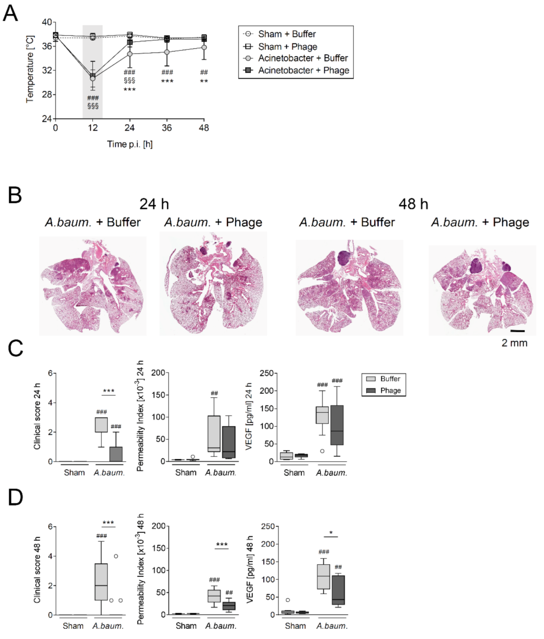
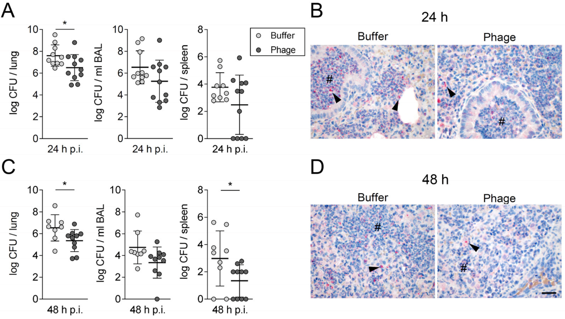
3.1. A Single Intratracheal Phage Application Mitigated the Severity of Murine Pneumonia Caused by Drug-Resistant A. baumannii Strain

Two independent batches of vB_AbaM_Acibel004 phage suspension were produced, and its virulence and host-specificity were confirmed prior testing in animals. Morphology and structural integrity of the prepared phage suspension was verified through electron microscopy analysis (Supplementary Materials Figure S2). Immunocompetent mice were intranasally challenged with 5 × 10⁸ CFU of A. baumannii RUH 2037 and developed clinically apparent pneumonia within 12 h characterized by a concomitant decline in body temperature (Figure 1A). Both sham and A. baumannii infected mice were subjected to intratracheal aerosolization with either buffer (control group) or phage suspension containing 5 × 10⁶ PFU/mL (phage-treated group). While the buffer-treated A. baumannii infected group had a lower body temperature until the end of the experiment (i.e., up to 48 h p.i.), normal body temperature was restored after phage treatment. Additionally, A. baumannii-infected mice exhibited significant weight loss. A marginally higher loss of body weight was noted in phage-treated mice only at 24 h p.i. when compared with the buffer-treated group (Supplementary Materials Figure S3). In contrast, no changes in body temperature or body weight were recorded in sham-infected animals regardless of treatment. Histopathological analysis of H&E-stained A. baumannii infected lungs revealed widely expansive bronchopneumonia (Figure 1B) with numerous infiltrating polymorphonuclear leukocytes (PMN) causing suppurative inflammation with alveolar edema and necrosis, as previously described [32]. Although the histopathologically determined total cell count remained unaltered by phage treatment (data not shown), spreading of inflammation to the periphery along bronchi and large vessels was less pronounced in phage-treated groups than in buffer-treated animals 24 h p.i. Severity and expansion of lesions were more pronounced 48 h p.i. and notably less prominent in lungs from phage-treated mice with less peripherally disseminated inflammation (Figure 1B). Clinical signs of murine pneumonia, regarding behavior and fur appearance, were milder after 12 h of phage treatment (Figure 1C,D).
Vascular endothelial growth factor (VEGF) among others promotes destabilization of cell-cell connections [37] and therefore contributes to development of pulmonary vascular leakage, represented by alveolar-capillary barrier permeability index [27]. Accordingly, alveolar VEGF-levels were found elevated in all infected mice (Figure 1C,D). However, there were lower pulmonary vascular leakage index and VEGF levels as well as improvement of clinical signs at 48 h p.i. in phage-treated animals (Figure 1D).
Consistent with the improved clinical condition of the infected mice, the bacterial burden in lungs was reduced in phage-treated animals by approximately 1 log10 at 24 h p.i. (Figure 2A). This was confirmed by the lower immunohistochemical detection of A. baumannii in lungs of phage-treated animals (Figure 2B). At 48 h p.i., phage-treatment was associated with lower bacterial counts in lungs and spleen, whereas bacterial counts in BAL remained unaltered (Figure 2C,D).

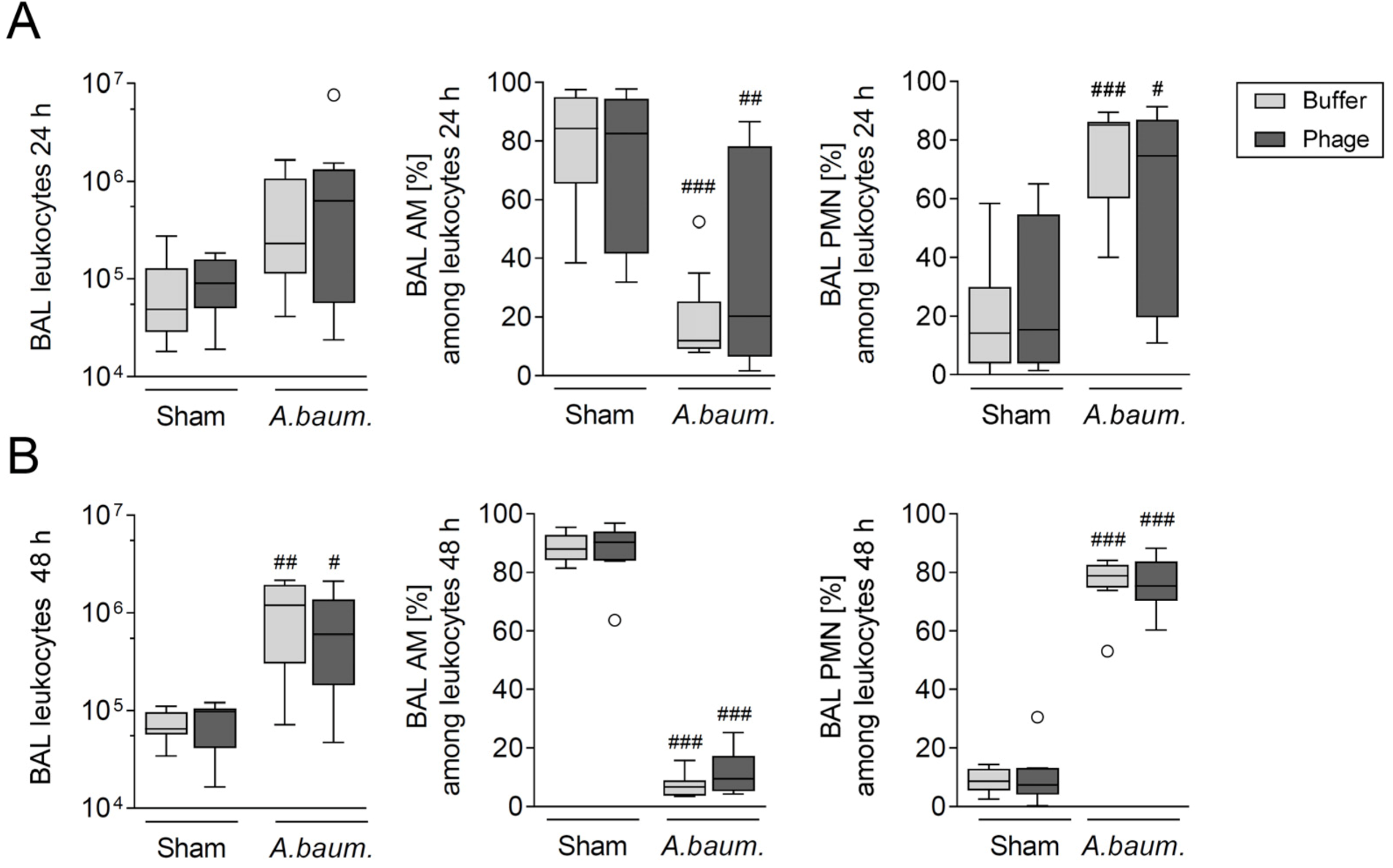
3.2. Phage Therapy Did Not Additionally Increase Lung Inflammation in Mice

Potential immune reactions due to locally acting or systemic dissemination of phage particles or components of phage-lysed bacteria remain a concern for therapeutic usage of phages. To test whether the presence of phages in vivo could stimulate a proinflammatory response, we analyzed changes in immune cell populations and inflammatory mediators in both sham and A. baumannii infected immunocompetent mice treated with phages or buffer.
As anticipated, A. baumannii infection resulted in mobilization of innate immune cells from vascular spaces to the site of infection. Influx of neutrophils into alveolar spaces was evident in terms of total numbers and proportional increase at 24 h (Figure 3A, Supplementary Materials Figures S4A and S5A) up to 48 h p.i. (Figure 3B, Supplementary Materials Figures S4B and S5B). At 24 h p.i., relative numbers of blood neutrophils had more than doubled, at the expense of relative lymphocyte numbers (Supplementary Materials Figure S6A). In response to PMN cell influx, relative numbers of alveolar macrophages (AM) in BAL declined proportionally (Figure 3A,B), while their total numbers remained stable and unaffected by infection (Supplementary Materials Figure S5A,B). At 48 h p.i., loss of pulmonary AM numbers was detected in lungs (Supplementary Materials Figure S4B), while infection-triggered recruitment of monocytes was most evident in BAL (Supplementary Materials Figure S5B). In blood, monocyte proportions tended to increase at 24 h p.i. (Supplementary Materials Figure S6A). At 48 h p.i., steady state of de novo monocyte egress from bone marrow and recruitment to lungs had likely emerged (Supplementary Materials Figure S6B). Of greatest relevance to the envisaged therapeutic usage in patients, absolute cell counts and proportions of innate immune cells in BAL and lungs as acquired by FACS analysis were not affected by phage treatment, irrespective of whether the animal was infected with A. baumannii or sham-infected (Figure 3, Supplementary Materials Figures S4 and S5). Likewise, quantification and differentiation of blood leukocytes showed no changes after phage application (Supplementary Materials Figure S6). Even 48 h p.i., no effect of phage-treatment on populations of innate immune cells in BAL, lungs, and blood was detected.
Furthermore, to exclude the possibility of phage treatment-specific changes in the release of pro-inflammatory mediators, levels of different cytokines and chemokines were determined in BAL fluid and plasma. Yet again, there were no differences found in sham-infected groups treated with either buffer or phages. As expected, CCL2, CCL3, CXCL1, and IL-6 levels were elevated in BAL fluid following A. baumannii infection. Most notably, when compared to buffer-treated group, there was no phage treatment-specific increase in cytokine and chemokine release at 24 h p.i. (Table 1). Whereas at 48 h p.i., cytokine levels were still increased in buffer-treated animals, phage-treated mice showed slightly attenuated cytokine levels. Notably, CCL-2 even showed significantly lower levels in BAL fluid of infected phage-treated animals compared to buffer treatment (Table 2). Likewise, systemic concentrations of G-CSF were found reduced in A. baumannii infected phage-treated mice when compared to the buffer-treated counterpart, while levels of CCL2 and CXCL1 were not affected significantly (Supplementary Materials Tables S3 and S4). Taken together, there was no phage treatment-specific induction of innate immune cells and inflammatory mediators in either A. baumannii or sham-infected immunocompetent animals.

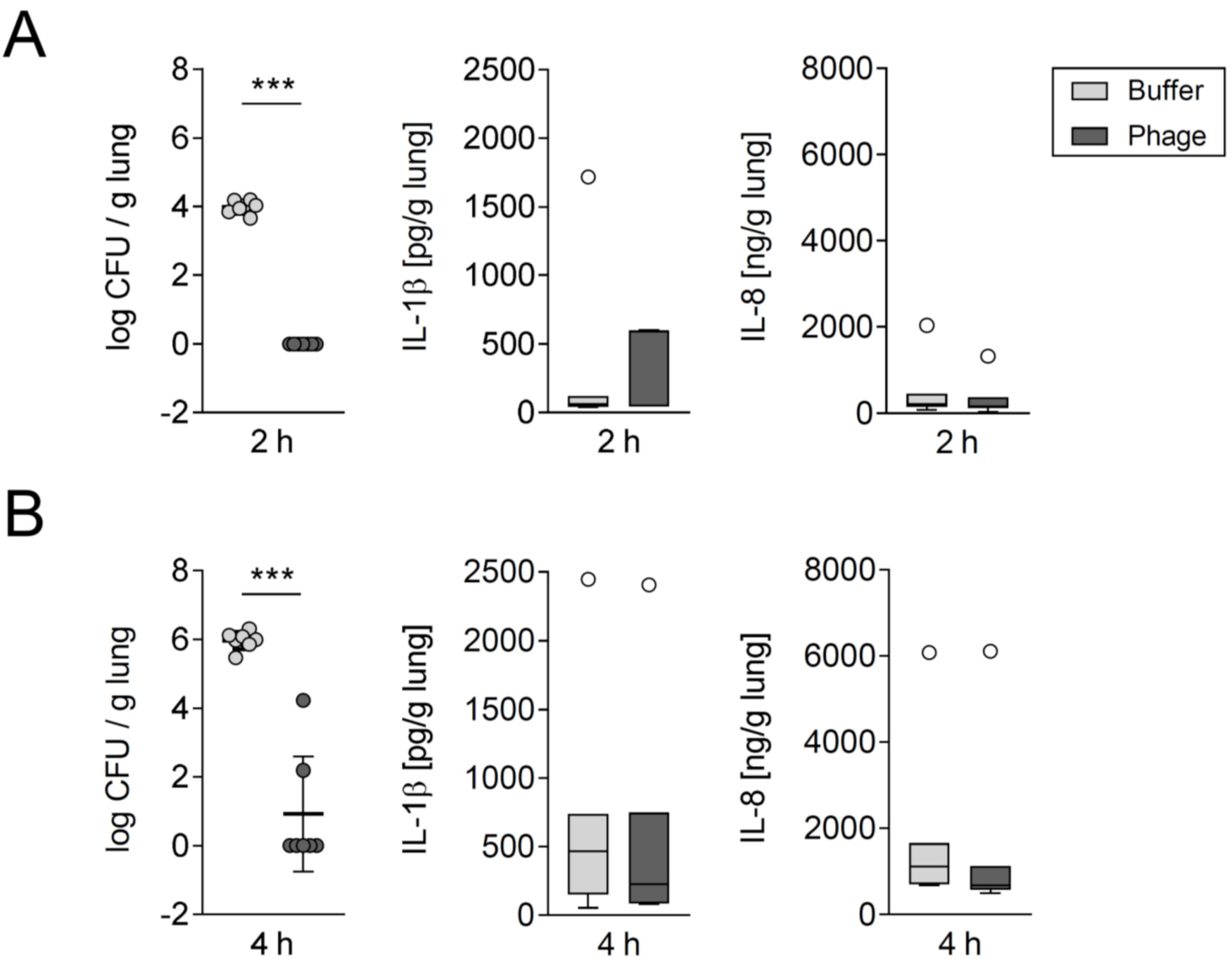
3.3. Phage Treatment Eliminated A. baumannii in Human Lung Tissue without Concomitantly Increasing Proinflammatory Cytokine Release

Above stated findings in mice were validated in an ex vivo model using excised human lung tissues [34]. Accordingly, human lung tissues were stimulated with 1 × 10³ CFU/mL MDR A. baumannii and treated with a single-dose of 5 × 10⁹ PFU/mL vB_AbaM_Acibel004 phage or buffer 30 min later. Remarkably, no viable bacteria could be isolated after 2 h of phage treatment under the conditions tested (Figure 4A). After two additional h of phage treatment, bacterial viability remained compromised, except for 2 samples showing detectable growth (Figure 4B). As a representative of proinflammatory response markers, the release of IL-1β and IL-8 was measured in infected human lung tissue. However, there was no significant increase upon phage treatment at both time points (Figure 4A,B).

4. Discussion

Multidrug-resistant A. baumannii is of serious concern and local outbreaks at intensive care units are difficult to manage due to decreasing treatment options. Phage therapy might provide an effective resource in this regard. Our results show that a single application of a lytic bacteriophage reduced the viability of multidrug-resistant A. baumannii clinical isolate under ex vivo and in vivo conditions. Additionally, the treatment with phages controlled the progression of murine pneumonia without any further aggravation of inflammatory response and adverse off-target side effects. Thus, our data reaffirm the potential of phage therapy to treat MDR A. baumannii infections.
Using the preclinical model developed here, we were able to accurately analyze the clinical success of our phage candidate against A. baumannii infection. Given the generally low virulence of most A. baumannii isolates for healthy mice [3], induction of full-blown pneumonia is rarely successful unless additional tools are employed, such as immunosuppression or exogenous mucin application. Consistently, our results showed a self-limiting and nonlethal A. baumannii lung infection in immunocompetent mice. However, it allowed to assess the efficacy of phage treatment and its impact on a fully functional immune system. Another key aspect of our model is the avoidance of neutropenia, induced by cyclophosphamide in similar preclinical evaluation studies [38,39,40], which is an uncommon risk factor for A. baumannii infection in patients [3,41]. This aspect is crucial as recently dependence of phage therapy success on the synergistic relationship of phages with innate immune cells was established [42]. Along this line, our study methods such as initiating treatment 12 h p.i. in immunocompetent mice aimed to mimic a more clinically relevant setting and are unique as previous studies evaluated phage efficacy in immunosuppressed mice either after simultaneous application or short time periods (2–4 h) following A. baumannii infection [38,39,40]. Initiating phage treatment at a later timepoint allowed for an assessment of efficacy of delayed phage application, interaction with a more progressed immune response to bacterial challenge, and influence on established pulmonary vascular barrier failure.
Besides the optimal timing, the route of application is also expected to influence the success of treatment. In this study, we chose intratracheal aerosolization of phages in infected mice considering the potential clinical relevance, although the preferable route of application for lung infections is not yet clearly defined. Carmody et al. found that systemic application is superior over inhalation of phages in controlling Burkholderia cenocepacia lung infection in a murine model [43], whereas, Semler and colleagues showed the opposite [44], highlighting the difficulty in defining an ideal route of phage application. Nevertheless, inhaled application might be favorable for patients with pulmonary infections [45,46]. Advantages lie in the effective delivery of the active agent to the site of infection, the easy and comfortable use for the patient and avoidance of systemic phage exposure [47,48]. The latter is of special relevance because systemic application might increase the risk of producing antibodies against phages [49,50] or adverse immune responses including fever [51]. Moreover, phages are quickly cleared from the bloodstream when the density of the host bacterium is insufficient for phage propagation, impairing efficacy of systemically applied phages [52]. Regardless, studies concerning pharmacodynamics, pharmacokinetics, and tissue penetration of phages are needed to identify an appropriate route of application [45].
Single application of phages significantly reduced the bacterial viability, although unable to sterilize A. baumannii entirely from alveolar spaces and lung parenchyma in vivo, as expected due to phage amplification at the site of infection. This is consistent with results from other studies, showing the inability to kill entire bacterial population through single dose of intratracheally or intravenously applied phages [53,54,55,56]. Even adequate antibiotic therapy as well as combination therapy with phages or phage lysins does not necessarily kill all bacteria at once [30,54]. Most likely, we failed to reach the ideal multiplicity of infection for the phage-host system used in our study. Repeated intratracheal aerosolization of the phage solution was impossible in our model as it would have required repeated sedations within a short period of time and a high cumulative liquid load to murine lungs. Nose-only-inhalation systems may be considered as alternative approach. Future studies should address the possibility of recurrent and long-term use of purified phages, particularly regarding efficacy and possible humoral immune responses, such as the role of neutralizing antibodies to phages. Additionally, recent data point towards a potential benefit of combination therapy utilizing conventional antimicrobial drugs and phages. Sublethal concentrations of antibiotics were shown to augment phage efficacy [57,58,59], while on the other hand phage application can trigger changes in bacteria that reduce their virulence or even restore their susceptibility against antibiotics [60,61,62,63]. However, unfavorable antagonistic interactions between certain antibiotics and phages have also been described and thus need to be considered [64]. At present, general recommendations regarding combination of antibiotic and phage therapy cannot be given and need further investigation.
Critically ill patients with MDR A. baumannii infection require rapid treatment [3], thus time-consuming de novo search, isolation, characterization, and purification of an active phage is less favorable [65]. Accordingly, phage vB_AbaM_Acibel004 [21] was selected as the only appropriate lytic phage against the used clinical isolate of A. baumannii from an existing collection of nine well-characterized A. baumannii phages. In addition to rapid phage selection, a controlled and standardized production process of phage formulation with possibility of large scale production is a key prerequisite for the approval of bacteriophages as human therapeutics [66]. Importantly, endotoxins and other bacterial components that inherently remain in the solution during phage production could eventually elicit severe immune responses upon phage application [67]. Thus, our candidate phage was obtained after purification according to quality requirements for a pharmaceutical grade active ingredient [25]. So far, there are no guidelines defined for maximum endotoxin levels in formulations for inhalation therapy in humans. However, the European Pharmacopoeia specifies an endotoxin level of 5 EU/kg body weight per h for intravenous application which must be considered prior seeking approval of phages as therapeutics in the European Union [68]. We succeeded in substantially reducing the endotoxin levels in our phage preparation, yet it still exceeded the above-mentioned standard. It is encouraging, however, the residual endotoxin present in our phage preparation did not elicit any marked proinflammatory immune response. Nevertheless, any additional steps intended to remove such residual endotoxins might inadvertently affect the final phage yield and production costs. The preparation of purified phages therefore should be considered as important step during preclinical development of phages for inhalation therapy.
Furthermore, increased release of endotoxins by rapid phage lysis of target bacteria and the possible associated immune response are among the main reservations against phage therapy. When applied locally in the alveolar space, endotoxin-mediated damage to the pulmonary vascular barrier could further exacerbate edema formation in the infected lung [69]. As expected, A. baumannii infection was associated with lung barrier integrity failure in our mouse model, but phage-mediated lysis of the bacteria did not result in increased barrier failure and was even associated with its improvement. In line with our findings, lower lung edema formation and therefore improved lung barrier integrity after phage therapy was described by Dufour et al. comparing the influence of single intranasal phage application with antibiotic treatment in Escherichia coli (E. coli)-induced pneumonia [53]. Accordingly, the same authors demonstrated in different experiments with enteropathogenic E. coli that the amount of endotoxins released by phage lysis was comparable to the amount of endotoxins released by bactericidal antibiotics [70]. In our study, phage application per se did not result in any signs of barrier failure at all, as the air-blood barrier of sham-infected and phage-treated animals was unaffected by the phage solution.
Intratracheal phage aerosolization in our study was associated with reduced spread of bacteria in the lungs, presumably leading to a more local infiltration of immune cells and subsequently more concentrated areas of lung inflammation with reduced barrier breakdown, thus resulting in an improved clinical state of treated animals. In a previous study, Prazak et al. had shown an increase of IL-1β production in phage-treated uninfected animals [54], which was already reported by others [71], indicating a possible pro-inflammatory effect of the phage preparation itself. However, we failed to observe such phage effects neither in vivo nor in human lungs ex vivo.
Our study has some limitations. Since we used a clinical nonmouse-adapted A. baumannii isolate, our infection model was self-limiting and nonlethal but therefore allowed evaluation of the unimpaired innate immune response in immunocompetent mice. Future studies may address ventilator-associated pneumonia caused by MDR A. baumannii and infection of animals pretreated with antibiotics to mimic the clinical situation even better. In addition, finding lytic phages for A. baumannii turned out to be more difficult than for other species as Schooley et al. reported after treating a patient with A. baumannii sepsis as a last resort [13]. Therefore, our approach of using preselected well-characterized phages would be of great benefit for the application of adequate therapy to patients infected with MDR A. baumannii.

5. Conclusions

Our results showed the outcome of pneumonia following A. baumannii lung infection in immunocompetent mice could be improved through single dose of phage administration. Most importantly, the lack of inflammatory or other adverse effects in our study encourage further efforts to develop phages for clinical application against MDR A. baumannii pulmonary infections.

Supplementary Materials

The following are available online at https://www.mdpi.com/article/10.3390/v14010033/s1, Table S1: Drug susceptibility profile of A. baumannii RUH 2037. Table S2: Murine clinical disease scoring system. Table S3: Levels of inflammatory mediators in plasma 24 h p.i. Table S4: Levels of inflammatory mediators in plasma 48 h p.i. Figure S1: Innate immune cell gating strategy. Figure S2: Lysis of Acinetobacter baumannii by phage vB_AbaM_Acibel004. Figure S3: Relative body weight curves of mice. Figure S4: Phage treatment had no impact on innate leukocyte counts in the lungs. Figure S5: Phage treatment had no impact on innate leukocyte counts in BAL. Figure S6: Phage treatment had no impact on innate leukocyte counts in blood.

Author Contributions

Conceptualization, S.-M.W., A.R., H.Z., A.C.H., C.R. and M.W.; methodology, validation, and investigation, S.-M.W., M.C.B., G.N., I.H.E.K., C.S., S.W., K.D., C.G., O.K., A.D.G., A.R., H.Z., M.R., J.L., S.H., A.C.H., C.R. and M.W.; formal analysis, S.-M.W., M.C.B., G.N., I.H.E.K., C.S., K.D., C.G. and J.L.; resources, C.S., A.D.G., A.R., H.Z., M.R., J.N., N.S., S.H., A.C.H., C.R. and M.W.; writing—original draft preparation, S.-M.W., M.C.B., G.N., G.K., K.D., J.L. and M.W.; writing—review and editing, S.-M.W., M.C.B., G.N., G.K., I.H.E.K., C.S., S.W., K.D., C.G., O.K., A.D.G., A.R., H.Z., M.R., J.N., J.L., N.S., S.H., A.C.H., C.R. and M.W.; visualization, S.-M.W., M.C.B., G.N., K.D., C.G., O.K., A.D.G., M.R., J.L. and M.W.; supervision, H.Z., A.C.H., C.R. and M.W.; project administration, S.-M.W., C.R. and M.W.; All authors have read and agreed to the published version of the manuscript.

Funding

This work was supported in parts by grants from the German Research Foundation (DFG) [SFB-TR84 C6, C9, B1, B6, Z1a, Z1b to M.W., N.S., S.H., A.D.G., A.C.H.]; by the Federal Ministry of Education and Research (BMBF) [e:Med CAPSyS 01ZX1604B and SYMPATH 01ZX1906A to N.S. and M.W. and Phage4Cure 16GW0141 to M.W.] and by the Agence nationale de la recherche (ANR) and the Federal Ministry of Education and Research (BMBF) [MAPVAP 16GW0247 to S.M.W., G.N., A.C.H. and M.W.]. M.C.B. received a fellowship from the Jürgen Manchot Foundation. S.H. and A.C.H. received funding by Charité 3R. The funding sources had no influence on study design or publication.

Institutional Review Board Statement

Animal procedures were approved by institutional (Charité—Universitätsmedizin Berlin) and governmental (Landesamt für Gesundheit und Soziales Berlin) authorities, approval number A 0299/15, approval date: 17 February 2016. The study involving human lung samples was approved by the Ethics Committee of Charité—Universitätsmedizin Berlin (EA2/079/13), approval date: 23 June 2013.

Informed Consent Statement

Informed consent was obtained from all subjects involved in the study.

Data Availability Statement

The complete susceptibility profile for A. baumannii RUH 2037 (DSM 101993) is publicly available at BacDive (https://bacdive.dsmz.de/strain/157916 doi:10.13145/bacdive157916.20201210.5, accessed on 29 November 2021).

Acknowledgments

We thank Ulrike Behrendt and Silke Dusatko, Charité—Universitätsmedizin Berlin, Berlin, Germany, Bettina Henze, DSMZ—German Collection of Microorganisms and Cell Cultures, Braunschweig, Germany, and Angela Linke, Department of Veterinary Pathology, Freie Universität Berlin, Berlin, Germany, for skillful technical support. We are grateful to Annemarie Overmann, TU Braunschweig, Braunschweig, Germany, for determination of phage spectra and Johannes Wittmann, DSMZ—German Collection of Microorganisms and Cell Cultures, Braunschweig, Germany, for fruitful discussions. We also thank Birgitt Gutbier and Katrin Reppe, Charité—Universitätsmedizin Berlin, Berlin, Germany, for managing in-lab animal facilities. We thankfully acknowledge Maya Merabishvili, Queen Astrid Military Hospital, Brussels, Belgium, for vB_AbaM_Acibel004 phage, Alex F. de Vos and Tom van der Poll, Amsterdam University Medical Centers, Amsterdam, the Netherlands, for A. baumannii isolate RUH 2037 and José Ramos-Vivas, Universidad Europea del Atlántico, Santander, Spain, for the antibody against A. baumannii. Parts of this work are incorporated into the doctoral thesis of Markus C. Brack.

Conflicts of Interest

S.M.W. reports nonfinancial support from Deutsche Gesellschaft für Pneumologie, non-financial support from Mukoviszidose e.V., personal fees and nonfinancial support from Schlütersche Verlagsgesellschaft. G.N. received funding for research from Biotest AG. M.W. received grants and personal fees from Actelion, Bayer Health Care, Biotest AG, Boehringer Ingelheim, NOXXON Pharma, Pantherna, Silence Therapeutics, Vaxxilon, grants from Quark Pharma, Takeda Pharma, Deutsche Gesellschaft für Pneumologie, European Respiratory Society, Marie Curie Foundation, Else Kröner–Fresenius–Stiftung, CAPNETZ STIFTUNG, International Max Planck Research School, personal fees from Aptarion, AstraZeneca, Berlin Chemie, Chiesi, GlaxoSmithKline, Novartis, Sinoxa and Teva, and has a patent “Means for inhibiting the expression of ANG2 US8829179B2” issued. The funders had no role in the design of the study; in the collection, analyses, or interpretation of data; in the writing of the manuscript, or in the decision to publish the results. All other authors declare that they have no competing interests.

Presentation of Results

The results presented here were reported in part as abstracts on scientific conferences: Herbsttagung der Sektionen Zellbiologie und Infektiologie und Tuberkulose der Deutschen Gesellschaft für Pneumologie und Beatmungsmedizin e.V., November 2017, Gießen, Germany, Georg Thieme Verlag KG; 2017; 59. Kongress der Deutschen Gesellschaft für Pneumologie und Beatmungsmedizin e.v., March 2018, Dresden, Germany, Pneumologie 2018; 72:S67-S68; European Respiratory Society International Congress, September 2018, Paris, France, Abstract No. PA5453, Respiratory infections, European Respiratory Society, 09152018:PA5453; American Thoracic Society International Conference, May, 2018, San Diego, California, USA, Abstract No. A7571; American Journal of Respiratory and Critical Care Medicine 2020;201:A7571.

References

  1. McCarthy, R. Antibiotic resistance: The ‘Other’ Pandemic Lurking behind COVID-19. Available online: https://bsac.org.uk/antibiotic-resistance-the-other-pandemic-lurking-behind-covid-19/ (accessed on 1 February 2021).
  2. Sader, H.S.; Castanheira, M.; Arends, S.J.R.; Goossens, H.; Flamm, R.K. Geographical and temporal variation in the frequency and antimicrobial susceptibility of bacteria isolated from patients hospitalized with bacterial pneumonia: Results from 20 years of the SENTRY Antimicrobial Surveillance Program (1997–2016). J. Antimicrob. Chemother. 2019, 74, 1595–1606. [Google Scholar] [CrossRef] [PubMed]
  3. Wong, D.; Nielsen, T.B.; Bonomo, R.A.; Pantapalangkoor, P.; Luna, B.; Spellberg, B. Clinical and Pathophysiological Overview of Acinetobacter Infections: A Century of Challenges. Clin. Microbiol. Rev. 2017, 30, 409–447. [Google Scholar] [CrossRef] [PubMed] [Green Version]
  4. Ben-Chetrit, E.; Wiener-Well, Y.; Lesho, E.; Kopuit, P.; Broyer, C.; Bier, L.; Assous, M.V.; Benenson, S.; Cohen, M.J.; McGann, P.T.; et al. An intervention to control an ICU outbreak of carbapenem-resistant Acinetobacter baumannii: Long-term impact for the ICU and hospital. Crit. Care 2018, 22, 319. [Google Scholar] [CrossRef] [PubMed] [Green Version]
  5. Vincent, J.-L.; Rello, J.; Marshall, J.; Silva, E.; Anzueto, A.; Martin, C.D.; Moreno, R.; Lipman, J.; Gomersall, C.; Sakr, Y.; et al. International study of the prevalence and outcomes of infection in intensive care units. JAMA 2009, 302, 2323–2329. [Google Scholar] [CrossRef] [Green Version]
  6. Shojaei, L.; Mohammadi, M.; Beigmohammadi, M.-T.; Doomanlou, M.; Abdollahi, A.; Feizabadi, M.M.; Khalili, H. Clinical response and outcome of pneumonia due to multi-drug resistant Acinetobacter baumannii in critically ill patients. Iran. J. Microbiol. 2016, 8, 288–297. [Google Scholar]
  7. McGann, P.; Courvalin, P.; Snesrud, E.; Clifford, R.J.; Yoon, E.-J.; Onmus-Leone, F.; Ong, A.C.; Kwak, Y.I.; Grillot-Courvalin, C.; Lesho, E.; et al. Amplification of aminoglycoside resistance gene aphA1 in Acinetobacter baumannii results in tobramycin therapy failure. mBio 2014, 5, e00915. [Google Scholar] [CrossRef] [Green Version]
  8. Pogue, J.M.; Lee, J.; Marchaim, D.; Yee, V.; Zhao, J.J.; Chopra, T.; Lephart, P.; Kaye, K.S. Incidence of and risk factors for colistin-associated nephrotoxicity in a large academic health system. Clin. Infect. Dis. Off. Publ. Infect. Dis. Soc. Am. 2011, 53, 879–884. [Google Scholar] [CrossRef] [Green Version]
  9. World Health Organisation. Global Priority List of Antibiotic-Resistant Bacteria to Guide Research, Discovery, and Development of New Antibiotics. Available online: https://www.who.int/medicines/publications/WHO-PPL-Short_Summary_25Feb-ET_NM_WHO.pdf?ua=1 (accessed on 29 November 2021).
  10. Kortright, K.E.; Chan, B.K.; Koff, J.L.; Turner, P.E. Phage Therapy: A Renewed Approach to Combat Antibiotic-Resistant Bacteria. Cell Host Microbe 2019, 25, 219–232. [Google Scholar] [CrossRef] [Green Version]
  11. Yen, M.; Cairns, L.S.; Camilli, A. A cocktail of three virulent bacteriophages prevents Vibrio cholerae infection in animal models. Nat. Commun. 2017, 8, 14187. [Google Scholar] [CrossRef] [Green Version]
  12. Pabary, R.; Singh, C.; Morales, S.; Bush, A.; Alshafi, K.; Bilton, D.; Alton, E.W.F.W.; Smithyman, A.; Davies, J.C. Antipseudomonal Bacteriophage Reduces Infective Burden and Inflammatory Response in Murine Lung. Antimicrob. Agents Chemother. 2016, 60, 744–751. [Google Scholar] [CrossRef] [Green Version]
  13. Schooley, R.T.; Biswas, B.; Gill, J.J.; Hernandez-Morales, A.; Lancaster, J.; Lessor, L.; Barr, J.J.; Reed, S.L.; Rohwer, F.; Benler, S.; et al. Development and Use of Personalized Bacteriophage-Based Therapeutic Cocktails To Treat a Patient with a Disseminated Resistant Acinetobacter baumannii Infection. Antimicrob. Agents Chemother. 2017, 61, e00954-17. [Google Scholar] [CrossRef] [Green Version]
  14. Law, N.; Logan, C.; Yung, G.; Furr, C.-L.L.; Lehman, S.M.; Morales, S.; Rosas, F.; Gaidamaka, A.; Bilinsky, I.; Grint, P.; et al. Successful adjunctive use of bacteriophage therapy for treatment of multidrug-resistant Pseudomonas aeruginosa infection in a cystic fibrosis patient. Infection 2019, 47, 665–668. [Google Scholar] [CrossRef]
  15. Jault, P.; Leclerc, T.; Jennes, S.; Pirnay, J.P.; Que, Y.-A.; Resch, G.; Rousseau, A.F.; Ravat, F.; Carsin, H.; Le Floch, R.; et al. Efficacy and tolerability of a cocktail of bacteriophages to treat burn wounds infected by Pseudomonas aeruginosa (PhagoBurn): A randomised, controlled, double-blind phase 1/2 trial. Lancet Infect. Dis. 2019, 19, 35–45. [Google Scholar] [CrossRef]
  16. Sarker, S.A.; Sultana, S.; Reuteler, G.; Moine, D.; Descombes, P.; Charton, F.; Bourdin, G.; McCallin, S.; Ngom-Bru, C.; Neville, T.; et al. Oral Phage Therapy of Acute Bacterial Diarrhea With Two Coliphage Preparations: A Randomized Trial in Children From Bangladesh. EBioMedicine 2016, 4, 124–137. [Google Scholar] [CrossRef] [Green Version]
  17. Wright, A.; Hawkins, C.H.; Anggård, E.E.; Harper, D.R. A controlled clinical trial of a therapeutic bacteriophage preparation in chronic otitis due to antibiotic-resistant Pseudomonas aeruginosa; a preliminary report of efficacy. Clin. Otolaryngol. 2009, 34, 349–357. [Google Scholar] [CrossRef]
  18. Iszatt, J.J.; Larcombe, A.N.; Chan, H.-K.; Stick, S.M.; Garratt, L.W.; Kicic, A. Phage Therapy for Multi-Drug Resistant Respiratory Tract Infections. Viruses 2021, 13, 1809. [Google Scholar] [CrossRef]
  19. Nale, J.Y.; Clokie, M.R. Preclinical data and safety assessment of phage therapy in humans. Curr. Opin. Biotechnol. 2021, 68, 310–317. [Google Scholar] [CrossRef]
  20. Dijkshoorn, L.; Aucken, H.; Gerner-Smidt, P.; Janssen, P.; Kaufmann, M.E.; Garaizar, J.; Ursing, J.; Pitt, T.L. Comparison of outbreak and nonoutbreak Acinetobacter baumannii strains by genotypic and phenotypic methods. J. Clin. Microbiol. 1996, 34, 1519–1525. [Google Scholar] [CrossRef] [Green Version]
  21. Merabishvili, M.; Vandenheuvel, D.; Kropinski, A.M.; Mast, J.; de Vos, D.; Verbeken, G.; Noben, J.-P.; Lavigne, R.; Vaneechoutte, M.; Pirnay, J.-P. Characterization of newly isolated lytic bacteriophages active against Acinetobacter baumannii. PLoS ONE 2014, 9, e104853. [Google Scholar] [CrossRef] [Green Version]
  22. Knapp, S.; Wieland, C.W.; Florquin, S.; Pantophlet, R.; Dijkshoorn, L.; Tshimbalanga, N.; Akira, S.; van der Poll, T. Differential roles of CD14 and toll-like receptors 4 and 2 in murine Acinetobacter pneumonia. Am. J. Respir. Crit. Care Med. 2006, 173, 122–129. [Google Scholar] [CrossRef]
  23. Grimm, H. Interpretationskriterien der Empfindlichkeitsprüfung von Ofloxacin mit dem Agardiffusionstest. Infection 1986, 14 (Suppl. S1), S16–S19. [Google Scholar] [CrossRef]
  24. Reimer, L.C.; Sarda Carbasse, J.; Koblitz, J.; Podstawka, A.; Overmann, J. Acinetobacter Baumannii Bouvet and Grimont 1986. 2020. Available online: https://bacdive.dsmz.de/strain/157916#ref64488 (accessed on 29 November 2021).
  25. Dannheim, A.; Hebecker, S.; Wienecke, S.; Ziehr, H. Bakteriophagen als Wirkstoffe zur Infektionsbehandlung—Was ist möglich? Pharmakon 2020, 8, 278–284. [Google Scholar] [CrossRef]
  26. Valentine, R.C.; Shapiro, B.M.; Stadtman, E.R. Regulation of glutamine synthetase. XII. Electron microscopy of the enzyme from Escherichia coli. Biochemistry 1968, 7, 2143–2152. [Google Scholar] [CrossRef]
  27. Berger, S.; Goekeri, C.; Gupta, S.K.; Vera, J.; Dietert, K.; Behrendt, U.; Lienau, J.; Wienhold, S.-M.; Gruber, A.D.; Suttorp, N.; et al. Delay in antibiotic therapy results in fatal disease outcome in murine pneumococcal pneumonia. Crit. Care 2018, 22, 287. [Google Scholar] [CrossRef] [Green Version]
  28. Müller-Redetzky, H.; Kellermann, U.; Wienhold, S.-M.; Gutbier, B.; Lienau, J.; Hellwig, K.; Reppe, K.; Letsiou, E.; Tschernig, T.; Scholz, M.; et al. Neutralizing Complement C5a Protects Mice with Pneumococcal Pulmonary Sepsis. Anesthesiology 2020, 132, 795–807. [Google Scholar] [CrossRef]
  29. Costa, D.L.; Lehmann, J.R.; Harold, W.M.; Drew, R.T. Transoral tracheal intubation of rodents using a fiberoptic laryngoscope. Lab. Anim. Sci. 1986, 36, 256–261. [Google Scholar]
  30. Doehn, J.M.; Fischer, K.; Reppe, K.; Gutbier, B.; Tschernig, T.; Hocke, A.C.; Fischetti, V.A.; Löffler, J.; Suttorp, N.; Hippenstiel, S.; et al. Delivery of the endolysin Cpl-1 by inhalation rescues mice with fatal pneumococcal pneumonia. J. Antimicrob. Chemother. 2013, 68, 2111–2117. [Google Scholar] [CrossRef] [Green Version]
  31. Dietert, K.; Nouailles, G.; Gutbier, B.; Reppe, K.; Berger, S.; Jiang, X.; Schauer, A.E.; Hocke, A.C.; Herold, S.; Slevogt, H.; et al. Digital Image Analyses on Whole-Lung Slides in Mouse Models of Acute Pneumonia. Am. J. Respir. Cell Mol. Biol. 2018, 58, 440–448. [Google Scholar] [CrossRef]
  32. Dietert, K.; Gutbier, B.; Wienhold, S.M.; Reppe, K.; Jiang, X.; Yao, L.; Chaput, C.; Naujoks, J.; Brack, M.; Kupke, A.; et al. Spectrum of pathogen- and model-specific histopathologies in mouse models of acute pneumonia. PLoS ONE 2017, 12, e0188251. [Google Scholar] [CrossRef]
  33. Lázaro-Díez, M.; Navascués-Lejarza, T.; Remuzgo-Martínez, S.; Navas, J.; Icardo, J.M.; Acosta, F.; Martínez-Martínez, L.; Ramos-Vivas, J. Acinetobacter baumannii and A. pittii clinical isolates lack adherence and cytotoxicity to lung epithelial cells in vitro. Microbes Infect. 2016, 18, 559–564. [Google Scholar] [CrossRef]
  34. Berg, J.; Zscheppang, K.; Fatykhova, D.; Tönnies, M.; Bauer, T.T.; Schneider, P.; Neudecker, J.; Rückert, J.C.; Eggeling, S.; Schimek, M.; et al. Tyk2 as a target for immune regulation in human viral/bacterial pneumonia. Eur. Respir. J. 2017, 50, 1601953. [Google Scholar] [CrossRef] [PubMed]
  35. Szymanski, K.V.; Toennies, M.; Becher, A.; Fatykhova, D.; N’Guessan, P.D.; Gutbier, B.; Klauschen, F.; Neuschaefer-Rube, F.; Schneider, P.; Rueckert, J.; et al. Streptococcus pneumoniae-induced regulation of cyclooxygenase-2 in human lung tissue. Eur. Respir. J. 2012, 40, 1458–1467. [Google Scholar] [CrossRef] [PubMed] [Green Version]
  36. Croghan, C.; Egeghy, P.P. Methods of dealing with values below the limit of detection using sas. Presented at the Southeastern SAS User Group, St. Petersburg, FL, USA, 22–24 September 2020; Available online: https://analytics.ncsu.edu/sesug/2003/SD08-Croghan.pdf (accessed on 29 November 2021).
  37. Broermann, A.; Winderlich, M.; Block, H.; Frye, M.; Rossaint, J.; Zarbock, A.; Cagna, G.; Linnepe, R.; Schulte, D.; Nottebaum, A.F.; et al. Dissociation of VE-PTP from VE-cadherin is required for leukocyte extravasation and for VEGF-induced vascular permeability in vivo. J. Exp. Med. 2011, 208, 2393–2401. [Google Scholar] [CrossRef] [PubMed] [Green Version]
  38. Jeon, J.; Ryu, C.-M.; Lee, J.-Y.; Park, J.-H.; Yong, D.; Lee, K. In Vivo Application of Bacteriophage as a Potential Therapeutic Agent To Control OXA-66-Like Carbapenemase-Producing Acinetobacter baumannii Strains Belonging to Sequence Type 357. Appl. Environ. Microbiol. 2016, 82, 4200–4208. [Google Scholar] [CrossRef] [Green Version]
  39. Hua, Y.; Luo, T.; Yang, Y.; Dong, D.; Wang, R.; Wang, Y.; Xu, M.; Guo, X.; Hu, F.; He, P. Phage Therapy as a Promising New Treatment for Lung Infection Caused by Carbapenem-Resistant Acinetobacter baumannii in Mice. Front. Microbiol. 2017, 8, 2659. [Google Scholar] [CrossRef] [Green Version]
  40. Cha, K.; Oh, H.K.; Jang, J.Y.; Jo, Y.; Kim, W.K.; Ha, G.U.; Ko, K.S.; Myung, H. Characterization of Two Novel Bacteriophages Infecting Multidrug-Resistant (MDR) Acinetobacter baumannii and Evaluation of Their Therapeutic Efficacy in Vivo. Front. Microbiol. 2018, 9, 696. [Google Scholar] [CrossRef] [Green Version]
  41. Freire, M.P.; de Oliveira Garcia, D.; Garcia, C.P.; Campagnari Bueno, M.F.; Camargo, C.H.; Kono Magri, A.S.G.; Francisco, G.R.; Reghini, R.; Vieira, M.F.; Ibrahim, K.Y.; et al. Bloodstream infection caused by extensively drug-resistant Acinetobacter baumannii in cancer patients: High mortality associated with delayed treatment rather than with the degree of neutropenia. Clin. Microbiol. Infect. 2016, 22, 352–358. [Google Scholar] [CrossRef]
  42. Roach, D.R.; Leung, C.Y.; Henry, M.; Morello, E.; Singh, D.; Di Santo, J.P.; Weitz, J.S.; Debarbieux, L. Synergy between the Host Immune System and Bacteriophage Is Essential for Successful Phage Therapy against an Acute Respiratory Pathogen. Cell Host Microbe 2017, 22, 38–47.e4. [Google Scholar] [CrossRef]
  43. Carmody, L.A.; Gill, J.J.; Summer, E.J.; Sajjan, U.S.; Gonzalez, C.F.; Young, R.F.; LiPuma, J.J. Efficacy of bacteriophage therapy in a model of Burkholderia cenocepacia pulmonary infection. J. Infect. Dis. 2010, 201, 264–271. [Google Scholar] [CrossRef] [Green Version]
  44. Semler, D.D.; Goudie, A.D.; Finlay, W.H.; Dennis, J.J. Aerosol phage therapy efficacy in Burkholderia cepacia complex respiratory infections. Antimicrob. Agents Chemother. 2014, 58, 4005–4013. [Google Scholar] [CrossRef] [Green Version]
  45. Bodier-Montagutelli, E.; Morello, E.; L’Hostis, G.; Guillon, A.; Dalloneau, E.; Respaud, R.; Pallaoro, N.; Blois, H.; Vecellio, L.; Gabard, J.; et al. Inhaled phage therapy: A promising and challenging approach to treat bacterial respiratory infections. Expert Opin. Drug Deliv. 2017, 14, 959–972. [Google Scholar] [CrossRef]
  46. Ruge, C.A.; Kirch, J.; Lehr, C.-M. Pulmonary drug delivery: From generating aerosols to overcoming biological barriers-therapeutic possibilities and technological challenges. Lancet. Respir. Med. 2013, 1, 402–413. [Google Scholar] [CrossRef]
  47. Chang, R.Y.K.; Wallin, M.; Lin, Y.; Leung, S.S.Y.; Wang, H.; Morales, S.; Chan, H.-K. Phage therapy for respiratory infections. Adv. Drug Deliv. Rev. 2018, 133, 76–86. [Google Scholar] [CrossRef]
  48. Golshahi, L.; Seed, K.D.; Dennis, J.J.; Finlay, W.H. Toward modern inhalational bacteriophage therapy: Nebulization of bacteriophages of Burkholderia cepacia complex. J. Aerosol Med. Pulm. Drug Deliv. 2008, 21, 351–360. [Google Scholar] [CrossRef]
  49. Hodyra-Stefaniak, K.; Kaźmierczak, Z.; Majewska, J.; Sillankorva, S.; Miernikiewicz, P.; Międzybrodzki, R.; Górski, A.; Azeredo, J.; Lavigne, R.; Lecion, D.; et al. Natural and Induced Antibodies Against Phages in Humans: Induction Kinetics and Immunogenicity for Structural Proteins of PB1-Related Phages. PHAGE 2020, 1, 91–99. [Google Scholar] [CrossRef]
  50. Biswas, B.; Adhya, S.; Washart, P.; Paul, B.; Trostel, A.N.; Powell, B.; Carlton, R.; Merril, C.R. Bacteriophage therapy rescues mice bacteremic from a clinical isolate of vancomycin-resistant Enterococcus faecium. Infect. Immun. 2002, 70, 204–210. [Google Scholar] [CrossRef] [Green Version]
  51. Aslam, S.; Lampley, E.; Wooten, D.; Karris, M.; Benson, C.; Strathdee, S.; Schooley, R.T. Lessons Learned From the First 10 Consecutive Cases of Intravenous Bacteriophage Therapy to Treat Multidrug-Resistant Bacterial Infections at a Single Center in the United States. Open Forum Infect. Dis. 2020, 7, ofaa389. [Google Scholar] [CrossRef]
  52. Capparelli, R.; Parlato, M.; Borriello, G.; Salvatore, P.; Iannelli, D. Experimental phage therapy against Staphylococcus aureus in mice. Antimicrob. Agents Chemother. 2007, 51, 2765–2773. [Google Scholar] [CrossRef] [Green Version]
  53. Dufour, N.; Delattre, R.; Chevallereau, A.; Ricard, J.-D.; Debarbieux, L. Phage Therapy of Pneumonia Is Not Associated with an Overstimulation of the Inflammatory Response Compared to Antibiotic Treatment in Mice. Antimicrob. Agents Chemother. 2019, 63, e00379-19. [Google Scholar] [CrossRef] [Green Version]
  54. Prazak, J.; Iten, M.; Cameron, D.R.; Save, J.; Grandgirard, D.; Resch, G.; Goepfert, C.; Leib, S.L.; Takala, J.; Jakob, S.M.; et al. Bacteriophages Improve Outcomes in Experimental Staphylococcus aureus Ventilator-associated Pneumonia. Am. J. Respir. Crit. Care Med. 2019, 200, 1126–1133. [Google Scholar] [CrossRef]
  55. Agarwal, R.; Johnson, C.T.; Imhoff, B.R.; Donlan, R.M.; McCarty, N.A.; García, A.J. Inhaled bacteriophage-loaded polymeric microparticles ameliorate acute lung infections. Nat. Biomed. Eng. 2018, 2, 841–849. [Google Scholar] [CrossRef]
  56. Chhibber, S.; Kaur, S.; Kumari, S. Therapeutic potential of bacteriophage in treating Klebsiella pneumoniae B5055-mediated lobar pneumonia in mice. J. Med. Microbiol. 2008, 57, 1508–1513. [Google Scholar] [CrossRef] [Green Version]
  57. Comeau, A.M.; Tétart, F.; Trojet, S.N.; Prère, M.-F.; Krisch, H.M. Phage-Antibiotic Synergy (PAS): Beta-lactam and quinolone antibiotics stimulate virulent phage growth. PLoS ONE 2007, 2, e799. [Google Scholar] [CrossRef] [Green Version]
  58. Kamal, F.; Dennis, J.J. Burkholderia cepacia complex Phage-Antibiotic Synergy (PAS): Antibiotics stimulate lytic phage activity. Appl. Environ. Microbiol. 2015, 81, 1132–1138. [Google Scholar] [CrossRef] [Green Version]
  59. Ryan, E.M.; Alkawareek, M.Y.; Donnelly, R.F.; Gilmore, B.F. Synergistic phage-antibiotic combinations for the control of Escherichia coli biofilms in vitro. FEMS Immunol. Med. Microbiol. 2012, 65, 395–398. [Google Scholar] [CrossRef] [Green Version]
  60. Mangalea, M.R.; Duerkop, B.A. Fitness Trade-Offs Resulting from Bacteriophage Resistance Potentiate Synergistic Antibacterial Strategies. Infect. Immun. 2020, 88, e00926-19. [Google Scholar] [CrossRef] [Green Version]
  61. Engeman, E.; Freyberger, H.R.; Corey, B.W.; Ward, A.M.; He, Y.; Nikolich, M.P.; Filippov, A.A.; Tyner, S.D.; Jacobs, A.C. Synergistic Killing and Re-Sensitization of Pseudomonas aeruginosa to Antibiotics by Phage-Antibiotic Combination Treatment. Pharmaceuticals 2021, 14, 184. [Google Scholar] [CrossRef]
  62. Wang, X.; Loh, B.; Gordillo Altamirano, F.; Yu, Y.; Hua, X.; Leptihn, S. Colistin-phage combinations decrease antibiotic resistance in Acinetobacter baumannii via changes in envelope architecture. Emerg. Microbes Infect. 2021, 10, 2205–2219. [Google Scholar] [CrossRef]
  63. Gordillo Altamirano, F.; Forsyth, J.H.; Patwa, R.; Kostoulias, X.; Trim, M.; Subedi, D.; Archer, S.K.; Morris, F.C.; Oliveira, C.; Kielty, L.; et al. Bacteriophage-resistant Acinetobacter baumannii are resensitized to antimicrobials. Nat. Microbiol. 2021, 6, 157–161. [Google Scholar] [CrossRef]
  64. Zuo, P.; Yu, P.; Alvarez, P.J.J. Aminoglycosides Antagonize Bacteriophage Proliferation, Attenuating Phage Suppression of Bacterial Growth, Biofilm Formation, and Antibiotic Resistance. Appl. Environ. Microbiol. 2021, 87, e0046821. [Google Scholar] [CrossRef]
  65. Mattila, S.; Ruotsalainen, P.; Jalasvuori, M. On-Demand Isolation of Bacteriophages Against Drug-Resistant Bacteria for Personalized Phage Therapy. Front. Microbiol. 2015, 6, 1271. [Google Scholar] [CrossRef] [PubMed] [Green Version]
  66. Luong, T.; Salabarria, A.-C.; Edwards, R.A.; Roach, D.R. Standardized bacteriophage purification for personalized phage therapy. Nat. Protoc. 2020, 15, 2867–2890. [Google Scholar] [CrossRef] [PubMed]
  67. Viertel, T.M.; Ritter, K.; Horz, H.-P. Viruses versus bacteria-novel approaches to phage therapy as a tool against multidrug-resistant pathogens. J. Antimicrob. Chemother. 2014, 69, 2326–2336. [Google Scholar] [CrossRef] [PubMed]
  68. Dawson, M. Endotoxin Limits for Parenteral Drug Products: BET White Paper. Available online: https://www.acciusa.com/pdfs/whitepapers/EndotoxinLimits_SeanJH-PR17-012.pdf (accessed on 10 October 2021).
  69. Perros, F.; Lambrecht, B.N.; Hammad, H. TLR4 signalling in pulmonary stromal cells is critical for inflammation and immunity in the airways. Respir. Res. 2011, 12, 125. [Google Scholar] [CrossRef] [PubMed] [Green Version]
  70. Dufour, N.; Delattre, R.; Ricard, J.-D.; Debarbieux, L. The Lysis of Pathogenic Escherichia coli by Bacteriophages Releases Less Endotoxin Than by β-Lactams. Clin. Infect. Dis. 2017, 64, 1582–1588. [Google Scholar] [CrossRef] [PubMed]
  71. van Belleghem, J.D.; Clement, F.; Merabishvili, M.; Lavigne, R.; Vaneechoutte, M. Pro- and anti-inflammatory responses of peripheral blood mononuclear cells induced by Staphylococcus aureus and Pseudomonas aeruginosa phages. Sci. Rep. 2017, 7, 8004. [Google Scholar] [CrossRef] [PubMed] [Green Version]
Figure 1. Phage therapy mitigated murine A. baumannii pneumonia. Mice were transnasally infected with 5 × 10⁸ CFU A. baumannii RUH 2037 or sham-infected (DPBS) and intratracheally treated with specific phage, vB_AbaM_Acibel004, or control solution (buffer) 12 h post infection (p.i.). At defined time points mice were sacrificed for BAL and blood sampling. Separate groups were sacrificed for histological analyses. (A) Body temperature curves of all mice subjected to analysis at 24 h (n = 11–12; sham-infected groups or n = 16–17; A. baumannii infected groups) and 48 h (n = 12; sham-infected groups or n = 17; A. baumannii infected groups) are shown combined (mean ± SD). Representative images of H&E-stained whole lung sections from A. baumannii infected buffer or phage-treated mice are shown at 24 h and 48 h p.i. (B); n = 3–4 (sham-infected groups), n = 6 (A. baumannii infected groups). Murine clinical disease score, permeability index and VEGF levels in BAL were assessed at 24 h (C) and 48 h p.i. (D). Data are presented as box plots depicting median, quartiles, and range excluding outliers (open circles) and analyzed using 2-way ANOVA and Tukey’s multiple comparisons test. C and D (clinical score), n = 11–12 (sham-infected groups) or n = 16–17 (A. baumannii infected groups; including 3 mice euthanized due to humane endpoints); C and D (permeability index and VEGF levels), n = 8 (sham-infected groups) or n = 8–11 (A. baumannii infected groups). Grey box in A marks time point of phage application. §/#/* indicate significant differences between groups at corresponding time point; § sham + phages vs. Acinetobacter + phages (A), # infected vs. corresponding sham group, *Acinetobacter + buffer vs. Acinetobacter + phages, * p < 0.05, ##/** p < 0.01 and ###/***/§§§ p < 0.001.
Viruses 14 00033 g001
Figure 2. Single intratracheal phage application reduced bacterial burden in mice. Colony forming units (CFU) of A. baumannii were quantified in BAL, homogenized half lungs and spleen at 24 h (A) and 48 h (C) post infection (p.i.). Representative images of immunohistochemically stained lung tissue sections to visualize A. baumannii at 24 h (B) and 48 h p.i. (D). Data are expressed as single values with mean ± SD. CFU data were logarithmized (Y = log(CFU+1)) and tested with unpaired student’s t-test; n = 9–11 (24 h p.i.); n = 8–10 (48 h p.i.) for each group. * p < 0.05 between indicated groups; n = 3–4 (sham-infected groups), n = 6 (A. baumannii infected groups) per time point for histology. #: neutrophils, arrowheads: bacteria, scale bar 50 μm.
Viruses 14 00033 g002
Figure 3. Phage therapy did not increase lung inflammation in mice. Numbers of leukocytes and percentages of alveolar macrophages (AM) and polymorphonuclear leukocytes (PMN) in BAL of mice were analyzed at 24 h (A) and 48 h p.i. (B) by flow cytometry. Data are presented as box plots depicting median, quartiles and range excluding outliers (open circles) and analyzed using 2-way ANOVA and Tukey´s multiple comparisons test; n = 8 (sham-infected groups); n = 8–11 (A. baumannii infected groups). # indicates significant difference between infected vs. corresponding sham group. # p < 0.05, ## p < 0.01 and ### p < 0.001.
Viruses 14 00033 g003
Figure 4. Phages lysed A. baumannii in human lung tissue without increasing proinflammatory cytokine release. Fresh human lung tissue was stimulated with 1 × 10³ CFU/mL A. baumannii RUH 2037. Thirty min after infection either phage vB_AbaM_Acibel004 or control solution (buffer) were injected. CFU and cytokine levels of IL-1β and IL-8 were determined 2 h (A) and 4 h (B) after treatment; n = 7, independent experiments. CFU data are expressed as single values with mean ± SD. CFU data were logarithmized (Y = log(CFU+1)) and analyzed with unpaired student’s t-test. *** p < 0.001 between indicated groups. Cytokine data are represented as box plots depicting median, quartiles and range excluding outliers (open circles) and analyzed using Mann-Whitney U Test.
Viruses 14 00033 g004
Table 1. Levels of inflammatory mediators in BAL fluid 24 h p.i.
Inflammatory
Mediator
Sham + Buffer
(n = 8)
Sham + Phage
(n = 8)
A. baum. + Buffer
(n = 10)
A. baum. + Phage
(n = 11)
CCL2 5.43
(11.70/5.43)
6.58
(9.73/5.72)
1859.06
(2381.62/533.71) #
778.07
(2586.93/220.98)
CCL3 1.75
(3.21/0.31)
1.25
(2.98/0.35)
1223.73
(2606.26/475.53) ##
445.54
(1415.85/80.79)
CXCL1 10.01
(18.72/4.14)
9.10
(16.04/4.67)
1948.90
(8826.17/1026.99) p = 0.0537
886.93
(2538.52/153.78)
IL-1β0.81
(1.09/0.81)
0.81
(0.81/0.81)
47.32
(428.05/17.64)
16.89
(103.86/4.12)
IL-6 14.94
(62.06/5.72)
9.25
(44.25/3.99)
7804.57
(23,592.32/5289.06) #
2969.75
(14,636.74/1045.34)
Data are shown as median levels (interquartile range, 75th/25th percentile) of inflammatory mediators (pg/mL) and analyzed using 2-way ANOVA and Tukey’s multiple comparisons test; # indicates significant difference between infected vs. corresponding sham group; # p < 0.05 and ## p < 0.01; A. baum., Acinetobacter baumannii; CCL2, chemokine (C-C motif) ligand 2; CCL3, chemokine (C-C motif) ligand 3; CXCL1, chemokine (C-X-C-motif) ligand 1; IL-1β, Interleukin 1-β; IL-6, Interleukin 6, p.i., post infection.
Table 2. Levels of inflammatory mediators in BAL fluid 48 h p.i.
Inflammatory
Mediator
Sham + Buffer
(n = 8)
Sham + Phage
(n = 8)
A. baum. + Buffer
(n = 8)
A. baum. + Phage
(n = 10)
CCL25.43
(6.29/4.77)
6.01
(6.58/5.43)
9772.22
(15525.95/4717.11) ###
2209.92
(4646.54/315.02) ***
CCL30.29
(0.66/0.29)
0.29
(0.52/0.24)
323.00
(428.44/296.65) #
263.22
(338.74/133.34)
CXCL1 4.14
(4.53/3.56)
4.14
(4.14/1.86)
521.75
(820.31/185.27) #
225.96
(361.35/51.88)
IL-1β0.81
(0.81/0.81)
0.81
(0.81/0.81)
21.75
(67.19/16.79) #
21.66
(42.19/12.54) #
IL-64.86
(8.86/2.76)
2.17
(3.87/0.95)
4237.34
(6621.55/3279.73) ###
3698.67
(4663.81/2628.84) ###
Data are shown as median levels (interquartile range, 75th/25th percentile) of inflammatory mediators (pg/mL) and analyzed using 2-way ANOVA and Tukey’s multiple comparisons test; # indicates significant difference between infected vs. corresponding sham group; * A. baumannii + buffer vs. A. baumannii + phages; # p < 0.05 and ###/*** p < 0.001; A. baum., Acinetobacter baumannii; CCL2, chemokine (C-C motif) ligand 2; CCL3, chemokine (C-C motif) ligand 3; CXCL1, chemokine (C-X-C-motif) ligand 1; IL-1 β, Interleukin 1-β; IL-6, Interleukin 6; p.i., post infection.
Publisher’s Note: MDPI stays neutral with regard to jurisdictional claims in published maps and institutional affiliations.

