Abstract
We conducted a systematic review in accordance with the Preferred Reporting Items for Systematic Reviews and Meta-Analyses (PRISMA) statement to ascertain why polyurethanes (PUs) have been used in the manufacture and design of cardiovascular devices. A complete database search was performed with PubMed, Scopus, and Web of Science as the information sources. The search period ranged from 1 January 2005 to 31 December 2019. We recovered 1552 articles in the first stage. After the duplicate selection and extraction procedures, a total of 21 papers were included in the analysis. We concluded that polyurethanes are being applied in medical devices because they have the capability to tolerate contractile forces that originate during the cardiac cycle without undergoing plastic deformation or failure, and the capability to imitate the behaviors of different tissues. Studies have reported that polyurethanes cause severe problems when applied in blood-contacting devices that are implanted for long periods. However, the chemical compositions and surface characteristics of polyurethanes can be modified to improve their mechanical properties, blood compatibility, and endothelial cell adhesion, and to reduce their protein adhesion. These modifications enable the use of polyurethanes in the manufacture and design of cardiovascular devices.
1. Introduction
In the treatment of different cardiovascular pathologies, it is generally necessary to replace the compromised structures and tissues. For decades, biomaterials for cardiovascular applications have been studied and developed to improve their biocompatibility with the human body when implanted, as well as their capability to mimic the behavior of living tissues. Polyurethanes (PUs) are a class of polymers that are used in the preparation of medical devices owing to their biocompatibility, degradability, and non-toxicity [1].
It has been established that PUs are produced by the condensation reaction of an isocyanate and a material with a hydroxyl functionality produced by using either a single type of monomer (homopolymers) or two types of monomers (copolymers). Copolymers are of the following types: random, alternating, segmented, block, and graft [1]. With regard to medical applications, PUs could be used in peritoneal dialysis, neurological leads, and infusion pumps. In addition, PUs could be used as pacemaker leads, vascular grafts, pacemaker lead insulation, probes, wound dressings, feeding tube, cannulas, catheters, or cardiovascular catheters [1].
Microbial colonization is likely to occur on material surfaces in some the PU applications mentioned above. This results in severe and generally life-threatening complications such as infections [1]. Polycarbonate-based PUs, thermoplastic medical-grade PUs, and fluorinated PUs are antimicrobial PUs that can be used in medical applications. Medical-grade PUs are classified into the following types based on hardness: CarbothaneTM, an aliphatic poly(carbonate)urethane; TecoflexTM, an aliphatic poly(ether)urethane; and TecothaneTM, an aromatic poly(ether)urethane [1]. These medical-grade PUs are potential materials for long-term implantation within a living body as “minimally invasive devices” because of their inherent properties, including having a low modulus of elasticity and high ultimate tensile strength [1]. It is established that the synthetic materials used for biomedical applications require remarkable mechanical properties, including blood compatibility, good biostability [2] and hemocompatibility, and the capability to minimize adverse rejection processes mediated by adhering cells or proteins [3].
Scientific publications contain information on the relationship between PU and its use in cardiovascular devices. However, of particular interest to our research group, we seek to develop a systematic review that would enable us to understand why PUs have been used in the manufacture and design of cardiovascular devices, with a focus on the past 15 years.
2. Materials and Methods
2.1. Review Design
The Preferred Reporting Items for Systematic Reviews and Meta-Analyses (PRISMA) framework was used as the guidelines for this systematic review [4]. This review was not previously registered.
2.2. Definition of the Research Question
The two elements format strategy was used for defining the research question. “Cardiovascular devices” and “polyurethane” were the key elements for P (population/phenomena) and O (outcomes), respectively. The research question was formulated as, “Why have PUs been used in the manufacture and design of cardiovascular devices?” This question follows the characteristics of a good research question: the feasible, interesting, novel, ethical, and relevant (FINER) criteria [5].
2.3. Eligibility Criteria
For this review, we selected the studies based on the type of article: journal articles and studies concerning the use of PUs in the manufacture and design of cardiovascular devices. We selected books, case reports, case series, conference, not-indexed documents, editorials, letters, manualx, miscellaneous (misc) documents, patents, proceedings, review articles, tech reports, theses, and dissertations as the exclusion criteria.
2.4. Information Sources, Search Strategy, and Study Selection
A complete database search was performed by two authors (K.N.-G. and M.F.V.) in November 2019. We used PubMed, Scopus, and Web of Science as the information sources for this review. The search parameters of the study selection were the year, publication state, and language. The search period ranged from 1 January 2005 to 31 December 2019, the publication state was “published”, and the language was limited to English.
The searches in Pubmed, Scopus, and Web of Science were carried out using the search queries [(polyurethane)) AND ((cardiovascular) AND (device))], [(ALL (polyurethane) AND ALL (“cardiovascular devices”))], and [polyurethane AND (cardiovascular AND device)], respectively.
The references were managed with Rayyan Qatar Computing Research Institute (QCRI) [6] to (1) eliminate duplicates and (2) exclude articles with insufficient information (both in the title and in the abstract); articles with keywords such as electrospun, electrospinning or electrospinning technique, drugs, blood vessels, blood contacting, urea, poly(etherurethane urea) (PEUU), poly(carbonate urethane) (PCU), polyhedral oligomeric silesquioxanes (POSS) or simulations; and articles with insufficient information on the use of PU as a material for constructing cardiovascular devices. Specifically, we excluded the articles using electrospun or electrospinning techniques for PU because the use of these techniques has no particular interest to our research group. The inclusion of articles that presented conflicts between two authors was resolved by having the articles evaluated by Luis Eduardo Díaz-Barrera (L.D) and Said Arévalo-Alquichire (S.A).
3. Results
3.1. Search Results
We used the PRISMA Flow Diagram [4] to summarize the search process (Figure 1). We recovered 1552 articles form the information sources (PubMed, Scopus, and Web of Science).
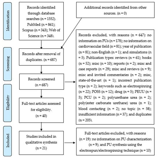
Figure 1.
Preferred Reporting Items for Systematic Reviews and Meta-Analyses (PRISMA) flow diagram.
The searches in PubMed, Scopus, and Web of Science yielded 861, 343, and 348 articles, respectively. In total, 647 articles were excluded after being managed with Rayyan QCRI [6]; 40 full-text articles were assessed for eligibility.
The reasons for the excluded records (n = 647) were: duplicates (n = 205); no information on PU (n = 178); no information on cardiovascular field (n = 81); year of publication (n = 81); no topic (n = 58); insufficient information (n = 37); Publication types: reviews (n = 61); books (n = 52); misc (n = 10); misc and case reports (n = 29); misc and reviews (n = 9); misc and invited commentaries (n = 2); report (n = 2); incorrect publication type (n = 2); misc, state of the art (n = 1); keywords such as electrospinning (n = 22); POSS (n = 12); drug (n = 5); PEUU (n = 5); PCU (n = 2); polyurethane urea (n = 2); blood-contacting (n = 2); poly(ester carbonate urethane) urea (n = 1); non-English (n = 1); and simulations (n = 5).
After a full-text revision, 19 articles were excluded owing to the following reasons: 9 did not contain information on PU characterization and 10 presented PU syntheses using the electrospun or electrospinning technique. A total of 21 papers were included in the analysis.
3.2. Characteristics and Results of Included Studies
The following data were tabulated from the included articles: author, year of publication, geographic setting, PU type or chemical composition, type of study (in vitro or in vivo) or type of cells, field of application, and main results (Table 1).

Table 1.
Characteristics and results of Included Studies.
Four of the studies were conducted in the USA [7,8,9,10], three in Italy [11,12,13], two each in India [14,15] and Iran [16,17], and one each in Australia [18], Canada [19], Colombia [20], Germany [3], South Korea [21], and Taiwan [22]. The collaborative studies that were conducted are as follows: Australia–France [23], Brazil–Germany [2]; and People’s Republic of China–Canada [24,25].
Five of the studies used commercial PUs: Elastogran [3], Elastollan® [14], ChronoFlex® [8], Elast-Eon™ [18], and NovoSorb™ [23]. Three of the studies used medical-grade PUs: Tecoflex® [10]; Tecothane™ [7,9].
Seventeen studies conducted in vivo tests with NIH/3T3 mouse fibroblast cell line [11], human umbilical vein endothelial cell (HUVEC) [2,14,16,23,25], L-929 fibroblast [16,20,22]; 3T3 [18,20], H9C2 cardiomyoblasts [12], endothelial colony forming cells (ECFCs) [3], smooth muscle cells (SMCs) [15], A10 SMCs [19], ECs [15], human microvascular endothelial cells (HMEC) [21], cardiomyocytes [13], or hBMSCs [17]. Two studies performed both in vivo and in vitro tests [7,9]. One study conducted only an in vivo test [24]. Two studies conducted neither in vivo nor in vitro tests [8,10]. The studies included in the analysis recommend the use of PUs in cardiovascular devices [22,24], hemodialysis [22], biomedical applications [18,20], blood-contacting devices [3,14], cardiovascular stents [8,15,17,23], heart valves [7,9], heart patches [12], tissue engineering [19,25], and vascular prostheses [21].
All the included studies presented modifications made to PUs with the aim of improving their properties. Six studies presented variations of the chemical compositions of PUs [11,15,16,17,20,24]. Five studies presented modifications to the material surface [7,9,14,21,23] and ten studies described different techniques to prepare PUs [2,3,8,10,12,13,18,19,22,25].
This study had limitations. We only used three databases (PubMed, Scopus, and Web of Science) and only articles in English were screened. The systematic review included articles that represent a small fraction of the literature on synthesis techniques for application in cardiovascular devices.
4. Discussion
PUs have found applications in biomedical field because of their properties, i.e., capability of sustaining the contractile forces that originate during the cardiac cycle without undergoing plastic deformation or failure [12], and the capability to imitate the behavior of different tissues [15,20,26,27,28]. The applications of PUs in this field include biostable implants [20] and cardiovascular implantable devices such as cardiac pacemakers [11,18,20], catheters [11,18], prostheses [11,21,29], cardiac assist devices [11], heart patches [12,16], heart valves [11,30] stent [8,15,17,23], and vascular grafts [11,18,31,32].
PUs are thermoplastic elastomers polymers that can be defined as segmented block copolymers, characterized by the presence of two micro-separated phases: soft and hard [11]. This morphology is related to their mechanical properties [8,11,29,33], including their high tensile strength [11], fatigue resistance [34], and elasticity [34]; good tear and abrasion resistance [11]; biodegradability [11]; blood compatibility [34,35]; and biocompatibility [8,11,29,33].
However, studies reported that PUs show severe problems [22] when applied in blood-contacting devices, such as the biodegradation that occurs during long-term implantation [29] by the adhesion of inflammatory cells [27], surface-induced thrombosis [2,22], and protein fouling [22] (which are known to participate in PU biodegradation); and the absence of endothelialization [2].
As a solution to this problem, PUs can be modified in terms of both their chemical composition [11,15,16,17,20,29,36] and surface functionalization to improve their mechanical properties, blood compatibility, and adhesion to ECs, and to reduce protein adhesion [2,3,7,18,24,32,36,37,38,39,40].
4.1. PU Modification in Terms of Chemical Composition
Our analysis revealed the use of silicone [11], PCL [12,16,17,19], PCL/PEG [12], castor oil (CO)/aliphatic diisocyanates [20], AgNO3, and carbon [15] to modify the chemical compositions.
4.1.1. Silicone
Silicone presents properties such as low surface energy [11]; hemocompatibility [11]; low toxicity [11,29]; remarkable thermal, oxidative, and hydrolytic stability [11]; high flexibility [11]; good biocompatibility [11,29,41]; and long-term biostability [29,41].
4.1.2. PCL
We have identified PCL as a bioresorbable and biocompatible polymer with good mechanical properties [12,16]. PU/PCL blends (copolymer and homopolymer) are well known for their low degradability, cell adhesion, and proliferation, which indicate good biocompatibility. They have been used as materials with elastic memory to achieve self-expansion within the range of the body temperature. Silvestri et al. illustrated that the shape recovery of PU/PCL blends is related to the elastic strain generated during deformation owing to the elasticity of the PU as a matrix phase in these blends [16]. Specifically, PU/PCL blends and PCL/PEG/PCL tri-blocks with different aliphatic and amino-acid-based chain extenders did not present toxicity. The authors recommend the use of the blend for tissue engineering (TE) applications, considering that their mechanical behavior and cell response depend on their chemical composition [12].
4.1.3. CO
A variation of the PU chemical composition with a CO/aliphatic diisocyanates blend is recommended in the included articles. Arévalo et al. used CO as the polyol because of its composition (ricinoleic acid), which presents a structure that enables the synthesis of cross-linked urethanes. Furthermore, it is a renewable source and has low toxicity. The CO/aliphatic diisocyanates (isophorone diisocyanate, IPDI) blend can be used to synthesize biomedical PUs because they do not promote the generation of carcinogenic products, such as the aromatic diamines, in in vivo conditions [20].
4.1.4. Nanomaterial Carbon Dot–Silver Nitrate
Meanwhile, Dura et al. developed a material incorporating a biocompatible nanomaterial carbon dot–silver nitrate (CD-Ag) in a smart polymer matrix for potential use as a stent. The study indicated that they attained a faster self-expansion of the material (in less than a minute), which prevented the migration of the device during in vivo deployment. The authors propose the use of both silver nitrate owing to its potent antibacterial activity, which prevents biofilm formation; and carbon dots owing to their highly polar peripheral groups, which enhance the mechanical properties of the nanocomposite [15].
4.2. PU Modification in Terms of Surface Functionalization
Ten studies described how the biomaterials interact with blood. Implantation begins with the blood–foreign material interaction [22,34].
At this stage, the plasma proteins are adsorbed onto the material surfaces, which occurs within a few seconds. This adsorption causes the adhesion of platelets, white blood cells, and a few red blood cells onto the plasma protein layer [34]. Yu et al. revealed that the aggregated platelets on the surface release materials such as adenosine diphosphate (ADP), which results in the formation of thrombin, an insoluble fibrin network, or thrombus [14,22]. Raut et al. explained that the thrombus may obstruct the blood flow and cause device failure. In addition, clots may be released into the systemic blood circulation from the devices that do not fail, resulting in an embolism [14]. Furthermore, Liu et al. highlighted that after the implantation, monocytes may be activated after adhesion to the biomaterials and release cytotoxic mediators, such as cytokines and reactive oxygen species (ROS) [34]. Monocytes have been recognized for their essential role in mediating inflammatory responses [37]. Du et al. described how thrombi that form inside a catheter lumen make the catheter unsuitable for biomedical uses, such as withdrawal of blood, delivery of fluids, or medication. Thrombi, which form outside the catheter, could permanently damage the vessel integrity, resulting in pain and swelling [42].
4.2.1. Surface Functionalization-Modified PU to Promote EC Adhesion and Proliferation
Surface coverage with ECs prevents the material from directly contacting with blood and can be considered as a solution for preventing thrombus formation in cardiovascular implants [2]. ECs are known to provide an antithrombogenic surface by producing antithrombogenic substances [40], such as prostacyclin (PGI2), tissue plasminogen activator (t-PA), heparin-like glycosaminoglycans, and thrombomodulin [21]. The use of ECM proteins such as Coll and Fn as coatings on synthetic polymers enhances endothelialization [23].
Stachelek et al. changed the PU formulation by including cholesterol (Chol). Cholesterol-modified polyurethane (PU-Chol) increased the adhesion of blood outgrowth endothelial cells (BOECs) compared with controls. This change in the adhesion rate is important because it is known that BOECs are an outgrowth of a circulating progenitor cell. These are present in peripheral blood, and thus represent an important potential source of autologous cells for seeding investigations [9].
Heparinized surfaces are clinically used to reduce thrombogenicity [36] as potent anticoagulants that interact strongly with antithrombin (AT) to prevent the formation of fibrin clots [39]. The presence of heparin on the surface positively affects EC growth and proliferation by binding and stabilizing cell growth factors (GFs) [40]. However, a disadvantage of immobilizing heparin onto a polymer surface is that the heparin has low bioactivity [38]. Klement et al. recommended the modification of a PU with an antithrombin–heparin (ATH) covalent complex, which displays the capability for rapid direct inhibition of thrombin [39].
A PU nanocomposite (polymer matrix with embedded nanoparticles of Au-Pt) is a PU modification that has been recommended by Hess et al. to improve cell adhesion, which are functions of the concentration of nanoparticles [3].
The surface functionalization of biomedical devices could be attained by changing the material topographies to enhance the adhesion and growth of ECs [2]. Our results indicate the use of different techniques to enhance surface functionalization, such as direct laser ablation technique (DLA) [2], polymeric endoaortic paving (PEAP) [10], pulsed laser ablation in liquid (PLAL) [3], thermally induced phase separation (TIPS) [13], and freeze-drying [25].
DLA is a technique that modifies the surface topography [2]. Cortella et al. showed the functionalization of PU by DLA-created microtopography, which improved the adhesion and proliferation rate of ECs [2].
Ashton et al. recommends an alternative method to enhance conventional endoaortic therapy to reduce the risk of endoleak. PEAP is a process where a polymer is endovascularly delivered and thermoformed to coat or pave the lumen of the aorta [10]. Meanwhile, the PLAL technique for solid targets is advantageous for the synthesis of biocompatible nanomaterials, because it generates nanoparticles without the need for chemical precursors, which potentially cause cell behavior side effects. Furthermore, the technique enables in situ functionalization with biomolecules and adsorption onto microparticle surfaces [3].
Vozzi et al. recommend the use of TIPS to fabricate oriented scaffolds. This technique modified the PU porosity by modulating the polymer concentration, quenching temperature, thermal gradient, and solvent type [13]. The authors indicate that fiber alignment supports cardiomyocytes in the generation of a tissue-like structure [13]. The last technique identified by us for enhancing surface functionalization was freeze-drying. Jiang et al. employed freeze-drying to develop 3-D porous scaffolds based on the prepared waterborne biodegradable PUs. With this technique, it is possible to obtain scaffolds with an appropriate pore diameter and distribution to enhance the adhesion and proliferation of ECs [25].
4.2.2. Use of Surface Functionalization-Modified PU to Enhance Biocompatibility, Bioactivity, Biodegradation Resistance, and Electrical Conductivity
Thampi et al. and Cortella et al. used surface functionalization with antithrombotic agents or immobilization of molecules, such as polyethylene oxide (PEO), heparin, albumin, and chitosan (CS), to improve the blood compatibility of biomedical devices [2,43]. Roth et al. and Yu et al. recommended the application of biologically active substances, such as anticoagulants [14], fibrinolytic enzymes or proteins [14], heparin [22], CS [22], and dextran sulfate (DS) [22], as surface modification substances [14,22] in order to enhance the compatibility of PUs with the blood components.
Another PU modification technique was presented by Gu et al. through the incorporation of PEG to enhance bioactivity. PEG chains were selected as hydrophilic and flexible spacers between the biological molecules and PU backbones [24]. Hess et al. recommended a PU modification with embedded nanoparticles of Au-Pt to improve biocompatibility, which are functions of the concentrations of nanoparticles [3]. Stachelek et al. recommended the use of modified PU with DBP to provide antioxidant activity to prevent biodegradation. The microscopy results showed that DBP-modified PU confers biodegradation resistance to surface cracking with dose-dependent DBP loading [7].
A different PU modification was presented for electrically conductive polymeric materials. These materials could be used in biomedical applications, such as for biosensors, drug delivery systems, biomedical implants, and TE. Kaur et al. recommended the incorporation of conductive fillers such as graphene to enhance the electrical conductivity without changing the polymeric characteristics [18].
5. Conclusions
PUs have found applications in medical devices because of their properties—their capability to tolerate contractile forces that originate during the cardiac cycle without undergoing plastic deformation or failure, and their ability to imitate the behaviors of different tissues. Although studies have reported that PUs suffer from severe problems when applied in blood-contacting devices that are implanted for long periods, they can be modified in terms of both the chemical composition and surface characteristics to improve their mechanical properties (blood compatibility and EC adhesion) and reduce protein adhesion. These modifications enable the use of PUs in the manufacture and design of cardiovascular devices.
Our analysis revealed the following:
- For stent design, it is important that the selected material displays self-expandable and shape memory behavior, which must be maintained at temperatures similar to those encountered in the human body. In addition, this expansion must be carried out as quickly as possible to prevent the migration of the stent during surgery. Modified PUs, such as those with added PCL or carbon dot–silver nanohybrid, show high modulus and tensile strength with low elongation and biocompatibility. Furthermore, these maintain self-expandable and shape memory behaviors. All these properties permit us to propose the use of these PUs as potential materials for stent implants.
- Among cardiovascular devices, heart patches and heart valves require the use of materials with appropriate properties such as strength and an elastomeric mechanical behavior to tolerate the contractile cardiac tissue and support its regeneration. For the design of heart patches and heart valves, it is important to create a structure that is similar to the muscle tissue. PUs are appropriate materials for cardiac applications because their biocompatibility and elastomeric behavior enable them to resist the cyclic heart stresses without deformation or failure. The PU structure can be modified to create anisotropic microstructures that may mimic the heart tissue function of different pore sizes. This could promote cell colonization, cell migration, nutrient supply, and vascularization.
- In general, for blood-contacting devices, the interactions between the material and blood generate cell responses that could favor the formation of thrombi. PUs provide a surface that can be modified to reduce non-specific protein adsorption and promote endothelial cell attachment and proliferation. This can enhance the biocompatibility and hemocompatibility. This implies that the fabrication of blood-contacting devices with modified PUs could decrease the numbers of repeated operations and deaths in cardiovascular surgery [3].
Author Contributions
Conceptualization, review, writing, and original draft preparation, K.N.-G.; review and editing, M.F.V. All authors have read and agreed to the published version of the manuscript.
Funding
We wish to thank the Universidad de La Sabana for financing the ING-202-2018 research project and MINCIENCIAS for the postdoctoral fellowships 811-2018.
Acknowledgments
Sincere thanks to Luis Eduardo Díaz-Barrera and Said Arévalo-Alquichire for their essential contributions to the article selection procedures.
Conflicts of Interest
The authors declare no conflict of interest.
Abbreviations
| AAK | alanine-alanine-lysine |
| ADP | adenosine diphosphate |
| AT | antithrombin |
| BD | 1,4-butane diol |
| BDI | 1,4-diisocyanatobutane |
| BDO | 1,4-butanediol |
| BOECs | blood outgrowth endothelial cells |
| Chol | cholesterol |
| CO | castor oil |
| Coll | collagen |
| CS | chitosan |
| DBP | di-tert-butylphenol |
| DBTDL | di-butyltindilaurate |
| DLA | direct laser ablation technique |
| D-PHI | elastomeric degradable/polar/hydrophobic/ionic |
| DS | dextran sulfate |
| ECFCs | endothelial colony forming cells |
| ECM | extracellular matrix |
| ECs | endothelial cells |
| Fn | fibronectin |
| GFs | growth factors |
| hBMSCs | human bone marrow mesenchymal stem cells |
| HDI | 1,6-diisocyanatohexane |
| HMEC | human microvascular endothelial cells |
| HPF | human plasma fibrinogen |
| HS | hard segment |
| HUVEC | human umbilical vein endothelial cell |
| IPDI | isophorone diisocyanate |
| LBL | layer-by-layer |
| MDI | 4,4′-methylenediphenyl diisocyanate |
| MDMs | monocyte-derived macrophages |
| MI | myocardial infarction |
| MISC | miscellaneous |
| PCL | polycaprolactone |
| PCU | poly(carbonate urethane) |
| PDMS | poly(dimethyl siloxane) |
| PEAP | polymeric endoaortic paving. |
| PEG | poly (ethylene glycol) |
| PEMs | polyelectrolyte multilayers |
| PEO | polyethylene oxide |
| PEUU | poly(etherurethane urea) |
| PLAL | pulsed laser ablation in liquid |
| PN | poly (ethylene glycol) bis(amine) |
| PO | poly (ethylene glycol) diglycidyl ether |
| POSS | polyhedral oligomeric silesquioxanes |
| PTFE | polytetrafluoroethylene |
| PTMEG | poly (oxytetramethylene) glycol |
| PTMO | poly (tetramethylene oxide) |
| PU | polyurethane |
| ROS | reactive oxygen species |
| SD | Sprague-Dawley |
| SMAs | shape memory alloys |
| SMCs | smooth muscle cells |
| SPEU | semi-microporous segmented polyurethane |
| TDI | 2, 4-2, 6-toluene diisocyanate |
| TE | tissue engineering |
| THP-1 | a human cell line |
| TIPS | thermally-induced phase separation |
| TPU | thermoplastic polyurethane |
| WBPU | biodegradable waterborne polyurethane |
References
- Szycher, M. Szycher’s Handbook of Polyurethanes, 2nd ed.; CRC Press: Boca Raton, FL, USA, 2013; ISBN 978-1-4398-6313-8. [Google Scholar]
- Cortella, L.R.X.; Cestari, I.A.; Guenther, D.; Lasagni, A.F.; Cestari, I.N. Endothelial cell responses to castor oil-based polyurethane substrates functionalized by direct laser ablation. Biomed. Mater. 2017, 12, 065010. [Google Scholar] [CrossRef] [PubMed]
- Hess, C.; Schwenke, A.; Wagener, P.; Franzka, S.; Laszlo Sajti, C.; Pflaum, M.; Wiegmann, B.; Haverich, A.; Barcikowski, S. Dose-dependent surface endothelialization and biocompatibility of polyurethane noble metal nanocomposites. J. Biomed. Mater. Res. Part A 2014, 102, 1909–1920. [Google Scholar] [CrossRef] [PubMed]
- Moher, D.; Liberati, A.; Tetzlaff, J.; Altman, D. The PRISMA Group Preferred reporting items for systematic reviews and meta-analyses: The PRISMA statement. PLoS Med. 2009, 6. [Google Scholar] [CrossRef] [PubMed]
- Hulley, S.; Cummings, S.; Browner, W.; Grady, D.; Newman, T. Designing Clinical Research: Fourth Edition; Lippincott Williams & Wilkins, a Wolters Kluwer Business: Philadelphia, PA, USA, 2013; ISBN 978-1-60831-804-9. [Google Scholar]
- Ouzzani, M.; Hammady, H.; Fedorowicz, Z.; Elmagarmid, A. Rayyan-a web and mobile app for systematic reviews. Syst. Rev. 2016, 5, 210. [Google Scholar] [CrossRef] [PubMed]
- Stachelek, S.J.; Alferiev, I.; Fulmer, J.; Ischiropouios, H.; Levy, R.J. Biological stability of polyurethane modified with covalent attachment of di-tert-butyl-phenol. J. Biomed. Mater. Res. Part A 2007, 82, 1004–1011. [Google Scholar] [CrossRef] [PubMed]
- Trigwell, S.; De, S.; Sharma, R.; Mazumder, M.K.; Mehta, J.L. Structural evaluation of radially expandable cardiovascular stents encased in a polyurethane film. J. Biomed. Mater. Res. Part B Appl. Biomater. 2006. [Google Scholar] [CrossRef]
- Stachelek, S.J.; Alferiev, I.; Connolly, J.M.; Sacks, M.; Hebbel, R.P.; Bianco, R.; Levy, R.J. Cholesterol-modified polyurethane valve cusps demonstrate blood outgrowth endothelial cell adhesion post-seeding in vitro and in vivo. Ann. Thorac. Surg. 2006. [Google Scholar] [CrossRef]
- Ashton, J.H.; Mertz, J.A.M.; Harper, J.L.; Slepian, M.J.; Mills, J.L.; McGrath, D.V.; Vande Geest, J.P. Polymeric endoaortic paving: Mechanical, thermoforming, and degradation properties of polycaprolactone/polyurethane blends for cardiovascular applications. Acta Biomater. 2011, 7, 287–294. [Google Scholar] [CrossRef]
- Silvestri, A.; Serafini, P.M.; Sartori, S.; Ferrando, P.; Boccafoschi, F.; Milione, S.; Conzatti, L.; Ciardelli, G. Polyurethane-based biomaterials for shape-adjustable cardiovascular devices. J. Appl. Polym. Sci. 2011, 122, 3661–3671. [Google Scholar] [CrossRef]
- Silvestri, A.; Sartori, S.; Boffito, M.; Mattu, C.; Di Rienzo, A.M.; Boccafoschi, F.; Ciardelli, G. Biomimetic myocardial patches fabricated with poly(ɛ-caprolactone) and polyethylene glycol-based polyurethanes. J. Biomed. Mater. Res. Part B Appl. Biomater. 2014, 102, 1002–1013. [Google Scholar] [CrossRef]
- Vozzi, F.; Logrand, F.; Cabiati, M.; Cicione, C.; Boffito, M.; Carmagnola, I.; Vitale, N.; Gori, M.; Brancaccio, M.; Del Ry, S.; et al. Biomimetic engineering of the cardiac tissue through processing, functionalization, and biological characterization of polyesterurethanes. Biomed. Mater. 2018, 13, 055006. [Google Scholar] [CrossRef] [PubMed]
- Raut, P.W.; Shitole, A.A.; Khandwekar, A.; Sharma, N. Engineering biomimetic polyurethane using polyethylene glycol and gelatin for blood-contacting applications. J. Mater. Sci. 2019, 54, 10457–10472. [Google Scholar] [CrossRef]
- Duarah, R.; Singh, Y.P.; Gupta, P.; Mandal, B.B.; Karak, N. High performance bio-based hyperbranched polyurethane/carbon dot-silver nanocomposite: A rapid self-expandable stent. Biofabrication 2016, 8, 045013. [Google Scholar] [CrossRef] [PubMed]
- Baheiraei, N.; Yeganeh, H.; Ai, J.; Gharibi, R.; Azami, M.; Faghihi, F. Synthesis, characterization and antioxidant activity of a novel electroactive and biodegradable polyurethane for cardiac tissue engineering application. Mater. Sci. Eng. C 2014, 44, 24–37. [Google Scholar] [CrossRef] [PubMed]
- Ajili, S.H.; Ebrahimi, N.G.; Soleimani, M. Polyurethane/polycaprolactane blend with shape memory effect as a proposed material for cardiovascular implants. Acta Biomater. 2009, 5, 1519–1530. [Google Scholar] [CrossRef]
- Kaur, G.; Adhikari, R.; Cass, P.; Bown, M.; Evans, M.D.M.; Vashi, A.V.; Gunatillake, P. Graphene/polyurethane composites: Fabrication and evaluation of electrical conductivity, mechanical properties and cell viability. RSC Adv. 2015, 5, 98762–98772. [Google Scholar] [CrossRef]
- Sharifpoor, S.; Simmons, C.A.; Labow, R.S.; Santerre, J.P. A study of vascular smooth muscle cell function under cyclic mechanical loading in a polyurethane scaffold with optimized porosity. Acta Biomater. 2010, 6, 4218–4228. [Google Scholar] [CrossRef]
- Arévalo, F.; Uscategui, Y.L.; Diaz, L.; Cobo, M.; Valero, M.F. Effect of the incorporation of chitosan on the physico-chemical, mechanical properties and biological activity on a mixture of polycaprolactone and polyurethanes obtained from castor oil. J. Biomater. Appl. 2016, 31, 708–720. [Google Scholar] [CrossRef]
- Cho, E.H.; Yang, Y.I.; Mun, C.W.; Kim, J.K. Tissue-engineered semi-microporous segmented polyetherurethane vascular prostheses. J. Biomater. Sci. Polym. Ed. 2005, 16, 775–790. [Google Scholar] [CrossRef]
- Yu, D.-G.; Lin, W.-C.; Lin, C.-H.; Yeh, Y.-H.; Yang, M.-C. Construction of antithrombogenic polyelectrolyte multilayer on thermoplastic polyurethane via layer-by-layer self-assembly technique. J. Biomed. Mater. Res. Part B Appl. Biomater. 2007, 83, 105–113. [Google Scholar] [CrossRef]
- Sgarioto, M.; Adhikari, R.; Gunatillake, P.A.; Moore, T.; Malherbe, F.; Nagel, M.-D.; Patterson, J. Properties and in vitro evaluation of high modulus biodegradable polyurethanes for applications in cardiovascular stents. J. Biomed. Mater. Res. Part B Appl. Biomater. 2014, 102, 1711–1719. [Google Scholar] [CrossRef] [PubMed]
- Gu, Y.; Sun, F.; Xie, X.; Wu, X.; Zhang, Z.; Guidoin, R.; Fu, Q.; Zhong, Y.; Zhao, C. Prenatal developmental safety of functional polyurethanes for cardiovascular implants. J. Biomed. Mater. Res. Part B Appl. Biomater. 2016, 104, 606–614. [Google Scholar] [CrossRef] [PubMed]
- Jiang, X.; Yu, F.; Wang, Z.; Li, J.; Tan, H.; Ding, M.; Fu, Q. Fabrication and characterization of waterborne biodegradable polyurethanes 3-dimensional porous scaffolds for vascular tissue engineering. J. Biomater. Sci. Polym. Ed. 2010, 21, 1637–1652. [Google Scholar] [CrossRef] [PubMed]
- Alperin, C.; Zandstra, P.W.; Woodhouse, K.A. Polyurethane films seeded with embryonic stem cell-derived cardiomyocytes for use in cardiac tissue engineering applications. Biomaterials 2005, 26, 7377–7386. [Google Scholar] [CrossRef] [PubMed]
- Grenier, S.; Sandig, M.; Mequanint, K. Polyurethane biomaterials for fabricating 3D porous scaffolds and supporting vascular cells. J. Biomed. Mater. Res. Part A 2007, 82, 802–809. [Google Scholar] [CrossRef] [PubMed]
- Herrmann, F.E.M.; Lehner, A.; Hollweck, T.; Haas, U.; Fano, C.; Fehrenbach, D.; Kozlik-Feldmann, R.; Wintermantel, E.; Eissner, G.; Hagl, C.; et al. In vitro biological and mechanical evaluation of various scaffold materials for myocardial tissue engineering. J. Biomed. Mater. Res. Part A 2014, 102, 958–966. [Google Scholar] [CrossRef]
- Briganti, E.; Losi, P.; Raffi, A.; Scoccianti, M.; Munaò, A.; Soldani, G. Silicone based polyurethane materials: A promising biocompatible elastomeric formulation for cardiovascular applications. J. Mater. Sci. Mater. Med. 2006, 17, 259–266. [Google Scholar] [CrossRef]
- Thierfelder, N.; Koenig, F.; Bombien, R.; Fano, C.; Reichart, B.; Wintermantel, E.; Schmitz, C.; Akra, B. In vitro comparison of novel polyurethane aortic valves and homografts after seeding and conditioning. ASAIO J. 2013. [Google Scholar] [CrossRef]
- Bergmeister, H.; Seyidova, N.; Schreiber, C.; Strobl, M.; Grasl, C.; Walter, I.; Messner, B.; Baudis, S.; Fröhlich, S.; Marchetti-Deschmann, M.; et al. Biodegradable, thermoplastic polyurethane grafts for small diameter vascular replacements. Acta Biomater. 2015, 11, 104–113. [Google Scholar] [CrossRef]
- Hu, Z.J.; Li, Z.L.; Hu, L.Y.; He, W.; Liu, R.M.; Qin, Y.S.; Wang, S.M. The in vivo performance of small-caliber nanofibrous polyurethane vascular grafts. BMC Cardiovasc. Disord. 2012, 12, 115. [Google Scholar] [CrossRef]
- Lehle, K.; Stock, M.; Schmid, T.; Schopka, S.; Straub, R.H.; Schmid, C. Cell-type specific evaluation of biocompatibility of commercially available polyurethanes. J. Biomed. Mater. Res. Part B Appl. Biomater. 2009, 90, 312–318. [Google Scholar] [CrossRef] [PubMed]
- Liu, X.; Xue, Y.; Sun, J. Indirect induction of endothelial cell injury by PU- or PTFE-mediated activation of monocytes. J. Biomater. Sci. Polym. Ed. 2010, 21, 1783–1797. [Google Scholar] [CrossRef]
- Ye, S.H.; Hong, Y.; Sakaguchi, H.; Shankarraman, V.; Luketich, S.K.; DAmore, A.; Wagner, W.R. Nonthrombogenic, biodegradable elastomeric polyurethanes with variable sulfobetaine content. ACS Appl. Mater. Interfaces 2014, 6, 22796–22806. [Google Scholar] [CrossRef]
- Bezuidenhout, D.; Davies, N.; Black, M.; Schmidt, C.; Oosthuysen, A.; Zilla, P. Covalent surface heparinization potentiates porous polyurethane scaffold vascularization. J. Biomater. Appl. 2010, 24, 401–418. [Google Scholar] [CrossRef] [PubMed]
- Xue, Y.; Liu, X.; Sun, J. PU/PTFE-stimulated monocyte-derived soluble factors induced inflammatory activation in endothelial cells. Toxicol. Vitr. 2010, 24, 404–410. [Google Scholar] [CrossRef] [PubMed]
- Boffito, M.; Di Meglio, F.; Mozetic, P.; Giannitelli, S.M.; Carmagnola, I.; Castaldo, C.; Nurzynska, D.; Sacco, A.M.; Miraglia, R.; Montagnani, S.; et al. Surface functionalization of polyurethane scaffolds mimicking the myocardial microenvironment to support cardiac primitive cells. PLoS ONE 2018, 13, e199896. [Google Scholar] [CrossRef]
- Klement, P.; Du, Y.J.; Berry, L.R.; Tressel, P.; Chan, A.K.C. Chronic performance of polyurethane catheters covalently coated with ATH complex: A rabbit jugular vein model. Biomaterials 2006, 27, 5107–5117. [Google Scholar] [CrossRef]
- Davoudi, P.; Assadpour, S.; Derakhshan, M.A.; Ai, J.; Solouk, A.; Ghanbari, H. Biomimetic modification of polyurethane-based nanofibrous vascular grafts: A promising approach towards stable endothelial lining. Mater. Sci. Eng. C 2017, 80, 213–221. [Google Scholar] [CrossRef]
- Spiller, D.; Mirtelli, C.; Losi, P.; Briganti, E.; Sbrana, S.; Counoupas, C.; Kull, S.; Tonlorenzi, S.; Soldani, G. In vitro evaluation of the PEtU-PDMS material immunocompatibility: The influence of surface topography and PDMS content. J. Mater. Sci. Mater. Med. 2009, 20, 2511–2520. [Google Scholar] [CrossRef]
- Du, Y.J.; Klement, P.; Berry, L.R.; Tressel, P.; Chan, A.K.C. In vivo rabbit acute model tests of polyurethane catheters coated with a novel antithrombin-heparin covalent complex. Thromb. Haemost. 2005, 94, 366–372. [Google Scholar] [CrossRef]
- Thampi, S.; Nandkumar, A.M.; Muthuvijayan, V.; Parameswaran, R. Differential Adhesive and Bioactive Properties of the Polymeric Surface Coated with Graphene Oxide Thin Film. ACS Appl. Mater. Interfaces 2017, 9, 4498–4508. [Google Scholar] [CrossRef] [PubMed]
© 2020 by the authors. Licensee MDPI, Basel, Switzerland. This article is an open access article distributed under the terms and conditions of the Creative Commons Attribution (CC BY) license (http://creativecommons.org/licenses/by/4.0/).