Case presentation
An 88-year-old man with a history of transient ischaemic attack, chronic kidney disease and primary hypertension presented with dyspnoea and was found to have large bilateral pulmonary emboli on a computed-tomography angiogram of the chest. A deep venous thrombosis (DVT) in the left lower extremity involving the popliteal, peroneal and gastrocnemius veins was found on venous duplex ultrasound. A transthoracic echocardiogram showed a thrombus in the right atrium, and a long thrombus in the left atrium extending via the left ventricle through the aortic valve (video 1) (You will find the video files in the multimedia collection of «Cardiovascular Medicine»: https://cardiovascmed.ch/online-only-content). A transoesophageal echocardiogram (TEE) showed a large thrombus entrapped in a patent foramen ovale (PFO) (video 2, Figure 1) with further extension of the same thrombus via the left ventricle through the aortic valve (videos 3 and 4, Figure 2 and Figure 3). Three-dimensional imaging taken during the TEE showed the entrapped thrombus passing through the PFO into the left atrium (Figure 4), and then through the mitral valve into the left ventricle (Figure 5 and Figure 6).
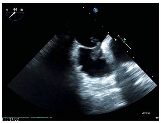
Figure 1.
Bicaval view showing a large thrombus entrapped in a patent formen ovale.
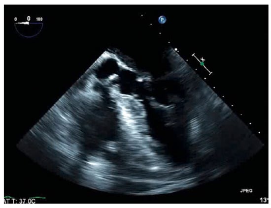
Figure 2.
Five-chamber view showing extension of the entrapped thrombus into the left ventricle.
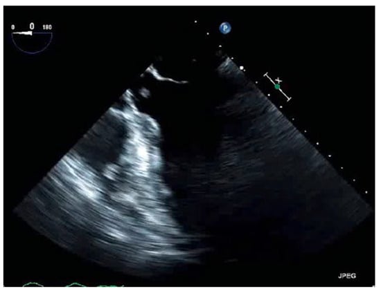
Figure 3.
Five-chamber view captured at the exact moment a piece of thrombus is about to pass through the aortic valve.
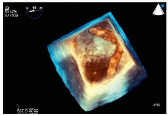
Figure 4.
3D view showing the thrombus passing through the patent foramen ovale into the left atrium.
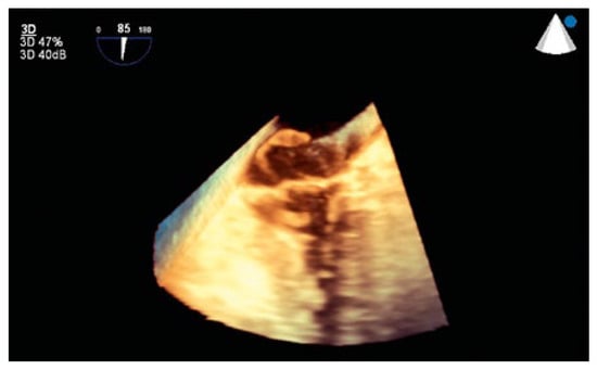
Figure 5.
3D view showing passage of the thrombus through the mitral valve.
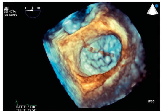
Figure 6.
3D view showing passage of the thrombus through the mitral valve.
The patient was not deemed a surgical candidate, given his multiple comorbidities and high risk of further embolisation. As there was no previous personal or family history of venous thromboembolism, congenital thrombophilia was not suspected. In the light of normal blood counts, a myeloproliferative disorder was not suspected. Further imaging studies for malignancy were all negative. The DVT was deemed to be unprovoked and, based on the extent of the thrombosis, the decision was made to start lifelong anticoagulation. He was treated initially with a heparin infusion for 5 days, then transitioned to apixaban. He remained stable during a 7-day hospital stay. He was seen in clinic 3 months after discharge and has done well with no noted complaints or signs of embolism.
Discussion
Evidence from autopsy reports suggest up to 35% of the population may possess a PFO [1]. We describe a case of an elderly gentleman with a DVT, bilateral pulmonary emboli and a large thrombus extending from the right atrium through the aortic valve via a PFO. There is no guideline or expert consensus on the best way to treat such a condition. A literature review of reported cases of thrombus entrapped in a PFO suggested either surgery or anticoagulation as treatment [2,3]. Surgery was the more popular option, and anticoagulation was preferred in cases where surgery was considered high risk [2,3]. Anticoagulation has been effective for our patient to date. Owing to the paucity of available data, long-term treatment for a thrombus entrapped in a PFO is unknown. The risk of a future embolus across the PFO and benefit of closure with this type of presentation are also unknown. Further research is necessary to answer these questions.
Mailing Address 945 Linden Line USA-Toledo OH 43615.
Acknowledgments
The patient gave written informed consent for publication.
Disclosure statement
No financial support and no other potential conflict of interest relevant to this article was reported.
References
- Wilmshurst, P.T.; de Belder, M.A. Patent foramen ovale in adult life. Br Heart J. 1994, 71, 209–212. [Google Scholar] [CrossRef] [PubMed]
- Baydoun, H.; Barakat, I.; Hatem, E.; et al. Thrombus in transit through patent foramen ovale. Case reports in cardiology 2013, 2013, 395879. [Google Scholar] [CrossRef] [PubMed]
- Fauveau, E.; Cohen, A.; Bonnet, N.; et al. Surgical or medical treatment for thrombus straddling the patent foramen ovale: Impending paradoxical embolism? Report of four clinical cases and literature review. Arch Cardiovasc Dis. 2008, 101, 637–644. [Google Scholar] [CrossRef] [PubMed]
© 2018 by the author. Attribution - Non-Commercial - NoDerivatives 4.0.