Summary
This case report describes a cause of precordial voltage loss with normal voltage in the peripheral leads in patients with obesity and/or temporal causes of increased intra-abdominal pressure. Important differential diagnoses were excluded with blood analyses, ECG and echocardiography. By placing the ECG-electrodes one or two intercostal spaces cranially we were able to detect the origin of the voltage loss. This case demonstrates that the standard position of the precordial ECG leads might not always correspond to the true anatomical position of the heart. If the probability of a cardiac or pericardial disease is excluded, this method might be sufficient as a basic diagnostic tool.
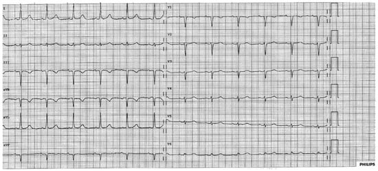
A 68-year-old, obese (body mass index 27 kg/m2) woman without prior cardiac history was admitted for an elective laparoscopic cholecystectomy. One hour after she was transferred to the intermediate care unit, she complained of chest pain and had a systolic blood pressure of >200 mm Hg. Physical examination was normal and the ECG (Figure 1) demonstrated low voltage in the precordial leads but normal voltage in the peripheral leads.

Figure 1.
A: Low voltage in the precordial leads with ECG electrodes positioned in the ordinary manner.
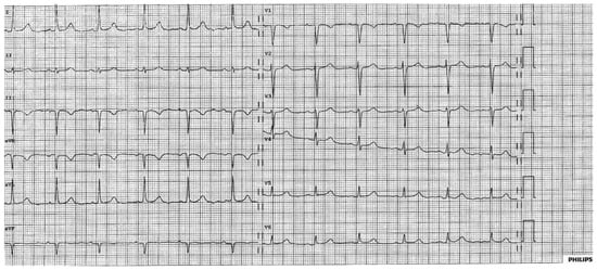
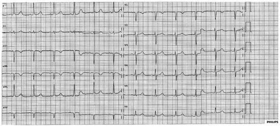
High-sensitivity troponin was slightly elevated at 0.024 µg/l (<0.014 µg/l) but showed no dynamic changes. Her only cardiovascular risk factor was hypertension. No ECG had been performed preoperatively. Echocardiography was normal, with a left ventricular ejection fraction of 60% and without left ventricular hypertrophy. Right ventricular function was normal and she had no pulmonary hypertension. We interpreted the low voltage as an artefact due to a shift of her anatomical heart axis and performed a modified ECG where the precordial leads were placed one (Figure 2) and two (Figure 3) intercostal spaces cranially. The modified ECG looked almost normal and virtually excluded another pathology.

Figure 2.
By placing the precordial leads one intercostal space cranially the low voltage could be reduced.

Figure 3.
By placing the precordial leads two intercostal spaces cranially the ECG normalised.
Our group has previously described a phenomenon of pseudo-voltage loss in the precordial leads in patients with ascites [1]. The shift of the anatomical axis owing to an increase in abdominal pressure leads to a shift of the electrical axis and, therefore, to low voltage on the ECG. By placing the precordial leads cranially the “modified” ECG returns to normal.
Importantly, alternative diagnoses that also lead to low voltage need to be considered. Pericardial effusion leads to a voltage loss mainly in the peripheral, rather than the precordial, leads and can be easily excluded with transthoracic echocardiography. Cardiac amyloidosis leads to voltage loss in all leads and careful echocardiography usually helps to establish this diagnosis. This case describes a less known but not uncommon cause of precordial voltage loss in patients with obesity and/or temporal causes of increased intra-abdominal pressure (ascites, laparoscopic procedures). We advise recording the ECG one and two intercostal spaces cranially in order to differentiate the cause of voltage loss in these patients.
Disclosure statement
No financial support and no other potential conflict of interest relevant to this article was reported.
References
- Cuculi, F.; Jamshidi, P.; Kobza, R.; Rohacek, M.; Erne, P. Precordial low voltage in patients with ascites. Europace 2008, 10, 96–98. [Google Scholar] [CrossRef] [PubMed]
© 2016 by the author. Attribution - Non-Commercial - NoDerivatives 4.0.