Abstract
We present a simplified, one-stage procedure to address combined ascending-descending aortic disease. This procedure consists of a hemiarch repair in hypothermic circulatory arrest and the antegrade delivery plus fixation of a stent graft in the descending aorta. This technique, known as “modified frozen elephant trunk” repair, has recently become popular for treating acute DeBakey type 1 dissections. We demonstrate, in a patient with a covered but complicated rupture of the descending aorta combined with an ascending aneurysm, that the indication for the procedure can be expanded and has technical advantages compared with the classical twostage approach.
Introduction
Thoracic endovascular aortic repair (TEVAR) is an established therapy for complicated type B aortic dissection and, in the case of descending aortic rupture, TEVAR is a valid and suitable procedure for definitive treatment or bridge to open surgery [,]. A one-stage approach with a “modified frozen elephant trunk” (MFET) repair has recently become popular for acute DeBakey type 1 aortic dissection and seems to be beneficial in cases with combined ascending and descending aortic disease [,].
Clinical summary
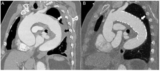
A 67-year-old male was hospitalised with acute thoracic pain. Computed tomography (CT) angiography revealed a covered rupture of the proximal descending aorta causing an intramural and perivascular haematoma as well as an ascending aneurysm with a maximal diameter of 55 mm extending into the proximal aortic arch (Figure 1A). After primarily surveillance with medical management, a CT scan on the following day showed further progression of the perivascular haematoma towards the distal part of the descending aorta. Therefore we decided to perform surgery.

Figure 1.
(A) Preoperative CT angiogram demonstrating the covered rupture of the proximal descending aorta (white arrow) with a progressive intramural and perivascular haematoma (black arrow). The CT scan also revealed an ascending aorta with maximal diameter measuring 55 mm. (B) CT scan angiography 6 months after surgery showing an excellent result with an ascending prosthesis without any leak or periprosthetic haematoma. The stent graft lies perfectly in the distal aortic arch and covers the ruptured area of the descending aorta (white arrow) without any remaining intramural or perivascular haematoma.
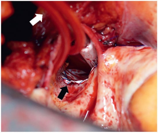
After median sternotomy, standard cardiopulmonary bypass (CPB) was initiated and the patient cooled. A guide-wire was introduced transfemorally and advanced into the descending aorta using transoesophageal echocardiographic guidance. After cross-clamping, the aorta was transsected at the sinotubular level and cardioplegic arrest was induced as previously described []. At the anticipated hypothermic circulatory arrest (HCA) temperature, 28 °C bladder, a bolus of thiopental (20 mg/kg) was given and the patient’s head packed in ice. CPB was stopped, and the aorta was opened and de-aired and antegrade cerebral perfusion catheters (ACP; Distal Perfusion Catheters, LeMaitre Vascular Inc., Burlington, MA, USA) were introduced into both carotid arteries (Figure 2). For cerebral perfusion, blood temperature was 20 °C as previously described []. A 37 mm × 15 cm conformable GoreTAG® aortic stent graft (WL Gore & Associates, Flagstaff, AZ, USA) was delivered antegradely over the guide wire and placed right distally to the left subclavian artery. Sizing of the stent was preoperatively determined from CT scan measurements at the level of the proximal descending aorta using the open-source viewer OsiriX (Pixmeo, Geneva, Switzerland). The proximal part of the stent graft was fixed with a 3-0 Surgipro IITM V-20 (CovidienTM) continuous suture (Figure 2). After resection of the diseased aortic tissue into the hemiarch, a Gelweave Ante-Flo prosthesis (Vascutek, Inchinnan Renfrewshire, Scotland, UK) was anastomosed using a running suture technique, which integrated the stent graft along the lesser curve. CPB was re-established in an antegrade fashion through the prosthesis side branch. The duration of HCA with ACP was 29 minutes. Proximal anastomosis was made at the supracoronary level as appropriate. The patient was rewarmed with a target bladder temperature of 36 °C. The postoperative course was uneventful; the patient was extubated 8 hours postoperatively and discharged after 7 days. A CT scan before discharge showed the stent in correct position and without endoleak. A CT check after 6 months showed an excellent result with an ascending prosthesis without any leak or periprosthetic haematoma, a descending stent without endoleak, and a completely resorbed haematoma at the descending aorta (Figure 1B).

Figure 2.
Intraoperative image during hypothermic circulatory arrest with opened aortic arch, antegrade cerebral perfusion catheters (white arrow) and the antegrade delivered stent located right distally of the left subclavian artery fixed with a 3-0 Surgipro IITM V-20 (CovidienTM) continuous suture (black arrow).
Discussion
Instead of the “classical” approach of a two-stage procedure composed of TEVAR and subsequent open aortic replacement, we decided on a simplified, one-stage procedure. Studies have revealed that such an approach, a “modified frozen elephant trunk” repair, effectively closes the false lumen at the stent level in acute DeBakey type 1 aortic dissections without increasing the operative morbidity and mortality [,]. The MFET facilitates addressing not only the ascending aortic disease, but the proximal and middle part of the descending aorta. Initial data in DeBakey type 1 aortic dissections shows that the MFET reduces the need for open distal aortic reoperations compared with the classical hemiarch repair in the mid-term []. While a two-stage procedure would have been possible in our patient, the antegrade “surgical” delivery of the stent graft has some technical advantages compared with TEVAR. The open delivery allows precise placement of the stent graft and the stent is implanted into the true aortic lumen without perfusion and pressure in the false lumen. Moreover the antegrade implantation allows proximal fixation (Figure 2), which leads to perfect modelling of the transition “aortic arch-stent graft”, reduces the endoleak risk and prevents any retrograde dissection into the aortic arch. The potential of this procedure to induce subsequent remodelling of the descending aorta is high, as shown in our patient in the check CT scan at 6 months after surgery (Figure 1B).
MFET repair is a promising approach for the treatment of a covered rupture or complicated type B aortic dissection combined with ascending aortic dilatation.
Disclosure statement
No financial support and no other potential conflict of interest relevant to this article was reported.
References
- Nienaber, C.A.; Kische, S.; Rousseau, H.; Eggebrecht, H.; Rehders, T.C.; Kundt, G.; et al. Endovascular repair of type B aortic dissection: Long-term results of the randomized investigation of stent grafts in aortic dissection trial. Circ. Cardiovasc. Interv. 2013, 6, 407–416. [Google Scholar] [CrossRef] [PubMed]
- Trimarchi, S.; Segreti, S.; Grassi, V.; Lomazzi, C.; de, V.C.; Rampoldi, V. Emergent treatment of aortic rupture in acute type B dissection. Ann. Cardiothorac. Surg. 2014, 3, 319–324. [Google Scholar] [PubMed]
- Preventza, O.; Cervera, R.; Cooley, D.A.; Bakaeen, F.G.; Mohamed, A.S.; Cheong, B.Y.; et al. Acute type I aortic dissection: Traditional versus hybrid repair with antegrade stent delivery to the descending thoracic aorta. J. Thorac. Cardiovasc. Surg. 2014, 148, 119–125. [Google Scholar] [CrossRef] [PubMed]
- Vallabhajosyula, P.; Szeto, W.Y.; Pulsipher, A.; Desai, N.; Menon, R.; Moeller, P.; et al. Antegrade thoracic stent grafting during repair of acute Debakey type I dissection promotes distal aortic remodeling and reduces late open distal reoperation rate. J Thorac Cardiovasc Surg. 2014, 147, 942–948. [Google Scholar] [CrossRef] [PubMed]
- Matt, P.; Albrecht, F.; Rueter, F.; Grapow, M.; Pargger, H.; Fassl, J.; et al. Hypothermic circulatory arrest using antegrade cerebral perfusion is safe for elective aortic arch surgery. Thorac. Cardiovasc. Surg. 2013, 61, 553–558. [Google Scholar] [CrossRef] [PubMed]
© 2016 by the author. Attribution - Non-Commercial - NoDerivatives 4.0.