Comparison of X-Ray Imaging and Computed Tomography Scan in the Evaluation of Knee Trauma


Abstract
1. Introduction
2. Materials and Method
Statistical Analysis
3. Results
4. Discussion
5. Conclusions
6. Limitation
Author Contributions
Funding
Conflicts of Interest
References
- Oei, E.H.; Nikken, J.J.; Ginai, A.Z.; Krestin, G.P.; Verhaar, J.A.; van Vugt, A.B.; Hunink, M.M. Acute knee trauma: Value of a short dedicated extremity MR imaging examination for prediction of subsequent treatment. Radiology 2005, 234, 125–133. [Google Scholar] [CrossRef] [PubMed]
- Teh, J.; Kambouroglou, G.; Newton, J. Investigation of acute knee injury. BMJ 2012, 344, e3167. [Google Scholar] [CrossRef] [PubMed]
- Mustonen, A.O.; Koskinen, S.K.; Kiuru, M.J. Acute knee trauma: Analysis of multidetector computed tomography findings and comparison with conventional radiography. ActaRadiol. 2005, 46, 866–874. [Google Scholar] [CrossRef]
- Pinto, A.; Berritto, D.; Russo, A.; Riccitiello, F.; Caruso, M.; Belfiore, M.P.; Papapietro, V.R.; Carotti, M.; Pinto, F.; Giovagnoni, A.; et al. Traumatic fractures in adults: Missed diagnosis on plain radiographs in the Emergency Department. Acta Bio Med. Atenei Parmensis 2018, 8 (Suppl. 1), 111–123. [Google Scholar] [CrossRef]
- Avci, M.; Kozaci, N.; Yuksel, S.; Etli, I.; Yilmaz, Y. Comparison of radiography and computed tomography in emergency department evaluation of ankle trauma. Ann. Med. Res. 2019, 26, 867–872. [Google Scholar] [CrossRef]
- Bengtzen, R.R.; Glaspy, J.N.; Steele, M.T. Knee Injuries. In Tintinalli’s Emergency Medicine—A Comprehensive Study Guide, 8th ed.; Tintinalli, J.E., Stapczynski, J.S., Ma, O.J., Yealy, D.M., Meckler, G.D., Cline, D.M., Eds.; McGraw-Hill Education: New York, NY, USA, 2016. [Google Scholar]
- Venkatasamya, A.; Ehlingerb, M.; Bierrya, G. Acute traumatic knee radiographs: Beware of lesions of little expression but of great significance. Diagn. Interv. Imaging 2014, 95, 551–560. [Google Scholar] [CrossRef] [PubMed][Green Version]
- Caracchini, G.; Pietragalla, M.; De Renzis, A.; Galluzzo, M.; Carbone, M.; Zappia, M.; Russo, A.; Greco, F.; Miele, V. Talar fractures: Radiological and CT evaluation and classification systems. ActaBiomed. 2018, 89 (Suppl. 1), 151–165. [Google Scholar] [CrossRef]
- Chen, Y.; Zhang, K.; Qiang, M.; Li, H.; Dai, H. Comparison of plain radiography and CT in postoperative evaluation of ankle fractures. Clin. Radiol. 2015, 70, e74–e82. [Google Scholar] [CrossRef]
- Olsson, O.; Isacsson, A.; Englund, M.; Frobell, R.B. Epidemiology of intra- and peri-articular structural injuries in traumatic knee joint hemarthrosis- data from 1145 consecutive knees with subacute MRI. Osteoarthr. Cartil. 2016, 24, 1890–1897. [Google Scholar] [CrossRef]
- Kozaci, N.; Ay, M.O.; Avci, M.; Turhan, S.; Donertas, E.; Celik, A.; Ararat, E.; Akgun, E. The comparison of point-of-care ultrasonography and radiography in the diagnosis of tibia and fibula fractures. Injury 2017, 48, 1628–1635. [Google Scholar] [CrossRef]
- Avci, M.; Kozaci, N.; Tulubas, G.; Caliskan, G.; Yuksel, A.; Karaca, A.; Doganay, F.; Etli, I. Comparison of Point-of-Care Ultrasonography and Radiography in the Diagnosis of Long-Bone Fractures. Medicina 2019, 55, e355. [Google Scholar] [CrossRef] [PubMed]
- Kozaci, N.; Ay, M.O.; Avci, M.; Beydilli, I.; Turhan, S.; Donertas, E.; Ararat, E. The comparison of radiography and point-of-care ultrasonography in the diagnosis and management of metatarsal fractures. Injury 2017, 48, 542–547. [Google Scholar] [CrossRef] [PubMed]
- Atilla, O.D.; Yesilaras, M.; Kilic, T.Y.; Tur, F.C.; Reisoglu, A.; Sever, M.; Aksay, E. The Accuracy of Bedside Ultrasonography as a Diagnostic Tool for Fractures in the Ankle and Foot. Acad. Emerg. Med. 2014, 21, 1058–1061. [Google Scholar] [CrossRef] [PubMed]
- Kozaci, N.; Ay, M.O.; Akcimen, M.; Sasmaz, I.; Turhan, G.; Boz, A. The effectiveness of bedside point-of-care ultrasonography in the diagnosis and management of metacarpal fractures. Am. J. Emerg. Med. 2015, 33, 1468–1472. [Google Scholar] [CrossRef] [PubMed]
- Kozaci, N.; Ay, M.O.; Akcimen, M.; Turhan, G.; Sasmaz, I.; Turhan, S.; Celik, A. Evaluation of the effectiveness of bedside point-of-care ultrasound in the diagnosis and management of distal radius fractures. Am. J. Emerg. Med. 2015, 33, 67–71. [Google Scholar] [CrossRef]
- Avcı, M.; Kozacı, N.; Beydilli, İ.; Yılmaz, F.; Eden, A.O.; Turhan, S. The comparison of bedside point-of-care ultrasound and computed tomography in elbow injuries. Am. J. Emerg. Med. 2016, 34, 2186–2190. [Google Scholar] [CrossRef] [PubMed]
- Mui, L.W.; Engelsohn, E.; Umans, H. Comparison of CT and MRI in patients with tibial plateau fracture: Can CT findings predict ligament tear or meniscal injury? Skeletal. Radiol. 2007, 36, 145–151. [Google Scholar] [CrossRef] [PubMed]
- Kumar, S.; Kumar, A.; Kumar, S.; Kumar, P. Functional Ultrasonography in Diagnosing Anterior Cruciate Ligament Injury as Compared to Magnetic Resonance Imaging. Indian J. Orthop. 2018, 52, 638–644. [Google Scholar] [CrossRef]
- Wicky, S.; Blaser, P.F.; Blanc, C.H.; Leyvraz, P.F.; Schnyder, P.; Meuli, R.A. Comparison between standard radiography and spiral CT with 3D reconstruction in the evaluation, classification and management of tibial plateau fractures. Eur. Radiol. 2000, 10, 1227–1232. [Google Scholar] [CrossRef]
- Hwang, J.S.; Koury, K.L.; Gorgy, G.; Sirkin, M.S.; Reilly, M.C.; Lelkes, V.; Adams, M.R. Evaluation of Intra-articular Fracture Extension After Gunshot Wounds to the Lower Extremity: Plain Radiographs Versus Computer Tomography. J. Orthop. Trauma 2017, 31, 334–338. [Google Scholar] [CrossRef]
- Lemburg, S.P.; Lilienthal, E.; Heyer, C.M. Growth plate fractures of the distal tibia: Is CT imaging necessary? Arch. Orthop. Trauma Surg. 2010, 130, 1411–1417. [Google Scholar] [CrossRef] [PubMed]
- Fleiss, J.L. Statistical Methods for Rates and Proportions, 2nd ed.; John Wiley & Sons Inc.: New York, NY, USA, 1981. [Google Scholar]
- Raschke, M.J.; Kittl, C.; Domnick, C. Partial proximal tibia fractures. EFORTOpenRev. 2017, 2, 241–249. [Google Scholar] [CrossRef] [PubMed]
- Draghi, F.; Urciuoli, L.; Alessandrino, F.; Corti, R.; Scudeller, L.; Grassi, R. Joint effusion of the knee: Potentialities and limitations of ultrasonography. J. Ultrasound 2015, 18, 361–371. [Google Scholar] [CrossRef] [PubMed]


| 1 | Detection presence of fracture (cortical deterioration) |
| 2 | Determine the type (fissure, linear, fragmented, torus) and localization of fracture. |
| 3 | Measure the degree of angulation of the fracture. |
| 4 | Measure the distance of stepping off. |
| 5 | Is there an extension of the fracture into the joint space or epiphyseal line? |
| 6 | Does the fracture include the epiphyseal line? (Growth plate fracture?) |
| 7 | Detect the presence of concomitant adjacent bone fracture. |
| 8 | Control of the joint space and the presence of joint dislocation. |
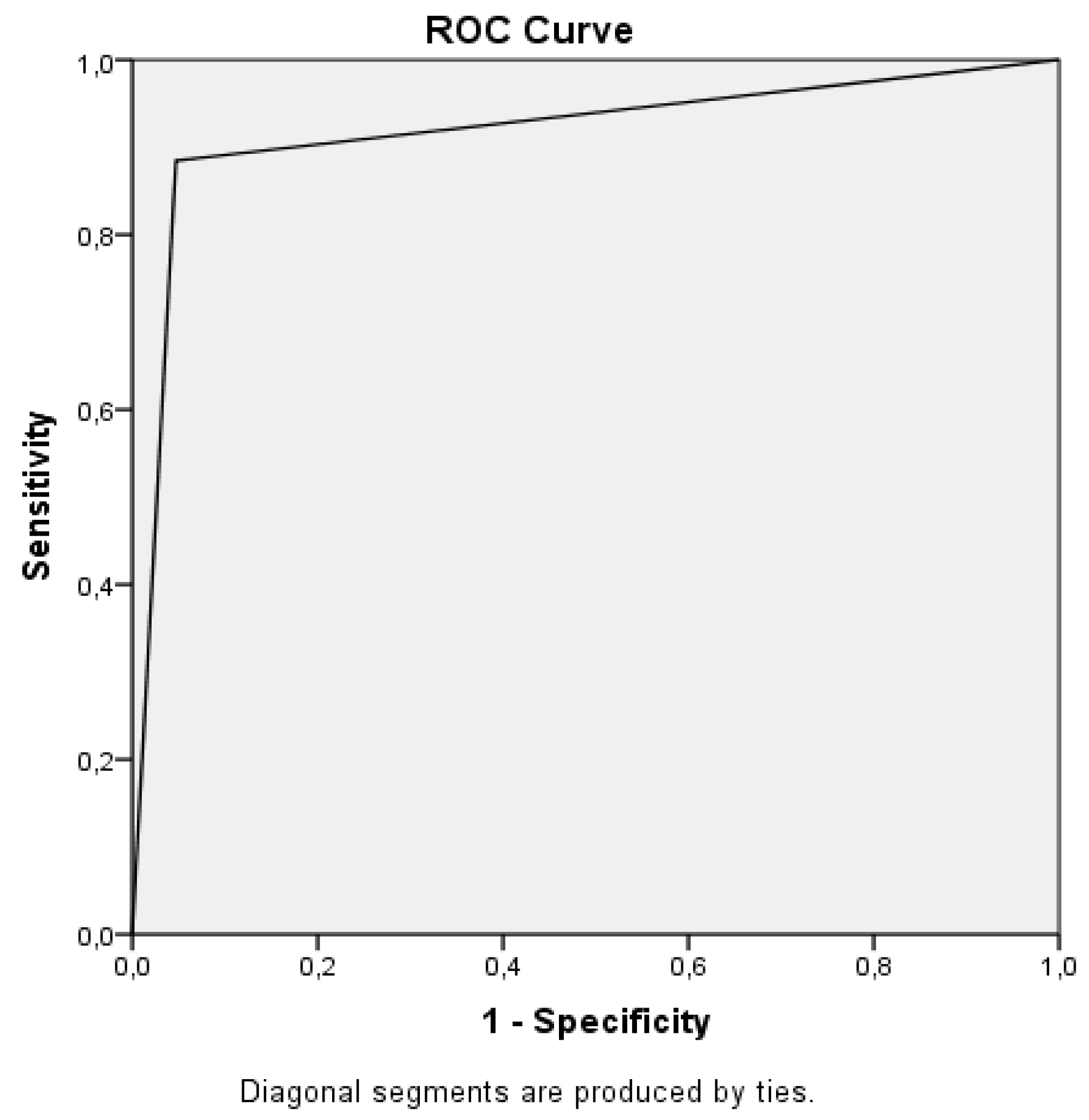
| Bone | XR, N (%) | CT, N (%) | Sensitivity/Specificity | AUC (95% CI) |
|---|---|---|---|---|
| Femur | 28 (5.1) | 42 (7.7) | 67/100 | 0.833 (0.746–0.921) |
| Tibia | 120 (21.9) | 130 (23.7) | 80/96 | 0.881 (0.839–0.923) |
| Patella | 48 (8.8) | 46 (8.4) | 100/100 | 0.998 (0.995–1.000) |
| Fibula | 18 (3.3) | 22 (4.1) | 82/100 | 0.909 (0.813–1.000) |
| Bone | XR, N (%) | CT, N (%) |
|---|---|---|
| Femur + patella | 2 (0.4) | 4 (0.7) |
| Femur + tibia | - | 4 (0.7) |
| Femur + fibula | - | 2 (0.4) |
| Tibia + patella | 2 (0.4) | 2 (0.4) |
| Tibia + fibula | 12 (2.2) | 12 (2.2) |
| Femur + fibula + tibia | - | 4 (0.7) |
| Type of Fracture | XR, N (%) | CT, N (%) | Sensitivity/Specificity | AUC (95% CI) | Kappavalue |
|---|---|---|---|---|---|
| Fissure | 48 (8.8) | 22(4.0) | 55/93 | 0.739 (0.609–0.868) | 0.305 |
| Linear | 40 (7.3) | 24 (4.4) | 58/95 | 0.767 (0.644–0.889) | 0.405 |
| Spiral | 24 (4.4) | 18(3.3) | 44/97 | 0.707 (0.556–0.858) | 0.357 |
| Fragmented | 68 (12.4) | 118 (21.5) | 58/100 | 0.788 (0.731–0.845) | 0.681 |
| Avulsion | 20 (3.6) | 26 (4.7) | 69/100 | 0.844 (0.736–0.952) | 0.773 |
| Fracture Characteristics | XR, N (%) | CT, N (%) | Sensitivity/Specificity | AUC (95% CI) | Kappa Values |
|---|---|---|---|---|---|
| Extension of the fracture into the joint space | 150 (27.4) | 176 (32.1) | 78/96 | 0.872 (0.834–0.910) | 0.782 |
| Growth plate fracture | 8 (1.5) | 8 (1.5) | 75/100 | 0.873 (0.693–1.000) | 0.746 |
| Angulation | 90 (16.4) | 118 (21.5) | 75/100 | 0.871 (0.823–0.918) | 0.811 |
| Stepping off | 94 (17.2) | 126 (23.0) | 71/99 | 0.852 (0.804–0.901) | 0.774 |
© 2019 by the authors. Licensee MDPI, Basel, Switzerland. This article is an open access article distributed under the terms and conditions of the Creative Commons Attribution (CC BY) license (http://creativecommons.org/licenses/by/4.0/).
Share and Cite
Avci, M.; Kozaci, N. Comparison of X-Ray Imaging and Computed Tomography Scan in the Evaluation of Knee Trauma. Medicina 2019, 55, 623. https://doi.org/10.3390/medicina55100623
Avci M, Kozaci N. Comparison of X-Ray Imaging and Computed Tomography Scan in the Evaluation of Knee Trauma. Medicina. 2019; 55(10):623. https://doi.org/10.3390/medicina55100623
Chicago/Turabian StyleAvci, Mustafa, and Nalan Kozaci. 2019. "Comparison of X-Ray Imaging and Computed Tomography Scan in the Evaluation of Knee Trauma" Medicina 55, no. 10: 623. https://doi.org/10.3390/medicina55100623
APA StyleAvci, M., & Kozaci, N. (2019). Comparison of X-Ray Imaging and Computed Tomography Scan in the Evaluation of Knee Trauma. Medicina, 55(10), 623. https://doi.org/10.3390/medicina55100623