In the original publication [1], there was a mistake in Figure 5. which was published. The image for the FGF-2 solution in vehicle 5 was mislabeled and required a replacement. A corrected version of Figure 5 appears below. The authors confirm that the scientific conclusions are unaffected. This correction was approved by the Academic Editor. The original publication has also been updated.
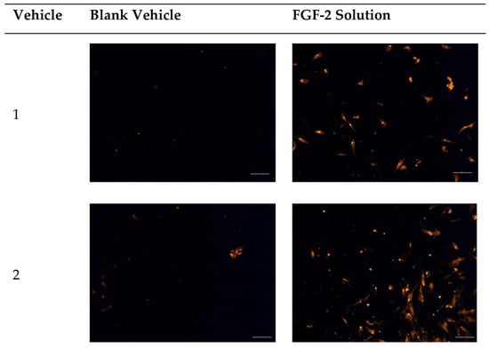
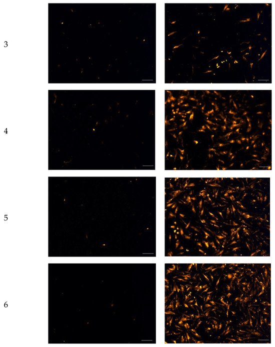
Figure 5.
Fluorescence micrographs showing chemotactic migration of human dermal fibroblast cells to the basal surface of a transwell membrane in response to FGF-2. Cells seeded on the apical transwell membrane were exposed to FGF-2 solutions or blank vehicles added to the basolateral chamber. Samples comprise blank vehicles or FGF-2 (50 ng/mL in the vehicle) in the vehicles: 1 (water only), 2 (methylcellulose (MC) 0.05% w/v), 3 (alanine 20 mM), 4 (human serum albumin (HSA) 1 mg/mL), 5 (MC 0.05% w/v and alanine 20 mM) and 6 (MC 0.05% w/v and HSA 1 mg/mL). Magnification: 200×, scale bar = 100 µm.
Reference
- Benington, L.; Mo, J.; Li, M.; Rajan, G.; Locher, C.; Lim, L.Y. In Vitro Assessment of Wound-Healing Efficacy of Stabilized Basic Fibroblast Growth Factor (FGF-2) Solutions. Pharmaceuticals 2024, 17, 247. [Google Scholar] [CrossRef]
Disclaimer/Publisher’s Note: The statements, opinions and data contained in all publications are solely those of the individual author(s) and contributor(s) and not of MDPI and/or the editor(s). MDPI and/or the editor(s) disclaim responsibility for any injury to people or property resulting from any ideas, methods, instructions or products referred to in the content. |
© 2024 by the authors. Licensee MDPI, Basel, Switzerland. This article is an open access article distributed under the terms and conditions of the Creative Commons Attribution (CC BY) license (https://creativecommons.org/licenses/by/4.0/).