Article

Morphological and Molecular Characterization of Arbuscular Mycorrhizal Fungi from the Rhizosphere of Date Palm (Phoenix dactylifera L.) in the Oasis of Figuig, Morocco

1
Laboratory of Bioresources, Biotechnology, Ethnopharmacology and Health, Faculty of Sciences, Université Mohamed Premier, BV Mohammed VI BP 717, Oujda 60000, Morocco
2
Institut Agro, Université Angers, National Research Institute for Agriculture, Food and Environment (INRAE), Institute of Research in Horticulture and Seeds (IRHS), Federative Research Structure for Plant Quality and Health (SFR QUASAV), 49000 Angers, France
3
Administrative Centre, Laboratory of Water Analysis of Figuig (L.A.E.F.), Municipality of Figuig, BP 121, Figuig 61000, Morocco
4
Department of Research in Drug Development, Faculty of Pharmacy, Université Libre de Bruxelles, Bvd du Triomphe, 1050 Brussels, Belgium
5
Laboratory of Plant Biotechnology, Université Libre de Bruxelles, 6041 Gosselies, Belgium
*
Author to whom correspondence should be addressed.
Diversity 2025, 17(10), 710; https://doi.org/10.3390/d17100710 (registering DOI)
Submission received: 22 August 2025 / Revised: 9 October 2025 / Accepted: 11 October 2025 / Published: 14 October 2025
(This article belongs to the Section Microbial Diversity and Culture Collections)

Abstract

This study presents the first molecular characterization of arbuscular mycorrhizal fungi (AMF) isolated from single-spore cultures in Morocco, specifically from the rhizosphere of date palm (Phoenix dactylifera L.) in the Figuig oasis. Nine indigenous AMF isolates were successfully established and identified through an integrative approach combining spore morphology with ribosomal DNA region sequencing (SSU–ITS–LSU). Morphological and phylogenetic analyses revealed that the isolates belonged mainly to the genera Rhizophagus and Glomus. These results provide new insights into AMF diversity in arid Moroccan ecosystems and establish a reference collection of indigenous isolates with potential applications. In particular, they open opportunities for developing bio-inoculants that can improve date palm growth, enhance resilience to environmental stresses, and contribute to sustainable agriculture and soil restoration in oasis systems.

1. Introduction

Arbuscular mycorrhizal fungi (AMF) are soil fungi that establish symbiotic associations with the roots of most agricultural and horticultural crops of importance, playing a critical role in plant mineral nutrition and resistance to biotic and abiotic stresses, soil structure, and ecosystem functioning [1,2,3]. Despite their presence in most if not all soils, the composition and distribution of AMF communities is influenced by host plants, vegetation types, soil properties, and climatic conditions [4,5,6,7,8]. Agricultural practices also drastically impact AMF community composition and population density [9,10,11].
In arid and semi-arid ecosystems, such as oases, AMF can play a pivotal role in plant adaptation to extreme environmental conditions, characterized by nutrient-poor soils, prolonged periods of drought and high temperatures [12,13,14]. The oasis of Figuig, located in southeastern Morocco, is characterized by an arid to hyper-arid climate, with annual rainfall generally under 100 mm and summer temperatures that frequently exceed 45 °C, creating a particularly challenging environment for agriculture [15]. This oasis, as most oases, is characterized by a three-tiered culture ecosystem dominated by date palms (Phoenix dactylifera L.), accompanied by fruit tree species such as pomegranate (Punica granatum L.), olive (Olea europaea L.), apricot (Prunus armeniaca L.), and fig (Ficus carica L.) and at the lower third vegetable crops such as onion (Allium cepa L.), pepper (Capsicum annuum L.), and tomato (Solanum lycopersicum L.), as well as forage crops [16,17]. Facing increasing challenges linked to desertification, soil salinity, and water scarcity, characterizing and conserving indigenous AMF strains in these oasis ecosystems emerges as a priority for sustainable agriculture.
Beyond its cultural and economic importance, the date palm represents a vital crop for millions of people living in desert regions [18,19,20,21]. However, global date production has experienced an alarming decline in certain regions, with some countries reporting losses of up to 30–40% over the past two decades due to biotic and abiotic stresses, including soil degradation and vascular wilt disease [22,23] leading to growing research interest and international academic cooperation aimed at ensuring its sustainability. Numerous studies have shown that date palms form symbiotic associations with AMF [24,25,26,27], thereby promoting seedling growth under low nutrient availability conditions [28,29]. Furthermore, AMF may contribute to biological control of Fusarium oxysporum f. sp. albedinis or Bayoud disease, one of the most aggressive threats to date palm cultivation in several oases in North Africa [26,30,31,32]. Applying AMF during the nursery stage has proven to be beneficial, allowing seedlings to establish symbiotic networks before field transplantation, thereby enhancing plant performance in harsh environments [33,34,35]. Given that AMF exhibit specific local adaptations to their ecosystems [36,37,38,39,40], it is essential to understand their natural diversity and distribution within the Figuig oasis to optimize their use in sustainable agriculture.
Despite the ecological significance of AMF, knowledge regarding their diversity in association with date palms remains limited, particularly in North African oases and notably in the oasis of Figuig. Only two studies have been conducted on AMF associated with date palms in this oasis [17,26], relying solely on morphological spore identification. However, molecular techniques provide a more comprehensive approach, enabling direct detection of AMF within colonized roots and facilitating taxonomic comparisons with public databases [41,42,43,44]. Advanced molecular methods, such as polymerase chain reaction (PCR) and high-throughput sequencing, have significantly improved AMF identification and characterization [45,46,47]. These approaches enable a deeper understanding of AMF diversity and ecological distribution [48]. In addition, research has refined phylogenetic reference data for AMF systematics, thus enhancing taxonomic analysis accuracy [49,50,51].
Within this context, the present study was conducted to characterize indigenous AMF associated with date palms in the Figuig oasis by combining morphological and molecular identification approaches. Documenting AMF biodiversity and establishing a collection of indigenous strains adapted to local conditions represent essential requirements for fundamental and applied research projects focused on these specific environments. This approach is similar to successful efforts conducted in arid and semi-arid regions such as the Arabian Peninsula [52,53,54], North Africa including Tunisia and Morocco [32,55,56] and other comparable ecosystems worldwide. Based on these considerations, we hypothesized that the rhizosphere of date palms in the Figuig oasis hosts a diverse assemblage of indigenous AMF adapted to local arid conditions, and the integration of morphological and molecular approaches would allow more accurate identification of these fungi, reducing potential mismatches inherent to morphology alone.

2. Materials and Methods

2.1. Soil Sampling

Soil samples were collected in March 2019 from 31 locations (plots) in the Figuig oasis (32°06′00″ N, 1°14′00″ W). The plots were randomly selected to maximize coverage of the area (Figure 1—see geographic coordinates of the sampling locations in Supplementary Material Table S1). On each plot, four date palms were randomly selected, and soil was sampled from four sub-sampling points around the base of each tree at a depth of 15–30 cm, corresponding to the active root zone. The upper 0–15 cm soil layer was excluded. These sub-samples were then mixed to create a composite sample representative of each plot. The composite samples were labeled accordingly, stored in sealed plastic bags, and kept at 4 °C without air-drying to preserve spore viability until further analysis, in line with standard AMF storage protocols that preserve spore viability [57].

2.2. Establishment of AMF Trap Cultures

Trap cultures were established within two weeks after sample collection. Soil samples containing naturally occurring root fragments of date palm were used as inoculum. In the laboratory, these root fragments were cut into small pieces and mixed with their respective soil samples to prepare the inoculum. This mixture was then combined with autoclaved Terra Green (calcined attapulgite clay; Oil Dri UK Ltd., Wisbech, UK), a substrate composed of two volumes of calcined clay, two volumes of quartz (0.4–0.8 mm), and one volume of quartz (1–2 mm). The substrate was sterilized in an autoclave MED 12 (JP Selecta, Barcelona, Spain) at 121 °C and 1.2 atm for two separate 1 h cycles on consecutive days. After sterilization, approximately 300 g of Terra Green was mixed with 100 g of the homogenized inoculum.
The planting setup consisted of a bottom layer of sterilized Terra Green, followed by the sampled soil containing the inoculum, and topped with another layer of sterilized Terra Green, following the method described by Walker [58]. This stratified arrangement created a controlled environment where the inoculum was enclosed between two sterile layers, ensuring physical stability, minimizing external contamination, and facilitating AMF-plant interactions.
Plantago lanceolata L. (Plantaginaceae) was selected as the trap plant due to its well-documented ability to promote AMF colonization and its adaptability to a wide range of soil conditions [59,60,61]. Studies have shown that P. lanceolata establishes strong symbiotic interactions with AMF, significantly improving root colonization and nutrient uptake [61,62,63].
Seeds of P. lanceolata seeds first were surface sterilized using a 10% (v/v) sodium hypochlorite solution for 10 min, with the addition of 0.5% (v/v) Tween 20. They were then thoroughly rinsed with autoclaved water to remove any remaining disinfectant. Approximately 20 seeds were sown per pot (1 L capacity, 12 cm in diameter and 10 cm in height). The pots were placed in Sun Bags (Product B7026, Sigma Aldrich, St. Louis, MO, USA) to prevent cross-contamination during incubation [64]. Trap cultures were grown for six months in the greenhouse under controlled conditions (temperature 25–30 °C, 50% relative humidity, and an 18/8 h light/dark photoperiod).

2.3. Establishment of AMF Monosporal Cultures

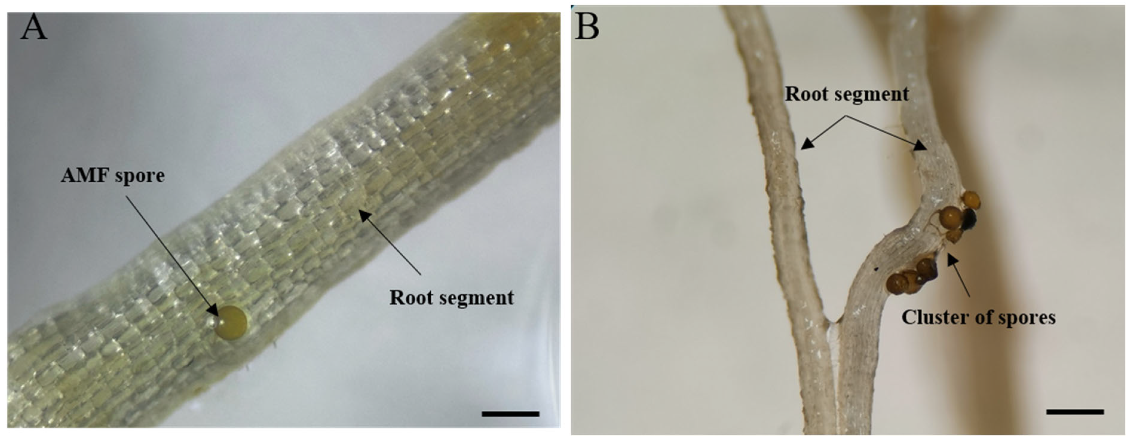
After six months of culture, AMF spores were extracted from the trap cultures using the wet sieving and decantation method [65,66], followed by sucrose flotation. Briefly, 50 g of soil was suspended in demineralized water and passed through 1 mm and 38 µm sieves. The retained fraction was centrifuged at 2000 rpm for 30 s, the pellet resuspended in a 1.4 M sucrose solution, and centrifuged again at 2000 rpm for 4 min. The supernatant containing spores was poured onto a 38 µm sieve, rinsed thoroughly with demineralized water, after which the spores were collected. The extracted spores were then used to establish single-spore cultures using the pipette tip technique described by Tchabi et al. [67]. In brief, under a binocular microscope (SZ61, Olympus, Tokyo, Japan), the spores were first sorted by morphotype and then carefully applied (one spore per plant) onto the root of one-week-old P. lanceolata seedlings (Figure 2). The seedlings were finally transplanted into autoclaved Terra Green (see above). The number of morphotypes isolated at each site is detailed in Supplementary Material Table S2.
The single-spore cultures were grown in 500 mL (8 cm in diameter and 7 cm in height) pots and placed in Sun Bags (Product B7026, Sigma Aldrich) to avoid cross-contamination, as described above. The cultures were incubated in a greenhouse under controlled conditions (25–30 °C, 50% relative humidity, and an 18/8 h light/dark photoperiod) for an additional six months. During this period, the development of AMF structures was regularly monitored to assess spore production and root colonization. At the end of the incubation period, spores were extracted and identified (see below). The details of single-spore-derived cultures, including the number of spores used, identified morphotypes, and culture success rates, are provided in Supplementary Material Table S3.

2.4. Morphological Analyses of AMF

Spores from successfully established single-morphotype cultures were isolated from the substrate using the method described by Sieverding [66]. The morphological characteristics of the spores, along with their subcellular structures, were analyzed by mounting the specimens in polyvinyl alcohol-lactic acid-glycerol (PVLG; [68] Koskey, 1983) and a mixture of PVLG with Melzer’s reagent [69]. Spore structures were described following the terminology described by Błaszkowski [70] and the species descriptions provided on the INVAM website (https://invam.ku.edu/species-descriptions (accessed on 15 August 2025). Photomicrographs were captured using an Olympus BH2-RFCA microscope (Olympus Optical GmbH, Hamburg, Germany) equipped with a digital camera. Image processing and detailed analyses were performed using ImageView software (version 64, 4.11.19271). All isolates and permanent slides containing mounted specimens have been deposited in the Laboratory for Research and Development Projects in the Figuig oasis (Morocco) for conservation and future reference.

2.5. Molecular Analyses and Phylogeny

Molecular analysis was performed following the protocols established by Krüger et al. [50] and Naumann et al. [71]. Deoxyribonucleic acid (DNA) was extracted from a single spore of the same morphotype by carefully crushing it with a fine needle in a 0.2 mL PCR tube containing 5 μL of nuclease-free water. The resulting extract was used as templates for the polymerase chain reaction (PCR). To obtain the large subunit (LSU) ribosomal DNA (rDNA) sequences, a nested PCR procedure was used, using the SSUmAf-LSUmAr and SSUmCf-LSUmBr primer pairs for the first and second nested PCR, respectively. These primer pairs are well established and have been used previously in similar studies [50]. The PCRs were performed using the Phusion High-Fidelity DNA Polymerase Kit (2 U/µL) from Thermoscientific. The final master mix contained 0.2 µL of Taq Phusion DNA polymerase (2 U/µL), 4 µL of buffer HF (5×), 0.4 µL of dNTPs, 0.1 µL of BSA (2 mg/mL), and 1 µL of each primer (10 µM). For the first PCR, an initial denaturation step was carried out at 99 °C for 5 min, followed by 40 cycles of denaturation at 99 °C for 10 s, annealing at 60 °C for 30 s, and elongation at 72 °C for 1 min. A final elongation step was performed at 72 °C for 10 min. The same conditions were applied to the nested PCR, except that only 30 cycles were performed, and the annealing temperature was set to 63 °C. Visualization of the PCR products was done by electrophoresis on 1.5% agarose gels in 1× TAE buffer, supplemented with 5 µL of Gelred (10,000×) for staining. To facilitate sequencing, the PCR products were cloned using the Zero Blunt TOPO PCR Cloning Kit (K280020) following the manufacturer’s protocol (Invitrogen, Thermo Fisher Scientific, Waltham, MA, USA). The cloned products were then transformed into competent E. coli cells using the One Shot TOP 10 Chemically Competent Cells Kit (C4040-03) following the manufacturer’s instructions (Invitrogen, Thermo Fisher Scientific). Confirmation of successful cloning was achieved through colony PCR using the nested PCR method described by Krüger et al. [50]. Colonies yielding PCR products of the expected size bands were selected for further analysis. Plasmid extraction was performed using the Plasmid Miniprep Kit 1, following the manufacturer’s instructions. The plasmids were subsequently subjected to sequencing using the ITS1/ITS3/ITS4 primers. Electropherograms generated from the sequencing reactions were analyzed using Sequencher (version 5.4.6, Gene Codes Corporation, Ann Arbor, MI, USA). To identify similar sequences, a BLASTn search (version 2.15.0) was performed against the National Center for Biotechnology Information (NCBI) DNA sequence database.
After sequencing by Eurofins Genomics (Konstanz, Germany), the resulting sequences were compared against the GenBank database using BLAST (version 2.15.0). The best matching sequences, along with reference alignments published by Krüger et al. [49], were used to construct the phylogenetic tree. Maximum Likelihood (ML) phylogenetic analyses were conducted in MEGA11 (version 11.0.13) under the Tamura–Nei model with a discrete gamma distribution (five categories) and a proportion of invariant sites. Node support was assessed with 500 bootstrap replicates. To strengthen the robustness of our inferences, complementary phylogenetic trees were reconstructed using Neighbor-Joining (NJ) and UPGMA, both based on Tamura–Nei distances, with node support evaluated from 500 (NJ) and 1000 (UPGMA) bootstrap replicates. Branches with <50% support were collapsed.
In addition, pairwise genetic distances for the SSU–ITS–LSU alignment were computed under the Kimura 2-parameter model. Distances within the range of conspecific sequences were considered intraspecific, whereas larger values approaching or exceeding those observed between species were considered interspecific.

3. Results

3.1. Establishment of the AMF Culture Collection

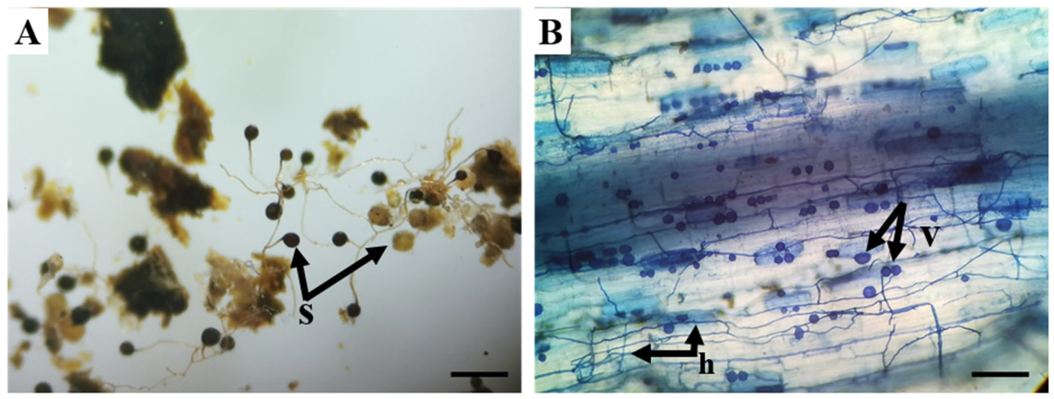
Thirteen trap cultures from field-collected soil samples were maintained for six months in the greenhouse to facilitate the development of AMF propagules. After this period, 10 trap cultures were deemed unsuccessful due to the absence of observable AMF structures in the roots of P. lanceolata. In contrast, the remaining 20 trap cultures were considered successful, as they exhibited well-developed mycorrhizal structures, including spores, extraradical hyphae, and vesicles (Figure 3).
From these 20 successful trap cultures, a total of 37 distinct morphotypes were obtained, each representing a potentially unique AMF taxon. They were used to establish 37 single-spore cultures with P. lanceolata. Details on the morphological identification of these 37 monoxenic AMF cultures, along with their corresponding soil origin identifiers, are provided in the Supplementary Information (Table S3).
The success of single-spore cultures varied across different morphotypes, with only 9 single-spore cultures (FIG3M1E, FIG4M2E, FIG5M1E, FIG6M3E, FIG8M1E, FIG14M1E, FIG16M1E, FIG18M2E, and FIG24M1E) establishing a successful mycorrhizal association (Table S3).

3.2. Morphological Identification of AMF Species from Single-Spore Cultures

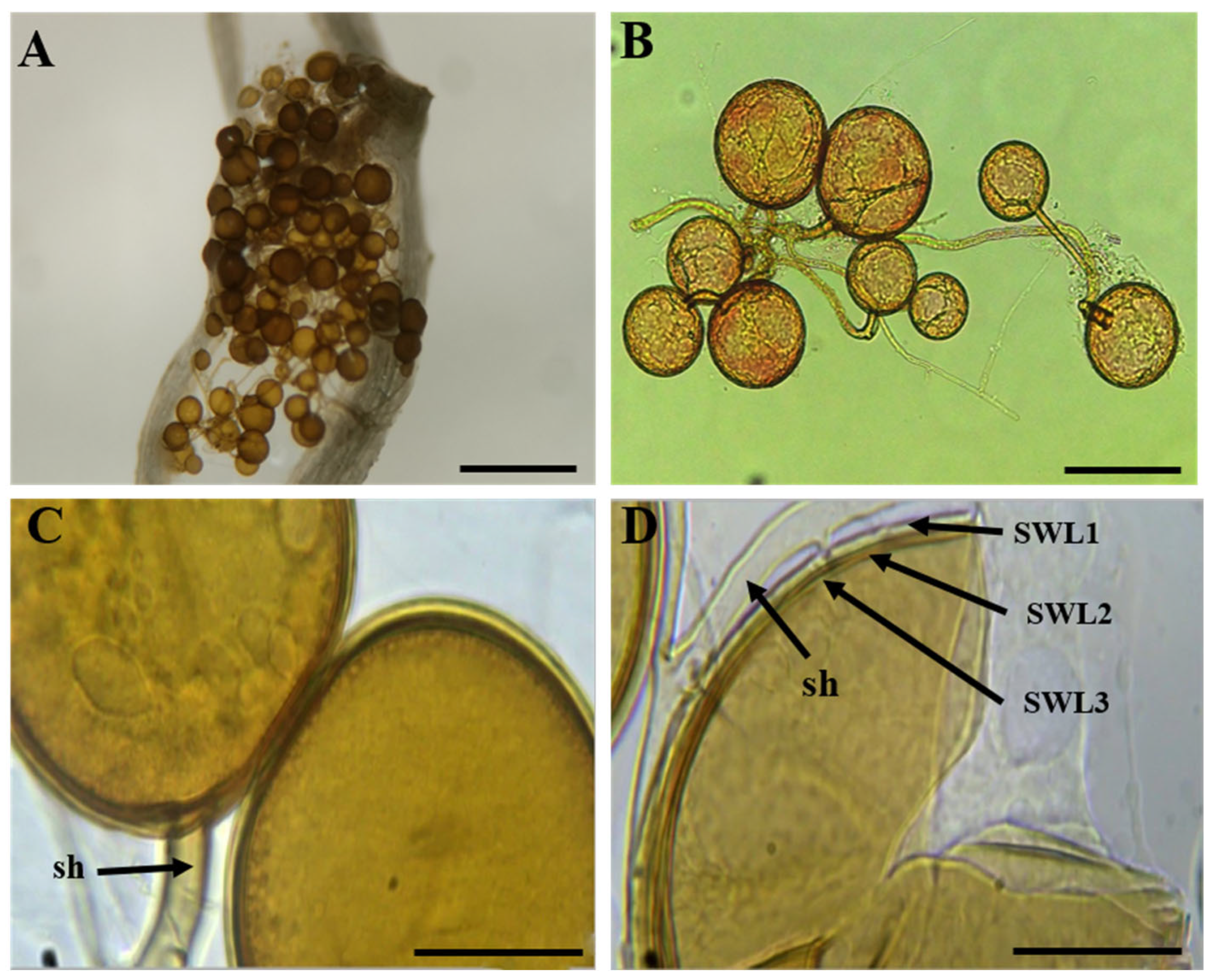
The morphological identification of AMF spores revealed that Rhizophagus sp. (4 out of 9) and Glomus sp. (3 out of 9) were the most frequently recovered genera, followed by Funneliformis sp. (1 out of 9) and Scutellospora sp. (1 out of 9). Isolates FIG14M1E, FIG16M1E, FIG24M1E, and FIG6M3E were identified as Rhizophagus sp., producing globose to subglobose spores ranging from 80 to 150 µm in diameter, hyaline to yellow-brown in color, with trilaminate spore walls and a straight cylindrical subtending hypha, a key morphological feature distinguishing this genus from Glomus (Figure 4), as described by Schüßler and Walker [72].
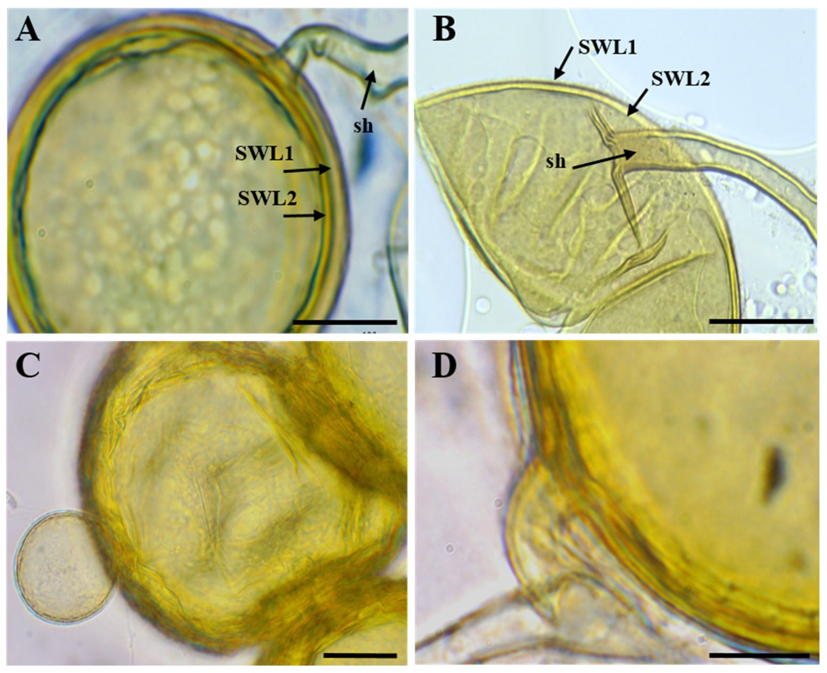
FIG3M1E, FIG8M1E and FIG18M2E were identified as Glomus sp., exhibiting globose spores ranging from 60 to 120 µm in diameter, with bilaminate walls (outer hyaline and inner yellowish to brown) and a cylindrical subtending hypha continuous with the inner wall (Figure 5), consistent with the morphological characteristics of Glomus species as revised by Redecker et al. [73].
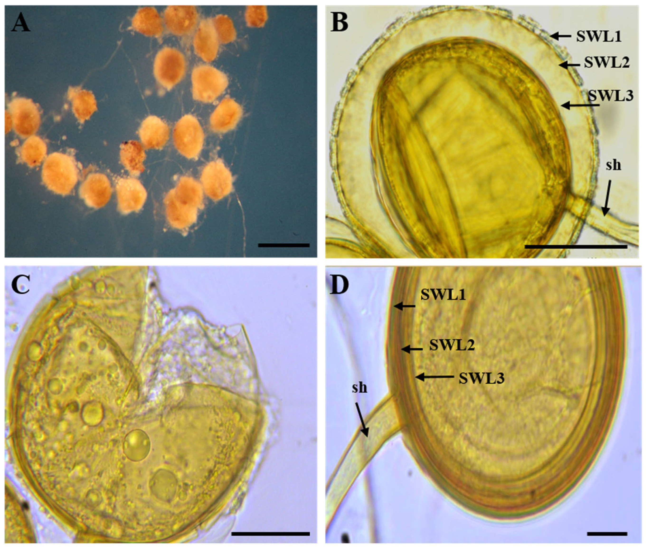
FIG04M2E was identified as Funneliformis sp., producing orange to yellow-brown globose to subglobose spores (≈90–140 µm) with a trilaminate wall and a persistent funnel- to cylindrical-shaped subtending hypha, a key diagnostic feature of this genus (Figure 6), as originally described by Oehl et al. [74].
Finally, FIG05M1E was identified as Scutellospora sp., producing large spores (120–220 µm) with a rigid outer wall, a thick laminate layer, and hyaline inner walls. Sporiferous saccules were also observed (Figure 7), consistent with the genus description by Walker and Sanders [75].

3.3. Molecular Characterization and Phylogenetic Analysis of AMF Isolates

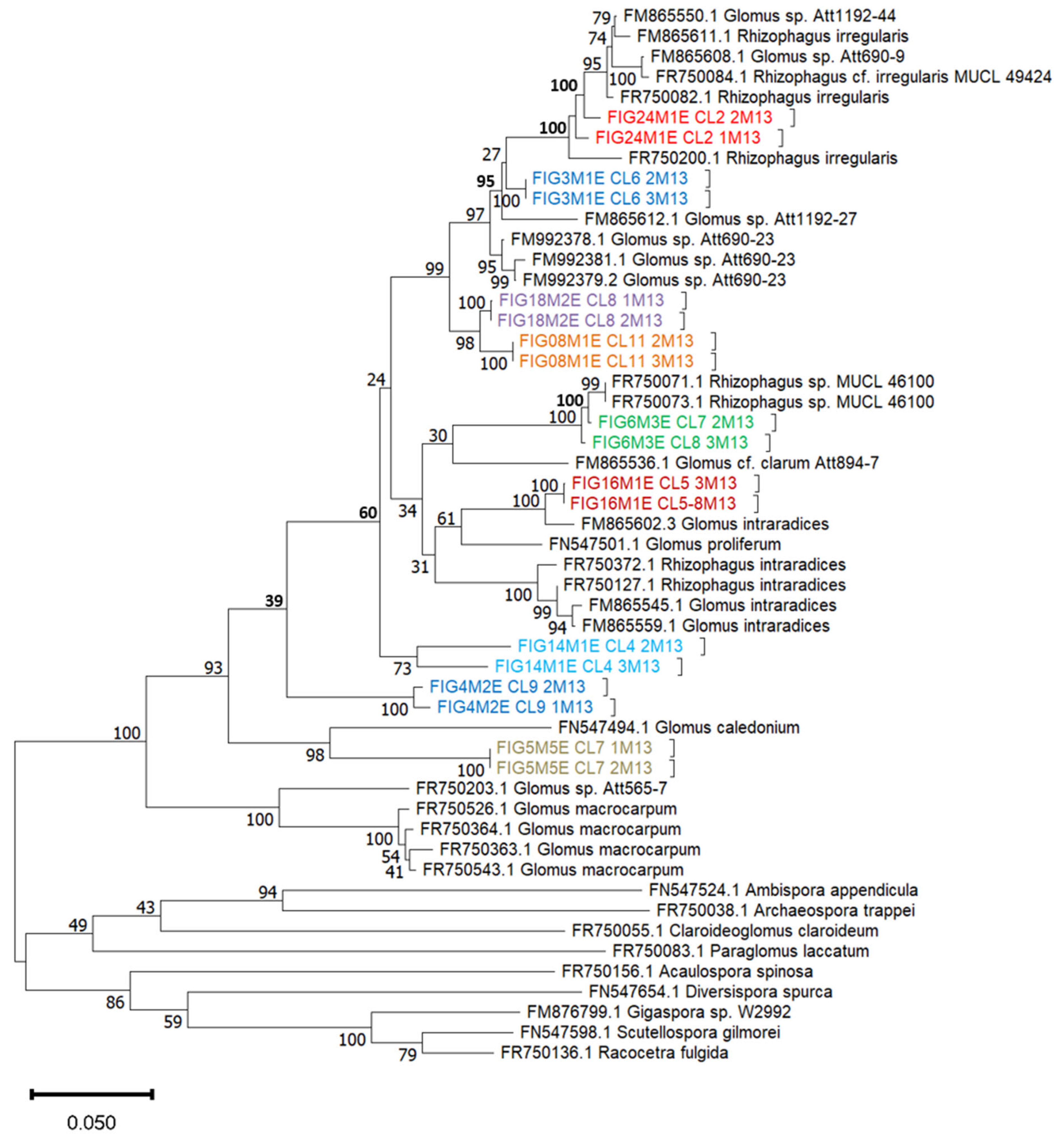
PCR-based approach targeting the SSU rDNA, ITS, and LSU rDNA regions, following the methodology described by Krüger et al. [50], was used for molecular identification of the 9 single-spore cultures. These regions were selected for their complementary phylogenetic resolution across taxonomic levels. The SSU rDNA region provides conserved sequences suitable for genus and family level classification, while the ITS region, known for its high variability, offers enhanced resolution at the species level. The LSU rDNA region, with its intermediate evolutionary rate, further refines phylogenetic placement at subgeneric levels. This multilocus strategy significantly improved the taxonomic resolution and allowed for a critical reassessment of the morphological identification initially assigned to each isolate. In addition to the Maximum Likelihood (ML) topology (Figure 8), phylogenetic reconstructions using Neighbor-Joining (NJ) and UPGMA yielded largely congruent trees, corroborating the major clades recovered (Supplementary Figures S1 and S2)
The phylogenetic tree (Figure 8) revealed that Glomus sp. was the main genus among the analyzed isolates. FIG3M1E, FIG8M1E, and FIG18M2E formed a well-supported clade (bootstrap values between 95% and 100%) within the G. intraradices/proliferum group, in full agreement with the morphological identification as Glomus sp.
FIG6M3E and FIG24M1E showed strong bootstrap support (100%), confirming their genetic affiliation with Rhizophagus sp. In the same clade, FIG14M1E also clustered within the Rhizophagus lineage, with a moderate bootstrap value (60%), which we now explicitly report. This relatively low support may reflect intraspecific divergence within Rhizophagus or the limited resolution of the SSU–ITS–LSU fragment. Consistent placements were also observed in the NJ and UPGMA analyses (Supplementary Figures S1 and S2), although bootstrap values remained low for this node. Additionally, FIG16M1E was also recovered within the Rhizophagus clade, with high bootstrap support, further supporting its placement within this genus.
However, FIG5M1E, was initially assigned to Scutellospora sp. based on spore wall features observed under light microscopy. However, the morphology of Scutellospora is markedly distinct from glomoid genera such as Glomus and Funneliformis, suggesting that the spores may have been atypical or degraded. Molecular analysis placed FIG5M1E within the G. caledonium clade with strong support (bootstrap 98%), underscoring the limitations of morphology alone and the need for molecular confirmation. Finally, FIG4M2E, initially identified as Funneliformis sp., also grouped within the G. caledonium lineage, but with weak bootstrap support (39%). This low support indicates a degree of phylogenetic uncertainty, which may arise from limited reference sequences or intra-genus variability. NJ and UPGMA analyses confirmed the same placement (Supplementary Figures S1 and S2), though again with modest support. These findings are further supported by the genetic distance analysis (Supplementary Table S4), which allowed us to distinguish intraspecific from interspecific divergence among the studied isolates and their closest relatives, and to highlight cases of potential cryptic diversity.
A comparative overview of molecular and morphological assignments for all isolates is provided in Supplementary Table S5.

4. Discussion

This study is the first conducted in Morocco, and more specifically in the Figuig oasis, to adopt a molecular approach for characterizing AMF isolated from single-spore cultures.
Previous studies in Morocco have relied exclusively on morphological approaches to identify and characterize AMF communities associated with various agricultural systems and natural ecosystems [32,76,77,78,79,80,81]. Our study identified the presence of both Glomus and Rhizophagus genera as the most represented AMF based on SSU–ITS–LSU rDNA sequences. This co-occurrence of Glomus and Rhizophagus is consistent with findings from similar studies conducted in other arid and semi-arid regions, including Tunisia [55], southern Arabia [52,53], and Algeria [82], where this genus is particularly adapted to challenging edaphic conditions such as high salinity and severe drought [32,83,84]. Unlike our study, which utilized molecular identification to characterize these isolates, several previous studies in Morocco, notably those by Meddich et al. [32,80], Ouahmane et al. [81], and Bouamri et al. [85], reported the presence of genera such as Sclerocystis sp., Acaulospora sp., G. monosporum, and G. clarum, but these identifications were based solely on morphological criteria. The unsuccessful establishment of some trap cultures in our study may be explained by factors such as low spore viability, microbial competition, or suboptimal growth conditions. Moreover, the variability observed among single-spore cultures is consistent with previous reports in AMF research, where spore germination, hyphal growth, and root colonization are influenced by spore maturity, host plant compatibility, and environmental conditions [54,86,87].
Our investigation underscores the limitations of relying solely on morphological traits for AMF identification, particularly when dealing with morphologically conserved genera. A well-documented case is the close morphological similarity between the genera Glomus and Rhizophagus, which often display overlapping spore sizes, wall structures, and hyphal attachments [49,72,88]. This phenotypic convergence makes it difficult to accurately distinguish between them using only morphological criteria [70,89]. Notably, Rhizophagus was historically included within Glomus and only established as a separate genus following molecular phylogenetic studies that revealed deep evolutionary divergence [49,72]. The genus Glomus, once considered a broad taxonomic group, has since undergone significant revision, leading to the delineation of several new genera within Glomeraceae based on DNA sequence data [73,74].
The phylogenetic resolution obtained for certain isolates, particularly FIG4M2E and FIG14M1E, remains moderate to low, with bootstrap values of 39% and 60%, respectively, indicating uncertainty regarding their precise taxonomic placement. Specifically, the FIG4M2E isolate forms a distinct but weakly supported group (bootstrap = 39%), closely related to G. caledonium and Glomus sp. (Att565-7), reflecting a potential lack of taxonomic precision due to high genetic variability or the underrepresentation of closely related sequences in current genetic databases. Conversely, the FIG14M1E isolate clusters within a moderately supported clade (60%) related to Rhizophagus sp., further indicating ambiguity in its precise affiliation.
It is well recognized that the exclusive use of the SSU rDNA region does not always provide sufficient resolution to effectively resolve phylogenetic relationships among closely related species [49]. Thus, molecular analyses incorporating ITS and LSU regions simultaneously generally yield superior resolution and finer distinctions among species within Glomeromycota. While SSU–ITS–LSU rDNA regions remain the most commonly used markers for identifying and differentiating AMF species [49], recent studies recommend the complementary use of additional genetic markers to improve phylogenetic resolution within the Glomeromycota. Among the most promising are β-tubulin (β-tub), elongation factor EF-1α, and mitochondrial genes such as COX1 and COX3, which offer higher variability and have proven useful for distinguishing closely related or cryptic taxa [90,91,92,93]. Moreover, next-generation sequencing (NGS) technologies and functional gene markers (e.g., phosphate transporters, SOD, GPX) have been proposed to uncover hidden diversity and better understand the ecological roles and adaptations of AMF [5,94,95,96,97].
These integrated approaches, combining multiple genetic markers and advanced technologies, are crucial for enhancing the reliability of taxonomic characterization and deepening our understanding of the phylogenetic diversity of AMF, particularly in the oasis, a unique ecosystem.

5. Conclusions

The integrated approach adopted in this study, combining morphological and molecular analyses, proved essential for improving taxonomic accuracy and improving our understanding of the actual AMF richness associated with the rhizosphere of date palm (P. dactylifera L.) in the Figuig oasis. The isolates obtained mainly belonged to the genera Glomus and Rhizophagus, both well known for their positive effects on plant growth, nutrient uptake, and stress tolerance in arid agroecosystems. Our findings also confirm the limitations of morphology-based identification and emphasize the added value of molecular tools in resolving taxonomic ambiguities. This integrative characterization provides a reference collection of indigenous isolates with potential applications in sustainable oasis agriculture. Future research should focus on evaluating the functional performance of these isolates in improving date palm resilience to biotic and abiotic stresses, as well as on developing scalable inoculum production systems for practical use. In addition, a subset of isolates exhibited comparatively elevated pairwise distances and weak phylogenetic support, suggesting potential cryptic diversity. These lineages may represent candidates for undescribed taxa and warrant further multilocus and detailed morphological analyses.

Supplementary Materials

The following supporting information can be downloaded at: https://www.mdpi.com/article/10.3390/d17100710/s1, Table S1: Geographical and experimental characteristics of trap cultures established from soil samples collected across different sites.; Table S2: Summary of original trap cultures and their subsequent subcultures.; Table S3: Morphological characteristics of the 37 single-spore AMF cultures. Isolate codes are structured as FIGXMYE, where FIGX refers to the trap culture identifier corresponding to the sampling site number, M indicates the morphotype number, and E denotes an isolate derived from a single spore. Rows highlighted in green indicate successful cultures.; Table S4: Summary of phylogenetic affiliations of AMF isolates and comparison with morphological identification.; Table S5: Estimates of evolutionary divergence between sequences. The number of base substitutions per site between sequences is shown. Analyses were conducted using the Tamura–Nei model in MEGA11.; Figure S1: Evolutionary relationships of AMF isolates and reference taxa inferred using the Neighbor-Joining (NJ) method. Bootstrap consensus tree generated from 500 replicates; branches with <50% support collapsed. Phylogenetic analyses conducted in MEGA11 [98,99,100,101]; Figure S2: Evolutionary relationships of AMF isolates and reference taxa inferred using the UPGMA method. Bootstrap consensus tree generated from 1000 replicates; branches with <50% support collapsed. Phylogenetic analyses conducted in MEGA11 [99,100,101,102].

Author Contributions

Conceptualization, E.G., A.H. and M.E.J.; methodology, E.G., C.G. and M.E.J.; software, C.G. and E.G.; validation, A.H., M.E.J. and K.C.; formal analysis, E.G.; investigation, E.G., M.A. and K.C.; resources, T.L.; data curation, C.G.; writing—original draft preparation, E.G. and M.E.J.; writing—review and editing, E.G., M.E.J., C.G. and A.H.; visualization, A.H., T.L. and K.C.; supervision, A.H. and M.E.J.; project administration, A.H. and M.E.J.; funding acquisition, A.H. and M.E.J. All authors have read and agreed to the published version of the manuscript.

Funding

This research was funded by the Academy of Research and Higher Education (ARES) as part of the Development Research Project (PRD-2022-Maroc) titled “A local and quality organic amendment to sustainably enrich Moroccan oasis soil”.

Data Availability Statement

Additional data are available on request.

Acknowledgments

We are deeply grateful to the Academy of Research and Higher Education (ARES) in Belgium for their invaluable support. We also warmly thank our partners—Mohammed First University and the Municipality of Figuig—for their dedicated collaboration and valuable contributions to this project.

Conflicts of Interest

The authors declare no conflicts of interest.

References

  1. Brundrett, M.C.; Tedersoo, L. Evolutionary History of Mycorrhizal Symbioses and Global Host Plant Diversity. New Phytol. 2018, 220, 1108–1115. [Google Scholar] [CrossRef]
  2. Magkourilou, E.; Bell, C.; Daniell, T.; Field, K. The Functionality of Arbuscular Mycorrhizal Networks across Scales of Experimental Complexity and Ecological Relevance. Funct. Ecol. 2024, 39, 1384–1399. [Google Scholar] [CrossRef]
  3. Wijayawardene, N.N.; Mešić, A.; Pošta, A.; Tkalčec, Z.; Thines, M. Outline of Fungi and Fungus-like Taxa–2021. Mycosphere 2022, 13, 53–453. [Google Scholar] [CrossRef]
  4. Bever, J.D.; Schultz, P.A.; Pringle, A.; Morton, J.B. Arbuscular Mycorrhizal Fungi: More Diverse than Meets the Eye, and the Ecological Tale of Why: The High Diversity of Ecologically Distinct Species of Arbuscular Mycorrhizal Fungi within a Single Community Has Broad Implications for Plant Ecology. Bioscience 2001, 51, 923–931. [Google Scholar] [CrossRef]
  5. Davison, J.; Moora, M.; Öpik, M.; Adholeya, A.; Ainsaar, L.; Bâ, A.; Burla, S.; Diedhiou, A.; Hiiesalu, I.; Jairus, T. Global Assessment of Arbuscular Mycorrhizal Fungus Diversity Reveals Very Low Endemism. Science 2015, 349, 970–973. [Google Scholar] [CrossRef]
  6. de Pontes, J.S.; Oehl, F.; Pereira, C.D.; de Toledo Machado, C.T.; Coyne, D.; da Silva, D.K.A.; Maia, L.C. Heterogeneity in Arbuscular Mycorrhizal Fungi and Plant Communities of the Brazilian Cerrado, Transitional Areas toward the Caatinga, and the Atlantic Forest. Microb. Ecol. 2024, 87, 29. [Google Scholar] [CrossRef]
  7. Větrovský, T.; Kolaříková, Z.; Lepinay, C.; Awokunle Holla, S.; Davison, J.; Fleyberková, A.; Gromyko, A.; Jelínková, B.; Kolařík, M.; Krüger, M. GlobalAMFungi: A Global Database of Arbuscular Mycorrhizal Fungal Occurrences from High-throughput Sequencing Metabarcoding Studies. New Phytol. 2023, 240, 2151–2163. [Google Scholar] [CrossRef] [PubMed]
  8. Wang, J.; Gao, X.; Wang, J.; Song, J.; Zhu, Z.; Zhao, J.; Liu, M.; Wang, Z. Host Plants Directly Determine the α Diversity of Rhizosphere Arbuscular Mycorrhizal Fungal Communities in the National Tropical Fruit Tree Field Genebank. Chem. Biol. Technol. Agric. 2024, 11, 20. [Google Scholar] [CrossRef]
  9. Bhattacharjee, S.; Rajput, V.; Biswal, B.; Basak, N.; Kumar, R. Impact of Seasonal Dynamics and Agronomic Practices on Soil Health Indicators: Arbuscular Mycorrhizal Fungi, Glomalin-Related Soil Protein, and Ergosterol. Eurasian Soil Sci. 2025, 58, 80. [Google Scholar] [CrossRef]
  10. Jansa, J.; Wiemken, A.; Frossard, E. The Effects of Agricultural Practices on Arbuscular Mycorrhizal Fungi. Geol. Soc. Lond. Spec. Publ. 2006, 266, 89–115. [Google Scholar] [CrossRef]
  11. Manoharan, L.; Rosenstock, N.P.; Williams, A.; Hedlund, K. Agricultural Management Practices Influence AMF Diversity and Community Composition with Cascading Effects on Plant Productivity. Appl. Soil Ecol. 2017, 115, 53–59. [Google Scholar] [CrossRef]
  12. Berza, B. The Bio-Irrigation Concept: The Plant Nutrient and Water Transferring Roles of the Common Mycorrhizal Networks in Arid and Semiarid Agriculture. In Management of Mycorrhizal Symbiosis for Mycoremediation and Phytostabilization; Elsevier: Amsterdam, The Netherlands, 2025; pp. 199–217. [Google Scholar]
  13. Li, X.; Wu, Y.; Huang, C.; Rahman, M.A.; Argaman, E.; Xiao, Y. Inoculation with Arbuscular Mycorrhizal Fungi in the Field Promotes Plant Colonization Rate and Yield. Eur. J. Agron. 2025, 164, 127503. [Google Scholar] [CrossRef]
  14. Oehl, F.; Laczko, E.; Bogenrieder, A.; Stahr, K.; Bösch, R.; van der Heijden, M.; Sieverding, E. Soil Type and Land Use Intensity Determine the Composition of Arbuscular Mycorrhizal Fungal Communities. Soil Biol. Biochem. 2010, 42, 724–738. [Google Scholar] [CrossRef]
  15. FAO. Les Ksour de Figuig: Cultures Osiennes et Pastorales Autour de la Gestion Sociale de Eau et des Terres: Dossier D’Inscription Du Site Figuig Comme Système Ingénieux Du Patrimoine Agricole Mondial (SIPAM); FAO: Rome, Italy, 2021. [Google Scholar]
  16. Houssni, M.; Kassout, J.; El Mahroussi, M.; Chakkour, S.; Kadiri, M.; Ater, M.; Petrisor, A.-I. Evaluation and Structuring of Agrodiversity in Oases Agroecosystems of Southern Morocco. Agriculture 2023, 13, 1413. [Google Scholar] [CrossRef]
  17. Gagou, E.; Chakroune, K.; Abbas, M.; Lamkami, T.; Hakkou, A. Evaluation of the Mycorrhizal Potential of Date Palm (Phoenix dactylifera L.) Rhizosphere Soils in the Figuig Oasis (Southeastern Morocco). J. Fungi 2023, 9, 931. [Google Scholar] [CrossRef]
  18. Al-Karmadi, A.; Okoh, A.I. An Overview of Date (Phoenix Dactylifera) Fruits as an Important Global Food Resource. Foods 2024, 13, 1024. [Google Scholar] [CrossRef] [PubMed]
  19. Al-Shahib, W.; Marshall, R.J. The Fruit of the Date Palm: Its Possible Use as the Best Food for the Future? Int. J. Food Sci. Nutr. 2003, 54, 247–259. [Google Scholar] [CrossRef]
  20. Soomro, A.H.; Marri, A.; Shaikh, N. Date Palm (Phoenix dactylifera): A Review of Economic Potential, Industrial Valorization, Nutritional and Health Significance. In Neglected Plant Foods of South Asia: Exploring and Valorizing Nature to Feed Hunger; Springer: Cham, Switzerland, 2023; pp. 319–350. [Google Scholar]
  21. Kalukuta Mahina, L.; Gagou, E.; Chakroune, K.; Hakkou, A.; El Jaziri, M.; Lamkami, T.; Van Pottelsberghe de la Potterie, B. Turning Waste into Wealth: The Case of Date Palm Composting. Sustainability 2025, 17, 7980. [Google Scholar] [CrossRef]
  22. El-Juhany, L.I. Degradation of Date Palm Trees and Date Production in Arab Countries: Causes and Potential Rehabilitation. Aust. J. Basic Appl. Sci. 2010, 4, 3998–4010. [Google Scholar]
  23. Zaid, A. Date Palm Cultivation; Food & Agriculture Organization of the United Nations: Rome, Italy, 2002; ISBN 92-5-138514-9. [Google Scholar]
  24. Ben Zineb, A.; Lamine, M.; Khallef, A.; Hamdi, H.; Ahmed, T.; Al-Jabri, H.; Alsafran, M.; Mliki, A.; Sayadi, S.; Gargouri, M. Harnessing Rhizospheric Core Microbiomes from Arid Regions for Enhancing Date Palm Resilience to Climate Change Effects. Front. Microbiol. 2024, 15, 1362722. [Google Scholar] [CrossRef] [PubMed]
  25. Dreyer, B.; Morte, A.; Pérez-Gilabert, M.; Honrubia, M. Autofluorescence Detection of Arbuscular Mycorrhizal Fungal Structures in Palm Roots: An Underestimated Experimental Method. Mycol. Res. 2006, 110, 887–897. [Google Scholar] [CrossRef]
  26. Gagou, E.; Bouchentouf, H.; Chakroune, K.; Abbas, M.; Lamkami, T.; El Jaziri, M.; Hakkou, A. Indigenous Arbuscular Mycorrhizal Fungi Consortium Enhances Growth and Protects Boufeggous Gharas Date Palm Against Fusarium Oxysporum f. Sp. Albedinis Infection in Figuig Oasis (Morocco). Int. J. Plant Biol. 2025, 16, 20. [Google Scholar] [CrossRef]
  27. Sabet, Y. Mycorrhisal Habit in the Date Palm (Phoenix dactylifera Linn.). Nature 1940, 145, 782–783. [Google Scholar] [CrossRef]
  28. Abdelaaziz, N.; Khardi, A.; Aboumadane, H.; Goutoutou, M.; Fatima, J. Enhancing Growth and Nutrient Uptake in Bouffegous Date Palm Variety with Seaweed Extracts and AMF/PGPR Combination in the Field. Agric. Biol. Res. 2024, 40, 1072–1077. [Google Scholar]
  29. Al-Whaibi, M.H.; Khaliel, A.S. The Effect of Mg on Ca, K and P Content of Date Palm Seedlings under Mycorrhizal and Non-Mycorrhizai Conditions. Mycoscience 1994, 35, 213–217. [Google Scholar] [CrossRef]
  30. Jaiti, F.; Kassami, M.; Meddich, A.; El Hadrami, I. Effect of Arbuscular Mycorrhization on the Accumulation of Hydroxycinnamic Acid Derivatives in Date Palm Seedlings Challenged with Fusarium Oxysporum f. Sp. Albedinis. J. Phytopathol. 2008, 156, 641–646. [Google Scholar] [CrossRef]
  31. Jaiti, F.; Meddich, A.; El Hadrami, I. Effectiveness of Arbuscular Mycorrhizal Fungi in the Protection of Date Palm (Phoenix dactylifera L.) against Bayoud Disease. Physiol. Mol. Plant Pathol. 2007, 71, 166–173. [Google Scholar] [CrossRef]
  32. Meddich, A.; Ait El Mokhtar, M.; Bourzik, W.; Mitsui, T.; Baslam, M.; Hafidi, M. Optimizing Growth and Tolerance of Date Palm (Phoenix dactylifera L.) to Drought, Salinity, and Vascular Fusarium-Induced Wilt (Fusarium oxysporum) by Application of Arbuscular Mycorrhizal Fungi (AMF). In Root Biology; Springer: Cham, Switzerland, 2018; pp. 239–258. [Google Scholar]
  33. Nzanza, B.; Marais, D.; Soundy, P. Effect of Arbuscular Mycorrhizal Fungal Inoculation and Biochar Amendment on Growth and Yield of Tomato. Int. J. Agric. Biol 2012, 14, 965–969. [Google Scholar]
  34. Ren, A.-T.; Zhu, Y.; Chen, Y.-L.; Ren, H.-X.; Li, J.-Y.; Abbott, L.K.; Xiong, Y.-C. Arbuscular Mycorrhizal Fungus Alters Root-Sourced Signal (Abscisic Acid) for Better Drought Acclimation in Zea mays L. Seedlings. Environ. Exp. Bot. 2019, 167, 103824. [Google Scholar] [CrossRef]
  35. Ren, L.; Wang, B.; Yue, C.; Zhou, S.; Zhang, S.; Huo, H.; Xu, G. Mechanism of Application Nursery Cultivation Arbuscular Mycorrhizal Seedling in Watermelon in the Field. Ann. Appl. Biol. 2019, 174, 51–60. [Google Scholar] [CrossRef]
  36. Antunes, P.M.; Koch, A.M.; Morton, J.B.; Rillig, M.C.; Klironomos, J.N. Evidence for Functional Divergence in Arbuscular Mycorrhizal Fungi from Contrasting Climatic Origins. New Phytol. 2011, 189, 507–514. [Google Scholar] [CrossRef] [PubMed]
  37. Lekberg, Y.; Koide, R.; Twomlow, S. Effect of Agricultural Management Practices on Arbuscular Mycorrhizal Fungal Abundance in Low-Input Cropping Systems of Southern Africa: A Case Study from Zimbabwe. Biol. Fertil. Soils 2008, 44, 917–923. [Google Scholar] [CrossRef]
  38. Marulanda-Aguirre, A.; Azcón, R.; Ruiz-Lozano, J.M.; Aroca, R. Differential Effects of a Bacillus megaterium Strain on Lactuca sativa Plant Growth Depending on the Origin of the Arbuscular Mycorrhizal Fungus Coinoculated: Physiologic and Biochemical Traits. J. Plant Growth Regul. 2008, 27, 10–18. [Google Scholar] [CrossRef]
  39. Wang, Q.; Liu, M.; Wang, Z.; Li, J.; Liu, K.; Huang, D. The Role of Arbuscular Mycorrhizal Symbiosis in Plant Abiotic Stress. Front. Microbiol. 2024, 14, 1323881. [Google Scholar] [CrossRef]
  40. Wang, Z.; Zhao, J.; Xiao, D.; Chen, M.; He, X. Higher Colonization but Lower Diversity of Root-Associated Arbuscular Mycorrhizal Fungi in the Topsoil than in Deep Soil. Appl. Soil Ecol. 2024, 194, 105195. [Google Scholar] [CrossRef]
  41. Krishnamoorthy, R.; Anandham, R.; Senthilkumar, M.; Sa, T. Diversity and Community Structure of Arbuscular Mycorrhizal Fungi in the Rhizosphere of Salt-Affected Soils. In Rhizosphere Microbes: Soil and Plant Functions; Springer: Singapore, 2020; pp. 453–470. [Google Scholar]
  42. Krishnamoorthy, R.; Premalatha, N.; Karthik, M.; Anandham, R.; Senthilkumar, M.; Gopal, N.; Selvakumar, G.; Sa, T. Molecular Markers for the Identification and Diversity Analysis of Arbuscular Mycorrhizal Fungi (AMF). In Molecular Markers in Mycology: Diagnostics and Marker Developments; Springer: Cham, Switzerland, 2017; pp. 177–199. [Google Scholar]
  43. Öpik, M.; Moora, M.; Liira, J.; Zobel, M. Composition of Root-colonizing Arbuscular Mycorrhizal Fungal Communities in Different Ecosystems around the Globe. J. Ecol. 2006, 94, 778–790. [Google Scholar] [CrossRef]
  44. Sharmah, D.; Jha, D.; Pandey, R. Molecular Approaches in Arbuscular Mycorrhizal Research: A Review. J. Phytol. 2010, 2, 75–90. [Google Scholar]
  45. Noceto, P.-A.; Durney, C.; Van Tuinen, D.; de Sousa, J.; Wipf, D.; Courty, P.-E. Arbuscular Mycorrhizal Fungal Communities Differ in Neighboring Vineyards of Different Ages. Mycorrhiza 2023, 33, 241–248. [Google Scholar] [CrossRef] [PubMed]
  46. Noceto, P.-A.; Bettenfeld, P.; Boussageon, R.; Hériché, M.; Sportes, A.; van Tuinen, D.; Courty, P.-E.; Wipf, D. Arbuscular Mycorrhizal Fungi, a Key Symbiosis in the Development of Quality Traits in Crop Production, Alone or Combined with Plant Growth-Promoting Bacteria. Mycorrhiza 2021, 31, 655–669. [Google Scholar] [CrossRef]
  47. Touré, G.; Nandjui, J.; Koné, A.; Kouadjo, A.; Ebou, A.; Tiho, S.; Zézé, A. Diversité Des Champignons Mycorhiziens à Arbuscules et Interactions Avec le Système Sol-Litière Dans un Écotone Forêt-Savane, Côte d’Ivoire. Côte D’ivoire-Etude Gest. Sols 2021, 28, 93–104. [Google Scholar]
  48. Rivaton, D. Soil Arbuscular Mycorrhizal Fungi Assessment in Organic Stockless Farming Systems: Focus on Phosphorus Nutrition; Newcastle University: Newcastle upon Tyne, UK, 2016. [Google Scholar]
  49. Krüger, M.; Krüger, C.; Walker, C.; Stockinger, H.; Schüßler, A. Phylogenetic Reference Data for Systematics and Phylotaxonomy of Arbuscular Mycorrhizal Fungi from Phylum to Species Level. New Phytol. 2012, 193, 970–984. [Google Scholar] [CrossRef] [PubMed]
  50. Krüger, M.; Stockinger, H.; Krüger, C.; Schüßler, A. DNA-based Species Level Detection of Glomeromycota: One PCR Primer Set for All Arbuscular Mycorrhizal Fungi. New Phytol. 2009, 183, 212–223. [Google Scholar] [CrossRef]
  51. Stockinger, H.; Krüger, M.; Schüßler, A. DNA Barcoding of Arbuscular Mycorrhizal Fungi. New Phytol. 2010, 187, 461–474. [Google Scholar] [CrossRef]
  52. Al-Yahya’ei, M.N.; Oehl, F.; Vallino, M.; Lumini, E.; Redecker, D.; Wiemken, A.; Bonfante, P. Unique Arbuscular Mycorrhizal Fungal Communities Uncovered in Date Palm Plantations and Surrounding Desert Habitats of Southern Arabia. Mycorrhiza 2011, 21, 195–209. [Google Scholar] [CrossRef]
  53. Symanczik, S.; Błaszkowski, J.; Chwat, G.; Boller, T.; Wiemken, A.; Al-Yahya’ei, M.N. Three New Species of Arbuscular Mycorrhizal Fungi Discovered at One Location in a Desert of Oman: Diversispora omaniana, Septoglomus nakheelum and Rhizophagus arabicus. Mycologia 2014, 106, 243–259. [Google Scholar] [CrossRef] [PubMed]
  54. Symanczik, S.; Błaszkowski, J.; Koegel, S.; Boller, T.; Wiemken, A.; Al-Yahya’Ei, M.N. Isolation and Identification of Desert Habituated Arbuscular Mycorrhizal Fungi Newly Reported from the Arabian Peninsula. J. Arid Land 2014, 6, 488–497. [Google Scholar] [CrossRef]
  55. Chebaane, A.; Symanczik, S.; Oehl, F.; Azri, R.; Gargouri, M.; Mäder, P.; Mliki, A.; Fki, L. Arbuscular Mycorrhizal Fungi Associated with Phoenix dactylifera L. Grown in Tunisian Sahara Oases of Different Salinity Levels. Symbiosis 2020, 81, 173–186. [Google Scholar] [CrossRef]
  56. El Hilali, R.; Symanczik, S.; Oehl, F.; Ouahmane, L.; Bouamri, R. Cultivation, Identification, and Application of Arbuscular Mycorrhizal Fungi Associated with Date Palm Plants in Drâa-Tafilalet Oasis. Rhizosphere 2022, 22, 100521. [Google Scholar] [CrossRef]
  57. Mahmoudi, N.; Cruz, C.; Mahdhi, M.; Mars, M.; Caeiro, M.F. Arbuscular Mycorrhizal Fungi in Soil, Roots and Rhizosphere of Medicago Truncatula: Diversity and Heterogeneity under Semi-Arid Conditions. PeerJ 2019, 7, e6401. [Google Scholar] [CrossRef]
  58. Walker, C. Methods for Culturing and Isolating Arbuscular Mycorrhizal Fungi. Mycorrhiza News 1999, 11, 2–4. [Google Scholar]
  59. Cotuna, O.; Sărăţeanu, V.; Durău, C.C. Influence of Arbuscular Mycorrhizae (AM) Colonization on Plant Growth: Plantago lanceolata L. Case Study. J. Food Agric. Environ. 2013, 11, 2005–2008. [Google Scholar]
  60. De Deyn, G.B.; Biere, A.; Van der Putten, W.; Wagenaar, R.; Klironomos, J. Chemical Defense, Mycorrhizal Colonization and Growth Responses in Plantago Lanceolata L. Oecologia 2009, 160, 433–442. Oecologia 2009, 160, 433–442. [Google Scholar] [CrossRef]
  61. Wang, Y.; Bao, X.; Li, S. Effects of Arbuscular Mycorrhizal Fungi on Rice Growth under Different Flooding and Shading Regimes. Front. Microbiol. 2021, 12, 756752. [Google Scholar] [CrossRef]
  62. Fontana, A.; Reichelt, M.; Hempel, S.; Gershenzon, J.; Unsicker, S.B. The Effects of Arbuscular Mycorrhizal Fungi on Direct and Indirect Defense Metabolites of Plantago lanceolata L. J. Chem. Ecol. 2009, 35, 833–843. [Google Scholar] [CrossRef]
  63. Romero, F.; Argüello, A.; de Bruin, S.; van der Heijden, M.G. The Plant–Mycorrhizal Fungi Collaboration Gradient Depends on Plant Functional Group. Funct. Ecol. 2023, 37, 2386–2398. [Google Scholar] [CrossRef]
  64. Walker, C.; Vestberg, M. A Simple and Inexpensive Method for Producing and Maintaining Closed Pot Cultures of Arbuscular Mycorrhizal Fungi. Agric. Food Sci. 1994, 3, 233–240. [Google Scholar] [CrossRef]
  65. Daniels, B.; Skipper, H. Methods for the Recovery and Quantitative Estimation of Propagules from Soil; American Phytopathological Society: St. Paul, MN, USA, 1982. [Google Scholar]
  66. Sieverding, E.; Friedrichsen, J.; Suden, W. Vesicular-Arbuscular Mycorrhiza Management in Tropical Agrosystems; Deutsche Gesellschaft für Technische Zusammenarbeit (GTZ): Eschborn, Germany, 1991; ISBN 3-88085-462-9. [Google Scholar]
  67. Tchabi, A.; Coyne, D.; Hountondji, F.; Lawouin, L.; Wiemken, A.; Oehl, F. Efficacy of Indigenous Arbuscular Mycorrhizal Fungi for Promoting White Yam (Dioscorea rotundata) Growth in West Africa. Appl. Soil Ecol. 2010, 45, 92–100. [Google Scholar] [CrossRef]
  68. Koskey, R. A Convenient, Permanent Slide Mounting Medium. Newsl. Mycol. Soc. Amer. 1983, 34, 59. [Google Scholar]
  69. Brundrett, M.C.; Abbott, L. Mycorrhizal Fungus Propagules in the Jarrah Forest: I. Seasonal Study of Inoculum Levels. New Phytol. 1994, 127, 539–546. [Google Scholar] [CrossRef]
  70. Błaszkowski, J.; Kovács, G.M.; Gáspár, B.K.; Balázs, T.K.; Buscot, F.; Ryszka, P. The Arbuscular Mycorrhizal Paraglomus majewskii Sp. Nov. Represents a Distinct Basal Lineage in Glomeromycota. Mycologia 2012, 104, 148–156. [Google Scholar] [CrossRef] [PubMed]
  71. Naumann, M.; Schüßler, A.; Bonfante, P. The Obligate Endobacteria of Arbuscular Mycorrhizal Fungi Are Ancient Heritable Components Related to the Mollicutes. ISME J. 2010, 4, 862–871. [Google Scholar] [CrossRef] [PubMed]
  72. Schüssler, A.; Walker, C. The Glomeromycota: A Species List with New Families and New Genera; CreateSpace Independent Publishing Platform: Scotts Valley, CA, USA, 2010. [Google Scholar]
  73. Redecker, D.; Schüßler, A.; Stockinger, H.; Stürmer, S.L.; Morton, J.B.; Walker, C. An Evidence-Based Consensus for the Classification of Arbuscular Mycorrhizal Fungi (Glomeromycota). Mycorrhiza 2013, 23, 515–531. [Google Scholar] [CrossRef]
  74. Oehl, F.; Sieverding, E.; Palenzuela, J.; Ineichen, K.; da Silva, G.A. Advances in Glomeromycota Taxonomy and Classification. IMA Fungus 2011, 2, 191–199. [Google Scholar] [CrossRef]
  75. Walker, C.; Sanders, F. Taxonomic Concepts in the Endogonaceae: III. The Separation of Scutellospora Gen. Nov. from Gigaspora Gerd. & Trappe. Mycotaxon 1986, 27, 169–182. [Google Scholar] [CrossRef]
  76. Abbas, Y.; Ducousso, M.; Abourouh, M.; Azcón, R.; Duponnois, R. Diversity of Arbuscular Mycorrhizal Fungi in Tetraclinis Articulata (Vahl) Masters Woodlands in Morocco. Ann. For. Sci. 2006, 63, 285–291. [Google Scholar] [CrossRef]
  77. Bouamri, R.; Dalpé, Y.; Serrhini, M.; Bennani, A. Arbuscular Mycorrhizal Fungi Species Associated with Rhizosphere of Phoenix dactylifera L. in Morocco. Afr. J. Biotechnol. 2006, 5, 510–516. [Google Scholar]
  78. Kachkouch, W.; Touhami, A.O.; Filali-Maltouf, A.; El Modafar, C.; Moukhli, A.; Oukabli, A.; Benkirane, R.; Douira, A. Arbuscular Mycorrhizal Fungi Species Associated with Rhizosphere of Olea Europaea L. in Morocco. J. Anim. Plant Sci. 2012, 15, 2275–2287. [Google Scholar]
  79. Lamaizi, S.; Meddich, A.; Akensous, F.-Z.; Hafidi, M. Arbuscular Mycorrhizal Fungi Application: Selected Case Studies Corroborating Sustainable Drought Mitigation and Enhanced Crop Productivity. In Sustainable Agriculture Under Drought Stress; Elsevier: Amsterdam, The Netherlands, 2025; pp. 385–399. [Google Scholar]
  80. Meddich, A.; Jaiti, F.; Bourzik, W.; El Asli, A.; Hafidi, M. Use of Mycorrhizal Fungi as a Strategy for Improving the Drought Tolerance in Date Palm (Phoenix dactylifera). Sci. Hortic. 2015, 192, 468–474. [Google Scholar] [CrossRef]
  81. Ouahmane, L.; Hafidi, M.; Thioulouse, J.; Ducousso, M.; Kisa, M.; Prin, Y.; Galiana, A.; Boumezzough, A.; Duponnois, R. Improvement of Cupressus Atlantica Gaussen Growth by Inoculation with Native Arbuscular Mycorrhizal Fungi. J. Appl. Microbiol. 2007, 103, 683–690. [Google Scholar] [CrossRef]
  82. Bencherif, K.; Boutekrabt, A.; Fontaine, J.; Laruelle, F.; Dalpe, Y.; Sahraoui, A.L.-H. Impact of Soil Salinity on Arbuscular Mycorrhizal Fungi Biodiversity and Microflora Biomass Associated with Tamarix articulata Vahll Rhizosphere in Arid and Semi-Arid Algerian Areas. Sci. Total Environ. 2015, 533, 488–494. [Google Scholar] [CrossRef]
  83. Evelin, H.; Kapoor, R.; Giri, B. Arbuscular Mycorrhizal Fungi in Alleviation of Salt Stress: A Review. Ann. Bot. 2009, 104, 1263–1280. [Google Scholar] [CrossRef] [PubMed]
  84. Ouhaddou, R.; Anli, M.; Ben-Laouane, R.; Boutasknit, A.; Baslam, M.; Meddich, A. The Importance of the Glomus Genus as a Potential Candidate for Sustainable Agriculture under Arid Environments: A Review. Int. J. Plant Biol. 2025, 16, 32. [Google Scholar] [CrossRef]
  85. Bouamri, R.; Dalpé, Y.; Serrhini, M. Effect of Seasonal Variation on Arbuscular Mycorrhizal Fungi Associated with Date Palm. Emir. J. Food Agric. 2014, 26, 977–986. [Google Scholar] [CrossRef]
  86. Alguacil, M.d.M.; Torrecillas, E.; Hernandez, G.; Roldan, A. Changes in the Diversity of Soil Arbuscular Mycorrhizal Fungi after Cultivation for Biofuel Production in a Guantanamo (Cuba) Tropical System. PLoS ONE 2012, 7, e34887. [Google Scholar] [CrossRef]
  87. de Novais, C.B.; Pepe, A.; Siqueira, J.O.; Giovannetti, M.; Sbrana, C. Compatibility and Incompatibility in Hyphal Anastomosis of Arbuscular Mycorrhizal Fungi. Sci. Agric. 2017, 74, 411–416. [Google Scholar] [CrossRef]
  88. Stockinger, H.; Walker, C.; Schüßler, A. ‘Glomus intraradices DAOM197198’, a Model Fungus in Arbuscular Mycorrhiza Research, Is Not Glomus intraradices. New Phytol. 2009, 183, 1176–1187. [Google Scholar] [CrossRef]
  89. Redecker, D.; Schüßler, A. 9 Glomeromycota. In Systematics and Evolution: Part A; Springe: Berlin/Heidelberg, Germany, 2014; pp. 251–269. [Google Scholar]
  90. Beaudet, D.; Terrat, Y.; Halary, S.; de la Providencia, I.E.; Hijri, M. Mitochondrial Genome Rearrangements in Glomus Species Triggered by Homologous Recombination between Distinct mtDNA Haplotypes. Genome Biol. Evol. 2013, 5, 1628–1643. [Google Scholar] [CrossRef]
  91. Nadimi, M.; Daubois, L.; Hijri, M. Mitochondrial Comparative Genomics and Phylogenetic Signal Assessment of mtDNA among Arbuscular Mycorrhizal Fungi. Mol. Phylogenetics Evol. 2016, 98, 74–83. [Google Scholar] [CrossRef]
  92. Stockinger, H.; Peyret-Guzzon, M.; Koegel, S.; Bouffaud, M.-L.; Redecker, D. The Largest Subunit of RNA Polymerase II as a New Marker Gene to Study Assemblages of Arbuscular Mycorrhizal Fungi in the Field. PLoS ONE 2014, 9, e107783. [Google Scholar] [CrossRef]
  93. Voříšková, A.; Jansa, J.; Püschel, D.; Krüger, M.; Cajthaml, T.; Vosátka, M.; Janoušková, M. Real-Time PCR Quantification of Arbuscular Mycorrhizal Fungi: Does the Use of Nuclear or Mitochondrial Markers Make a Difference? Mycorrhiza 2017, 27, 577–585. [Google Scholar] [CrossRef] [PubMed]
  94. Chen, X.; Aili, Y.; Ma, X.; Wang, H.; Dawuti, M. Mycorrhizal Fungal Colonization Promotes Apparent Growth and Physiology of Alhagi sparsifolia Seedlings under Salt or Drought Stress at Vulnerable Developmental Stage. Plant Growth Regul. 2024, 102, 267–278. [Google Scholar] [CrossRef]
  95. Corradi, N.; Ruffner, B.; Croll, D.; Colard, A.; Horák, A.; Sanders, I.R. High-Level Molecular Diversity of Copper-Zinc Superoxide Dismutase Genes among and within Species of Arbuscular Mycorrhizal Fungi. Appl. Environ. Microbiol. 2009, 75, 1970–1978. [Google Scholar] [CrossRef] [PubMed]
  96. Liu, W.; Zhang, Y.; Jiang, S.; Deng, Y.; Christie, P.; Murray, P.J.; Li, X.; Zhang, J. Arbuscular Mycorrhizal Fungi in Soil and Roots Respond Differently to Phosphorus Inputs in an Intensively Managed Calcareous Agricultural Soil. Sci. Rep. 2016, 6, 24902. [Google Scholar] [CrossRef] [PubMed]
  97. Öpik, M.; Davison, J.; Moora, M.; Zobel, M. DNA-Based Detection and Identification of Glomeromycota: The Virtual Taxonomy of Environmental Sequences. Botany 2014, 92, 135–147. [Google Scholar] [CrossRef]
  98. Saitou, N.; Nei, M. The neighbor-joining method: A new method for reconstructing phylogenetic trees. Mol. Biol. Evol. 1987, 4, 406–425. [Google Scholar] [CrossRef]
  99. Felsenstein, J. Confidence limits on phylogenies: An approach using the bootstrap. Evolution 1985, 39, 783–791. [Google Scholar] [CrossRef] [PubMed]
  100. Tamura, K.; Nei, M. Estimation of the number of nucleotide substitutions in the control region of mitochondrial DNA in humans and chimpanzees. Mol. Biol. Evol. 1993, 10, 512–526. [Google Scholar] [CrossRef]
  101. Tamura, K.; Stecher, G.; Kumar, S. MEGA11: Molecular Evolutionary Genetics Analysis Version 11. Mol Biol Evol. 2021, 38, 3022–3027. [Google Scholar] [CrossRef]
  102. Sneath, P.H.A.; Sokal, R.R. Numerical Taxonomy; Freeman: San Francisco, CA, USA, 1973. [Google Scholar]
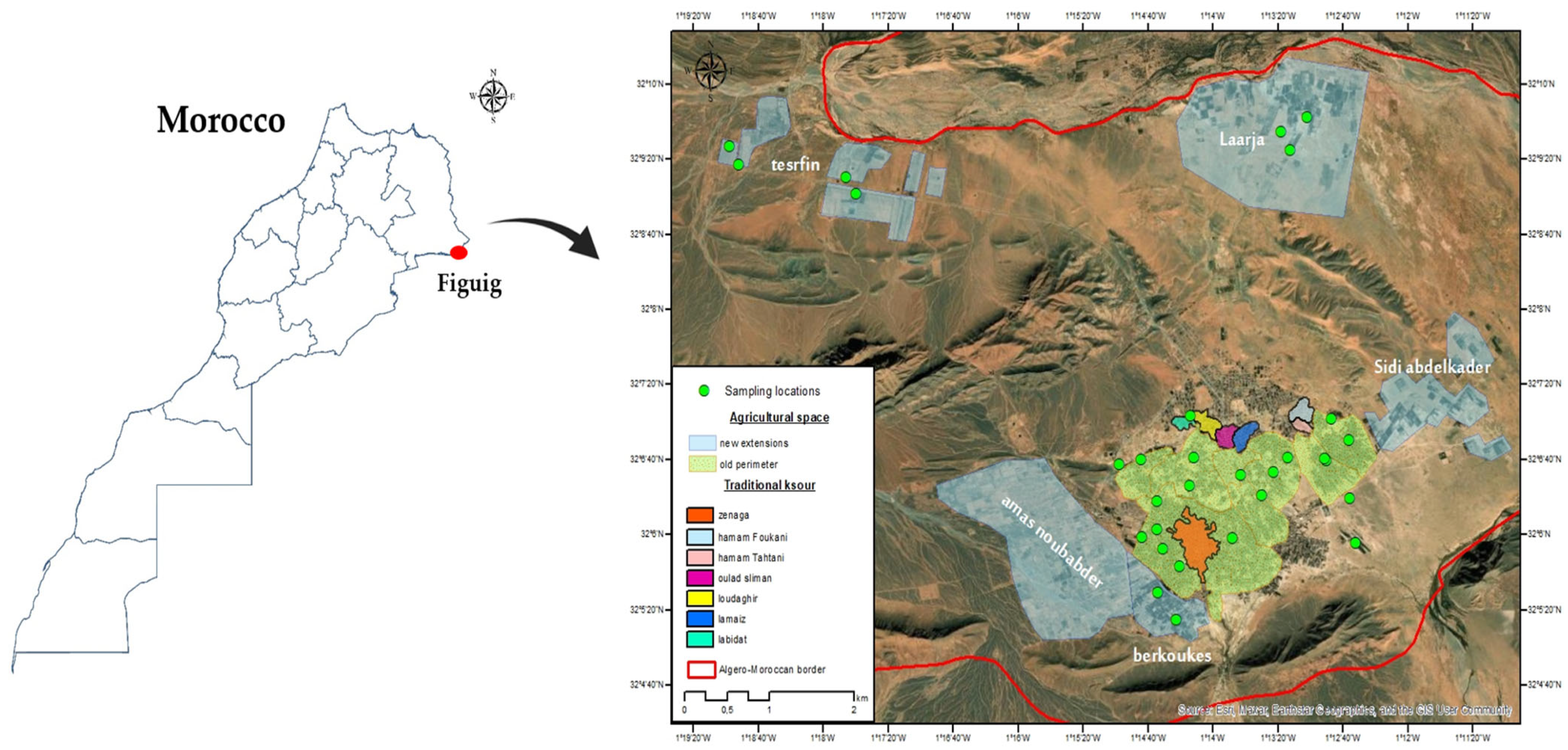
Figure 1. Geographical distribution of soil sampling sites in the Figuig oasis, Morocco (location of traditional ksour—fortified villages).
Diversity 17 00710 g001
Figure 2. Inoculation of P. lanceolata seedlings with single AMF spores under a stereomicroscope. (A) AMF single spore carefully isolated and applied onto the root surface of P. lanceolata. (B) Cluster of AMF spores of same morphotype attached and applied onto the root. Scale bars: (A) = 200 µm; (B) = 500 µm.
Diversity 17 00710 g002
Figure 3. Microscopic observation of AMF structures confirming successful colonization in trap cultures. (A) AMF spores extracted from successfully established trap cultures, showing various morphotypes with associated hyphal structures. (B) Stained root section of P. lanceolata exhibiting characteristic AMF colonization structures, including intraradical hyphae (h), spores (s) and vesicles (v). Scale bars: (A) = 500 µm; (B) = 500 µm.
Diversity 17 00710 g003
Figure 4. Morphological characteristics of Rhizophagus sp. spores isolated from the rhizosphere of P. dactylifera in the Figuig oasis. (A) Extraradical spores from pot culture forming clusters attached to roots; (B) intact mature spores mounted in PVLG + Melzer’s reagent, globose to subglobose, with variable diameters (80–150 µm); (C) spore in PVLG showing straight subtending hypha (sh) with continuous wall connection; (D) crushed spore in PVLG displaying three distinct wall layers (SWL1–SWL3) and subtending hypha attachment. Scale bars: (A) = 500 µm; (B) = 100 µm; (C) = 50 µm; (D) = 50 µm.
Diversity 17 00710 g004
Figure 5. Morphological characteristics of Glomus sp. spores isolated from the rhizosphere of P.dactylifera in the Figuig oasis. (A) Extraradical spores associated with hyphae from pot culture; (B) Crushed spore in PVLG showing two distinct wall layers (SWL1–SWL2) and a subtending hypha (sh) continuous with the innermost wall layer; (C) Intact spore in PVLG with cylindrical subtending hypha (sh) and bilaminate wall (SWL1–SWL2); (D) Cluster of mature spores, globose to subglobose, yellow-brown to dark brown in color, with diameters ranging from 60 to 120 µm, observed crushed in PVLG + Melzer’s reagent. Scale bars: (A) = 100 µm; (B) = 100 µm; (C) = 50 µm; (D) = 100 µm.
Diversity 17 00710 g005
Figure 6. Morphological characteristics of Funneliformis sp. spores isolated from the rhizosphere of P.dactylifera in the Figuig oasis. (A) Mature spores with peridium from pot culture, aggregated in clusters with orange coloration; (B) intact spore in PVLG showing three distinct wall layers (SWL1–SWL3) and a cylindrical subtending hypha (sh); (C) crushed spore in PVLG revealing internal compartments and cytoplasmic content; (D) spore wall in PVLG + Melzer’s reagent showing clear stratification of three wall layers (SWL1–SWL3) and subtending hypha attachment. Scale bars: (A) = 200 µm; (B) = 50 µm; (C) = 50 µm; (D) = 50 µm.
Diversity 17 00710 g006
Figure 7. Morphological characteristics of Scutellospora sp. spores isolated from the rhizosphere of P.dactylifera in the Figuig oasis. (A) Intact mature spore in PVLG showing external wall stratification; (B) crushed spore in PVLG with subtending hypha (sh) and bilaminate wall layers (SWL1–SWL2); (C) sporiferous saccule in PVLG + Melzer’s reagent attached to a spore, showing hyaline structure; (D) close-up in PVLG + Melzer’s reagent highlighting separation between rigid outer wall and laminate wall. Scale bars: (A) = 50 µm; (B) = 50 µm; (C) = 50 µm; (D) = 50 µm.
Diversity 17 00710 g007
Figure 8. Maximum likelihood phylogenetic tree of AMF isolates based on SSU–ITS–LSU rDNA sequences. Using the maximum likelihood (ML) method, the evolutionary relationships between AMF isolates and reference sequences from GenBank were analyzed. The reference consensus sequences (in black) are as described by Krüger et al. [49]. The sequences of our indigenous isolates are shown in color. Bootstrap values (%) are indicated at the nodes to assess the robustness of each clade. The scale bar represents the number of substitutions per site. The tree was generated, visualized, and edited using MEGA 11.
Diversity 17 00710 g008
Disclaimer/Publisher’s Note: The statements, opinions and data contained in all publications are solely those of the individual author(s) and contributor(s) and not of MDPI and/or the editor(s). MDPI and/or the editor(s) disclaim responsibility for any injury to people or property resulting from any ideas, methods, instructions or products referred to in the content.