Article

Epibenthic Dinoflagellates in the Southern Gulf of California: Species Composition and Abundance

by
Yuri B. Okolodkov
1,*,
Ismael Gárate-Lizárraga
2,
Victor A. Cervantes-Urieta
3,
Manuel E. Martínez-Cruz
1 and
Citlalli Galicia-García
4
1
Laboratorio de Planctología y Botánica Marina, Instituto de Ciencias Marinas y Pesquerías, Universidad Veracruzana, Mar Mediterráneo 314, Fracc. Costa Verde, Boca del Río 94294, Veracruz, Mexico
2
Laboratorio de Fitoplancton, Departamento de Plancton y Ecología Marina, Instituto Politécnico Nacional, Centro Interdisciplinario de Ciencias Marinas, Av. Instituto Politécnico Nacional, s/n, Col. Playa Palo de Santa Rita, La Paz 23096, Baja California Sur, Mexico
3
Facultad de Ciencias Ambientales, Universidad Autónoma de Guerrero, Carretera Cayaco-Puerto Marqués, Ejido Llano Largo 56-58, Acapulco 39906, Guerrero, Mexico
4
Departamento de Ingeniería Química y Bioquímica, Instituto Tecnológico de Boca del Río-Tecnológico Nacional de México, Km 12, Carretera Veracruz-Córdoba, Boca del Río 94290, Veracruz, Mexico
*
Author to whom correspondence should be addressed.
Diversity 2025, 17(10), 674; https://doi.org/10.3390/d17100674
Submission received: 21 July 2025 / Revised: 10 September 2025 / Accepted: 22 September 2025 / Published: 26 September 2025
(This article belongs to the Special Issue Diversity, Phylogeny and Ecology of Marine Microorganisms)

Abstract

Bahía de La Paz is the largest bay in the southern Gulf of California. This bay is an important area with a variety of commercial fish species and other natural resources and recreational activities. Epibenthic dinoflagellates are common inhabitants of harbors, inlets and semi-enclosed coastal lagoons; they produce potent toxins that may negatively affect human health and marine biota. The purpose of the present study was to identify potentially harmful epibenthic dinoflagellates growing on macroalgae from different coastal sites of the bay to determine their species composition, abundances, seasonal distributions, interannual and spatial variations. A total of 153 quantitative samples were collected in 2015–2019 (at 10 sites during four samplings in May, June and December) mainly from macroalgae. About 23 dinoflagellate species from the genera Prorocentrum, Ostreopsis, Sinophysis, Gambierdiscus, Fukuyoa, Amphidinium, Blixaea, Bysmatrum, Cabra, Coolia, Durinskia and Plagiodinium were found as epiphytes on at least 58 macroalgal species of 42 genera. Toxigenic genera, such as Gambierdiscus, Ostreopsis, Coolia and Prorocentrum, were widespread throughout the study area. Playa El Tecolote and Playa Costa Baja were the best habitats for dinoflagellates; therefore, the two locations can be considered the beaches with the greatest risk to human health.

1. Introduction

Harmful algal blooms (HABs) have been increasing globally in extension, species diversity and economic impact [1,2,3,4,5], although it was recently shown that the perceived global trends are poorly evidenced and that they should be considered regional [6]. Epibenthic dinoflagellates (mostly sand-dwelling, but also epiphytic and epizoic) [7] are found globally and include many toxigenic species (including the causative agents of ciguatera fish poisoning—CFP) that negatively impact human health and marine animals. In addition to the impact caused by epiphytic dinoflagellates blooms, macroalgal blooms can also have deleterious effects. These can produce a large amount of biomass that can be stranded on beaches along the coastline where it decomposes, triggering severe ecological impacts. Joniver et al. (2021) [8] pointed out that macroalgal blooms had a clear negative effect on aquatic plants (including seagrasses), fish and benthic invertebrates. Similarly, the decomposition of micro- and macroalgae blooms on various beaches in Bahía de La Paz may cause unpleasant smells impacting public access to the beaches, as well as ecological, economic, and human-health issues.
Marine benthic dinoflagellates have been much less studied than marine planktonic ones, and their role in marine food webs is still poorly understood. Over the past two decades, many benthic dinoflagellate species have attracted increasing attention as the causative agents of benthic harmful algal blooms (BHABs) and related syndromes, such as CFP and diarrhetic shellfish poisoning (DSP) in humans through consumption of seafood. Less than 10% of the total number of the known dinoflagellates appear to be benthic [9,10]. In general, 244 dinoflagellate species from 63 genera had been described by 2023 [7]. To our knowledge, marine dinoflagellates species have been found in all the oceans of the planet except the Antarctic Ocean in a range of habitats such as sand-dwelling, epiphytic on macroalgae, seagrasses and mangrove roots and floating detritus, in tide pools and subtidal areas, as epizoic beings attached to corals and other invertebrates, and also epilithic.
Epibenthic dinoflagellates are common inhabitants of harbors, inlets and semi-enclosed coastal lagoons. The known toxic benthic dinoflagellates reported for Latin America are within the genera Gambierdiscus R. Adachi et Y. Fukuyo, Fukuyoa F. Gómez, D.X. Qiu, R.M. Lopes et Senjie Lin, Ostreopsis Johs. Schmidt, Prorocentrum Ehrenberg, Coolia Meunier and Amphidinium Hulburt [11,12]; also see references in them. Such benthic harmful algal blooms (BHABs) have been described from tropical and subtropical coastal environments [13]. Few studies have been conducted on the BHABs along the Baja California Peninsula. Macroalgal assemblages in Bahía de La Paz usually comprise the phyla Rhodophyta, Ochrophyta (class Phaeophyceae) and Chlorophyta, with the families Rhodomelaceae and Ceramiaceae being the most abundant [14].
There are a few investigations regarding epibenthic dinoflagellates in the study area [15,16,17,18,19]. About 50% of them are master’s theses dealing with the species composition, toxins, allelopathy, morphological and molecular characteristics of individual species, with an emphasis on Coolia, Gambierdiscus, Ostreopsis and Prorocentrum spp. All 240 ciguatera cases (including 200 in the La Paz area) reported from 1984 to 2013 in the Mexican Pacific have been documented in the state of Baja California Sur; the toxicity was assessed by mouse bioassay, as well as by immunoassay and HPLC [20].
Some benthic dinoflagellates have been reported in this bay because of the resuspension processes [21,22,23]. This process has favored the proliferation (blooms) of few benthic dinoflagellate species, such as Prorocentrum rhathymum, P. lima, Blixaea quinquecornis and Amphidinium cf. carterae, without consequences for the marine biota. Nevertheless, some of these blooms have contributed substantially to the phytoplankton biomass (13.20 to 17.75 mg/m chlorophyll-a) [22].
The goal of the present study was to identify potentially harmful benthic dinoflagellates growing on macroalgae from different coastal sites in the southern Gulf of California to carry out an assessment of their specific composition, cell abundances, seasonal distribution and their relationship with the macroalgal substrate, thus evaluating the risk of CFP in the study area.

2. Materials and Methods

2.1. Study Area

Bahía de La Paz is located on the Gulf of California western coast (24°28′27″ N, 110°33′2″ W), delimited by Isla San José to the north (24°58′23″ N, 110°36′52″ W), the Baja California Peninsula to the west and south, and Isla Espiritu Santo and Isla La Partida to the east (Figure 1). Bahía de La Paz is the largest bay on the Baja California Peninsular side of the Gulf of California. The bay constantly exchanges water with the latter via a northern and a southern opening [24]. Bahía de La Paz is subject to two main wind patterns. Southerly and southeasterly winds, locally called Coromuel, prevail from late spring to early autumn, with magnitudes of 4 m/s combined with frequent calm periods. Strong and persistent northerly and northwesterly winds prevail in late autumn and winter, reaching velocities of 12 m/s. Based on the temperature in the 0–25 m surface layer, two seasons are distinguished: the cold season (December to May, 22.5 ± 4.4 °C) and the warm season (July to November, 28.4 ± 1.9 °C) [25]. Similarly, in another study of the surface water, the temperate season (April to June, 23.55 ± 1.35 °C) and the warm season (July to September, 30.25 ± 0.95 °C) are distinguished [26].
Recently, the surface sediments of the eastern coast of Bahía de La Paz were studied, and the seafloor-unit-classification proposal was made [27]. This region corresponds to sites 3 to 7 in our study area. The surface sediments at our sampling sites were coarse (0.5–1 mm) and very coarse (1–2 mm) sands. Sites 3 to 6 (Figure 1) are within the Punta Coyote zone, while site 7 (Playa El Tecolote) belongs to Canal de San Lorenzo; the latter is characterized by coarse to very coarse sands with more than 90% CaCO3 and the presence of bioclasts formed by mollusk, coral, rhodolith and foraminifera fragments. At sites 7 and 2 (Playa El Califin), rocky substrate was common. The southern area of the bay, called Laguna de La Paz, separated from the rest of the bay by El Mogote sand barrier, which contains a wide range of sediments from coarse sands to fine limes of fluvial and eolic origin, was not sampled in this study.

2.2. Sampling

Before sampling, a visual survey of the area was conducted. With the aim of studying the epibenthic dinoflagellate taxocoenosis, 153 quantitative samples were collected from different substrates (mainly green, brown and red macroalgae attached to the sea bottom, as well as sediments, mangrove roots and floating macroalgae) at 10 sites (corresponding to 8 sites on the map below) along the western coast of the southern Gulf of California (9 sites in Bahía de La Paz from Playa El Califin to Playa El Tecolote and a site beyond the bay) between 09:15 and 13:30 on 22 May 2015; 15, 21 and 22 June 2016; 16, 23 and 30 June 2018; and 7 December 2019. Macroalgae were collected manually at nine selected sites in Bahía de La Paz and a site in Ensenada de Muertos by snorkeling (Figure 1). Recurrent sampling in different years was performed only at Playa El Tecolote. At Balandra, samples were taken at two separate sites. Additionally, three mangrove root samples were collected at Playa El Tesoro I and II, the two inlets surrounded by mangroves.
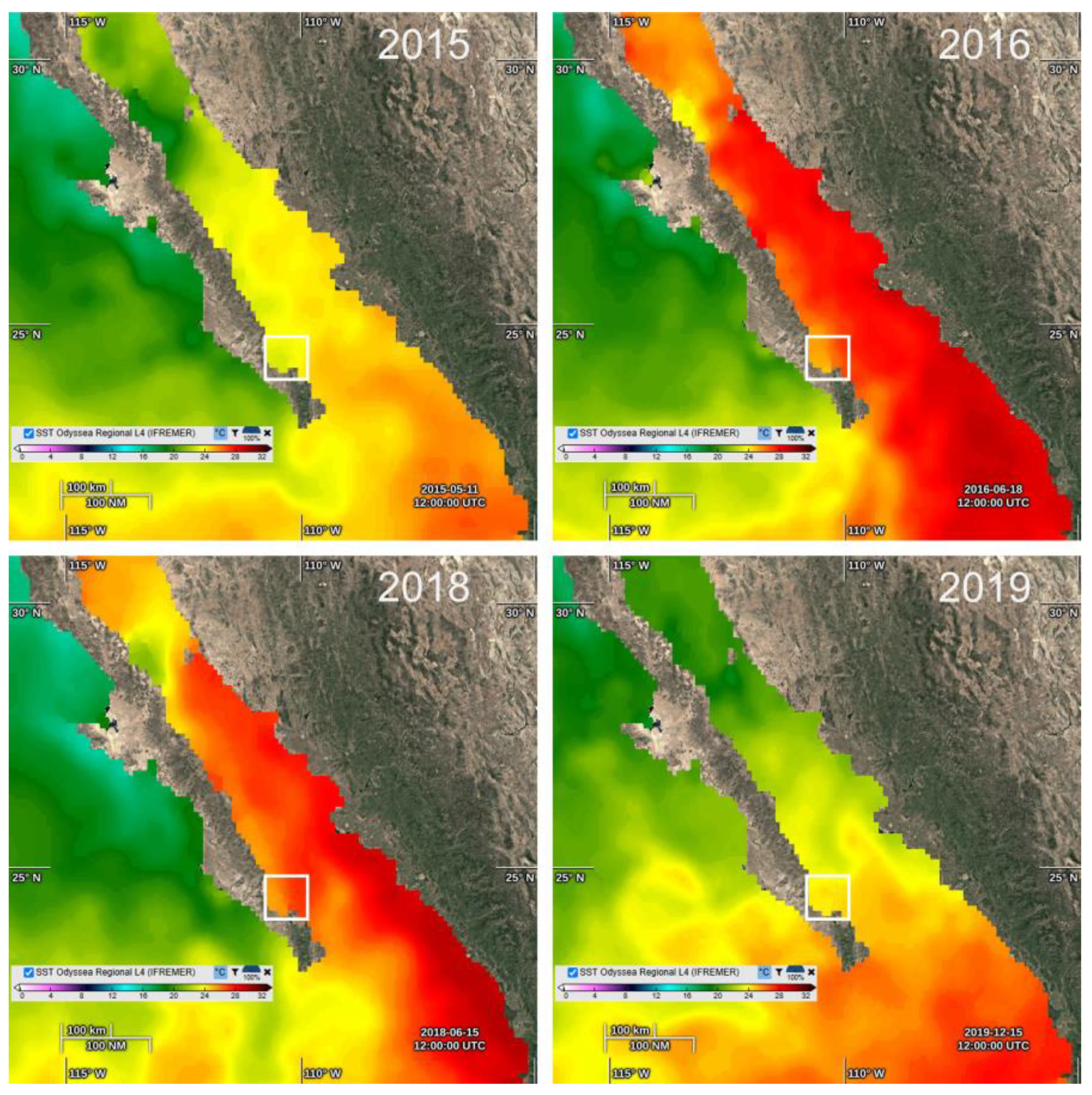
The samples were taken in shallow water (down to 1.5 m depth) with a temperature of 21.5–25.0 °C and salinity of 35–36. Additionally, the data of the sea surface temperature were obtained from online resources using the Ocean Virtual Laboratory [28] (Figure 2). Level 4 interpolated temperature products are from ODYSSEA/Ifremer. Macroalgae were detached gently from the sea bottom, manually or using a knife, and fixed with formalin (stock solution of 37%) to a final concentration of 4%.

2.3. Laboratory Analyses

Before cell counting in a 1 mL Sedgwick–Rafter chamber, a sample was vigorously shaken for one minute. Cells were counted using an Olympus CKX41 (Olympus Optical Co., Ltd., Tokyo, Japan) inverted microscope equipped with the phase-contrast objectives; the whole chamber or a part of it was scanned, depending on the total dinoflagellate cell abundance. Smaller cells of the genera Amphidinium, Cabra Sh. Murrat et D.J. Patterson emend. Chomérat, Couté et Nézan and Plagiodinium M.A. Faust et Balech were counted using the 40× objective, while larger cells of other genera were counted using the 10× objective. The results were expressed as the number of cells per gram (cells/g) of wet weight substrate (WWS). For taxonomic identification of dinoflagellates, both light and scanning electron microscopy were used together with the specialized literature [7,29,30,31]. The nomenclature of the algal taxa was updated following [7,32].
An epifluorescence compound microscope Axio Scope.A.1 (Carl Zeiss, Oberkochen, Germany) equipped with an Axiocam 506 color six-megapixel digital camera was used to observe the cell shape and size, as well as the thecal plate arrangement. Staining was performed with Calcofluor White M2R 0.2% [33] in epifluorescence, and Trypan Blue 0.2% [34,35] in bright field.
A JEOL JSM-7600F field emission scanning electron microscope (FESEM; JEOL, Ltd., Tokyo, Japan) was used at 5 kV at a working distance of 17.2–20.6 mm in the LEI regime to study the thecal morphology of dinoflagellate species in some samples. The samples were preliminarily washed in distilled water, followed by dehydration in a series of ethanol solutions of increasing concentration (30, 50, 70, 90 and 100%—twice in the latter). Subsequently, cells were air dried on 0.5″ aluminum mounts and sputter-coated with gold–palladium using a Polaron SC7640 high-resolution sputter coater (Quorum Technologists, Newhaven, SXE, UK).
Morphological features of the entire macroalgal thalli were observed using a stereomicroscope of low magnification Motic SMZ-168 (Kowloon, Hong Kong, China). When necessary, cross-sections of algal thalli were made and observed using a compound Motic B3 microscope (Kowloon, Hong Kong, China). Specimens were identified using the specialized literature [36,37,38,39]. The status of the current names for each species, as well as the synonyms, were checked using AlgaeBase [32] and a checklist of macroalgae [40].

2.4. Data Analysis

All analyses were performed in an open-source environment in R (version 4.2.2) and RStudio (1.6.4) [41]. Statistical differences between different cell abundances of epiphytic dinoflagellates, different sampling sites and years of study were analyzed using a two-level permutational multivariate test (PERMANOVA) [42]. Alpha diversity was estimated using the ecological descriptors: Shannon diversity, Pielou evenness, Simpson dominance and richness and evaluated with two-way PERMANOVA. Differences in the species assemblage composition between sites and years (beta diversity) were analyzed using Bray–Curtis dissimilarity on log-transformed abundance data. Structural patterns in taxonomic composition were investigated by performing a non-metric multidimensional scaling (NMDS) analysis on the beta diversity dissimilarity matrix [43].
A one-way analysis of similarities (ANOSIM) test was performed to determine whether the species composition of epiphytic dinoflagellates assemblages differed significantly between study years. Similarity percentage analysis (SIMPER) was then used to determine dinoflagellate taxa that accounted for the largest observed differences between NMDS assemblage patterns. Species contributing > 2% of the average dissimilarity within a single assemblage pattern by the mean of each pairwise comparison between years were considered potential “indicator” taxa that are more representative.
An interaction analysis based on UpSetR visualization matrices was used to assess and identify the years that shared or showed exclusivity of cell abundances and the different genera of epiphytic dinoflagellates across the years. The effect of different sampling sites was analyzed using a two-way cluster analysis and visualized in a full-linkage heatmap for each sampling site and years with spatial variation. The abundances of different species were first log-transformed (log + 1). Different clusters were selected with more than 30% similarity. Analysis and visualization were performed using the heatmap function of the ‘stats’ (version 4.6.0) package.
Dinoflagellate–macroalgae association was evaluated using a differential ternary composition graph showing the distribution of dinoflagellates among the three major taxa of macroalgae: Chlorophyta, Rhodophyta and Ochrophyta (Phaeophyceae). The graph was constructed using the ggtern function and package with confidence intervals of 95%.

3. Results

3.1. Species Composition of Macroalgae

In the sampling sites during the period of 2015–2019, no seagrasses were found, so samples of the epiphytic dinoflagellates were taken only from macroalgae. Species composition of macroalgae differed between years and sites (Table 1). The number of species of Rhodophyta was more than twice of that of Phaeophyceae and Chlorophyta combined. Many taxa were identified only to the generic level and some to the family level. In the genera Laurencia J.V. Lamouroux, Dictyota J.V. Lamouroux and Padina Adanson, two or three morphologically different species were identified in each of them. In total, at least 58 macroalgal species of 42 genera and 26 families were distinguished: 33 Rhodophyta species of 26 genera, 13 Phaeophyceae species of 10 genera, and 12 Chlorophyta of 6 genera. The thalli of all the examined species served as a habitat for epiphytic dinoflagellates.

3.2. Species Composition of Dinoflagellates

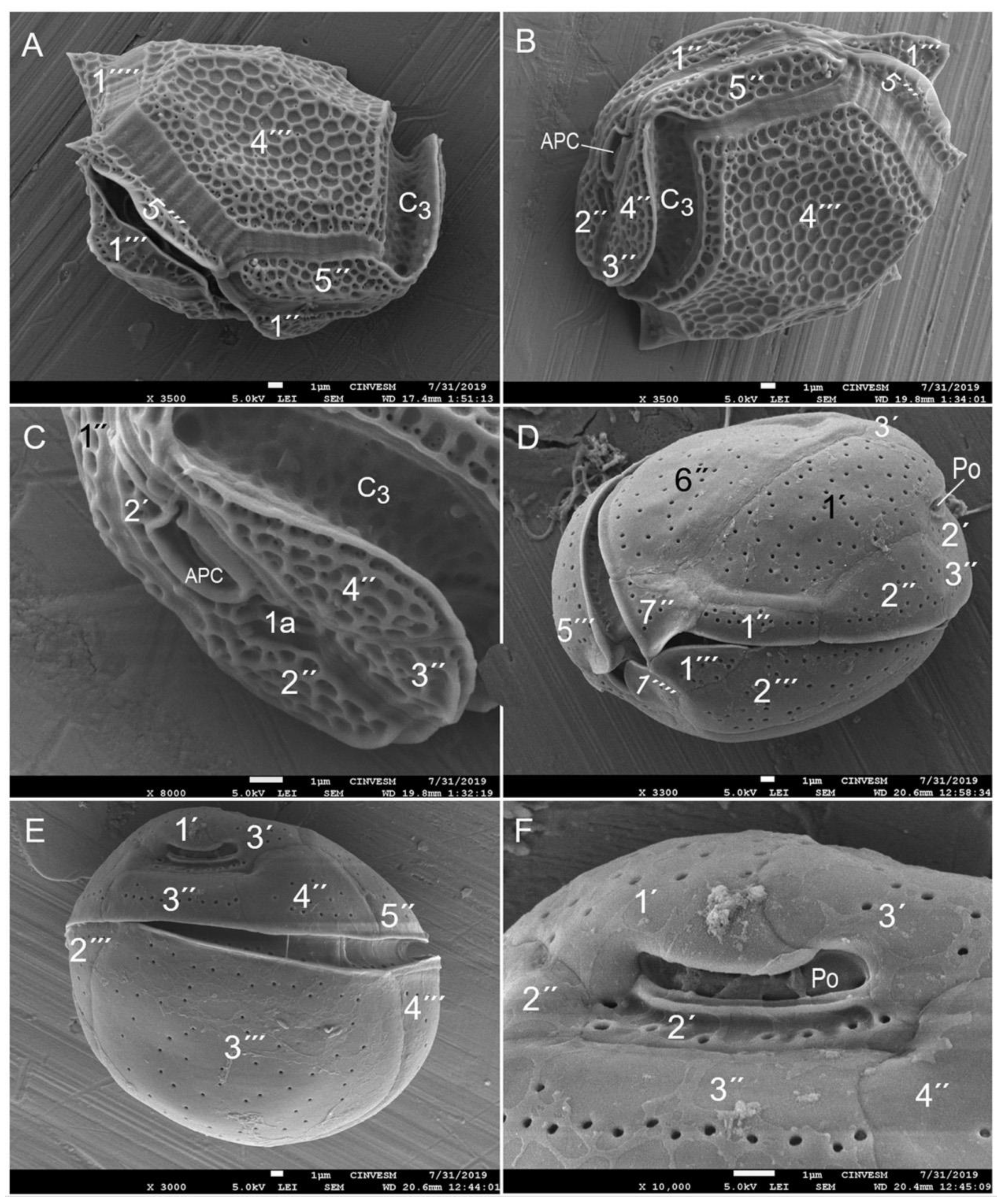
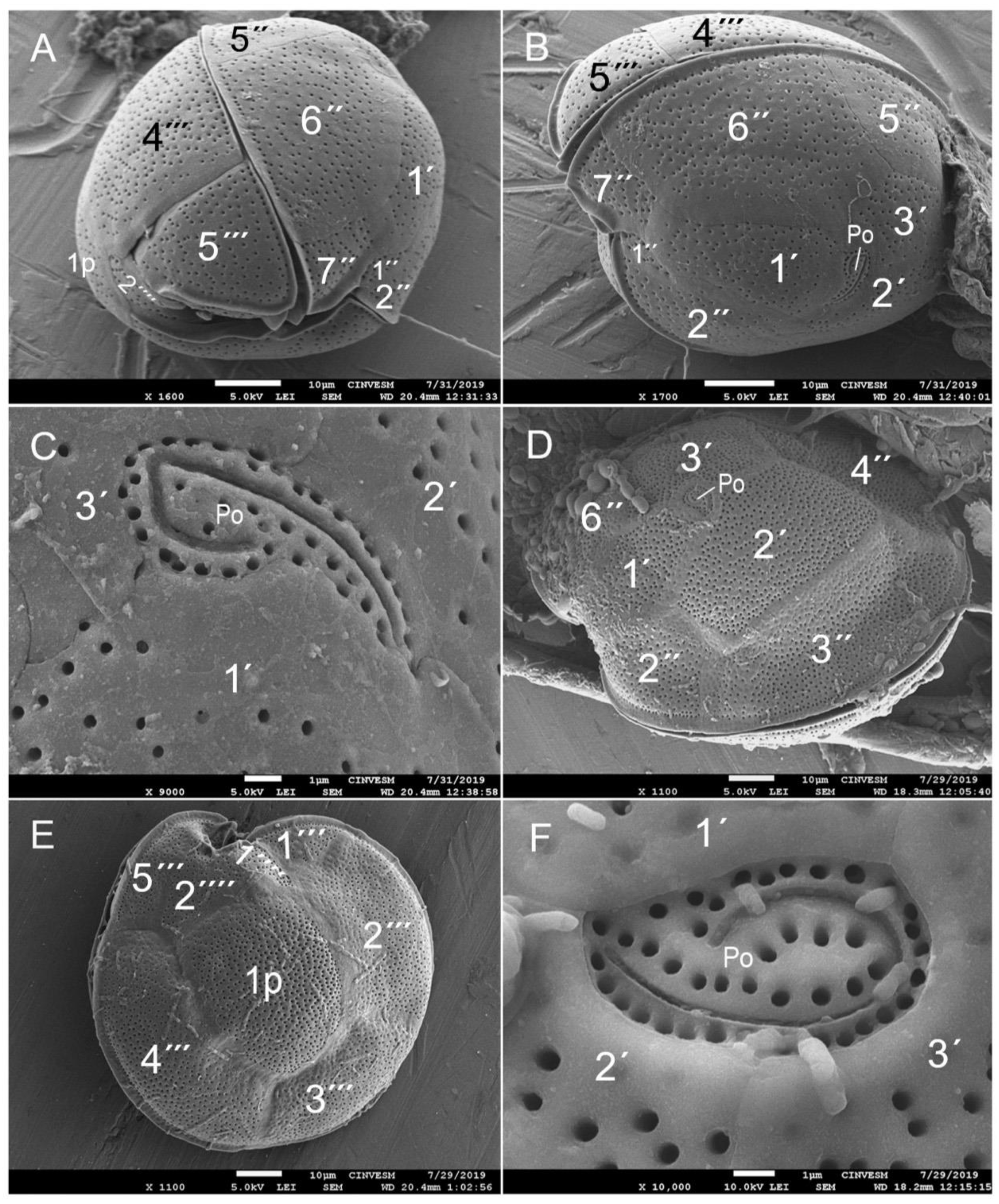
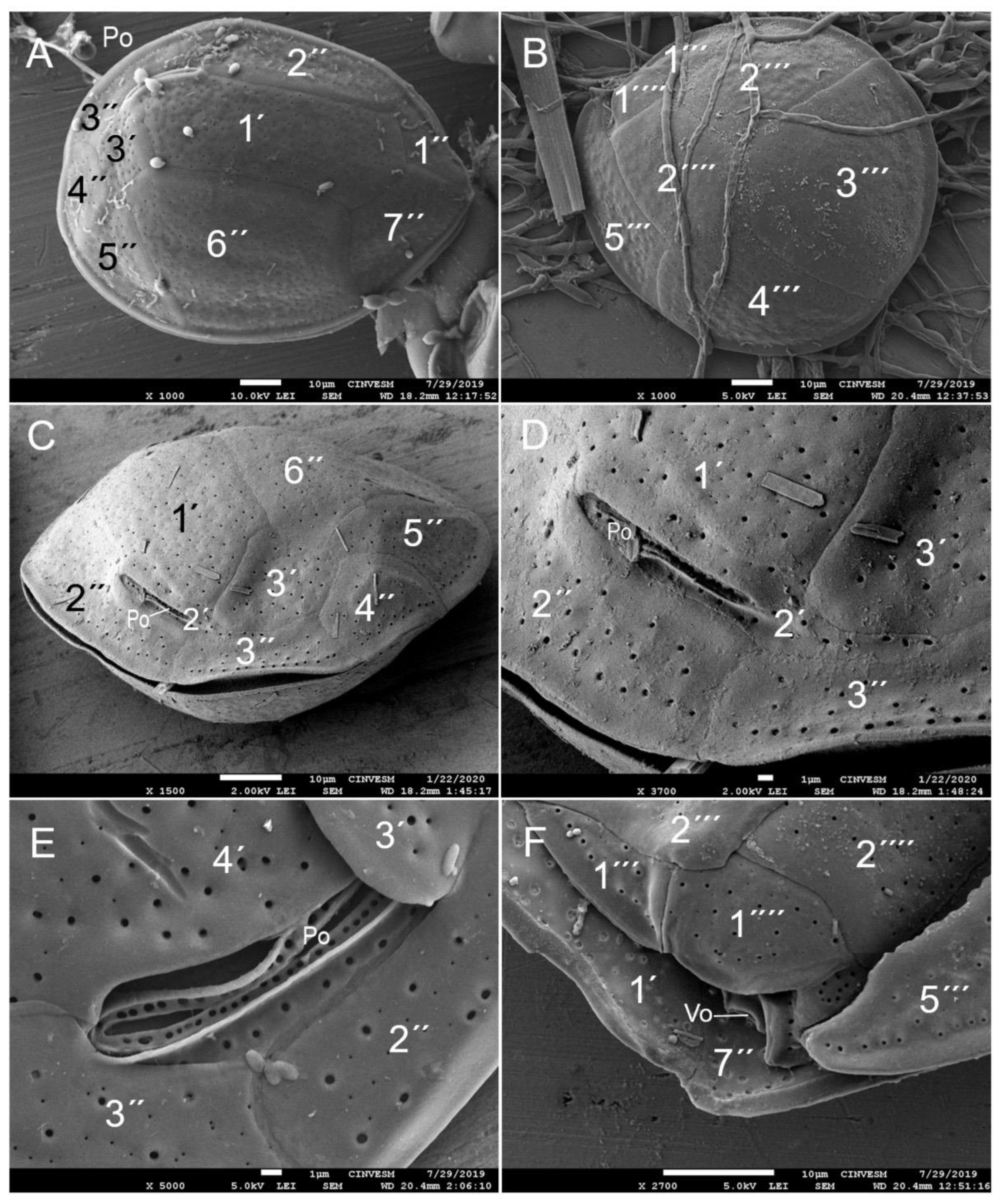
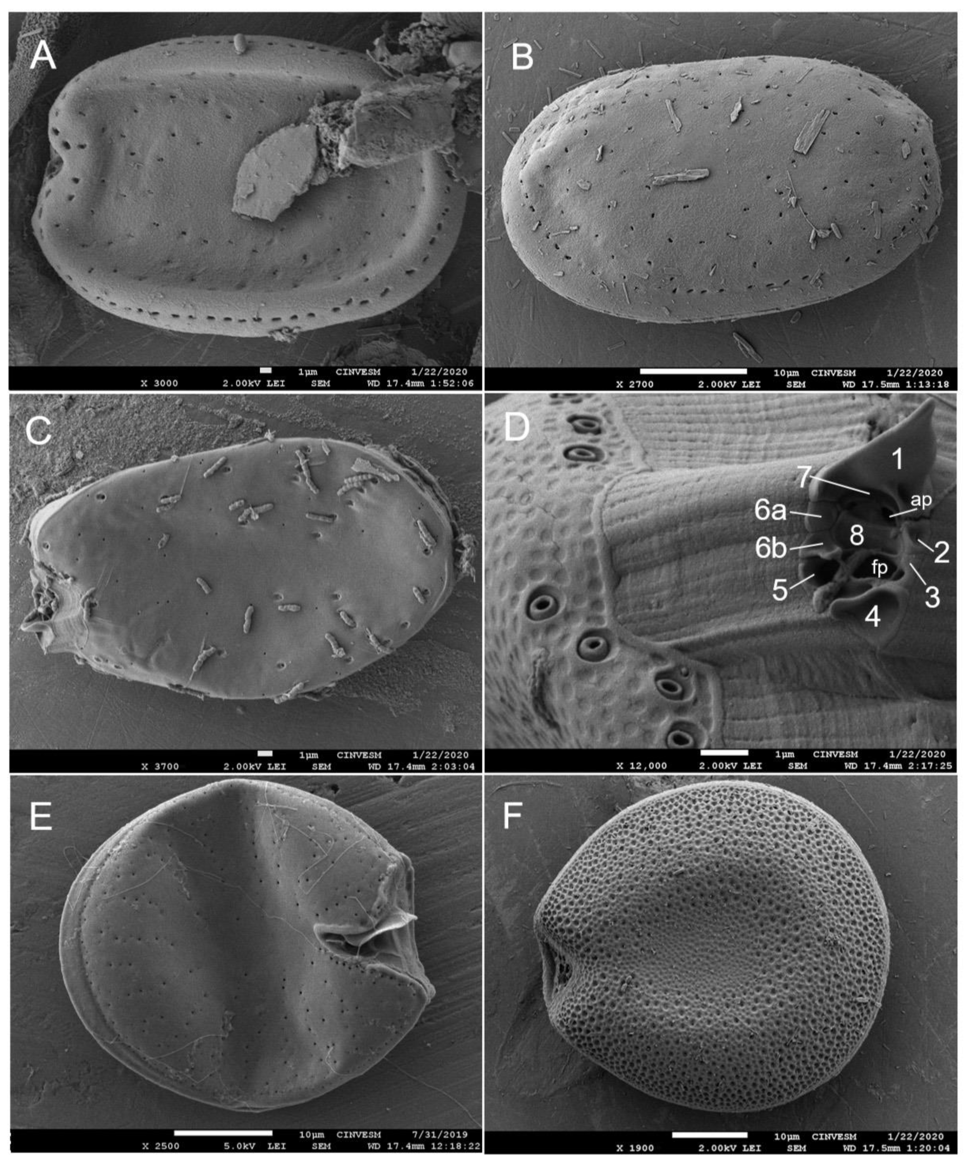
In total, about 23 dinoflagellate species from the genera Prorocentrum (8), Ostreopsis (3), Sinophysis Nie et Wang (3), Gambierdiscus (2), Fukuyoa, Amphidinium, Blixaea Gottschling, Bysmatrum, Cabra, Coolia Meunier, Durinskia S. Carty et E.R. Cox and Plagiodinium (one species each) were found (Figure 3, Figure 4, Figure 5 and Figure 6). Microscopic observations did not allow us to identify some taxa to species level.

3.3. Abundance of Dinoflagellates

The maximum cell abundance was observed at Playa El Califin in June 2016: 14,441 cells/g of macroalgal wet weight (WWS: wet weight substrate; Padina sp. with attached cyanobacterial clumps). Usually, Prorocentrum lima, P. rhathymum and P. sculptile were dominant (up to 76%). Gambierdiscus was dominant only once (64%; Playa El Tecolote, December 2019). Occasionally, Ostreopsis dominated (up to 94%; Playa El Tecolote, December 2019). In all 13 macroalgal samples taken at Playa El Tecolote in 2019, Gambierdiscus and Ostreopsis occurred; their abundance reached 5697 cells/g MWW; Ostreopsis contributed up to 5195 cells/g. Coolia reached rather high abundances on both sides of Bahía de La Paz (1979 cells/g at Playa El Califin on the west coast of the bay and 1433 cells/g at Playa El Tecolote on the east coast, with up to 42% of the dominance in May–June).

3.4. Interannual and Spatial Changes in Cell Abundance of Dinoflagellates

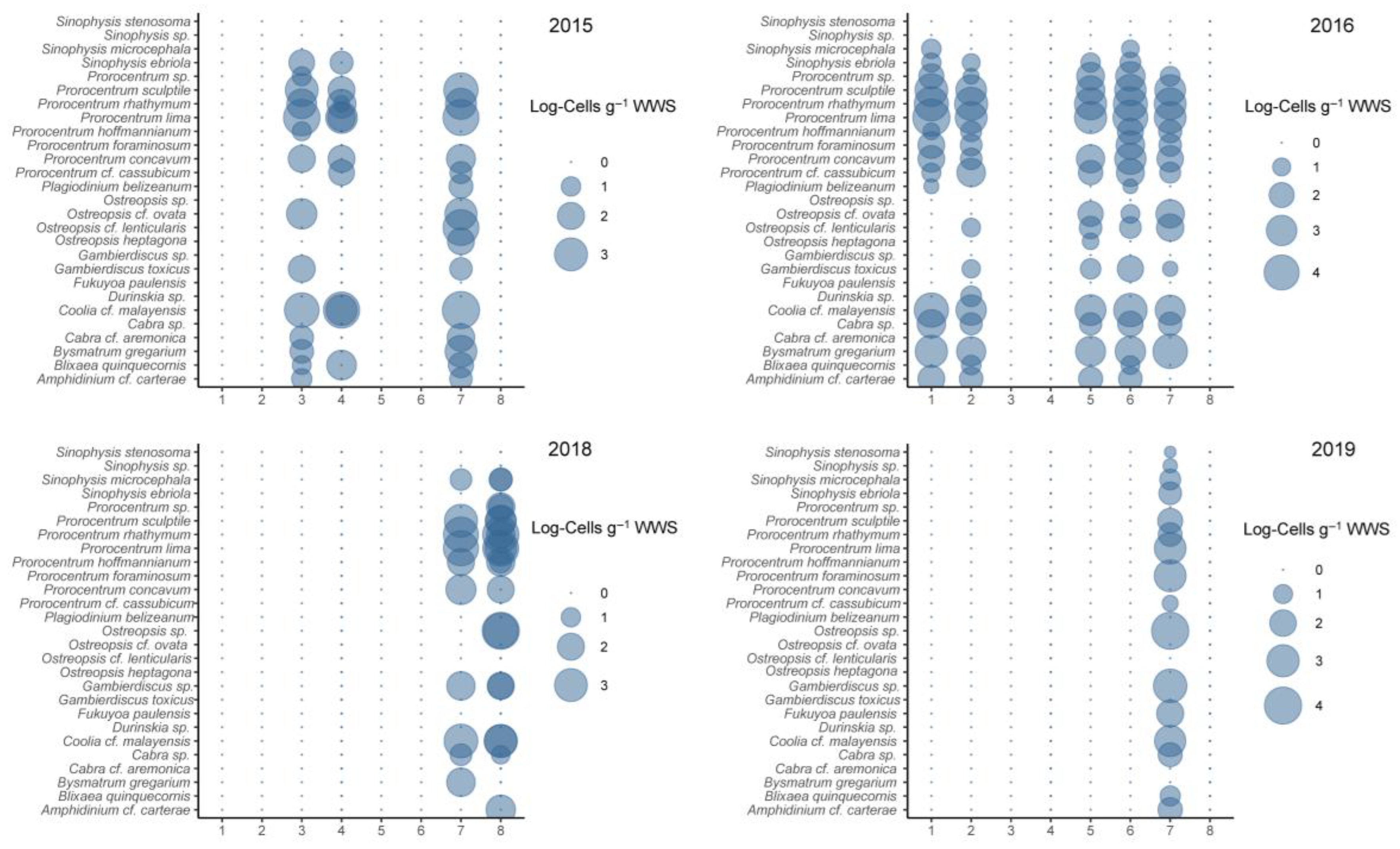
The results of the PERMANOVA analysis indicated that interannual (r2 = 0.02, p = 0.001) and spatial variation (r2 = 0.12, p = 0.001) induced changes in the abundance and structure of epiphytic dinoflagellates. In May 2015, the cell abundance was represented mainly by Coolia cf. malayensis, with 13,828 cells/g WWS (~3 orders of magnitude) (Figure 7), followed by Ostreopsis cf. lenticularis and Prorocentrum sculptile with 3669 cells/g and 2872 cells/g WWS, respectively. The lowest cell abundance was observed for Prorocentrum sp. and P. hoffmannianum with 7 to 8 cells/g WWS. Sites 7 and 3 showed the highest abundances, whereas site 4 showed the lowest cell abundance. Prorocentrum lima and P. rhathymum showed the highest abundances with 48,327 and 20,133 cells/g WWS in June 2016, in addition to 7438 cells/g and 7486 cells/g WWS in June 2018, followed by Coolia sp. with 14,064 cells/g WWS and Bysmatrum gregarium with 12,537 cells/g WWS in June 2016. Similarly, in 2016, sites 1 and 7 hosted the highest abundances. In June 2018, site 8 showed the highest abundance of epiphytic dinoflagellates. Ostreopsis and Gambierdiscus were the dominant genera in December 2019, with 10,172 cells/g and 1699 cells/g WWS, respectively. The least represented were Sinophysis stenosoma and Prorocentrum cf. cassubicum, with 1.9 cells/g and 3.8 cells/g WWS.

3.5. Seasonal Variation in Epibenthic Dinoflagellates

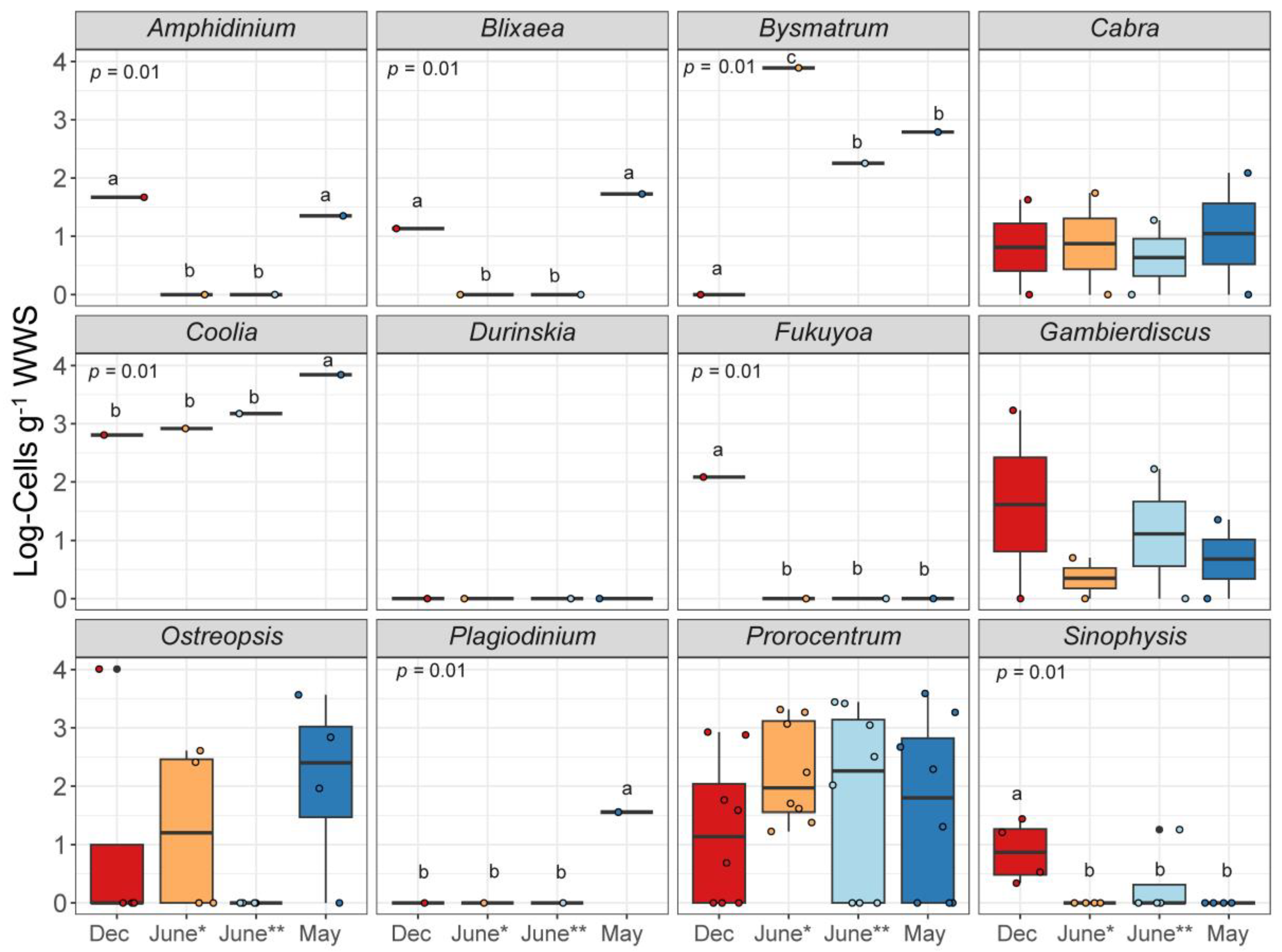
The genera Blixaea, Bysmatrum, Coolia, Fukuyoa, Plagiodinium and Sinophysis showed seasonal variation at the Tecolote site (H = 10.46, p = 0.01), respectively. However, no preference was observed for any season (month) in terms of increasing or decreasing abundance, so their behavior is differential throughout the months of study. The maximum variability was detected for the genera Prorocentrum, although it is associated with the richness within this genus. Bysmatrum showed a maximum abundance of four orders of magnitude in June 2016, followed by Coolia in May 2015 (Figure 8).

3.6. Alpha and Beta Diversity of Epiphytic Dinoflagellates

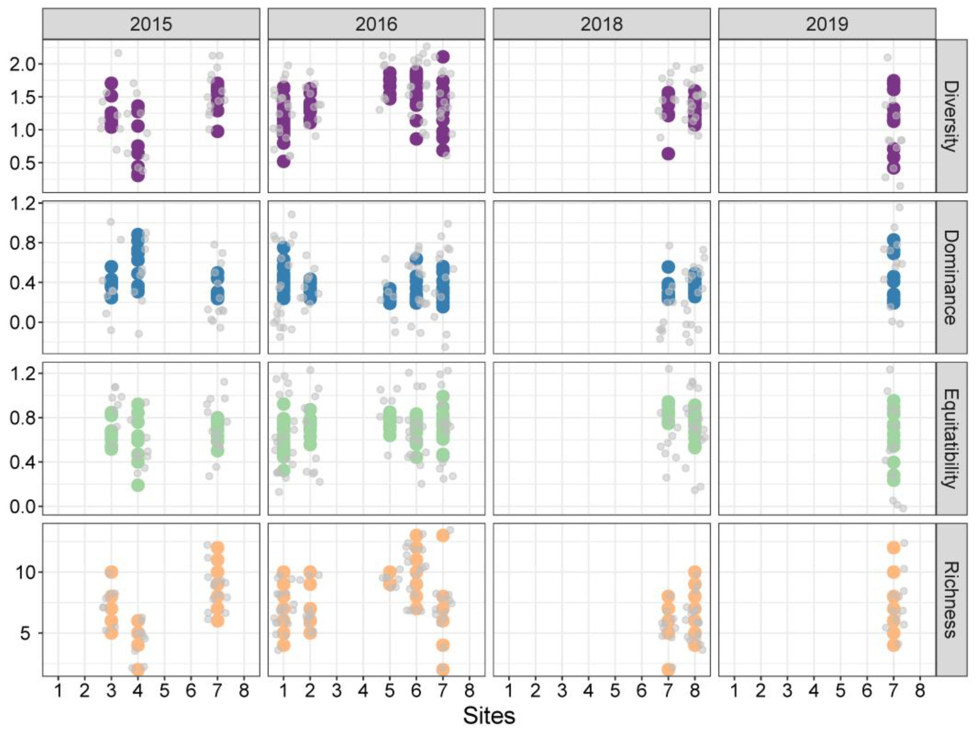
Shannon diversity (r2 = 0.11, p = 0.001), richness (r2 = 0.10, p = 0.001) and Simpson dominance (r2 = 0.08, p = 0.002) were influenced by interannual changes at all sampling sites (Table 2). During the four years of study, a total of 27 epiphytic dinoflagellate species from 12 genera of epiphytic dinoflagellates were observed. Six genera (Amphidinium, Prorocentrum, Coolia, Ostreopsis, Gambierdiscus and Fukuyoa) include potentially toxigenic species. Prorocentrum was the most represented genus (eight species), followed by Ostreopsis and Sinophysis genera with three species each. In June 2016, the highest richness (20 species) was observed (sites 6 and 7), while in June 2018 only 13 species were found (site 8). In May 2015 and December 2019, 16 and 17 species of epiphytic dinoflagellates, respectively, were observed (at site 7 in particular) (Figure 9).
The highest species diversity was observed in 2016, particularly at site 7, with values of 2.1 (maximal) and 1.7 (minimal), while the lowest diversity related to an increase in observed dominance occurred in 2015 at site 4 with diversity values of 0.3 and 0.4 and dominance of 0.82 and 0.88. In 2018 and 2019, the values of diversity and dominance were 0.2 and 0.8, respectively (Figure 9).
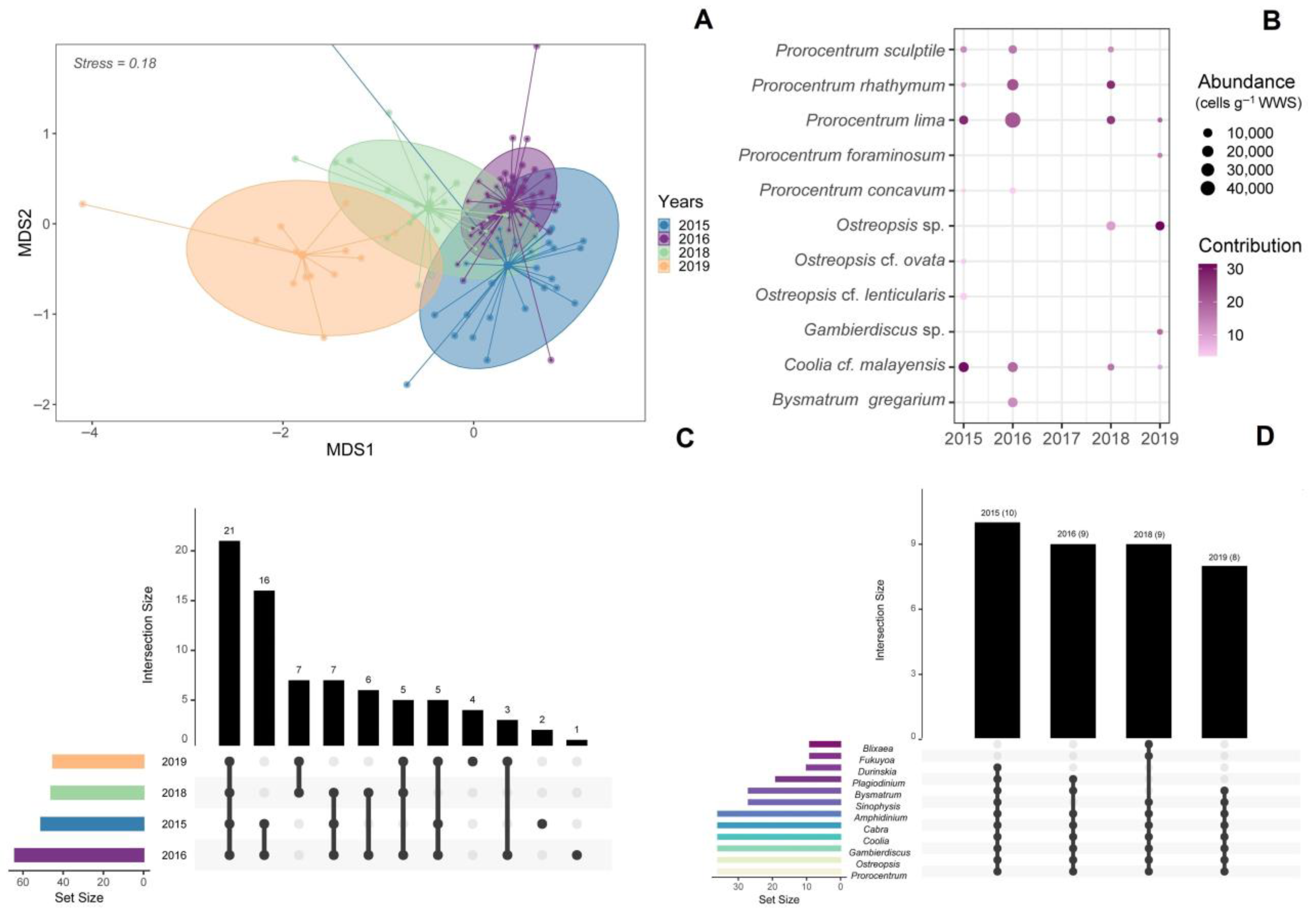
The ANOSIM analysis associated with the NMDS (Figure 10A) indicated that the epiphytic dinoflagellate taxocoenosis differed in the study years (dissimilarity > 0.65, Stres = 0.18, R = 0.57, p = 0.001), translating into the different patterns observed (p = < 0.001, Table 3).
SIMPER analysis indicated a total of 11 epiphytic dinoflagellate species that had contributed significantly to the composition of the different patterns observed. In 2015, Coolia cf. malayensis, Prorocentrum lima and P. sculptile contributed up to 70% of the taxocoenosis, while in 2016 and 2018, P. rhathymum, P. lima and Coolia cf. malayensis contributed up to 60.5% and 69%, respectively. In 2019, taxocoenosis was represented mainly by Ostreopsis sp., P. lima and Gambierdiscus sp. (68.2%; Figure 10B). Interaction analysis based on UpSetR visualization matrices (Figure 10C,D) indicated that Amphidinium cf. carterae, Coolia cf. malayensis, P. lima, P. rhathymum and P. sculptile were the only species with maximum abundances (together contributing up to 66,079 cells/g WWS) shared during the entire study period. In 2016, the highest number of shared abundances with seven interactions was observed, while in 2015, only four similar abundances were shared. The UpSetR indicated 10 of the 12 reported genera in 2015, 9 in 2018, and 8 genera in 2016 and 2019.
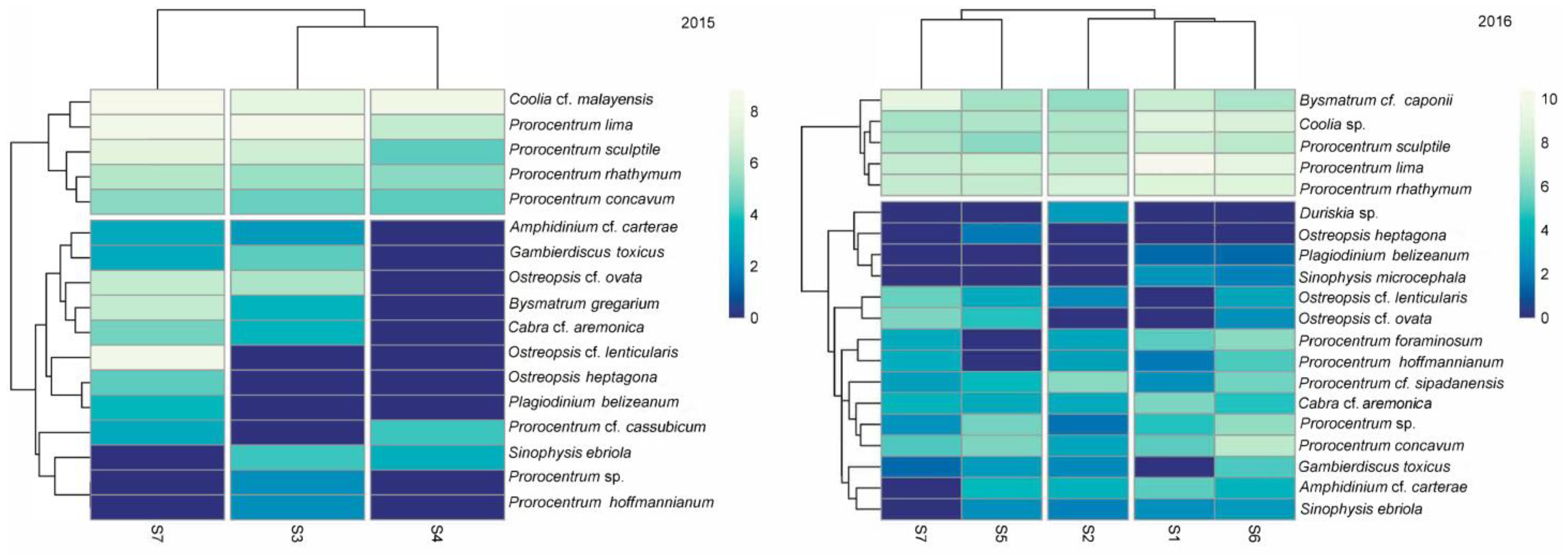
The Clustering-Heatmap applied to the years with the greatest spatial variability (2015 and 2016) indicated that in May 2015 sites 3 and 4 showed similar cell abundances. Similarly, in June 2016, sites 1, 2 and 7 formed the first similar group, and the second cluster was formed by sites 5 and 6. Prorocentrum lima, P. rhathymum, P. sculptile, P. concavum and Coolia cf. malayensis were the key species in the clusters in both years (Figure 11).

3.7. Dinoflagellate-Macroalgae Associations

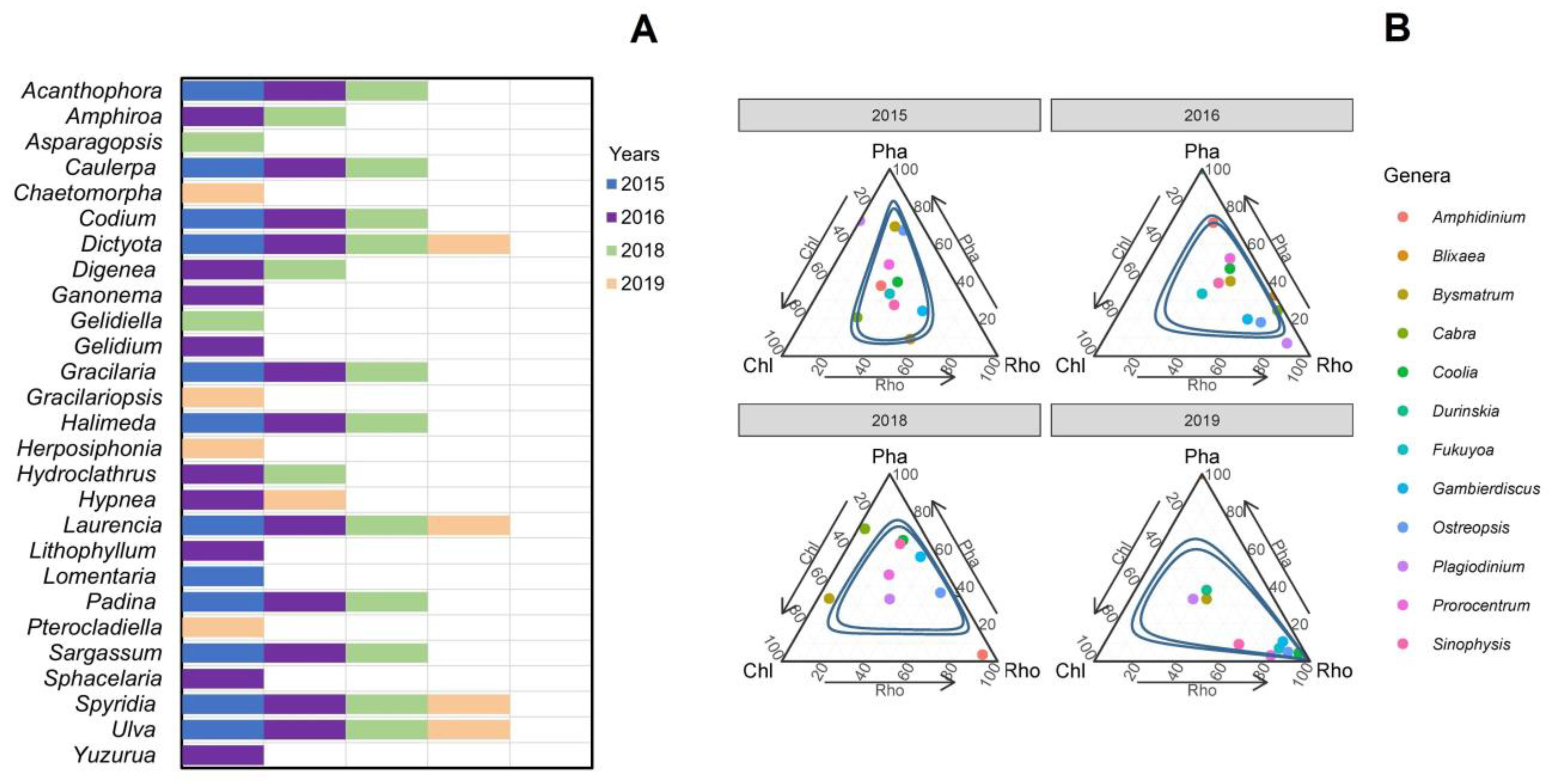
A total of 43 genera of macroalgae were identified; however, only the dominant genera (27) in the samples were plotted in Figure 12A. Of these, 17 belong to Rhodophyta, 5 to Phaeophyceae and 5 to Chlorophyta. Dictyota, Laurencia, Spyridia and Ulva showed the most extended distributions during the study period. The ternary graph revealed that 95% of the epiphytic dinoflagellate taxocenosis showed preferences for Rhodophyta (Spyridia filamentosa, Hypnea valentiae and Laurencia cf. masonii), mainly in 2016 and 2019 (Figure 12B).

4. Discussion

Regarding the data on sea surface temperature in the months of sampling (Figure 2), the temperature conditions were similar in June 2016 and 2018 and also in May 2015 and December 2019.

4.1. Dinoflagellate-Macroalgae Associations

During the study period, species of red algae as substrates were more than twice those of brown and green algae (Table 1), unlike in the shallow coastal zone of the southern Gulf of Mexico, where green algae and seagrasses are the most common substrate for epiphytic dinoflagellates [44,45,46,47]. In general, for the macroalgal flora of Bahía de La Paz, a similar prevalence of the red algae over brown and green algae was found [14].
In 2016, Blixaea, Cabra, Plagiodinium, Ostreopsis and Gambierdiscus were the genera that showed specific associations in the mutualistic spectrum, while in 2019 they were Duriskia, Fukuyoa, Gambierdiscus, Ostreopsis, Prorocentrum and Sinophysis. In 2015, the brown algae Sargassum sp., Padina sp. and Dictyota sp. were the preferred hosts for species of Plagiodinium, Ostreopsis and Bysmatrum. In 2018, no significant associations in comparison to other years were revealed. Therefore, epiphytic dinoflagellates in Bahía de La Paz did not show preferences for any specific macroalgae, and at the 95% confidence level they demonstrated generalist behavior (Figure 12B).
The preference of epiphytic dinoflagellates (individual species or the entire dinoflagellate taxocoenoses) for macrophyte substrate has been previously discussed in the literature; however, more studies are necessary. Some publications are dedicated to only one or two seagrass host species [44,45]. Others investigated individual dinoflagellate species, with a special emphasis on the CFP-causative species of the genus Gambierdiscus [46,47,48,49,50]. Apart from Gambierdiscus, Ostreopsis and Coolia are another two genera that have served as subjects of numerous studies in tropical and subtropical waters [7,50]; also see references in these reviews. Among toxigenic genera, Amphidinium has remained the least studied, morphologically, taxonomically and ecologically.
Various authors investigated epiphytic dinoflagellates in a range of macrophytes, most of which were macroalgal assemblages/taxocoenoses [51,52,53,54,55,56,57,58,59,60,61,62,63,64,65,66,67,68,69,70,71]. Conclusions of the authors who worked in different geographical regions differ as the species composition of macrophytes does. In some publications, an emphasis is put on the preference of macroalgal substrate in terms of major taxonomic groups (red, brown or green algae) or the preference between macroalgae and seagrass. Other authors study only one or several host or epiphytic species and compare the dinoflagellate cell abundances in different parts of the leaves (seagrasses) or thalli (macroalgae). Some investigations compare cell abundance between different morpho-functional types of macroalgae, considering the weight to surface area ratio. Rare studies are focused on the allelopathic effect of macroalgal extracts on epiphytic dinoflagellates or, in general, on allelopathic interactions between a macrophyte and dinoflagellates. Finally, sampling procedures and protocols of cell counting also affect the reliability of the results obtained. Selection between a natural or an artificial substrate to study cell abundances and preferences for substrate has also been under discussion (for references, see [45,71]).

4.2. Potentially Toxic Dinoflagellates

Although in the present study a number of species were not identified to the species level and toxicological analyses were not performed, the following taxa can be listed as potentially toxic: Gambierdiscus cf. toxicus, Gambierdiscus sp., Fukuyoa paulensis (ciguatoxins and maitotoxins); the Prorocentrum lima complex spp., the P. hoffmannianum complex spp., P. concavum (okadaic acid, diarrhetic shellfish toxins–dinophysistoxins and their analogs and others); Ostreopsis cf. ovata, O. cf. lenticularis, O. cf. heptagona (palytoxin, palytoxin-like compounds and ovatoxins); Coolia cf. malayensis (cooliatoxin, gambierone and yessotoxin analogs) and Amphidinium cf. carterae (species of this genus produce different cytotoxic, antibacterial and antifungal compounds). The toxins produced by these genera pose a major threat to marine ecosystems and human health [72]. The most recent reviews of benthic dinoflagellate toxins, including numerous references, can be found in [7,72,73].
The differences in the species composition of benthic dinoflagellates between the sampling sites, as well as between years, did not show significant changes. This may be due to fact that the samples were not always taken in the same months. Similarly, the taxocoenosis that was observed in this study, not only at the generic level but also at the species level, is comparable to that in studies performed in other regions.

4.3. Areas with the Highest Potential Toxic Impact

Macroalgae are exposed to tidal action, wind and upwelling events. These conditions can cause the algae to break free from their substrate and be released to the water column. Similarly, epiphytic dinoflagellates can detach from macroalgae and cause blooms. Some of the dinoflagellate species reported in this study, such as Amphidinium cf. carterae, Blixaea quinquecornis, Prorocentrum lima and P. rhathymum [22,23], have caused blooms in coastal areas in Bahía de La Paz. The occurrence of blooms in the bay could represent a risk for aquaculture (shrimp farming and the fattening of tuna in captivity), touristic activities and human health.
In comparison to other studies, herein, benthic dinoflagellate abundances were expressed as cells per gram of the macroalgal wet weight. The abundance values of potentially toxic dinoflagellates found in our study are moderate (Figure 7 and Figure 10B), and they do not develop as a bloom according to the data reported from elsewhere, e.g., 1.7 × 10⁶ cells/g of Ostreopsis ovata in the northern Adriatic Sea [74]. However, the possibility of the occurrence of a potential bloom of some of the aforementioned toxic species should not be excluded. Usually, benthic dinoflagellate blooms in the Bahía de La Paz have occurred during the upwelling season [22,23].
Although the abundances of toxic benthic dinoflagellates in Bahía de La Paz were not so high, the possibility exists of a feasible benthic bloom occurring. This fact could have a significant impact on marine fauna as well as on human health, as has happened in other parts of the bay. Among all sampling sites, Playa El Tecolote can be considered a locality of maximum risk for human health (Figure 1), probably due to the richest subaquatic vegetation (Caulerpa J.V. Lamour., Codium Stackhouse, Halimeda J.V. Lamour., Dictyota J.V. Lamour., Padina Adanson, Sargassum C. Agardh; Acanthophora J.V. Lamour., Hypnea J.V. Lamour., Laurencia J.V. Lamour., Spyridia Harvey, Corallinaceae gen. sp., etc.) inhabited by potentially toxigenic epiphytic dinoflagellates.
Playa Costa Baja, closest to the City of La Paz, a recreational zone with human activities and characterized by diverse macroalgae, had abundances of epiphytic dinoflagellates up to 1526 cells/g (Padina) and 2139 cells/g (Codium), with Prorocentrum lima as the dominant species (55.6–57.9%), followed by Coolia cf. malayensis (up to 25.7% of the dominance).
Both localities are popular sites for human recreational activities. Therefore, these are preferential zones for establishing a regular all-year round monitoring of the CFP-causative (Gambierdiscus and Fukuyoa) and other toxigenic species (Amphidinium, Coolia, Ostreopsis and Prorocentrum) that can impact human health or diverse invertebrate and vertebrate marine fauna. Other recreational beaches, such as Balandra, also have the potential for CFP, although low to moderate abundances of Coolia, Ostreopsis and Gambierdiscus species were detected there.

5. Conclusions

The same level of cell abundances in May–June and December can be explained by similar seasonal characteristics (warm and dry), particularly the moderate water temperature and low precipitation. Little variation in both water temperature and salinity during the studied period was possibly one of the reasons why a great similarity was observed between the benthic dinoflagellate taxocoenoses of the sampling sites. High dinoflagellate abundance in Playa El Tecolote in December 2019 was one of the most unexpected findings. Toxigenic genera, such as Gambierdiscus, Ostreopsis, Coolia and Prorocentrum, are widespread throughout the study area. Playa El Tecolote and Playa Costa Baja can be considered the localities of maximum risk for human health, probably due to the richest subaquatic vegetation as a habitat for epiphytic toxigenic dinoflagellates tending to prefer red and brown algae growing on a range of substrates (sand, rocks and dead corals).

Author Contributions

Conceptualization, methodology, resources, investigation, writing—original draft preparation, writing—review and editing, visualization, supervision, project administration: Y.B.O. and I.G.-L.; methodology, validation, formal analysis, investigation, writing—original draft preparation, writing—review and editing, visualization: V.A.C.-U.; methodology, validation, formal analysis, investigation, data curation: M.E.M.-C.; methodology, formal analysis, investigation: C.G.-G. All authors have read and agreed to the published version of the manuscript.

Funding

This research received no external funding.

Institutional Review Board Statement

Not applicable.

Data Availability Statement

Data are contained within the article.

Acknowledgments

We thank Dora A. Huerta-Quintanilla and Patricia Quintana-Owen (CINVESTAV-IPN, Merida) for technical and financial support for FESEM observations (projects FOMIX-Yucatán No. 108160 and CONACyT LAB-2009-01 No. 123913, Mexico), Ciro Arista de la Rosa (IPN-CICIMAR) for logistic support in sampling in 2016, Natalia A. Okolodkova (Mexico City, Mexico) for technical help with the map, Marcia M. Gowing (Seattle, WA, USA) for improving the writing style, and three anonymous reviewers for their critical comments and advice. I.G.-L. is a COFAA fellow and obtained financial support from the IPN projects SIP-20150537, SIP-20190272 and SIP-20250016.

Conflicts of Interest

The authors declare no conflicts of interest.

Abbreviations

The following abbreviations are used in this manuscript:
HABsharmful algal blooms
CFPciguatera fish poisoning
BHABsbenthic harmful algal blooms
DSPdiarrhetic shellfish poisoning
HPLChigh-performance liquid chromatography
IfremerInstitut Français de Recherche pour l’Exploitation de la Mer
WWSwet weight substrate
FESEMfield emission scanning electron microscope
PERMANOVApermutational multivariate analysis of variance
NMDSnon-metric multidimensional scaling
ANOSIManalysis of similarities
SIMPERsimilarity percentage analysis
UpSetRa package for visualization of set intersections
CINVESTAV-IPNCentro de Investigación y de Estudios Avanzados del Instituto Politécnico Nacional
FOMIXFondos Mixtos
CONACyTConsejo Nacional de Ciencia y Tecnología (Mexico)
COFAAComisión de Operación y Fomento de Actividades Académicas del IPN

References

  1. Anderson, D.M. Toxic algal bloom and red tides: A global perspective. In Red tides: Biology, Environmental Science and Technology; Okaichi, T., Anderson, D.M., Nemoto, T., Eds.; Elsevier: New York, NY, USA, 1989; pp. 11–16. [Google Scholar]
  2. Anderson, D.M. HABs in a changing world: A perspective on harmful algal blooms, their impacts, and research and management in a dynamic era of climatic and environental change. In Harmful Algae 2012: Proceedings of the 15th International Conference on Harmful Algae; Kim, H.-G., Reguera, B., Hallegraeff, G.M., Eds.; International Scociety for the Study of Harmful Algae, and IOC of UNESCO: Paris, France, 2014; pp. 3–17. [Google Scholar]
  3. Smayda, T.J. Novel and nuisance phytoplankton booms in the sea: Evidence for a global epidemic. In Toxic Marine Phytoplankton; Graneli, E., Sundström, B., Edler, L., Anderson, D.M., Eds.; Elsevier: New York, NY, USA, 1990; pp. 29–40. [Google Scholar]
  4. Hallegraeff, G. A review of harmful algal blooms and their apparent global increase. Phycologia 1993, 32, 79–99. [Google Scholar] [CrossRef]
  5. Anderson, D.M.; Glibert, P.M.; Burkholder, J.M. Harmful algal blooms and eutrophication: Nutrient sources, composition, and consequences. Estuaries 2002, 25, 704–726. [Google Scholar] [CrossRef]
  6. Hallegraeff, G.M.; Anderson, D.M.; Belin, C.; Dechraoui Bottein, M.-Y.; Bresnan, E.; Chinain, M.; Enevoldsen, H.; Iwataki, M.; Karlson, B.; McKenzie, C.H.; et al. Perceived global increase in algal blooms in attributable to intensified monitoring bloom impacts. Commun. Earth Environ. 2021, 2, 117. [Google Scholar] [CrossRef] [PubMed]
  7. Hoppenrath, M.; Chomérat, N.; Horiguchi, T.; Murray, S.A.; Rhodes, L. Marine Benthic Dinoflagellates—Their Relevance for Science and Society, 2nd ed.; Senckenberg Gesellschaft für Naturforschung: Frankfurt am Main, Germany, 2023. [Google Scholar]
  8. Joniver, C.F.H.; Photiades, A.; Moore, P.J.; Winters, A.L.; Woolmer, A.; Adams, J.M.M. The global problem of nuisance macroalgal blooms and pathways to its use in the circular economy. Algal Res. 2021, 58, 102407. [Google Scholar] [CrossRef]
  9. Taylor, F.J.R.; Hoppenrath, M.; Saldarriaga, J.F. Dinoflagellate diversity and distribution. Biodivers. Conserv. 2008, 17, 407–418. [Google Scholar] [CrossRef]
  10. Hoppenrath, M. Dinoflagellate taxonomy—A review and proposal of a revised classification. Mar. Biodivers. 2017, 47, 381–403. [Google Scholar] [CrossRef]
  11. Okolodkov, Y.B.; Durán-Riveroll, L.M.; Band-Schmidt, C.J.; Leyva-Valencia, I.; Gárate-Lizárraga, I.; Cembella, A.D. A review on marine benthic dinoflagellates in Mexico. Hidrobiológica 2022, 32, 183–210. [Google Scholar] [CrossRef]
  12. Mafra, L.L., Jr.; Sunesen, I.; Pires, E.; Nascimento, S.M.; Álvarez, G.; Mancera-Pineda, J.E.; Torres, G.; Carnicer, O.; Huamaní-Galindo, J.A.; Sánchez-Ramirez, S.; et al. Benthic harmful microalgae and their impacts in South America. Harmful Algae 2023, 127, 102478. [Google Scholar] [CrossRef]
  13. Alabarces Álvarez, E.; Klemm, K.; Hoppenrath, M.; Cembella, A.; John, U.; Karlton, B. Temporal and spatial distribution of epibenthic dinoflagellates in the Kattegat-Skagerrak, NE Atlantic—Focus on Prorocentrum lima and Coolia monotis. Harmful Algae 2002, 118, 102312. [Google Scholar] [CrossRef]
  14. Piñón-Gimate, A.; Chávez-Sánchez, T.; Mazariegos-Villarreal, A.; Balart, E.F.; Serviere-Zaragoza, E. Species richness and composition of macroalgal assemblages of a disturbed coral reef in the Gulf of California, Mexico. Acta Bot. Mex. 2020, 127, e1653. [Google Scholar] [CrossRef]
  15. Hernández-Castro, J.E. Dinoflagelados y Toxinas Lipofílicas en Bancos Naturales de Bivalvos al sur de la Bahía de la Paz, B.C.S., México. Master’s Thesis, Instituto Politécnico Nacional, Centro Interdisciplinario de Ciencias Marinas, La Paz, Mexico, 2017. [Google Scholar]
  16. Sepúlveda-Villarraga, M. Dinoflagelados Potencialmente Tóxicos Asociados a Macroalgas en la Bahía de la Paz. Master’s Thesis, Instituto Politécnico Nacional, Centro Interdisciplinario de Ciencias Marinas, La Paz, Mexico, 2017. [Google Scholar]
  17. Herrera-Herrera, N.V. Interacciones Alelopáticas de Dinoflagelados Epibentónicos de los Géneros Amphidinium y Coolia (Dinophyceae) en Condiciones de Cultivo. Master’s Thesis, Instituto Politécnico Nacional, Centro Interdisciplinario de Ciencias Marinas, La Paz, Mexico, 2022. [Google Scholar]
  18. Ramos-Santiago, A.E. Taxonomía Morfológica y Molecular de Dinoflagelados Epibentónicos del Golfo de California. Master’s Thesis, Instituto Politécnico Nacional, Centro Interdisciplinario de Ciencias Marinas, La Paz, Mexico, 2023. [Google Scholar]
  19. Espinoza-Espíritu, V.A. Morfología, Filogenia Molecular y Toxicología de Sepas del Género Ostreopsis (Dinophyceae) Aisladas de la Región Central y Sur Oeste del Golfo de California. Master’s Thesis, Centro de Investigaciones Biológicas del Noroeste, S.C., La Paz, Mexico, 2024. [Google Scholar]
  20. Núñez-Vásquez, E.J.; Almazán-Becerril, A.; López-Cortés, D.J.; Heredia-Tapia, A.; Hernández-Sandoval, F.E.; Band-Schmidt, C.J.; Bustillos-Guzmán, J.J.; Gárate-Lizárraga, I.; García-Mendoza, E.; Salinas-Zavala, C.A.; et al. Ciguatera in Mexico (1984–2013). Mar. Drugs 2019, 17, 13. [Google Scholar] [CrossRef] [PubMed]
  21. Heredia-Tapia, A.; Arredondo-Vega, B.O.; Núñez-Vásquez, E.J.; Yasumoto, T.; Yasuda, M.; Ochoa, J.L. Isolation of Prorocentrum lima (Syn. Exuviaella lima) and diarrhetic shellfish poisoning (DSP) risk assessment in the Gulf of California, Mexico. Toxicon 2002, 40, 1121–1127. [Google Scholar] [CrossRef] [PubMed]
  22. Gárate-Lizárraga, I.; Muñetón-Gómez, M.S. Bloom of Peridinium quinquecorne Abé in la Ensenada de la Paz, Gulf of California (July 2003). Acta Bot. Mex. 2008, 83, 33–47. [Google Scholar] [CrossRef]
  23. Gárate-Lizárraga, I.; González-Armas, R.; Verdugo-Díaz, G.; Okolodkov, Y.B.; Pérez-Cruz, B.; Díaz-Ortíz, J.A. Seasonality of the dinoflagellate Amphidinium cf. carterae (Dinophyceae: Amphidiniales) in Bahía de La Paz, Gulf of California. Mar. Pollut. Bull. 2019, 146, 532–541. [Google Scholar] [CrossRef]
  24. Gómez-Valdés, J.G.; Delgado, J.A.; Dworak, J.A. Overtides, compound tides, and tidal residual current in Ensenada de la Paz lagoon, Baja California Sur. Mexico. Geofís. Int. 2003, 42, 623–634. [Google Scholar] [CrossRef]
  25. Martínez-López, A.; Cervantes-Duarte, R.; Reyes-Salinas, A.; Valdez-Holguín, J.E. Cambio estacional de clorofila a en la Bahía de La Paz, B.C.S., México. Hidrobiológica 2001, 11, 45–52. [Google Scholar]
  26. Cervantes-Duarte, R.; Aguirre-Bahena, F.; Reyes-Salinas, A.; Valdez-Holguín, J.E. Caracterización hidrológica de una laguna costera de Baja California Sur, México. Oceánides 2001, 16, 93–105. [Google Scholar]
  27. Urcádiz-Cásarez, F.J.; Cruz-Escalona, V.H.; Nava-Sánchez, E.H.; Ortega-Rubio, A. Clasificación de unidades del fondo marino a partir de la distribución espacial de los sedimentos superficiales de la Bahía de La Paz, Golfo de California. Hidrobiológica 2017, 17, 399–409. [Google Scholar]
  28. Ocean Virtual Laboratory. Available online: https://ovl.oceandatalab.com/ (accessed on 15 July 2025).
  29. Litaker, R.W.; Vandersea, M.W.; Faust, M.A.; Kibler, S.R.; Chinain, M.; Holmes, M.J.; Holland, W.C.; Tester, P.A. Taxonomy of Gambierdiscus including four new species, Gambierdiscus caribaeus, Gambierdiscus carolinianus, Gambierdiscus carpenteri and Gambierdiscus ruetzleri (Gonyaulacales, Dinophyceae). Phycologia 2009, 48, 344–390. [Google Scholar] [CrossRef]
  30. Chomérat, N.; Couté, A.; Nézan, E. Further investigations on the sand-dwelling genus Cabra (Dinophyceae, Peridiniales) in South Brittany (northwestern France), including the description of C. aremonica sp. nov. Mar. Biodiv. 2010, 40, 131–142. [Google Scholar] [CrossRef]
  31. Hoppenrath, M.; Chomérat, N.; Horiguchi, T.; Schweikert, M.; Nagahaama, Y.; Murray, S. Taxonomy and phylogeny of the benthic Prorocentrum species (Dinophyceae)—A proposal and review. Harmful Algae 2013, 27, 1–28. [Google Scholar] [CrossRef]
  32. Guiry, M.D.; Guiry, G.M. AlgaeBase. World-Wide Electronic Publication; National University of Ireland: Galway, Ireland, 2025; Available online: https://www.algaebase.org (accessed on 15 July 2025).
  33. Fritz, L.; Triemer, R.E. A rapid simple technique utilizing Calcofluor White M2R for the visualization of dinoflagellate thecal plates. J. Phycol. 1985, 21, 662–664. [Google Scholar] [CrossRef]
  34. Lebour, M.V. The Dinoflagellates of Northern Seas; The Marine Biological Association of the United Kingdom: Plymouth, UK, 1925. [Google Scholar]
  35. Taylor, F.J.R. Dinoflagellates. Identification problems. Some specific preparations, Chapter 6. In Phytoplankton Manual. Monographs on Oceanographic Methodology; Sournia, A., Ed.; UNESCO: Paris, France, 1978; pp. 143–147. [Google Scholar]
  36. Abbot, I.A.; Hollenberg, G.J. Marine Algae of California; Stanford University Press: Stanford, CA, USA, 1992. [Google Scholar]
  37. Littler, D.S.; Littler, M.M. Caribbean Reef Plants. An Identification Guide to the Reef Plants of the Caribbean, Bahamas, Florida and Gulf of Mexico; Offshore Graphics, Inc.: Washington, DC, USA, 2000. [Google Scholar]
  38. Norris, J.N. Marine Algae of the Northern Gulf of California: Chlorophyta and Phaeophyceae; Smithsonian Institution Scholarly Press: Washington, DC, USA, 2010; Volume 94, pp. 1–274. [Google Scholar] [CrossRef]
  39. Norris, J.N. Marine Algae of the Northern Gulf of California II: Rhodophyta; Smithsonian Institution Scholarly Press: Washington, DC, USA, 2014; Volume 96, pp. 1–555. [Google Scholar] [CrossRef]
  40. Wynn, M.J. A Checklist of Benthic Marine Algae of the Tropical and Subtropical Western Atlantic, 4th ed.; Nova Hedwigia. Beiheft, 145; Borntraeger Science Publishers: Stuttgart, Germany, 2017; Volume 145, pp. 1–203. [Google Scholar]
  41. Posit Team. RStudio: Integrated Development Environment for R; Posit Software, PBC: Boston, MA, USA, 2023; Available online: http://www.posit.co/ (accessed on 3 February 2025).
  42. Anderson, M.J. Permutational multivariate analysis of variance (PERMANOVA). In Wiley StatRref: Statistics Reference Online; Balakrishnan, N., Colton, T., Everitt, B., Piegorsch, W., Ruggeri, F., Teugels, J.L., Eds.; John Wiley & Sons, Ltd.: Hoboken, NJ, USA, 2014; pp. 1–15. [Google Scholar] [CrossRef]
  43. Legendre, P.; Legendre, L. Numerical Ecology. Developments in Environmental Modeling, 3rd ed.; Elsevier: Amsterdam, The Netherlands, 2012; Volume 24. [Google Scholar]
  44. Turki, S. Distribution of toxic dinoflagellates along the leaves of seagrass Posidonia oceanica and Cymodocea nodosa from the Gulf of Tunis. Cah. Biol. Mar. 2005, 46, 29–34. [Google Scholar]
  45. Boisnoir, A.; Pascal, P.-Y.; Cordonnier, S.; Lemée, R. Depth distribution of benthic dinoflagellates. J. Sea Res. 2018, 135, 74–83. [Google Scholar] [CrossRef]
  46. Yasumoto, T.; Inoue, A.; Bagnis, R. Ecological survey of a toxic dinoflagellate associated with ciguatera. In Toxic Dinoflagellate Blooms; Taylor, D.L., Seliger, H., Eds.; Elsevier/North-Holland: New York, NY, USA, 1979; pp. 221–224. [Google Scholar]
  47. Withers, N.W. Toxin production, nutrition, and distribution of Gambierdiscus toxicus (Hawaiian strain). In Proceedings of the Fourth International Coral Reef Symposium, Manila, Philippines, 18–22 May 1981. [Google Scholar]
  48. Shimizu, Y.; Shimizu, H.; Scheuer, P.; Hokama, Y.; Oyama, O.; Miyahara, J.T. Gambierdiscus toxicus, a ciguatera causing dinoflagellate from Hawaii. Bull. Jpn. Soc. Sci. Fish 1982, 48, 811–813. [Google Scholar] [CrossRef]
  49. Mustapa, N.I.; Yong, H.L.; Lee, L.K.; Lim, Z.F.; Lim, H.C.; Teng, S.; Luo, Z.; Gu, H.; Leaw, C.P.; Lim, P.T. Growth and epiphytic behavior of three Gambierdiscus species (Dinophyceae) associated with various macroalgal substrates. Harmful Algae 2019, 89, 101671. [Google Scholar] [CrossRef]
  50. Parsons, M.L.; Aligizaki, K.; Dechraoui Bottein, M.-Y.; Fraga, S.; Morton, S.L.; Penna, A.; Rhodes, L. Gambierdiscus and Ostreopsis: Reassessment of the state of knowledge of their taxonomy, geography, ecophysiology, and toxicology. Harmful Algae 2012, 14, 107–129. [Google Scholar] [CrossRef]
  51. Bomber, J.W.; Rubio, M.G.; Norris, D.R. Epiphytism of dinoflagellates associated with the disease ciguatera: Substrate specificity and nutrition. Phycologia 1989, 28, 360–368. [Google Scholar] [CrossRef]
  52. Mitchell, L.E. Ecological Studies of Benthic Dinoflagellates Associated with Ciguatera in the Florida Keys: The 0-38 Micrometer Size Fraction. Master’s Thesis, Florida Institute of Technology, Melbourne, FL, USA, 1985. [Google Scholar]
  53. Bomber, J.W.; Norris, D.R.; Mitchell, L.E. Benthic dinoflagellates associated with ciguatera from the Florida Keys. II. Temporal, spatial and substrate heterogeneity of Prorocentrum lima. In Toxic Dinoflagellates; Anderson, D.M., White, A.W., Baden, D.G., Eds.; Elsevier Science: New York, NY, USA, 1989; pp. 45–50. [Google Scholar]
  54. Ichinotsubo, D.; Asahina, A.Y.; Titus, E.; Chun, S.; Hong, T.W.; Shirai, J.L.; Hokama, Y. Survey for ciguatera fish poisoning in west Hawaii. Mem. Queensl. Mus. 1994, 34, 513–522. [Google Scholar]
  55. Morton, S.L.; Faust, M. Survey of toxic epiphytic dinoflagellates from the Belizean barrier reef ecosystem. Bull. Mar. Sci. 1997, 61, 899–906. [Google Scholar]
  56. Vila, M.; Garcés, E.; Masó, M. Potentially toxic epiphytic dinoflagellate assemblages on macroalgae in the NW Mediterranean. Aquat. Microb. Ecol. 2001, 26, 51–60. [Google Scholar] [CrossRef]
  57. Delgado, G.; Lechuga-Devéze, C.H.; Popowski, G.; Troccoli, L.; Salinas, C.A. Epiphytic dinoflagellates associated with ciguatera in the northwestern coast of Cuba. Rev. Biol. Trop. (Int. J. Trop. Biol.) 2006, 54, 299–310. [Google Scholar] [CrossRef] [PubMed]
  58. Mohammad-Noor, N.; Daugbjerg, N.; Moestrup, Ø.; Anton, A. Marine epibenthic dinoflagellates from Malaysia—A study of live cultures and preserved samples based on light and scanning electron microscopy. Nordic J. Bot. 2007, 24, 629–690. [Google Scholar] [CrossRef]
  59. Parsons, M.L.; Preskitt, L.B. A survey of epiphytic dinoflagellates from the coastal waters of the island of Hawai‘i. Harmful Algae 2007, 6, 658–669. [Google Scholar] [CrossRef]
  60. Okolodkov, Y.B.; Campos-Bautista, G.; Gárate-Lizárraga, I.; González-González, J.A.G.; Hoppenrath, M.; Arenas, V. Seasonal changes of benthic and epiphytic dinoflagellates in the Veracruz reef zone, Gulf of Mexico. Aquat. Microb. Ecol. 2007, 47, 223–237. [Google Scholar] [CrossRef]
  61. Aguilar-Trujillo, A.C.; Okolodkov, Y.B.; Herrera-Silveira, J.A.; Merino-Virgilio, F.d.C.; Galicia-García, C. Taxocoenosis of epibenthic dinoflagellates in the coastal waters of the northern Yucatan Peninsula before and after the harmful algal bloom event in 2011–2012. Mar. Pollut. Bull. 2017, 119, 396–406. [Google Scholar] [CrossRef]
  62. Kim, H.S.; Yih, W.; Kim, J.H.; Myung, G.; Heong, H.J. Abundance of epiphytic dinoflagellates from coastal waters off Jeju Island, Korea during autumn 2009. Ocean Sci. J. 2011, 46, 205–209. [Google Scholar] [CrossRef]
  63. Selina, M.S.; Morozova, T.V.; Vyshkvartsev, D.I.; Orlova, T.Y. Seasonal dynamics and spatial distribution of epiphytic dinoflagellates in Peter the Great Bay (Sea of Japan) with special emphasis on Ostreopsis species. Harmful Algae 2014, 32, 1–10. [Google Scholar] [CrossRef]
  64. Almazán-Becerril, A.; Escobar-Morales, S.; Rosiles-González, G.; Valadez, F. Benthic epiphytic dinoflagellates from the northern portion of the Mesoamerican Reef System. Bot. Mar. 2015, 58, 115–128. [Google Scholar] [CrossRef]
  65. Hachani, M.A.; Dhib, A.; Fathalli, A.; Ziadi, B.; Turki, S.; Aleya, L. Harmful epiphytic dinoflagellate assemblages on macrophytes in the Gulf of Tunis. Harmful Algae 2018, 77, 29–42. [Google Scholar] [CrossRef]
  66. Irola-Sansores, E.D.; Delgado-Pech, B.; García-Mendoza, E.; Núñez-Vázquez, E.J.; Olivos-Ortiz, A.; Almazán-Becerril, A. Population dynamics of benthic-epiphytic dinoflagellates on two macroalgae from coral reef systems of the northern Mexican Caribbean. Front. Mar. Sci. 2018, 5, 487. [Google Scholar] [CrossRef]
  67. Yong, H.L.; Mustapa, N.I.; Lee, L.K.; Lim, Z.F.; Tan, T.H.; Usup, G.; Gu, H.; Litaker, R.W.; Tester, P.A.; Lim, P.T.; et al. Habitat complexity affects benthic harmful dinoflagellate assemblages in the fringing reef of Rawa Island, Malaysia. Harmful Algae 2018, 78, 56–68. [Google Scholar] [CrossRef]
  68. Boisnoir, A.; Pascal, P.-Y.; Cordonnier, S.; Lemée, R. Spatio-temporal dynamics and biotic substrate preferences of benthic dinoflagellates in the Lesser Antilles, Caribbean Sea. Harmful Algae 2019, 81, 18–29. [Google Scholar] [CrossRef]
  69. Boisnoir, A.; Pascal, P.-Y.; Marro, S.; Lemée, R. First spatial distribution of potentially toxic benthic dinoflagellates in the Lesser Antilles (Guadeloupe and Martinique), Caribbean Sea. Bot. Mar. 2019, 62, 309–322. [Google Scholar] [CrossRef]
  70. Lee, L.K.; Lim, Z.F.; Gu, H.; Chan, L.L.; Litaker, R.W.; Tester, P.A.; Leaw, C.P.; Lim, P.T. Effects of substratum and depth on benthic harmful dinoflagellate assemblages. Sci. Rep. 2020, 10, 11251. [Google Scholar] [CrossRef] [PubMed]
  71. Lim, A.S.; Jeong, H.J. Benthic dinoflagellates in Korean waters. Algae 2021, 36, 91–109. [Google Scholar] [CrossRef]
  72. Tang, J.-Q.; Shen, Q.-H.; Han, Y.-Y.; Wu, Y.; He, X.-F.; Li, D.-W.; Huang, Y. Analysis of research status and trends on marine benthic dinoflagellate toxins: A bibliometric study based on web of science database and VOSviewer. Environ. Res. 2023, 238, 117179. [Google Scholar] [CrossRef]
  73. Lundholm, N.; Bernard, C.; Churro, C.; Escalera, L.; Hoppenrath, M.; Iwataki, M.; Larsen, J.; Mertens, K.; Moestrup, Ø.; Murray, S. (Eds.) IOC-UNESCO Taxonomic Reference List of Harmful Micro Algae; World Register of Marine Species: Oostende, Belgium, 2009. [Google Scholar] [CrossRef]
  74. Totti, C.; Accoroni, S.; Cerino, F.; Cucchiari, E.; Romagnoli, T. Ostreopsis ovata bloom along the Conero Riviera (northern Adriatic Sea): Relationships with environmental conditions and substrata. Harmful Algae 2010, 9, 233–239. [Google Scholar] [CrossRef]
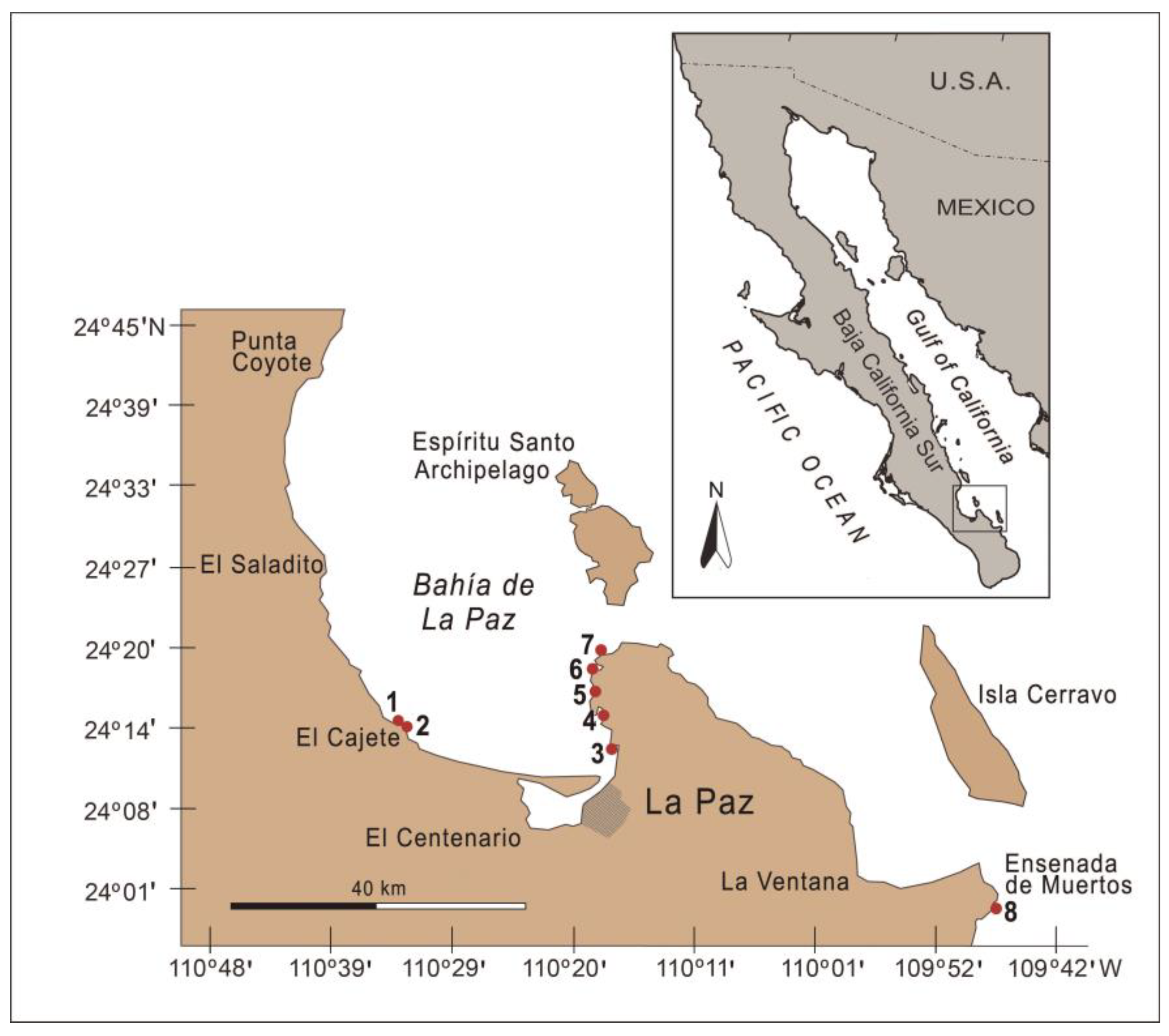
Figure 1. Sampling sites in Bahía de La Paz and adjacent waters in 2015–2019: 1—Agua de Yepiz, 2—Playa El Califin, 3—Costa Baja, 4—Playa El Tesoro (El Tesoro I and El Tesoro II), 5—Playa Las Brujas, 6—Balandra (Balandra-1 and Balandra-2), 7—Playa El Tecolote, 8—Playa Ensenada de Muertos (Bahía de los Sueños).
Diversity 17 00674 g001
Figure 2. Sea surface temperature (SST) on 11 May 2015, 18 June 2016, 15 June 2018 and 15 December 2019 at 12:00. Temperature data were extrapolated regionally to an L4 level. The red box within the maps indicates the sampling area for this study.
Diversity 17 00674 g002
Figure 3. Cabra cf. aremonica (AC) and Coolia cf. malayensis (DF): (A) cell in right-side-ventral view; (B) cell in apical-right-side-ventral view; (C) epitheca and cingulum; (D) cell in apical-ventral view; (E) left-side-dorsal view; (F) fragment of the epitheca showing the Po and adjacent plates. Symbols for thecal plates: 1′–3′—the apical plates; 1″–7″—the precingular plates; 1a—the anterior intercalary plate; 1‴–5‴—the postcingular plates; 1″″—the first posterior plate; APC—the apical pore complex; c3—the third cingular plate; Po—the pore plate. Scale bars: 1 μm.
Diversity 17 00674 g003
Figure 4. Fukuyoa paulensis (AC) and Gambierdiscus toxicus (DF): (A) cell in right-side view; (B) cell in apical-right-side view; (C) the Po and adjacent plates; (D) cell in apical view; (E) cell in antapical view; (F) the Po and adjacent plates. Symbols for thecal plates: 1′–3′—apical plates; 1″–7″—precingular plates; 1‴–5‴—postcingular plates; 1″″, 2″″ and 1p—antapical plates; Po—the pore plate. Scale bars: 10 μm in (A,B,D,E); 1 μm in (C,F).
Diversity 17 00674 g004
Figure 5. Ostreopsis cf. lenticularis: (A) cell in apical view; (B) cell in antapical view; (C) cell in apical-dorsal view; (D,E) the Po and adjacent plates; (F) fragment of the theca showing plates adjacent to the sulcal area in antapical-ventral view. Symbols for thecal plates: 1′–4′—apical plates; 1″–7″—precingular plates; 1‴–5‴—postcingular plates; 1″″ and 2″″—posterior plates; Po—the pore plate; Vo—ventral opening. Scale bars: 10 μm in (AC,F); 1 μm in (D,E).
Diversity 17 00674 g005
Figure 6. Prorocentrum species: (A) the Prorocentrum lima complex sp. 1 (cell in right-side view); (B) the Prorocentrum lima complex sp. 2 (cell in left-side view); (C) P. rhathymum (cell in left-side view); (D) P. rhathymum (the apical zone showing the trichocyst pores and the sagittal suture on the left plate, and the periflagellar area on the right plate); (E) P. sculptile (cell in right-side view); (F) P. hoffmannianum (cell in left-side view). Symbols for thecal plates: 1–8—platelets of the periflagellar area; ap—the accessory pore, fp—the flagellar pore. Scale bars: 10 μm in (A,B,E); 1 μm in (C,D,F).
Diversity 17 00674 g006
Figure 7. Interannual and spatial variation in the abundance of epiphytic dinoflagellates in Bahía de La Paz and adjacent waters in 2015–2019, showing the highest abundances in 2016 and 2019.
Diversity 17 00674 g007
Figure 8. Seasonal variation in the main genera of epibenthic dinoflagellates. The total abundance of dinoflagellates was represented at the Tecolote site in Bahía de La Paz, Mexico. On the X-axis, May–May (2015), Jun *–June (2016), Jun **–June (2018), Dec–December (2019). Kruskal–Wallis analysis was performed to explore statistically significant differences. The letters above the boxplots indicate significant differences at the 95% level, based on Wilcox multiple comparisons.
Diversity 17 00674 g008
Figure 9. Interannual and spatial variation in the associated ecological descriptors in the coastal waters of Bahía de La Paz and adjacent waters in 2015–2019. The colored points show the dispersion of the data and the gray points indicate the modeled distribution. The highest species richness and diversity were observed in 2016 and 2019.
Diversity 17 00674 g009
Figure 10. Analysis of beta diversity of epiphytic dinoflagellates from Bahía de La Paz and adjacent waters in 2015–2019. (A) Non-metric multidimensional scaling (NMDS) plot for epiphytic dinoflagellate taxocoenoses over four years of study. (B) Contribution of species during the different years of study. (C) UpSetR annual intersection map of cell density. Black bars indicate the number of interactions based on total dinoflagellate cell abundance. (D) Shared genera of epiphytic dinoflagellates and shared species richness.
Diversity 17 00674 g010
Figure 11. The Clustering-Heatmap of the similarity-based relationship between epiphytic dinoflagellates (Y-axis) and the different sampling sites in this study (X-axis).
Diversity 17 00674 g011
Figure 12. Interannual relationship between epiphytic dinoflagellate species and macroalgae. (A) Composition of the macroalgae identified in this study. (B) Ternary composition graph between dinoflagellates and macroalgae. The X-axis indicates the year when the different macroalgae were present. The genera Dictyota, Laurencia, Spyridia and Ulva were reported for all the study years. Red algae showed greater association with dinoflagellates compared to green and brown algae. Abbreviations: Chl—Chlorophyta, Ph—Phaeophyceae, Rh—Rhodophyta.
Diversity 17 00674 g012
Table 1. Macroalgal taxa found in Bahía de La Paz and adjacent waters in the southern Gulf of California in 2015–2019 at ten sampling sites. Sampling sites: 1—Agua de Yepiz, 2—Playa El Califin, 3—Costa Baja, 4a—Playa El Tesoro I, 4b—Playa El Tesoro II, 5—Playa Las Brujas, 6a—Balandra-1, 6b—Balandra-2, 7—Playa El Tecolote, 8—Playa Ensenada de Muertos (Bahía de los Sueños); also see Figure 1. Identification was performed based on the entire individuals or thallus fragments.
TaxaMay 2015June 2016June 2018December 2019
Rhodophyta
Acanthophora spicifera (Vahl) Børgesen3, 4b1, 2, 6a,b, 77
Amphiroa cf. vanbosseae Me. Limoine 7
Amphiroa sp. 787
Asparagopsis sp. 8
Bostrychia radicans (Montagne) Montagne4a *
Callithamnion sp. 7
Centroceras sp.4a
Ceramium sp. 2, 777
Champia sp. 7
Corallinaceae gen. sp.4b, 7
Crouania sp. 7
Digenea simplex (Wulfen) C. Agardh 5, 88
Ganonema cf. farinosum (J.V. Lamouroux)
K.-C. Fan et Y.-C. Wang 7
Gayliella sp. 2
Gelidiella sp. 8
Gelidium sp. 6a
Gigartinaceae gen. sp. 8
Gracilaria sp.28
(?)Gracilariopsis sp. 7
Herposiphonia cf. littoralis Hollenberg 7
Herposiphonia sp. 7
Hypnea valentiae (Turner) Montagne 1, 2, 6b, 7
Hypnea sp.35, 887
Laurencia cf. masonii Setchell et N.L. Gardner 2
Laurencia spp.3, 71, 2, 5, 6a,b, 7, 877
Lithophyllum sp. 7
Lomentaria sp.4b
Neosiphonia sp.317, 87
(?)Pterocladia sp. 7
Spyridia filamentosa (Wulfen) Harvey3, 72, 6b, 7
Spyridia sp.4a,b, 72, 5, 6a, 877
Wrangelia sp. 7
Yuzurua sp.6a
Phaeophyceae
Chnoospora sp. 8
Colpomenia sp. 7
Dictyopteris sp. 7
Dictyota spp.71, 2, 787
Hydroclathrus clathratus (C. Agardh) M. Howe 1, 2, 6b
Hydroclathrus sp. 7
Myrionema sp. 1, 2 **
Padina mexicana E.Y. Dawson var. erecta
Avila-Ortíz15
Padina spp.3, 72, 87, 8
Sargassum agardhianum Farlow 1, 2, 7
Sargassum sp.3, 71, 5, 87, 87
Sphacelaria furcigera Kützing 1, 2 ***
(?)Taonia sp. 1
Chlorophyta
Caulerpa racemosa (Forsskål) J. Agardh 5, 88
Caulerpa sertularioides (S.G. Gmelin) R. Howe 28
Chaetomorpha cf. californica Collins 7
Cladophora cf. stimpsonii Harvey 7
Cladophora sp. 87
Codium cuneatum (Setchell) N.L. Gardner 1, 2, 6b
Codium sp.3, 4b, 76a,b, 77, 8
Halimeda sp.3, 76a, 777
Ulva clathrata (Roth) C. Agardh 6b
Ulva intestinalis Linnaeus 7
Ulva sp.1 (laminar) 5, 6b, 7, 87, 87
Ulva sp.2 (tubular)4b6b 7
Note: * as epiphyte on mangrove roots; ** as epiphyte on Sargassum agardhianum; *** as epiphyte on S. agardhianum, Padina sp. and Hypnea valentiae.
Table 2. Summary of the permutational multivariate analysis of variance (PERMANOVA) based on Bray–Curtis dissimilarities and applied to the taxocoenosis structure of epiphytic dinoflagellates observed at 10 sampling sites for four years. Bold numbers indicate a significant effect in the models. Asterisks indicate the interaction between the models.
FactorPseudo-Fp (Perm)R2
Density
Years1.360.0010.02
Sites3.230.0010.12
Years * Sites3.620.9120.01
Shannon diversity
Years0.450.5080.003
Sites7.600.0070.05
Years * Sites15.780.0010.11
Richness
Years8.090.0010.15
Sites4.470.0320.03
Years * Sites15.280.0010.10
Equitability index
Years14.090.0010.10
Sites4.260.0100.03
Years * Sites2.760.1060.01
Simpson index (λ)
Years4.290.0480.03
Sites6.350.0160.04
Years * Sites11.280.0020.08
Table 3. ANOSIM tests and interannual pairwise comparison of epiphytic dinoflagellate taxocoenoses in the study area.
GroupsGlobal RSignificance Level (%)
All years0.5700.0001
Year 2015 vs. 20160.4630.0006
Year 2015 vs. 20180.3860.0006
Year 2015 vs. 20190.7950.0006
Year 2016 vs. 20180.4850.0012
Year 2016 vs. 20190.960.0006
Year 2018 vs. 20190.6380.0006
Disclaimer/Publisher’s Note: The statements, opinions and data contained in all publications are solely those of the individual author(s) and contributor(s) and not of MDPI and/or the editor(s). MDPI and/or the editor(s) disclaim responsibility for any injury to people or property resulting from any ideas, methods, instructions or products referred to in the content.