Abstract
This study aimed to characterize the transcriptional response of Porphyromonas gingivalis biofilms to treatment with xanthohumol and curcumin. A validated dynamic in vitro biofilm model, based on microbial growth under flow and shear conditions resembling the oral cavity, was used to develop mature biofilms of P. gingivalis on sterile ceramic calcium hydroxyapatite discs. Transcriptional profiles of biofilms, treated and untreated with both extracts, were obtained through RNA-Sequencing (RNA-Seq). The biofilm development and the lack of phenotypic effects from sublethal concentrations of xanthohumol and curcumin were confirmed via Scanning Electron Microscopy (SEM) and Confocal Laser Scanning Microscopy (CLSM). Reverse transcription quantitative PCR (RT-qPCR) was employed to verify differentially expressed genes identified by RNA-Seq. Xanthohumol and curcumin caused extensive reprogramming of P. gingivalis biofilm gene expression. Out of 1,973 genes, xanthohumol activated 173 and repressed 286, whereas curcumin activated 170 and repressed 163. These changes affected genes involved in membrane integrity, oxidative stress, transmembrane transport, and virulence, suggesting a mechanism of action that involves membrane disruption.
1. Introduction
Subgingival biofilms are collections of microorganisms closely associated and organized within an extracellular matrix, produced by the microbes themselves, which allows them to anchor to biotic or abiotic surfaces [,]. The various activities of these microbial communities, as well as the qualitative and quantitative changes in their composition, have been linked to the initiation and progression of periodontal diseases [,]. In fact, according to the 2018 Classification of Periodontal and Peri-implant Diseases and Conditions, dental biofilm-induced gingivitis and periodontitis are among the most important conditions listed [,].
One of the main contributors to the pathobiology of these conditions is Porphyromonas gingivalis, a Gram-negative anaerobic bacterium, considered a late colonizer in biofilm formation. Its pathogenicity is closely linked with its abundance within the biofilm, the expression of specific virulence factors, and its resistance to host inflammatory and immune responses [,,,,,]. In fact, the growth of P. gingivalis within the subgingival biofilm triggers the expression of a set of genes, mainly related to the cell wall, oxidative stress, virulence, transport, membrane proteins, and quorum sensing [,,].
Although the primary approaches to controlling dental and subgingival biofilms are mechanically based, the use of chemical agents, whose mechanism of action is related to the inhibition of the previously described processes, represents a promising strategy for controlling the development and progression of periodontal and peri-implant diseases. Among these chemical agents, specific phytochemicals (natural substances of vegetal origin) have shown activity against recognized periodontal pathogens [,,].
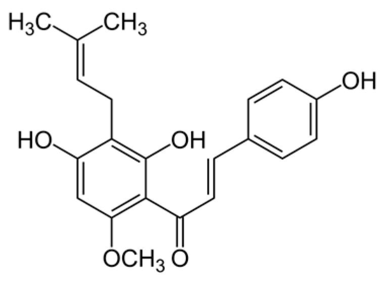
A phytochemical of interest is xanthohumol (XN), a prenylated flavonoid (C21H22O5) from the female flowers of the hop plant (Humulus lupulus), which exhibits anti-inflammatory and antioxidant properties [] (Figure 1). This extract has demonstrated a strong antimicrobial effect [,,,].
Figure 1.
Chemical structure of xanthohumol.
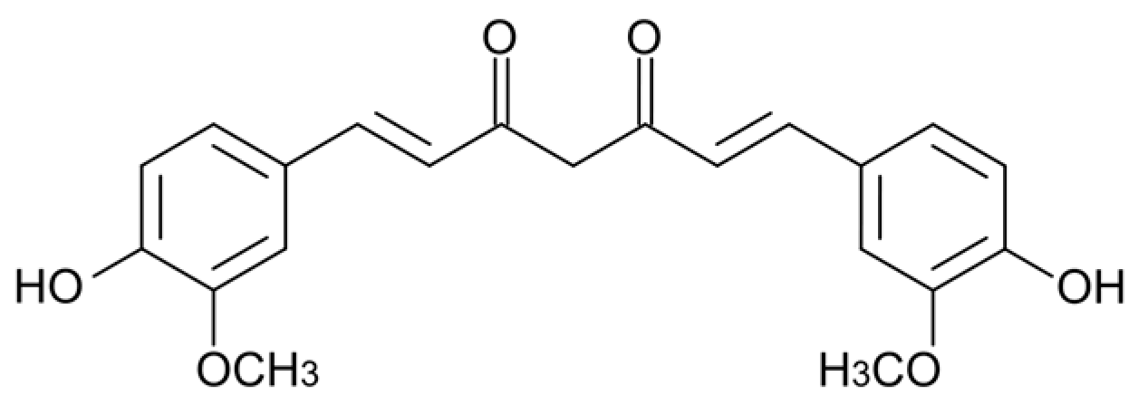
A second promising phytochemical is curcumin (Cur), an aromatic polyphenol (C21H20O6) found in the rhizomes of turmeric (Curcuma longa) (Figure 2). This extract has antioxidant and anti-inflammatory properties [,,], as well as bactericidal and fungicidal activity [,].
Figure 2.
Chemical structure of curcumin.
Recent research highlights the antimicrobial potential of XN and Cur against P. gingivalis. XN significantly reduces the viability and biomass of multispecies biofilms, developed on implant surfaces in vitro models, with an efficacy comparable to or better than chlorhexidine [,]. Cur also shows strong inhibitory effects on P. gingivalis: it disrupts key metabolic pathways and enzyme activities involved in bacterial growth [], decreases adhesion and biofilm formation while downregulating major virulence genes [], and inhibits gingipain activity, resulting in a notable reduction in biofilm []. Overall, these findings suggest that both compounds have antimicrobial potential for their use as adjuncts in the treatment of periodontitis. However, the mechanism of action remains unclear. Comparing the gene expression profiles of P. gingivalis biofilms with and without XN and Cur could reveal how both phytochemicals function as either antimicrobials or host response modulators. The complete genome characterization of P. gingivalis had been a significant milestone for gene expression studies of this bacterial species [,].
Therefore, this study aimed to characterize the differential gene expression of P. gingivalis in sessile growth with and without XN and Cur in a validated dynamic in vitro biofilm model.
2. Results
2.1. Minimum Inhibitory Concentrations of Xanthohumol and Curcumin Against P. gingivalis
The minimum inhibitory concentrations (MICs) of XN and Cur against P. gingivalis were established at 50 and 500 µM, respectively.
2.2. Scanning Electron Microscopy and Confocal Laser Scanning Microscopy to Monitor P. gingivalis Biofilm Development and the Effect of Treatments
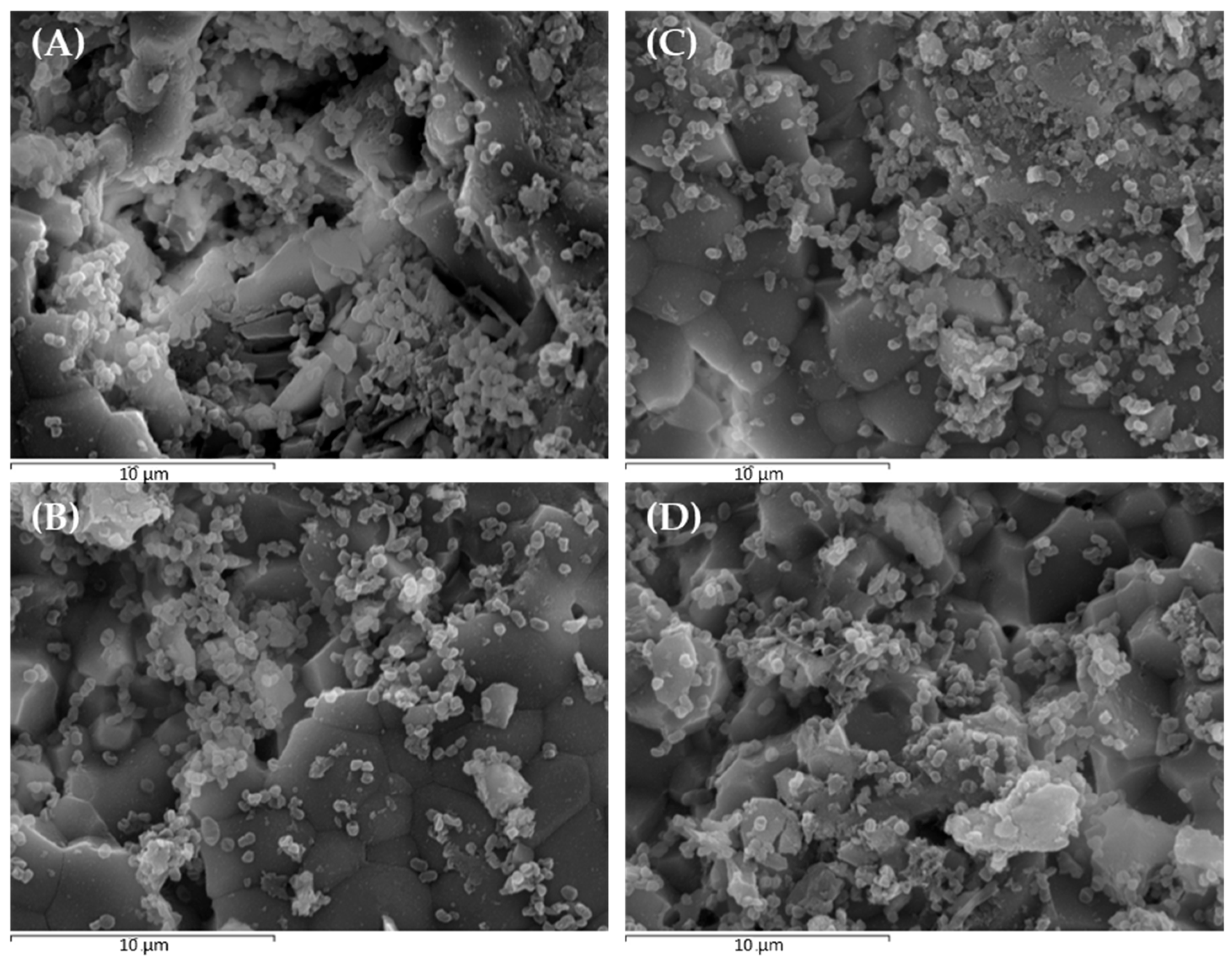
Scanning electron microscopy (SEM) images revealed the formation of a mature monospecies biofilm of P. gingivalis, where cocco-bacillary forms gathered in a highly organized three-dimensional structure (Figure 3A). Treatments with sublethal concentrations of dimethyl sulfoxide (DMSO), XN, and Cur, used in this study, did not alter the structure or organization of the developed biofilms (Figure 3B–D, respectively).
Figure 3.
Scanning electron microscopy (SEM) images at 5000× magnification showing: (A) negative control biofilms incubated with phosphate-buffered saline; (B) negative control biofilms incubated with 0.25% dimethyl sulfoxide; (C) biofilms treated with 50 µM xanthohumol; and (D) biofilms treated with 500 µM curcumin (scale bar = 10 µm).
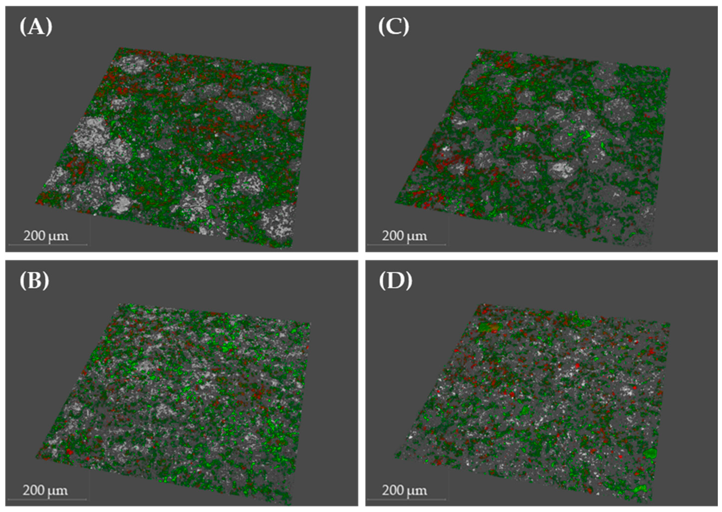
Similarly, confocal laser scanning microscopy (CLSM) analysis showed no effect on bacterial density, cell viability, or biofilm roughness (Table 1 and Figure 4).
Table 1.
Bacterial density, viability, percentage and roughness coefficient (Ra*) obtained by quantification of CLSM images.
Figure 4.
Confocal laser scanning microscopy (CLSM) images: (A) negative control biofilms incubated with phosphate-buffered saline (PBS); (B) negative control biofilms incubated with 0.25% dimethyl sulfoxide (DMSO); (C) biofilms treated with 50 µM xanthohumol (XN); and (D) biofilms treated with 500 µM curcumin (Cur). LIVE/DEAD® BacLight Kit was used to stain live bacteria (green), dead bacteria (red), and disc surfaces (white). (scale bar = 200 µm).
The minor differences among groups were not statistically significant.
2.3. Comparative Analysis of RNA-Sequencing-Obtained Transcriptomes
The obtained transcriptomes revealed 1973 genes with at least 10 counts in each experimental replicate, representing 91.55% of the total genes in the P. gingivalis ATCC 33277 strain []. The comparison between the transcriptional profiles after biofilm incubation in PBS and in 0.25% DMSO showed no gene regulation changes caused by the solvent at the selected concentration, with only one gene being overexpressed (Supplementary Table S1). Therefore, differential gene expression in the presence or absence of 50 µM XN and 500 µM Cur was evaluated by comparing the transcriptomes obtained after incubation with each extract against the profile obtained with 0.25% DMSO.
As shown in Supplementary Table S1 and in Figure 5, XN induced the transcriptional activation of 173 genes and the repression of 286 (out of 1973 genes, representing 8.8% and 14%, respectively). Cur activated the expression of 170 genes and repressed the transcription of 163 (8.6% and 8.3%, respectively).
Figure 5.
Proportion of upregulated and downregulated genes of P. gingivalis in response to exposure to xanthohumol (XN) and curcumin (Cur), compared to dimethyl sulfoxide (DMSO) as control.
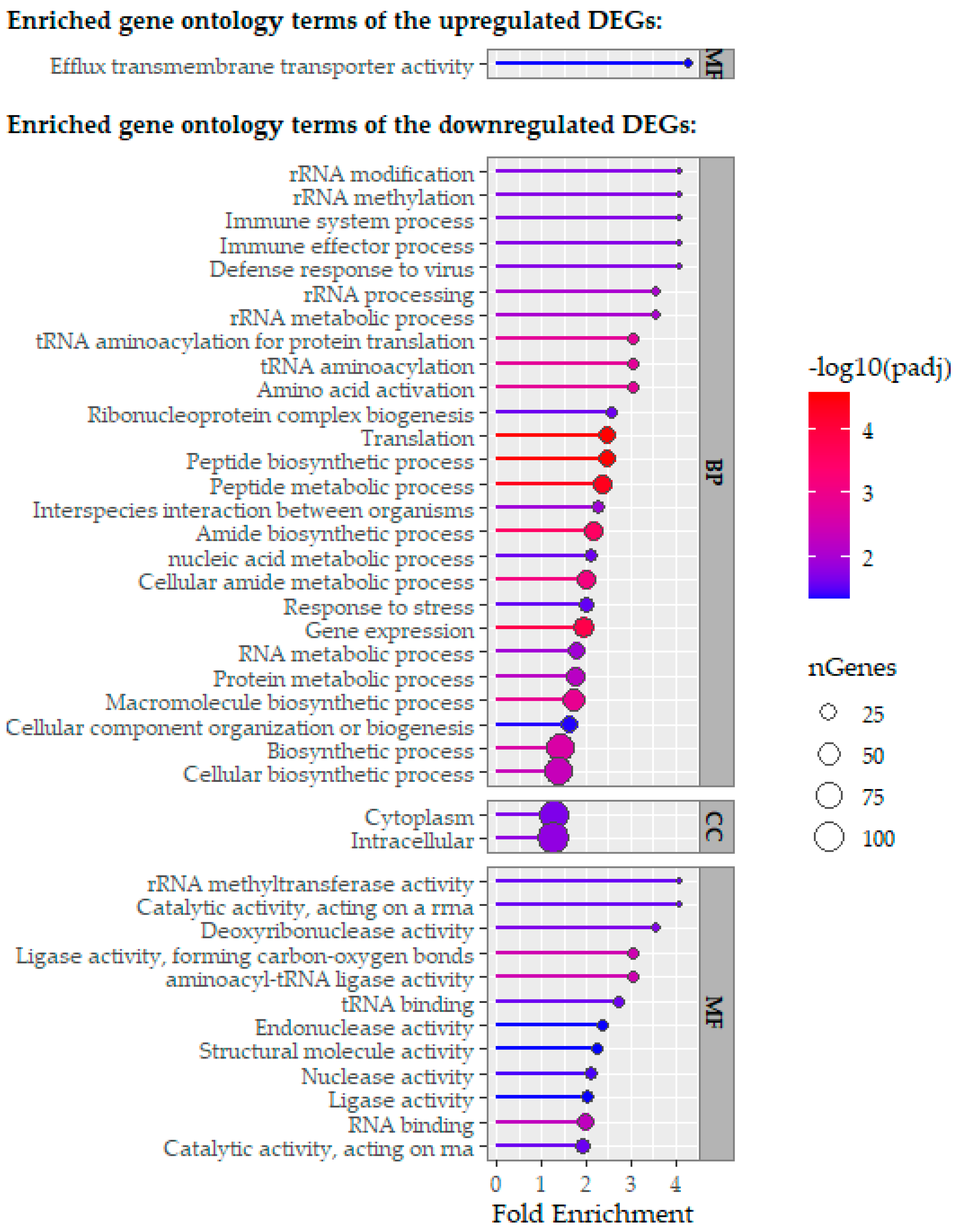
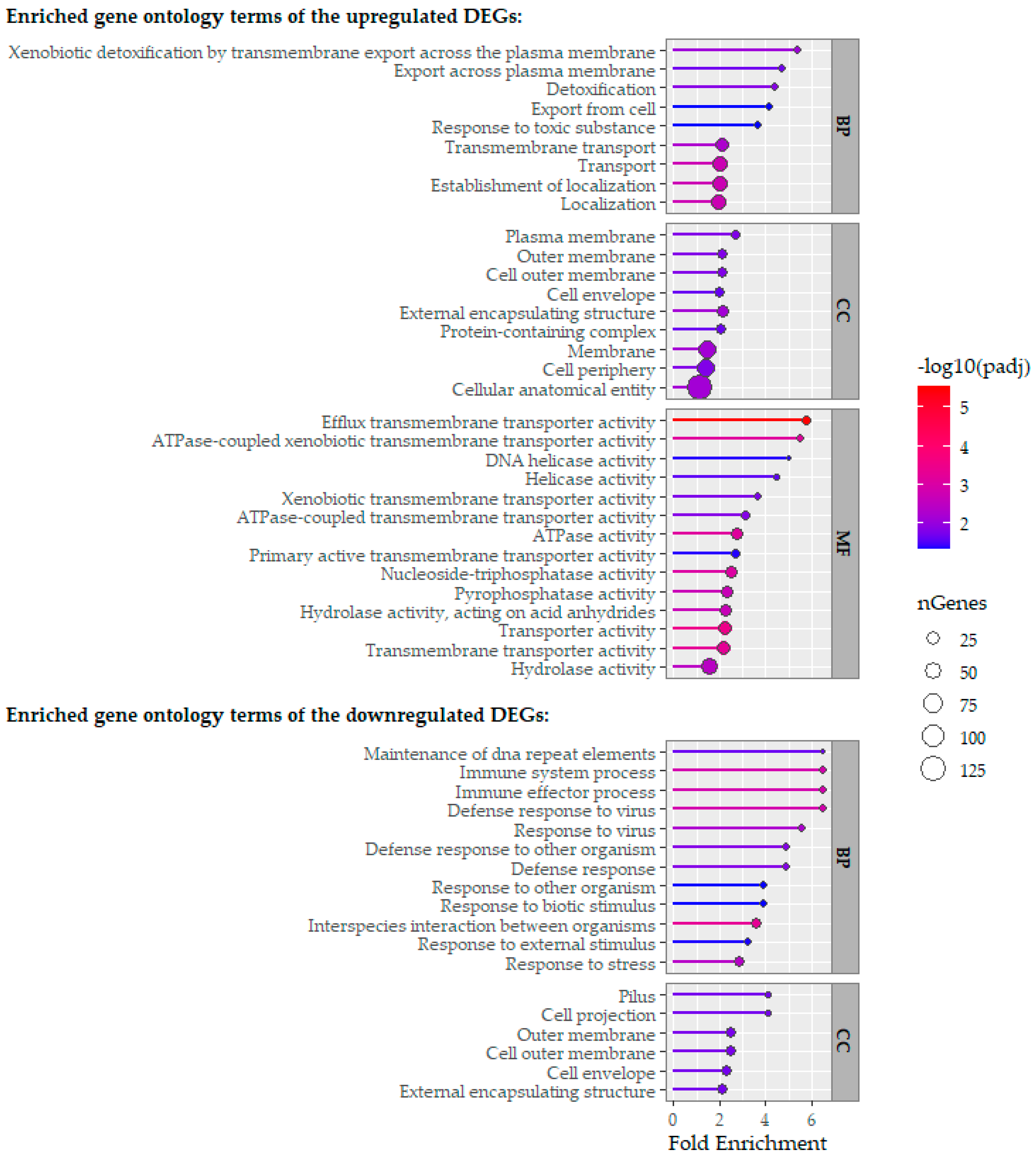
Differentially expressed genes across the tested conditions were classified into Gene Ontology (GO) categories: Biological Process (BP), Cellular Component (CC), and Molecular Function (MF) (Figure 6 and Figure 7).
Figure 6.
Functional categorization of significantly upregulated and downregulated genes of P. gingivalis in response to xanthohumol exposure, grouped by Gene Ontology (GO) categories: Biological Process (BP), Cellular Component (CC), and Molecular Function (MF). Enriched GO terms (p-value < 0.05) were identified using the hypergeometric test in ShinyGO after differential expression analysis with DESeq2.
Figure 7.
Functional categorization of significantly upregulated and downregulated genes of P. gingivalis in response to curcumin exposure, grouped according to Gene Ontology (GO) categories: Biological Process (BP), Cellular Component (CC), and Molecular Function (MF). Enriched GO terms (p-value < 0.05) were identified using the hypergeometric test implemented in ShinyGO, following differential expression analysis with DESeq2.
Regarding the genes transcriptionally activated by Cur, transcripts involved in localization, transmembrane export, and response to toxic substances were overrepresented. Conversely, genes repressed by Cur treatment mainly related to stress response regulation and interactions with other microbial agents. For genes activated by XN, there was a strong enrichment in transport-related functions. In contrast, genes repressed by XN exposure showed significant enrichment in processes involving polypeptide and protein biosynthesis, intracellular translation, and RNA metabolism.
A total of 89 genes (4.5% of the P. gingivalis genome) were commonly upregulated by both extracts. Similarly, 140 genes (7.1%) were downregulaled under both conditions (Supplementary Table S1).
Principal Component Analysis (PCA) verified the quality and consistency of the data (Supplementary Figure S1). The samples are grouped according to the established experimental conditions, separated by the treatment variable, which confirms that the observed differences in gene expression are due to the experimental factor and not technical variability or noise. No outliers, batch effects, or technical errors were identified.
The estimated statistical power at n = 3 biological replicates per group was 0.75 for upregulated and 0.77 for downregulated genes in both the Cur vs. DMSO and XN vs. DMSO comparisons, further supporting the robustness of the differential expression results (Supplementary Figure S2).
To validate the sequencing results, the expression of twelve selected differentially expressed genes between target conditions was analyzed by reverse transcription quantitative PCR (RT-qPCR) (Supplementary Table S2). The differential gene expression patterns observed were consistent across both techniques.
3. Discussion
SEM and CLSM analyses confirmed the formation of a typical monotypic P. gingivalis biofilm with normal structure, complexity, and cell viability. Exposure to sublethal concentrations of XN and Cur used in this study did not affect any of these parameters (Figure 3 and Figure 4), thus ruling out artefacts that could bias the interpretation of the gene expression data.
The current study, based on RNA sequencing analysis, has demonstrated a significant impact of XN and Cur on the transcriptional profile of P. gingivalis after 60 s of exposure. This treatment duration matches the typical exposure time used during oral rinsing procedures or implant decontamination. Additionally, previous studies in which we showed the bactericidal effects of XN and Cur in subgingival biofilm models were conducted using this same treatment duration []. XN upregulated 8.8% and downregulated 14% of the genes, while Cur upregulated 8.6% and downregulated 8.3% (Figure 5).
XN exposure triggered the upregulation of genes related to transmembrane proteins, indicating a direct bacterial response to this compound. The increase in transcriptional expression of this gene cluster is consistent with the previously reported affinity of XN for specific bacterial membrane lipids, such as phosphatidylglycerol and cardiolipin (key components of the lipid bilayer), which in turn alters membrane integrity and function and induces oxidative stress through an increase in intracellular reactive oxygen species (ROS) []. This mechanism is consistent with the strong bactericidal effect of XN against P. gingivalis. Thus, these findings suggest a mechanism of action for XN involving membrane disturbance and oxidative stress in P. gingivalis. The ability of XN to destabilize the membrane structure of anaerobic bacteria and affect their viability by disrupting ion transport also supports this approach [,,]. The notable downregulation of genes involved in polypeptide and protein biosynthesis, translation, and RNA metabolism in response to XN indicates a conserved bacterial stress response to phenolic compounds. This transcriptional pattern matches that seen in Lactobacillus plantarum after exposure to hydroxytyrosol, another phenolic compound that inhibits protein synthesis and RNA metabolism []. Furthermore, other polyphenols, such as ferulic and gallic acids, cause changes in Gram-negative bacterial membranes, promote ROS buildup, and inhibit vital biosynthetic pathways, including translation and RNA metabolism, thereby contributing to their bacteria-killing effects [].
Like XN, Cur also activated genes related to transmembrane transport and detoxification, indicating an adaptive response by P. gingivalis aimed at maintaining cell viability under stress [,]. One proposed antibacterial mechanism of Cur is disrupting the plasma membrane and causing morphological changes due to cell wall alterations [,]. The activation of stress response genes coding for membrane- and wall-associated proteins supports this idea. Additionally, the upregulation of transmembrane transport genes may result from altered metabolism of several amino acids essential for P. gingivalis [], which primarily uses amino acids rather than sugars as carbon sources derived from protein degradation []. This interpretation is reinforced by recent studies indicating that Cur disrupts bacterial membrane integrity and modulates membrane-associated processes, supporting the membrane as a central antimicrobial target [,,]. Another suggested mechanism of Cur is inhibiting P. gingivalis growth by blocking dipeptidyl peptidase (DPP) activity []; however, the present study showed the opposite, with transcriptional activation of genes related to DPP7 and DPP III activity. Likewise, no inhibition of GTPase activity, reported in Bacillus subtilis [], was observed here, possibly due to the low concentration of Cur used.
The present study also showed the inhibition of expression of genes related to gingipain production (rgpA and porT) and fimbriae formation (fimA, fimC, and fimD) in response to both Cur and XN. These gene groups are closely associated with P. gingivalis virulence. It has been suggested that P. gingivalis virulence can be reduced using Cur, due to its ability to inhibit the expression of gingipain and biofilm-related genes controlled by quorum sensing [,,]. Downregulation of these same genes also occurs when P. gingivalis is exposed to higher concentrations of Cur or for longer incubation periods []. In the case of XN, the genes vimE and vimF, which are responsible for synthesizing the structural lipopolysaccharide, were also suppressed, potentially decreasing biofilm virulence.
Thus, both XN and Cur downregulate well-established P. gingivalis virulence genes and biofilm formation-related genes, although no downregulation of the luxS gene (quorum sensing system of P. gingivalis []) was detected. Comparatively, chlorhexidine (CHX), the reference antiseptic in managing periodontal diseases [], shows similar action by targeting the bacterial cell membrane, where it electrostatically binds to membrane phosphates, increases permeability, and causes leakage of essential intracellular components []. This comparison is especially relevant, as CHX acts through physical membrane disruption, while XN and Cur regulate gene expression at the transcriptional level, targeting virulence factors (e.g., fimbriae, gingipains) and membrane integrity—effects not reported for CHX. This suggests that, beyond direct antimicrobial action, these compounds may reduce P. gingivalis pathogenicity, a critical factor in modulating periodontal inflammatory responses [,]. Additionally, XN and Cur exhibit greater cytocompatibility than CHX, maintain similar antimicrobial efficacy, and have the potential to lessen the cytotoxic effects of P. gingivalis outer membrane vesicles [,]. Therefore, amid the rising levels of antibiotic resistance [], our findings highlight the potential of phytochemicals as alternative strategies for preventing and treating periodontal and peri-implant diseases.
This study has limitations that must be acknowledged for accurate interpretation of the results. The experimental conditions focused only on P. gingivalis in a biofilm-sessile state. It is important to note that the transcriptional profile of P. gingivalis varies significantly between sessile and planktonic growth []. Specifically, genes involved in transport, membrane proteins, and envelope components are overexpressed in biofilm growth, while stress response genes are often under expressed. Moreover, growth in complex polymicrobial communities, such as those found in the subgingival periodontal pocket, further alters gene expression, particularly regarding oxidative stress response, cell envelope structure, transposon activity, and metabolism []. These conditions were not simulated in this study, which was limited to a monospecies biofilm model. It should also be noted that sublethal concentrations of XN and Cur were used in this study to maintain enough cell viability for detecting induced transcriptional changes, which might otherwise be hidden under high mortality conditions. Therefore, the concentrations used here do not match those suggested for therapeutic purposes. In fact, previous research has shown that lethal concentrations of both compounds significantly reduce the microbial density in treated biofilms, leading to a strong bactericidal effect [,]. Finally, factors such as host immune response and patient physiological status, both of which can greatly influence P. gingivalis gene expression, were not considered in this in vitro biofilm model.
In conclusion, despite the considerations mentioned, transcriptomic analysis showed that both XN and Cur cause extensive reprogramming of P. gingivalis gene expression, impacting genes related to membrane integrity, oxidative stress, transmembrane transport, and virulence. These effects suggest a mechanism of action involving membrane disruption and a potential induction of oxidative stress, leading to transcriptional adaptive responses and suppression of key virulence factors. Therefore, both compounds emerge as promising therapeutic options for managing periodontal and peri-implant diseases.
4. Materials and Methods
4.1. Microbial Strains and Culture Conditions
The bacterial strain Porphyromonas gingivalis ATCC 33277 was used. It was cultured on blood agar plates (Blood Agar Oxoid No. 2; Oxoid, Basingstoke, UK), supplemented with 5% (v/v) sterile horse blood (Oxoid), 5.0 mg/L haemin (Sigma, St. Louis, MO, USA), and 1.0 mg/L menadione (Merck, Darmstadt, Germany) at 37 °C for 24–72 h under anaerobic conditions (10% H2, 10% CO2, and N2 balance).
Pure cultures of P. gingivalis were obtained during its exponential growth phase under anaerobic conditions in protein-enriched brain heart infusion (BHI) medium (Becton, Dickinson and Company, Franklin Lakes, NJ, USA). The medium was supplemented with 2.5 g/L mucin (Oxoid), 1.0 g/L yeast extract (Oxoid), 0.1 g/L cysteine (Sigma), 2.0 g/L sodium bicarbonate (Merck), 5.0 mg/L haemin (Sigma), 1.0 mg/L menadione (Merck), and 0.25% (v/v) glutamic acid (Sigma). Bacterial concentration was measured spectrophotometrically to develop a P. gingivalis suspension containing 108 colony-forming units (CFU)/mL.
4.2. Minimum Inhibitory Concentrations of Xanthohumol and Curcumin Against P. gingivalis
To select the optimum concentration of XN and Cur, at which the growth of the strain of interest was not affected, MICs assays against P. gingivalis were conducted [].
Isolated colonies of P. gingivalis were grown in a protein-enriched BHI-modified medium, adjusted to pH 7.2 (Becton, Dickinson and Company, Franklin Lakes, NJ, USA), which was supplemented with 2.5 g/L mucin (Oxoid), 1.0 g/L yeast extract (Oxoid), 0.1 g/L cysteine (Sigma), 2.0 g/L sodium bicarbonate (Merck), 5.0 mg/L haemin (Sigma), 1.0 mg/L menadione (Merck), and 0.25% (v/v) glutamic acid (Sigma), at 37 °C under anaerobic conditions (10% H2, 10% CO2 and N2 equilibrium).
The exponential growth phase was identified spectrophotometrically, with cultures consistently below an optical density (OD550nm) of 1.2. Once exponential growth was achieved, 200 μL of inoculum were transferred to a 24-well microplate, resulting in a final concentration of 106 CFU/mL. Then, XN (NATECO® GmbH & Co., Wolnzach, Germany) and Cur (Sigma-Aldrich®, Steinheim, Germany) were added at final concentrations of 6.25, 12.5, 25, 50, 100, 250, 500, and 1000 µM for both phytochemicals, using PBS as the negative control. These microplates were incubated for 24 h at 37 °C under anaerobic conditions. The minimum inhibitory concentrations (MICs) of each extract were determined on blood agar plates, on which 100 µL of each suspension was seeded. The plates were incubated for 72 h at 37 °C under anaerobic conditions. The lowest concentrations of XN and Cur that produced at least a 90% inhibition of bacterial growth were considered the MICs for P. gingivalis, with a lower concentration range selected to avoid effects on cell viability.
MIC assays were performed in triplicate, with appropriate controls for contamination.
4.3. In Vitro Dynamic Monospecies Biofilm Model
A validated dynamic biofilm in vitro model was used []. The system includes a sterile vessel where the modified BHI medium flows via a peristaltic pump. The bioreactor (Lambda Minifor© bioreactor, LAMBDA Laboratory Instruments, Sihlbruggstrasse, Switzerland) maintains the culture medium under the stable conditions of the oral cavity (temperature of 37 °C, pH of 7.2, and an anaerobic environment with 10% H2, 10% CO2, and N2 as the balance).
The vessel was inoculated with 5 mL of the previously described P. gingivalis suspension, containing 108 CFU/mL, and incubated for 12 h under the specified conditions. When the culture reached the exponential phase, a second peristaltic pump was activated at a steady flow rate of 30 mL/h to initiate continuous culture and transfer the culture to the Robbins device, where sterile calcium hydroxyapatite (HA) ceramic discs [7 mm diameter (SD = 0.2) and 1.8 mm thick] (Clarkson Chromatography Products, Williamsport, PA, USA) were placed. The discs were maintained for 72 h under the same conditions inside the Robbins device until mature monospecies biofilms of P. gingivalis formed on the HA discs. Three Robbins devices were used sequentially to increase the sample size of each test [].
4.4. Experimental Groups
After incubation for 72 h, the discs were removed from the Robbins device and treated in microplate wells for 60 s, containing 1 mL of XN (50 µM) and 1 mL of Cur (500 µM), both resuspended in 0.25% (v/v) DMSO (AppliChen GinbH, Darmstadt, Germany). One mL of PBS and 1 mL of 1% DMSO (v/v) were used as negative control (PBS) and to discard the possible impact of the solvent at the concentration used (DMSO).
Subsequently, the discs were washed sequentially in 2 mL of sterile PBS three times (immersion time per rinse, 10 s), to remove any unattached bacteria. Immediately after the treatments, samples were flash-frozen in liquid nitrogen to halt any transcriptional activity occurring after the intervention.
For each condition, the protocol was repeated over nine discs in six independent sets of experiments, thus analyzing a total of 54 discs (n = 54) for RNA sequencing (RNA-Seq) and one disc per set of experiment for CLSM and SEM (n = 6).
4.5. Scanning Electron Microscopy
Samples were extracted from the Robbins device, washed, treated with PBS, DMSO, XN, or Cur, and dried using increasing concentrations of ethanol. They were then coated with gold before analysis, following a previously standardized protocol by Blanc et al. []. The samples were examined using a JSM 6400 electron microscope (JSM6400, JEOL, Tokyo, Japan).
This analysis was performed at the National Centre of Electron Microscopy (In-stalación Científico-Técnico Singular; ICTS) at the Moncloa Campus of the Complutense University of Madrid (Madrid, Spain).
4.6. Confocal Laser Scanning Microscopy
Samples were extracted, washed, treated with PBS, DMSO, XN, or Cur, and stained using the LIVE/DEAD® BacLightTM bacterial viability kit solution, following the protocol described by Blanc et al. [].
A Leica® LCS SP8 STED 3X inverted microscope (Mannheim, Germany) was used to capture non-invasive confocal images of the formed biofilms. COMSTAT 2.1 software [www.comstat.dk (accessed on February and March 2025)] was employed to calculate the biofilm biomass in cubic micrometres per square micrometre (µm3/µm2) and the roughness coefficient (Ra*) from the CLSM data. The analysis was conducted at the Margarita Salas Biological Research Centre (Centro de Investigaciones Biológicas, Consejo Superior de Investigaciones Científicas—CIB-CSIC), located on the Moncloa Campus of the Universidad Complutense de Madrid (Madrid, Spain).
4.7. Total RNA Isolation
After the four treatments (PBS, DMSO, XN, and Cur), the discs were rinsed in 2 mL of sterile PBS three times for 10 s each to remove cells not attached to the biofilm. They were then vortexed in 1 mL of PBS for 2 min at full power to disaggregate the biofilm, followed by centrifugation at 13,000 rpm for 3 min at room temperature to obtain sample pellets. Eighteen pellets from each replicate of each treatment (PBS, DMSO, XN, and Cur), obtained from two different experimental sets, were sequentially and cumulatively resuspended in 1 mL of sterile PBS. This process represented each biological replicate for each condition.
The RNeasy Protect Bacteria Kit (Qiagen, Hilden, Germany) was used to extract total RNA from the samples, following the manufacturer’s instructions. The quantity and quality of the RNA obtained were evaluated using the Agilent 2100 Bioanalyzer (Agilent Technologies, CA, USA). All samples used in this study had an A260nm/A280nm ratio of at least 2.0.
4.8. RNA Sequencing
DNA libraries were prepared from the depleted RNA using the NEBNEXT Ultradirectional RNA Library Prep Kit (New England Biolabs, MA, USA), following the manufacturer’s instructions. In brief, the depleted RNA was fragmented and converted into double-stranded cDNA. Illumina sequencing adapters were then attached to the cDNAs, and the library was enriched through limited polymerase chain reaction (PCR). DNA fragments with 300–450 bp inserts were then sequenced in parallel at the Genomics Unit (Complutense University of Madrid).
Raw sequencing reads were initially evaluated with FastQC (version 0.12.1) [] to analyze quality metrics such as base quality scores, duplication levels, and GC content. Then, filtering and trimming were carried out with AfterQC (version 0.9.6) [], which automatically removed low-quality reads, trimmed adapters, and corrected sequencing errors.
High-quality reads were aligned to the Porphyromonas gingivalis ATCC 33277 reference genome (GenBank accession: GCA_000010505.1, ASM1050v1) using Bowtie2 (version 2.5.2) []. The genome was indexed with the bowtie2-build algorithm. Paired-end reads were aligned in global mode, and only those mapping concordantly and uniquely were kept for downstream analysis. Alignment files in SAM format were converted to compressed BAM format using SAMtools (version 1.17) [], and the alignments were visualized with the Integrative Genomics Viewer (IGV) [].
To estimate rRNA contamination, reads were aligned to rRNA databases using SortMeRNA (version 4.3.6) [], and the proportion of rRNA-derived reads was then quantified.
Gene-level quantification was carried out using featureCounts [] from the Subread package (version 2.0.5), assigning reads to genomic features based on the annotation file linked to the reference genome. Only fragments that mapped concordantly and exactly once were counted. The resulting count matrix was then used for differential gene expression analysis.
The gene count matrix was imported into R (version 4.3.3) for differential expression analysis using DESeq2 (version 1.42.1) []. Genes with more than 10 reads in at least three samples were retained for downstream analysis, resulting in a filtered dataset of 1973 genes (91.55% of the original gene set). This filtering step discarded 182 genes (8.45%) due to low or absent expression. Count data were normalized using the median-of-ratios method, and a negative binomial generalized linear model was fitted. Wald tests were used to assess differential expression, with p-values adjusted via the Benjamini–Hochberg method. Genes with an adjusted p-value < 0.05 and a log2 fold change (LFC) > 0.585 (upregulated) or <−0.60 (downregulated) were considered significantly differentially expressed.
Exploratory analysis was conducted using principal component analysis (PCA) to examine sample grouping by experimental condition and assess the consistency of replicates.
To gain insights into the biological functions linked to the differentially expressed genes, Gene Ontology (GO) enrichment analysis was conducted using the web-based tool ShinyGO (version 0.81) [http://bioinformatics.sdstate.edu/go/ (accessed on January 2025)]. This analysis categorized regulated genes into three GO groups: Biological Process (BP), Cellular Component (CC), and Molecular Function (MF), identifying significantly overrepresented functional terms (p < 0.05).
Power calculations were performed to evaluate the ability of the RNA-Seq experimental design to detect differentially expressed genes under each treatment condition (Curcumin vs. DMSO and Xanthohumol vs. DMSO). Analyses were based on empirical mean expression levels and dispersion estimates from the DESeq2 model, and genes were classified as upregulated (log2 fold change > 0.585) or downregulated (log2 fold change < −0.6) using the same thresholds applied in the DESeq2 differential expression analysis. For each comparison, power was estimated as a function of sample size (2–8 replicates per group) using the RNASeqPower package (version 1.42.0), with an alpha level of 0.05 and the global coefficient of variation derived from normalized counts. Power curves were computed separately for up- and downregulated genes to account for potential asymmetries in expression magnitude or dispersion, providing a realistic assessment of the experimental design.
4.9. Reverse Transcriptase Quantitative Polymerase Chain Reaction Validation
To confirm the differential expression results by RT-qPCR, twelve genes were chosen (three overexpressed and three repressed with XN, and three overexpressed and three repressed with Cur, always compared to the profile obtained with the DMSO control).
The cDNA was synthesized from 1 μg of total RNA using the PrimeScriptTM RT Reagent Kit (Takara, Kusatsu, Japan), according to the manufacturer’s instructions.
Specific primers were designed using the Universal Probe Library Roche (Roche Diagnostics) software tool (https://www.sigmaaldrich.com/JP/ja/product/roche/upl71thru80?srsltid=AfmBOor-3e0nXRaA3AMLJ3-IbesGZZOUgYZDOyJWuEwop6EtJ7LBJzWR (accessed on 21 October 2025)) (Supplementary Table S3). Primers and probes were supplied by Life Technologies Invitrogen (Carlsbad, CA, USA), Applied Biosystems (Carlsbad, CA, USA), and Roche (Roche Diagnostic GmbH, Mannheim, Germany). All quantifications were normalized to the 16S rRNA gene of P. gingivalis.
The qPCR reaction was performed using 1 μL of each cDNA per well and 9 μL of a mixture containing 10 μM of each primer, 5 μL of Fast Enzyme ExTaq (SYBR Green) (Takara, Kusatsu, Japan), and nuclease-free water (Roche) to reach a final volume of 10 μL on LightCycler 480 Multiwell-384 optical plates (Roche). PCR were carried out on a LightCycler® 480 II thermal cycler (Roche Diagnostic GmbH, Mannheim, Germany). The amplification programme included an initial cycle at 95 °C for 3 min, followed by 40 cycles of 95 °C for 10 s, 60 °C for 20 s, and 72 °C for 1 s, and one final cycle at 95 °C for 5 s, 65 °C for 1 min, and 97 °C. The results were analyzed using the comparative Ct method (ΔΔCt) []. Each gene was tested in triplicate and compared with the results obtained from RNA-Seq analysis.
4.10. Statistical Analysis
Bacterial biomass reported after analysis by CLSM was expressed in µm3/µm2. Data are shown as means and SDs, and the Shapiro–Wilk goodness-of-fit test was used to assess data normality. When the two datasets compared showed a normal distribution, a T-test with Welch’s correction was applied. Statistically significant differences were considered for p-values <0.05. Present data were analyzed with the GraphPad Prism version 8.0.1 software.
Supplementary Materials
The following supporting information can be downloaded at: https://www.mdpi.com/article/10.3390/ijms262311315/s1.
Author Contributions
E.B., C.C., D.H., and M.S. designed the study. E.B. performed the experiments. C.C. and E.B. analyzed and interpreted the collected data and performed the statistical analysis. E.B. drafted the manuscript. C.C., D.H., and M.S. critically revised the manuscript. All authors have read and agreed to the published version of the manuscript.
Funding
This research received no external funding. Funds from the ETEP Research Group were utilized for this investigation.
Institutional Review Board Statement
Not applicable.
Informed Consent Statement
Not applicable.
Data Availability Statement
All data are available within the article and its Supplementary Materials.
Acknowledgments
The authors would like to acknowledge the technical support of M.G. Elvira and M.T. Seisdedos, at the Confocal and Fluorescence Microscopy Service of Margarita Salas Biological Research Center (Superior Center for Scientific Research), A.M. Vicente, at the ICTS National Centre of Electron Microscopy (University Complutense, Madrid, Spain), R. Ayuso, at the Platform of Mechanic Workshops (University Complutense, Madrid, Spain) and P. Botías and J. García-Cantalejo, at Genomic Unit (University Complutense, Madrid, Spain). We also thank J. Arroyo and R. García, at the Faculty of Pharmacy (University Complutense, Madrid, Spain), for the loan of xanthohumol.
Conflicts of Interest
The authors declare no conflicts of interest.
References
- Jakubovics, N.S.; Goodman, S.D.; Mashburn-Warren, L.; Stafford, G.P.; Cieplik, F. The dental plaque biofilm matrix. Periodontology 2000 2021, 86, 32–56. [Google Scholar] [CrossRef] [PubMed]
- Ray, R.R. Biofilm architecture and dynamics of the oral ecosystem. BioTechnologia 2024, 105, 395–402. [Google Scholar] [CrossRef]
- Filoche, S.; Wong, L.; Sissons, C.H. Oral biofilms: Emerging concepts in microbial ecology. J. Dent. Res. 2010, 89, 8–18. [Google Scholar] [CrossRef]
- Bloch, S.; Hager-Mair, F.F.; Andrukhov, O.; Schaffer, C. Oral streptococci: Modulators of health and disease. Front. Cell. Infect. Microbiol. 2024, 14, 1357631. [Google Scholar] [CrossRef]
- Papapanou, P.N.; Sanz, M.; Buduneli, N.; Dietrich, T.; Feres, M.; Fine, D.H.; Flemmig, T.F.; Garcia, R.; Giannobile, W.V.; Graziani, F.; et al. Periodontitis: Consensus report of workgroup 2 of the 2017 World Workshop on the Classification of Periodontal and Peri-Implant Diseases and Conditions. J. Periodontol. 2018, 89 (Suppl. 1), S173–S182. [Google Scholar] [CrossRef]
- Berglundh, T.; Armitage, G.; Araujo, M.G.; Avila-Ortiz, G.; Blanco, J.; Camargo, P.M.; Chen, S.; Cochran, D.; Derks, J.; Figuero, E.; et al. Peri-implant diseases and conditions: Consensus report of workgroup 4 of the 2017 World Workshop on the Classification of Periodontal and Peri-Implant Diseases and Conditions. J. Periodontol. 2018, 89 (Suppl. 1), S313–S318. [Google Scholar] [CrossRef]
- Socransky, S.S.; Haffajee, A.D. Dental biofilms: Difficult therapeutic targets. Periodontol. 2000 2002, 28, 12–55. [Google Scholar] [CrossRef] [PubMed]
- Limoli, D.H.; Jones, C.J.; Wozniak, D.J. Bacterial Extracellular Polysaccharides in Biofilm Formation and Function. Microbiol. Spectr. 2015, 3, 223–247. [Google Scholar] [CrossRef]
- Sanz, M.; Beighton, D.; Curtis, M.A.; Cury, J.A.; Dige, I.; Dommisch, H.; Ellwood, R.; Giacaman, R.A.; Herrera, D.; Herzberg, M.C.; et al. Role of microbial biofilms in the maintenance of oral health and in the development of dental caries and periodontal diseases. Consensus report of group 1 of the Joint EFP/ORCA workshop on the boundaries between caries and periodontal disease. J. Clin. Periodontol. 2017, 44 (Suppl. 18), S5–S11. [Google Scholar] [CrossRef]
- Xu, W.; Zhou, W.; Wang, H.; Liang, S. Roles of Porphyromonas gingivalis and its virulence factors in periodontitis. Adv. Protein Chem. Struct. Biol. 2020, 120, 45–84. [Google Scholar] [CrossRef] [PubMed]
- Lamont, R.J.; Kuboniwa, M. The polymicrobial pathogenicity of Porphyromonas gingivalis. Front. Oral Health 2024, 5, 1404917. [Google Scholar] [CrossRef]
- Cui, Z.; Wang, P.; Gao, W. Microbial dysbiosis in periodontitis and peri-implantitis: Pathogenesis, immune responses, and therapeutic. Front. Cell. Infect. Microbiol. 2025, 15, 1517154. [Google Scholar] [CrossRef]
- Sanchez, M.C.; Romero-Lastra, P.; Ribeiro-Vidal, H.; Llama-Palacios, A.; Figuero, E.; Herrera, D.; Sanz, M. Comparative gene expression analysis of planktonic Porphyromonas gingivalis ATCC 33277 in the presence of a growing biofilm versus planktonic cells. BMC Microbiol. 2019, 19, 58. [Google Scholar] [CrossRef] [PubMed]
- Romero-Lastra, P.; Sanchez, M.C.; Llama-Palacios, A.; Figuero, E.; Herrera, D.; Sanz, M. Gene expression of Porphyromonas gingivalis ATCC 33277 when growing in an in vitro multispecies biofilm. PLoS ONE 2019, 14, e0221234. [Google Scholar] [CrossRef]
- Romero-Lastra, P.; Sanchez, M.C.; Ribeiro-Vidal, H.; Llama-Palacios, A.; Figuero, E.; Herrera, D.; Sanz, M. Comparative gene expression analysis of Porphyromonas gingivalis ATCC 33277 in planktonic and biofilms states. PLoS ONE 2017, 12, e0174669. [Google Scholar] [CrossRef]
- Al-Maweri, S.A.; Nassani, M.Z.; Alaizari, N.; Kalakonda, B.; Al-Shamiri, H.M.; Alhajj, M.N.; Al-Soneidar, W.A.; Alahmary, A.W. Efficacy of aloe vera mouthwash versus chlorhexidine on plaque and gingivitis: A systematic review. Int. J. Dent. Hyg. 2020, 18, 44–51. [Google Scholar] [CrossRef]
- Cai, H.; Chen, J.; Panagodage Perera, N.K.; Liang, X. Effects of Herbal Mouthwashes on Plaque and Inflammation Control for Patients with Gingivitis: A Systematic Review and Meta-Analysis of Randomised Controlled Trials. Evid.-Based Complement. Altern. Med. 2020, 2020, 2829854. [Google Scholar] [CrossRef] [PubMed]
- Halboub, E.; Al-Maweri, S.A.; Al-Wesabi, M.; Al-Kamel, A.; Shamala, A.; Al-Sharani, A.; Koppolu, P. Efficacy of propolis-based mouthwashes on dental plaque and gingival inflammation: A systematic review. BMC Oral Health 2020, 20, 198. [Google Scholar] [CrossRef]
- Rozalski, M.; Micota, B.; Sadowska, B.; Stochmal, A.; Jedrejek, D.; Wieckowska-Szakiel, M.; Rozalska, B. Antiadherent and antibiofilm activity of Humulus lupulus L. derived products: New pharmacological properties. Biomed Res. Int. 2013, 2013, 101089. [Google Scholar] [CrossRef]
- Shinada, K.; Tagashira, M.; Watanabe, H.; Sopapornamorn, P.; Kanayama, A.; Kanda, T.; Ikeda, M.; Kawaguchi, Y. Hop bract polyphenols reduced three-day dental plaque regrowth. J. Dent. Res. 2007, 86, 848–851. [Google Scholar] [CrossRef] [PubMed]
- Cermak, P.; Olsovska, J.; Mikyska, A.; Dusek, M.; Kadleckova, Z.; Vanicek, J.; Nyc, O.; Sigler, K.; Bostikova, V.; Bostik, P. Strong antimicrobial activity of xanthohumol and other derivatives from hops (Humulus lupulus L.) on gut anaerobic bacteria. APMIS 2017, 125, 1033–1038. [Google Scholar] [CrossRef]
- Sleha, R.; Radochova, V.; Mikyska, A.; Houska, M.; Bolehovska, R.; Janovska, S.; Pejchal, J.; Muckova, L.; Cermak, P.; Bostik, P. Strong Antimicrobial Effects of Xanthohumol and Beta-Acids from Hops against Clostridioides difficile Infection In Vivo. Antibiotics 2021, 10, 392. [Google Scholar] [CrossRef] [PubMed]
- Leonida, M.D.; Belbekhouche, S.; Benzecry, A.; Peddineni, M.; Suria, A.; Carbonnier, B. Antibacterial hop extracts encapsulated in nanochitosan matrices. Int. J. Biol. Macromol. 2018, 120, 1335–1343. [Google Scholar] [CrossRef] [PubMed]
- Hatcher, H.; Planalp, R.; Cho, J.; Torti, F.M.; Torti, S.V. Curcumin: From ancient medicine to current clinical trials. Cell. Mol. Life Sci. 2008, 65, 1631–1652. [Google Scholar] [CrossRef]
- Marchiani, A.; Rozzo, C.; Fadda, A.; Delogu, G.; Ruzza, P. Curcumin and curcumin-like molecules: From spice to drugs. Curr. Med. Chem. 2014, 21, 204–222. [Google Scholar] [CrossRef]
- Banez, M.J.; Geluz, M.I.; Chandra, A.; Hamdan, T.; Biswas, O.S.; Bryan, N.S.; Von Schwarz, E.R. A systemic review on the antioxidant and anti-inflammatory effects of resveratrol, curcumin, and dietary nitric oxide supplementation on human cardiovascular health. Nutr. Res. 2020, 78, 11–26. [Google Scholar] [CrossRef]
- Praditya, D.; Kirchhoff, L.; Bruning, J.; Rachmawati, H.; Steinmann, J.; Steinmann, E. Anti-infective Properties of the Golden Spice Curcumin. Front. Microbiol. 2019, 10, 912. [Google Scholar] [CrossRef]
- Rai, M.; Ingle, A.P.; Pandit, R.; Paralikar, P.; Anasane, N.; Santos, C.A.D. Curcumin and curcumin-loaded nanoparticles: Antipathogenic and antiparasitic activities. Expert Rev. Anti-Infect. Ther. 2020, 18, 367–379. [Google Scholar] [CrossRef] [PubMed]
- Alonso-Espanol, A.; Bravo, E.; Ribeiro-Vidal, H.; Virto, L.; Herrera, D.; Alonso, B.; Sanz, M. The Antimicrobial Activity of Curcumin and Xanthohumol on Bacterial Biofilms Developed over Dental Implant Surfaces. Int. J. Mol. Sci. 2023, 24, 2335. [Google Scholar] [CrossRef]
- Alonso-Espanol, A.; Bravo, E.; Carrillo de Albornoz, A.; Martinez, M.; Doll-Nikutta, K.; Winkel, A.; Stiesch, M.; Herrera, D.; Alonso, B.; Sanz, M. Antimicrobial Effect and Cytocompatibility After Using Different Decontamination Methods on Titanium Implant Surfaces: An In Vitro Study. Clin. Oral Implant. Res. 2025, 36, 626–639. [Google Scholar] [CrossRef]
- Murai, H.; Kuboniwa, M.; Kakiuchi, M.; Matsumura, R.; Hirata, Y.; Amano, A. Curcumin inhibits growth of Porphyromonas gingivalis by arrest of bacterial dipeptidyl peptidase activity. J. Oral Microbiol. 2024, 16, 2373040. [Google Scholar] [CrossRef] [PubMed]
- Kumbar, V.M.; Peram, M.R.; Kugaji, M.S.; Shah, T.; Patil, S.P.; Muddapur, U.M.; Bhat, K.G. Effect of curcumin on growth, biofilm formation and virulence factor gene expression of Porphyromonas gingivalis. Odontology 2021, 109, 18–28. [Google Scholar] [CrossRef]
- Izui, S.; Sekine, S.; Murai, H.; Takeuchi, H.; Amano, A. Inhibitory effects of curcumin against cytotoxicity of Porphyromonas gingivalis outer membrane vesicles. Arch. Oral Biol. 2021, 124, 105058. [Google Scholar] [CrossRef]
- Naito, M.; Hirakawa, H.; Yamashita, A.; Ohara, N.; Shoji, M.; Yukitake, H.; Nakayama, K.; Toh, H.; Yoshimura, F.; Kuhara, S.; et al. Determination of the genome sequence of Porphyromonas gingivalis strain ATCC 33277 and genomic comparison with strain W83 revealed extensive genome rearrangements in P. gingivalis. DNA Res. 2008, 15, 215–225. [Google Scholar] [CrossRef]
- Cheng, W.; Xu, T.; Cui, L.; Xue, Z.; Liu, J.; Yang, R.; Qin, S.; Guo, Y. Discovery of Amphiphilic Xanthohumol Derivatives as Membrane-Targeting Antimicrobials against Methicillin-Resistant Staphylococcus aureus. J. Med. Chem. 2023, 66, 962–975. [Google Scholar] [CrossRef]
- Betancur, M.; López, J.; Salazar, F. Antimicrobial activity of compounds from hop (Humulus lupulus L.) following supercritical fluid extraction: An overview. Chil. J. Agric. Res. 2023, 83, 499–509. [Google Scholar] [CrossRef]
- Kolodziejczak, A.; Dziedzic, M.; Algiert-Zielinska, B.; Mucha, P.; Rotsztejn, H. A Novel Look at Mechanisms and Applications of Xanthohumol (XN) in Dermatology and Cosmetology. Int. J. Mol. Sci. 2024, 25, 11938. [Google Scholar] [CrossRef]
- Reveron, I.; Plaza-Vinuesa, L.; Santamaria, L.; Oliveros, J.C.; Rivas, B.L.; Munoz, R.; Lopez de Felipe, F. Transcriptomic Evidence of Molecular Mechanisms Underlying the Response of Lactobacillus Plantarum WCFS1 to Hydroxytyrosol. Antioxidants 2020, 9, 442. [Google Scholar] [CrossRef] [PubMed]
- Borges, A.; Ferreira, C.; Saavedra, M.J.; Simoes, M. Antibacterial activity and mode of action of ferulic and gallic acids against pathogenic bacteria. Microb. Drug Resist. 2013, 19, 256–265. [Google Scholar] [CrossRef]
- Nakao, R.; Takatsuka, A.; Mandokoro, K.; Narisawa, N.; Ikeda, T.; Takai, H.; Ogata, Y. Multimodal inhibitory effect of matcha on Porphyromonas gingivalis. Microbiol. Spectr. 2024, 12, e0342623. [Google Scholar] [CrossRef]
- Ding, J.; Tan, L.; Wu, L.; Li, J.; Zhang, Y.; Shen, Z.; Zhang, C.; Zhao, C.; Gao, L. Regulation of tryptophan-indole metabolic pathway in Porphyromonas gingivalis virulence and microbiota dysbiosis in periodontitis. NPJ Biofilms Microbiomes 2025, 11, 37. [Google Scholar] [CrossRef]
- Confessor, M.V.A.; Agreles, M.A.A.; Campos, L.A.D.; Neto, A.F.S.; Borges, J.C.; Martins, R.M.; Scavuzzi, A.M.L.; Lopes, A.C.S.; Kretzschmar, E.A.D.; Cavalcanti, I.M.F. Olive oil nanoemulsion containing curcumin: Antimicrobial agent against multidrug-resistant bacteria. Appl. Microbiol. Biotechnol. 2024, 108, 241. [Google Scholar] [CrossRef]
- Janesomboon, S.; Sawaengwong, T.; Muangsombut, V.; Vanaporn, M.; Santanirand, P.; Kritsiriwuthinan, K.; Gundogdu, O.; Chantratita, N.; Nale, J.Y.; Korbsrisate, S.; et al. Synergistic antibacterial activity of curcumin and phage against multidrug-resistant acinetobacter baumannii. Sci. Rep. 2025, 15, 8959. [Google Scholar] [CrossRef] [PubMed]
- Beganovic, S.; Wittmann, C. Medical properties, market potential, and microbial production of golden polyketide curcumin for food, biomedical, and cosmetic applications. Curr. Opin. Biotechnol. 2024, 87, 103112. [Google Scholar] [CrossRef]
- Singh, A.K.; Yadav, S.; Sharma, K.; Firdaus, Z.; Aditi, P.; Neogi, K.; Bansal, M.; Gupta, M.K.; Shanker, A.; Singh, R.K.; et al. Correction: Quantum curcumin mediated inhibition of gingipains and mixed-biofilm of Porphyromonas gingivalis causing chronic periodontitis. RSC Adv. 2018, 9, 91. [Google Scholar] [CrossRef] [PubMed]
- Ribeiro-Vidal, H.; Sanchez, M.C.; Alonso-Espanol, A.; Figuero, E.; Ciudad, M.J.; Collado, L.; Herrera, D.; Sanz, M. Antimicrobial Activity of EPA and DHA against Oral Pathogenic Bacteria Using an In Vitro Multi-Species Subgingival Biofilm Model. Nutrients 2020, 12, 2812. [Google Scholar] [CrossRef] [PubMed]
- Blanc, V.; Isabal, S.; Sanchez, M.C.; Llama-Palacios, A.; Herrera, D.; Sanz, M.; Leon, R. Characterization and application of a flow system for in vitro multispecies oral biofilm formation. J. Periodontal Res. 2014, 49, 323–332. [Google Scholar] [CrossRef]
- Bravo, E.; Arce, M.; Ribeiro-Vidal, H.; Herrera, D.; Sanz, M. The Impact of Candida albicans in the Development, Kinetics, Structure, and Cell Viability of Biofilms on Implant Surfaces-An In Vitro Study with a Validated Multispecies Biofilm Model. Int. J. Mol. Sci. 2024, 25, 3277. [Google Scholar] [CrossRef]
- Chen, S.; Huang, T.; Zhou, Y.; Han, Y.; Xu, M.; Gu, J. AfterQC: Automatic filtering, trimming, error removing and quality control for fastq data. BMC Bioinform. 2017, 18, 80. [Google Scholar] [CrossRef]
- Langmead, B.; Salzberg, S.L. Fast gapped-read alignment with Bowtie 2. Nat. Methods 2012, 9, 357–359. [Google Scholar] [CrossRef]
- Danecek, P.; Bonfield, J.K.; Liddle, J.; Marshall, J.; Ohan, V.; Pollard, M.O.; Whitwham, A.; Keane, T.; McCarthy, S.A.; Davies, R.M.; et al. Twelve years of SAMtools and BCFtools. Gigascience 2021, 10, giab008. [Google Scholar] [CrossRef] [PubMed]
- Robinson, J.T.; Thorvaldsdottir, H.; Turner, D.; Mesirov, J.P. igv.js: An embeddable JavaScript implementation of the Integrative Genomics Viewer (IGV). Bioinformatics 2023, 39, btac830. [Google Scholar] [CrossRef] [PubMed]
- Kopylova, E.; Noe, L.; Touzet, H. SortMeRNA: Fast and accurate filtering of ribosomal RNAs in metatranscriptomic data. Bioinformatics 2012, 28, 3211–3217. [Google Scholar] [CrossRef] [PubMed]
- Liao, Y.; Smyth, G.K.; Shi, W. featureCounts: An efficient general purpose program for assigning sequence reads to genomic features. Bioinformatics 2014, 30, 923–930. [Google Scholar] [CrossRef]
- Love, M.I.; Huber, W.; Anders, S. Moderated estimation of fold change and dispersion for RNA-seq data with DESeq2. Genome Biol. 2014, 15, 550. [Google Scholar] [CrossRef]
- Schmittgen, T.D.; Livak, K.J. Analyzing real-time PCR data by the comparative C(T) method. Nat. Protoc. 2008, 3, 1101–1108. [Google Scholar] [CrossRef]
Disclaimer/Publisher’s Note: The statements, opinions and data contained in all publications are solely those of the individual author(s) and contributor(s) and not of MDPI and/or the editor(s). MDPI and/or the editor(s) disclaim responsibility for any injury to people or property resulting from any ideas, methods, instructions or products referred to in the content. |
© 2025 by the authors. Licensee MDPI, Basel, Switzerland. This article is an open access article distributed under the terms and conditions of the Creative Commons Attribution (CC BY) license (https://creativecommons.org/licenses/by/4.0/).