Abstract
Sleep deprivation (SD) has been revealed to provoke anxiety-like behavior. Phytochemicals and nanotechnology-based interventions have emerged as promising alternatives due to their pleiotropic activity and enhanced bioavailability. Here we investigated the effect of sodium selenite, Epigallocatechin-3-gallate (EGCG), and EGCG–Selenium nanoparticles (SeNPs) on SD-provoked cortical impairment and tried to recognize the possible underlying mechanisms in addition to in silico analysis of EGCG. SD was provoked in rats utilizing a modified multiple platform model. We performed an in silico analysis of EGCG docked on Bcl2 and MMP2. Forty animals were divided into five groups of eight animals each: animals were given saline orally for 8 days (control); animals were given saline orally for 8 days, and on day 7 animals were exposed to 24 h of SD (24 h SD); animals were given Na2SeO3 orally with 0.5 mg/kg/day for 8 days, and on day 7 animals were exposed to 24 h of SD (24 h SD/Na2SeO3); animals were given 100 mg/kg/day EGCG orally for 8 days, and on day 7 animals were exposed to 24 h of SD (24 h SD/EGCG); animals were given SeNPs biosynthesized using EGCG and 0.5 mg/kg/day orally for 8 days, and on day 7 animals were exposed to 24 h of SD (24 h SD/EGCG-SeNPs). Behavioral tests were performed, including the sucrose preference test and the open-field test. Neurotransmitters (norepinephrine, serotonin, and dopamine), monoamine oxidase, ACh, GABA, AChE, neurotropic and glial markers (BDNF and GFAP), as well as neuro-inflammatory, oxidative stress, and apoptotic markers were assessed. Interestingly, sodium selenite, EGCG, and EGCG-SeNP employment mitigated cognitive functions and cortical histopathological alterations in SD-subjected rats. These potential impacts elicited by sodium selenite, EGCG, and EGCG-SeNPs may be related to their impact of elucidating corticosterone increase, cortical neurotransmitter decrease, and neurotropic and glial markers alterations, while also inhibiting the inflammatory and apoptotic axis and upregulating Nrf2 antioxidant cascade. These results prove the neuroprotective potential of sodium selenite, EGCG, and EGCG-SeNPs, especially EGCG-SeNPs in sleep deprivation-subjected rats by ameliorating cortical neuroinflammation, prooxidant alterations, and apoptotic events likely caused by modulating the NOS-2/Nrf2 axis.
Keywords:
sleep deprivation; inflammation; neurotransmitter; Nrf2; cerebral cortex; oxidative stress; neurotropic; glial 1. Introduction
Sleep deprivation (SD) is a pervasive modern stressor with profound consequences on brain health, behavior, and overall physiology. Acute or chronic loss of sleep is closely linked to cognitive deficits, impaired memory consolidation, mood dysregulation, and heightened vulnerability to neurodegenerative disorders [1]. Mechanistically, SD induces a cascade of pathological processes, including oxidative stress, neuroinflammation, apoptosis, neurotransmitter imbalance, and neuroendocrine dysfunction, which together drive structural and functional brain alterations [2]. Despite its clinical relevance, therapeutic strategies that effectively mitigate the neurotoxic impact of SD remain limited, warranting exploration of novel, multi-targeted interventions [3].
Oxidative stress is considered a central mediator of SD-induced neuropathology. Elevated levels of lipid peroxidation products such as MDA and the reactive free radical NO are consistently reported in SD models, indicating enhanced oxidative burden [4]. This redox imbalance is further aggravated by the depletion of endogenous antioxidants, including GSH, and the reduced activity of enzymatic scavengers such as SOD and CAT [5]. Transcriptional dysregulation of the antioxidant defense regulator Nrf2 also contributes to diminished cellular resilience, promoting oxidative damage to neuronal membranes, proteins, and DNA [6].
Parallel to oxidative stress, neuroinflammation plays a pivotal role in SD-related brain injury. Pro-inflammatory cytokines such as TNF-α, IL-6, and IL-1β are markedly elevated in experimental SD, while inducible nitric oxide synthase (NOS2) activation exacerbates nitrosative stress [7]. This inflammatory milieu facilitates microglial activation and amplifies neuronal damage, impairing synaptic integrity and cognitive performance [8].
Apoptotic signaling further accelerates neurodegeneration under SD. Increased production of pro-apoptotic proteins Bax and caspase-3, together with suppression of the anti-apoptotic marker Bcl-2, has been shown in sleep-deprived brains [9]. These molecular alterations contribute to neuronal shrinkage, synaptic loss, and functional decline, particularly in hippocampal and cortical regions that regulate learning, memory, and emotional processing [10].
Disruption of neurotransmitter systems is another defining feature of SD. Monoamines such as serotonin (5-HT), dopamine (DA), and norepinephrine (NE) are depleted, contributing to impaired mood regulation, arousal, and cognitive flexibility. In parallel, cholinergic dysfunction is evident, with reduced acetylcholine (ACh) levels and increased activity of acetylcholinesterase (AChE) and monoamine oxidase (MAO), which are enzymes responsible for neurotransmitter degradation [11]. Moreover, altered expression of glutamatergic receptors, such as Grin1, further destabilizes excitatory–inhibitory balance, leading to excitotoxicity and synaptic failure [12,13].
Neurotrophic and glial markers also undergo marked alterations. BDNF, a critical regulator of synaptic plasticity, neuronal survival, and memory processes, is downregulated following SD, correlating with impaired cognitive and affective outcomes [14]. At the same time, elevated expression of GFAP reflects reactive astrogliosis and glial activation, which intensify neuro-inflammatory cascades and reduce neurotrophic support [15].
Neuroendocrine response to SD is characterized by hyperactivation of the hypothalamic–pituitary–adrenal (HPA) axis [16,17]. Increased corticosterone levels in rodents, analogous to cortisol in humans, serve as a biochemical hallmark of sleep loss-induced stress [18]. Sustained corticosterone elevation is associated with neuronal atrophy, reduced neurogenesis, and exacerbation of oxidative and inflammatory injury within limbic brain regions [19].
Given the multifactorial pathogenesis of SD-induced neurotoxicity, therapeutic approaches targeting a single pathway are often inadequate. Phytochemicals and nanotechnology-based interventions have emerged as promising alternatives due to their pleiotropic activity and enhanced bioavailability [20]. Epigallocatechin-3-gallate (EGCG), the principal catechin in green tea, has potent antioxidant, anti-inflammatory, and neuroprotective properties, including the ability to cross the blood–brain barrier, scavenge reactive species, inhibit prooxidant enzymes, and upregulate BDNF expression [21,22].
Selenium, a vital trace element with recognized roles in redox homeostasis and neuroprotection, has likewise demonstrated efficacy in mitigating oxidative and inflammatory injury [23]. Importantly, the biosynthesis of selenium nanoparticles (SeNPs) using EGCG not only exploits the reducing and stabilizing capacities of EGCG but also confers synergistic neuroprotective properties [24]. These EGCG-SeNPs exhibit superior bioavailability, controlled release, and enhanced antioxidant, anti-inflammatory, and anti-apoptotic activity compared to either agent alone [25,26].
Based on these observations, we hypothesized that EGCG-mediated SeNPs could provide a robust therapeutic strategy against SD-induced neuropathology. By simultaneously modulating oxidative stress, inflammatory mediators, apoptotic signaling, neurotransmitter dynamics, neurotrophic and glial markers, and corticosterone levels, EGCG-SeNPs hold the potential to restore neuronal homeostasis and mitigate the deleterious consequences of sleep deprivation.
2. Results
2.1. In Silico Analysis
2.1.1. Docking Score
Molecular docking analysis revealed that epigallocatechin gallate (EGCG) exhibited favorable binding interactions with both anti-apoptotic BCL-2 and metalloproteinase MMP-2. The Glide XP G-scores were −4.033 kcal mol−1 for BCL-2 and −6.749 kcal mol−1 for MMP-2, indicating good and high binding affinities, respectively. EGCG formed specific hydrogen bonds with key residues in the BCL-2 active site (Leu 121, Glu 160, Asn 163, and Arg 164) and established hydrogen-bonding interactions with critical residues in the MMP-2 binding pocket (Phe 80, Pro 466, Gly 505, and Pro 506). These findings demonstrate EGCG’s strong molecular recognition and potential dual inhibitory activity against both therapeutic targets (Table 1 and Figure 1).
Table 1.
Molecular docking results of EGCG with anti-apoptotic BCL-2 and Metalloproteinase MMP-2.
Figure 1.
Molecular docking visualization of EGCG binding interactions. (A) 2D interaction and (B) 3D interactions of anti-apoptotic BCL-2 and (C) 2D interaction and (D) 3D interactions of Metalloproteinase MMP-2.
2.1.2. Absorptivity Measurement of Nanomaterials
The adsorption properties of nano-selenium and EGCG–nano-selenium with BCL2 and MMP2 structures were investigated using the BIOVIA Materials Studio 2024 software suite. The lowest energy poses were initially determined using the adsorption locator module, which employs Forcite and COMPASS III force fields. The total energy, adsorption energy, rigid adsorption energy, and deformation energy were then calculated for each system. As shown in the results, the BCL2-EGCG–nano-selenium complex exhibited a total energy of 1.21 × 10⁵ and an adsorption energy of −8.16 × 10⁴. The deformation energy for this complex was calculated to be −1.026 × 10⁴. In comparison, the BCL2–nano-selenium system showed a total energy of 2.49 × 10⁴ and an adsorption energy of −5.44 × 10⁴, with a deformation energy of −1.19 × 10⁴. For the MMP2-EGCG–nano-selenium complex, the total energy was 1.36 × 10⁵ and the adsorption energy was −1.01 × 10⁵, with a deformation energy of −3.21 × 10⁴. The MMP2–nano-selenium system had a total energy of 3.64 × 10⁴ and an adsorption energy of −7.69 × 10⁴, with a deformation energy of −3.62 × 10⁴. The rigid adsorption energy values for the complexes were also determined, ranging from −22.15 to 6.015. The results indicate that the EGCG–nano-selenium ligand consistently exhibited more negative adsorption energies compared to the nano-selenium ligand alone when interacting with both BCL2 and MMP2, suggesting a more favorable and stable binding interaction (Table 2 and Figure 2).
Table 2.
Comparative adsorption energetics of nano-selenium and EGCG-functionalized nano-selenium adsorbed on BCL-2 and MMP-2.
Figure 2.
Absorptivity models of BCL2 and MMP2 proteins with SeNPs and EGCG-functionalized SeNPs: (A) BCL2-SeNPs, (B) BCL2-EGCG-SeNPs, (C) MMP2-SeNPs, and (D) MMP2-EGCG-SeNPs.
2.2. SeNPs-EGCG Characterization
2.2.1. Transmission Electron Microscope (TEM), Zeta Size, and Zeta Potential
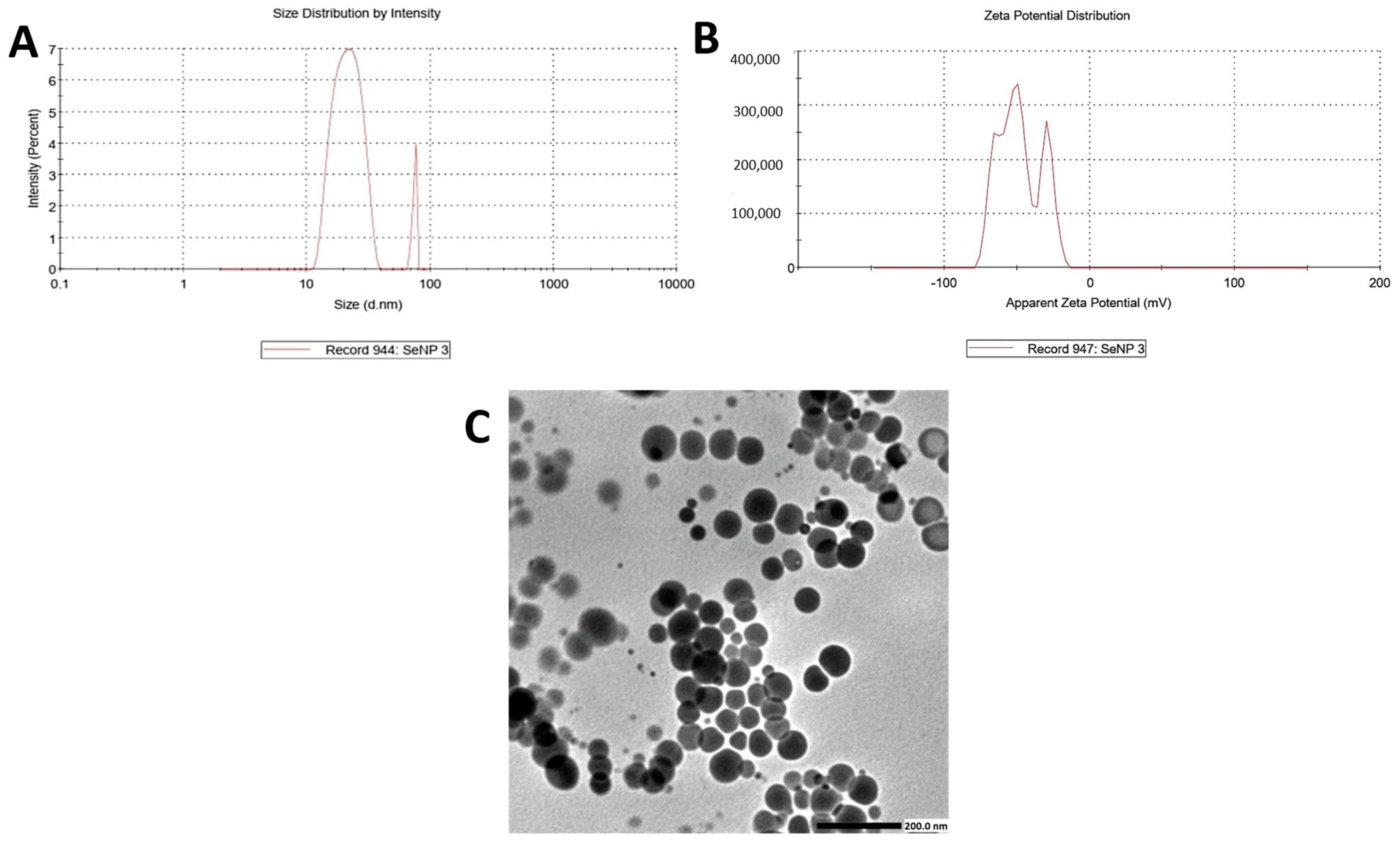
Transmission electron microscopy (TEM) analysis revealed that the EGCG-assisted biosynthesis of selenium nanoparticles (SeNPs) yielded predominantly spherical structures with a relatively uniform morphology. The particle sizes were mainly distributed within the 30–60 nm range, consistent with nanoscale dimensions (Figure 3). While the nanoparticles exhibited adequate dispersion across several regions, partial aggregation was also observed, likely attributable to the intrinsic high surface energy of SeNPs, even in the presence of EGCG functioning as a dual reducing and stabilizing agent. These results show the successful green synthesis of SeNPs, characterized by controlled geometry, narrow size distribution, and stable nanoscale features.
Figure 3.
(A) Zeta size, (B) zeta potential, and (C) TEM of selenium nanoparticles showing successful preparation (Scale bar = 200 nm).
2.2.2. UV–Vis Spectroscopic Characterization
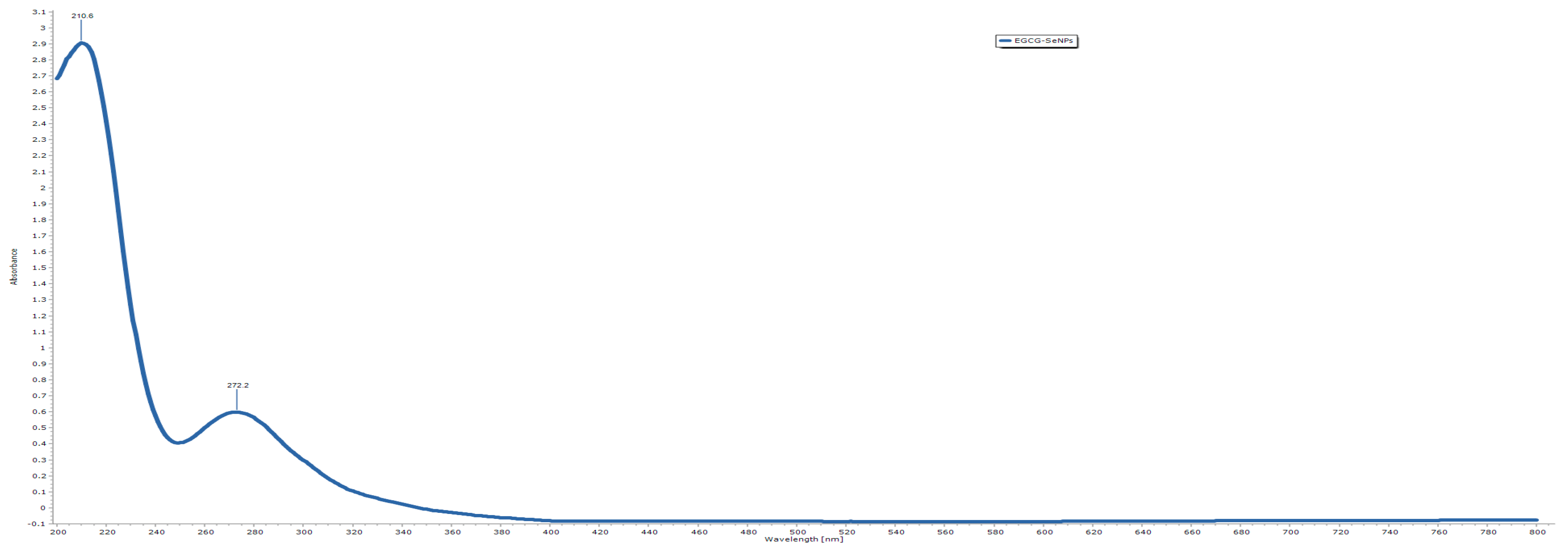
The optical absorption spectrum of the biosynthesized EGCG-SeNPs exhibited a prominent absorption peak at 210.6 nm, accompanied by a secondary band at 272.2 nm (Figure 4). The sharp peak at 210.6 nm corresponds to the π → π* transition of aromatic rings within EGCG, while the minor band near 272.2 nm reflects the n → π* transition associated with phenolic functional groups, confirming the interaction of EGCG molecules with selenium ions. The absence of additional peaks beyond 300 nm and the gradual decline in absorbance across the visible region indicate the formation of stable, well-dispersed SeNPs without aggregation.
Figure 4.
UV–vis result of selenium nanoparticles, showing successful preparation.
2.2.3. FT-IR Analysis
The FTIR analysis was conducted to confirm the molecular structure of EGCG and to elucidate the nature of the interaction between EGCG and selenium in the nanocomposite. The spectra for both pure EGCG and the EGCG-SeNPs complex display several characteristic peaks corresponding to the major functional groups present in the EGCG molecule. A comparison of the two spectra reveals distinct shifts in peak positions and changes in intensity, providing evidence for the successful formation of the complex.
FTIR Peaks of Pure EGCG
The FTIR spectrum of pure EGCG shows its characteristic functional group peaks (Table 3). A broad and strong absorption band observed at 3241 cm−1 is attributed to the O-H stretching vibration, arising from the multiple phenolic hydroxyl groups present in the EGCG structure. The broadness of this peak is indicative of strong intermolecular and intramolecular hydrogen bonding. The peaks at 2918 cm−1 and 2849 cm−1 correspond to the C-H stretching vibrations of the aliphatic -CH2 and -CH3 groups within the flavan-3-ol backbone. A prominent and sharp peak at 1704 cm−1 is assigned to the C=O stretching vibration of the ester carbonyl group in the gallate moiety of EGCG. The region between 1600 and 1450 cm−1, with peaks at 1610 cm−1 and 1448 cm−1, is associated with the aromatic C=C stretching vibrations of the benzene rings. Furthermore, the peaks observed at 1242 cm−1 and 1053 cm−1 are characteristic of the C-O stretching vibrations from the phenolic and ester groups, respectively. These peaks collectively confirm the chemical structure of the EGCG sample.
Table 3.
FTIR peak assignments for pure EGCG.
FTIR Peaks and Interaction in the EGCG-SeNPs Complex
The FTIR spectrum of the EGCG–nano-Se complex shows significant changes compared to that of pure EGCG, confirming the interaction between EGCG and the selenium nanoparticles (Table 4). The most notable change is observed in the broad O-H stretching band. In the complex, this band shifts from 3241 cm−1 to a lower wavenumber of approximately 3225 cm−1 and becomes broader. This red-shift and broadening suggest that the phenolic hydroxyl groups of EGCG are involved in the coordination with the nano-selenium surface, likely through the formation of Se-O bonds or enhanced hydrogen bonding, which weakens the O-H bond. Similarly, the C=O stretching vibration of the ester group shifts from 1704 cm−1 to 1698 cm−1, indicating that the carbonyl oxygen may also participate in the stabilization of the nanocomposite. The peaks corresponding to C-O stretching vibrations at 1242 cm−1 and 1053 cm−1 also shift to lower wavenumbers (e.g., to 1235 cm−1 and 1048 cm−1), further supporting the involvement of oxygen-containing groups in the interaction. The aromatic C=C stretching peaks show minor shifts, suggesting that the core aromatic structure remains largely intact, but the electronic environment is altered due to the complex formation. Overall, the observed shifts in the key functional group peaks provide strong spectroscopic evidence that EGCG successfully acts as a capping and reducing agent, binding to the surface of selenium nanoparticles through its hydroxyl and carbonyl groups (Figure 5).
Table 4.
Comparison of FTIR peaks between EGCG and EGCG-SeNPs.
Figure 5.
FTIR peaks between EGCG and EGCG-SeNPs.
2.3. Sodium Selenite, EGCG, and EGCG-SeNPs Enhance the Performance of Sleep Deprivation-Subjected Rats in the Open-Field Test
Three tests were applied to evaluate the impact of sodium selenite, EGCG, and EGCG-SeNPs on depression-like behavior using the OFT. The results from these tests revealed that sodium selenite, EGCG, and EGCG-SeNPs improved sucrose preference, rearing and grooming time, and alleviated anhedonic-like behavior in sleep deprivation-subjected rats.
As illustrated in (Figure 6), SD caused a remarkable reduction in rearing, grooming, and sucrose-intake frequencies as compared with the negative control group. The co- treatment with sodium selenite, EGCG, and EGCG-SeNPs significantly increased rearing and grooming frequencies and sucrose intake, in comparison with the sleep deprivation-subjected rats. For more accuracy, we found that EGCG-SeNPs markedly raised the rearing and grooming frequencies, as well as sucrose intake, versus the sodium selenite- and EGCG-co-treated group. Additionally, EGCG-SeNPs increased rearing and grooming frequencies, making them similar to those of the control animals as there was no remarkable variance between the EGCG-SeNP-co-treated group and normal control group.
Figure 6.
Effect of selenium selenite, EGCG, and EGCG-SeNPs on (A) grooming, (B) rearing, and (C) sucrose intake in sleep deprivation-subjected rats (N = 8). Data represented as mean ± SEM; one way analysis of variance (ANOVA) was performed, followed by Tukey’s multiple comparison post hoc test to evaluate intergroup differences. $ Significant to the normal control group; # significant to sleep deprivation group; @ significant to EGCG-SeNPs.
2.4. The Effects of EGCG-SeNPs on Brain Monoamine Contents in the Sleep Deprivation-Subjected Rats
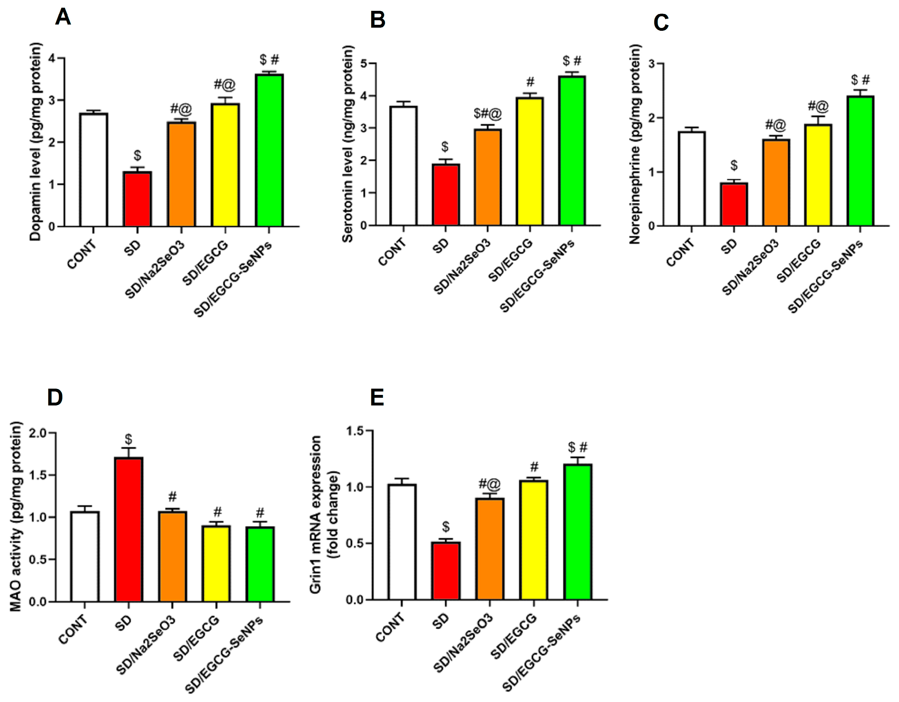
As represented in Figure 7, there were significant differences in cortical dopamine, serotonin, norepinephrine, and Grin 1 gene expression between different groups. Sleep deprivation significantly decreased dopamine, serotonin, norepinephrine, and Grin 1 gene expression, along with considerable increases in monoamine oxidase activity in the prefrontal cortex as compared to the normal control rats. Indeed, co-treatment with sodium selenite, EGCG, and EGCG-SeNPs significantly increased dopamine, serotonin, norepinephrine, and Grin 1 gene expression with significant decline in monoamine oxidase activity in the prefrontal cortex as compared with the sleep-deprived-rats. Moreover, EGCG-SeNP co-administration markedly elevated dopamine and norepinephrine versus sodium selenite- and EGCG-co-treated animals, and upregulated serotonin and Grin 1 gene expression versus sodium selenite-co-treated animals.
Figure 7.
Impact of selenium selenite, EGCG, and EGCG-SeNPs on (A) dopamine, (B) serotonin, (C) norepinephrine, (D) MAO, and (E) Grin 1 in sleep deprivation-challenged rats. In each group, n = 8 and data are represented as mean ± SEM; one way analysis of variance (ANOVA) was applied, followed by Tukey’s multiple comparison post hoc test to evaluate intergroup differences. $ Significant to the normal control group; # significant to sleep deprivation group; @ significant to EGCG-SeNPs.
Additionally, EGCG-SeNPs significantly increased dopamine, serotonin, norepinephrine, and Grin 1 gene expression in the cortex versus the normal control group.
So, these findings confirm sodium selenite, EGCG, and particularly EGCG-SeNPs’ ability to enhance the aforementioned cortical neurotransmitters, overcoming the depression manifested in sleep deprivation-subjected rats.
2.5. EGCG-SeNPs Can Restore AChE Activity, ACh and GABA Levels in Sleep Deprivation-Subjected Rats
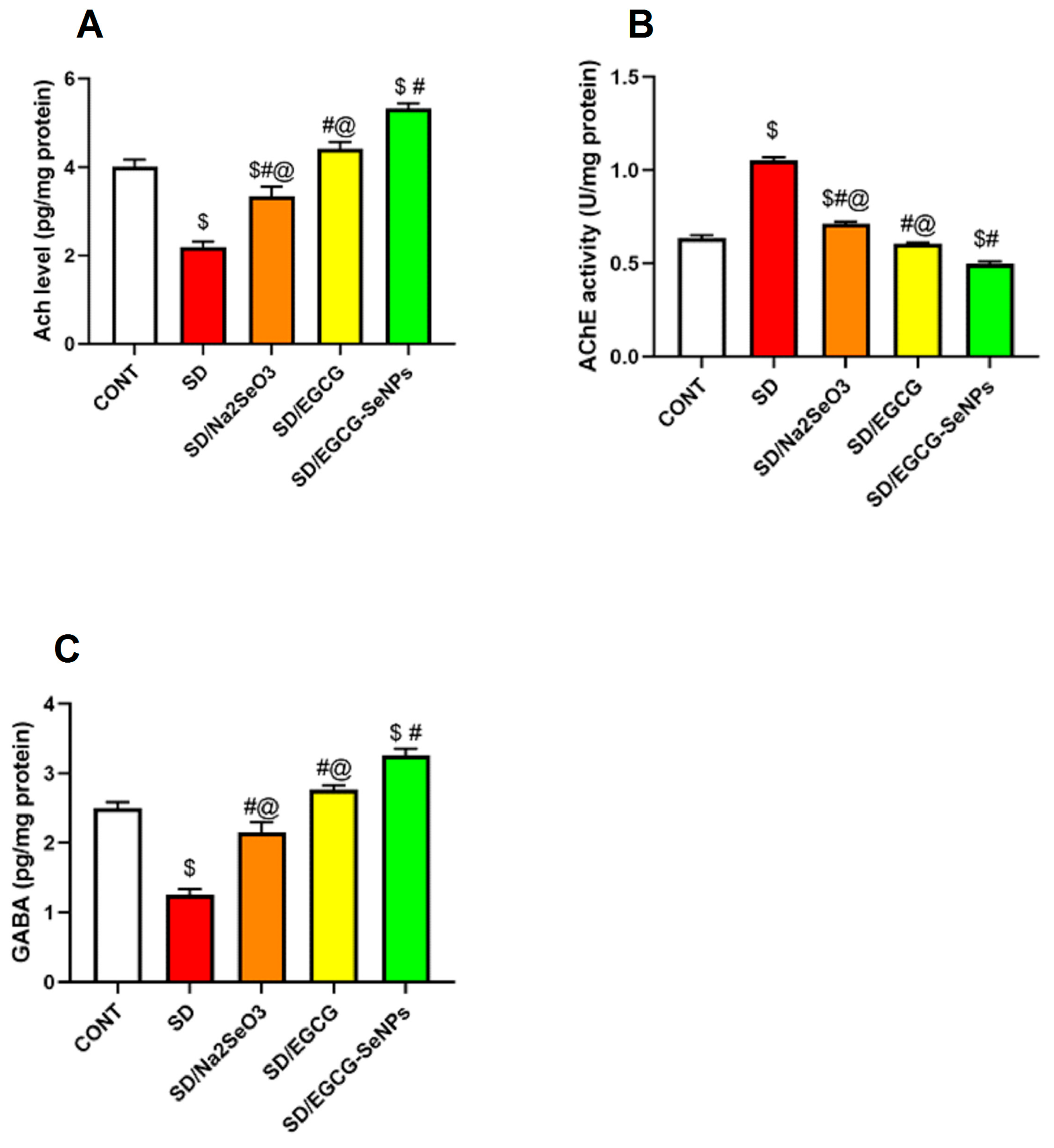
To investigate the neurotransmitter alterations correlated with sleep deprivation, brain values of acetylcholine and its degrading enzyme acetylcholinesterase were examined. It is known that cholinergic transmission in the cortex is associated with cognitive impairment. Meanwhile, the amount of GABA was determined. Figure 6 shows that the levels of cortical acetylcholine, acetylcholine esterase activity, and GABA differed significantly between groups. Cortical acetylcholine and GABA values were substantially lower than those in the control group; moreover, acetylcholine esterase activity was significantly elevated. By administering sodium selenite, EGCG, and EGCG-SeNPs to sleep deprivation-subjected rats, these neurotransmitter changes were reversed, as detected by a remarkable increase in acetylcholine and GABA which was accompanied by a substantial decline in acetylcholine esterase. Notably, EGCG-SeNP co-treatment exerts significant increase in acetylcholine and GABA which sees a significant decrease in acetylcholine esterase activity when compared to sodium selenite- and EGCG-co-treated rats and normal control rats (Figure 8).
Figure 8.
Selenium selenite, EGCG, and EGCG-SeNPs’ effect on (A) acetylcholine level, (B) acetylcholine esterase activity, and (C) γ-aminobutyric acid (GABA) in sleep deprivation-subjected rats. In each group, n = 8 and data are represented as mean ± SEM; one way analysis of variance (ANOVA) was performed, followed by Tukey’s multiple comparison post hoc test to evaluate intergroup differences. $ Significant to the normal control group; # significant to sleep deprivation group; @ significant to EGCG-SeNPs.
According to these data, sodium selenite, EGCG, and EGCG-SeNPs can improve neurotransmitter changes, so engaging in rescuing the cognitive impairment associated with sleep deprivation is significantly linked to EGCG-SeNPs.
2.6. Sodium Selenite, EGCG, and EGCG-SeNPs Can Ameliorate the Cortical Oxidative Stress in the Sleep Deprivation-Subjected Rats
As seen in Figure 9, sleep deprivation resulted in a remarkable increase in oxidative stress responses in the rat brain. This process was characterized by a significant rise in MDA content and NO values in the prefrontal cortex. Compared to those in the control group, the expression levels of genes implicated in the antioxidant system, including Nrf2 and its downstream effectors GSH, GPx, SOD, and catalase, were significantly reduced.
Figure 9.
Effect of sodium selenite, EGCG, and EGCG-SeNPs on (A) MDA, (B) NO, (C) Nrf2, (D) GSH, (E) GPx, (F) SOD, and (G) CAT in sleep deprivation-subjected rats. Data are represented as mean ± SEM (N = 8). In the analysis, one way analysis of variance (ANOVA) was performed, followed by Tukey’s multiple comparison post hoc tests to evaluate intergroup differences. $ Significant to the normal control group; # significant to sleep deprivation group; @ significant to EGCG-SeNPs.
Compared with the SD group, the administration of sodium selenite, EGCG, and EGCG-SeNPs to rats that were subjected to SD successfully reduced oxidative stress responses, resulting in a remarkable drop in brain MDA content and NO level in the prefrontal cortex. This effect was further validated by the pronounced upregulation of antioxidant Nrf2 gene expression and the subsequently increase in GSH content, as well as GPx, catalase, and SOD activities.
A comparative analysis of the three compounds revealed that EGCG-SeNP administration resulted in superior antioxidative stress effects. Compared with sodium selenite-co-treated rats, it significantly decreased NO and increased Nrf2 gene expression, GPx, and catalase activities. Additionally, it increased GSH content and SOD activity versus the EGCG-co-treated group. Moreover, for GSH content, GPx, catalase, and SOD activities in the EGCG-SeNP co-group revealed a substantial rise compared to the normal control animals.
These findings illuminate the role of sodium selenite, EGCG, and EGCG-SeNPs in mitigating oxidative stress and activating the Nrf2 cascade, contributing at least partially to the amelioration of cognitive impairment induced by SD.
2.7. Sodium Selenite, EGCG, and EGCG-SeNPs Can Abrogate Inflammation in the Sleep Deprivation-Subjected Rats
Chronic SD has been found to decrease cognitive performance, affect mood states, and simultaneously dysregulate inflammatory and stress hormones.
In our work, mRNA expression of NOS2 showed a rising trend in the prefrontal cortex of the SD group compared to the normal group. However, versus the sleep deprivation group, sodium selenite, EGCG, and EGCG-SeNPs showed a tendency to decline the mRNA expression of NOS2 (Figure 10). In the same context, TNF-α, IL-1β, and IL-6 levels remarkably elevated in the prefrontal cortex of the SD group, compared to the normal group. A marked decrease in TNF-α, IL-1β, and IL-6 levels was observed with the sodium selenite-, EGCG-, and EGCG-SeNP-co-treated groups compared to the SD group. Additionally, the EGCG-SeNP-co-treated group exerts more powerful anti-inflammatory action that exceeds that of the normal control rats and other co-treated groups as it considerably downregulated NOS2, TNF-α, and IL-6, compared to the sodium selenite-co-treated group, and decreased IL-1β versus the sodium selenite- and EGCG-co-treated group. And additionally, it significantly decreased IL-1β and IL-6 compared to the normal control group.
Figure 10.
Selenium selenite, EGCG, and EGCG-SeNPs altered (A) NOS2 gene expression, (B) TNF-α, (C) IL-1β, (D) IL-6, and (E) corticosterone in sleep deprivation-subjected rats. In each group, n = 8 and data are represented as mean ± SEM; one way analysis of variance (ANOVA) was performed, followed by Tukey’s multiple comparison post hoc test to evaluate intergroup differences. $ Significant to the normal control group; # significant to sleep deprivation group; @ significant to EGCG-SeNPs.
Regarding corticosterone level, stress factor was substantially elevated in the SD group compared to the normal control group. Co-administration of sodium selenite, EGCG, and EGCG-SeNPs resulted in a remarkable decrease in corticosterone level compared to the sleep deprivation group, confirming the ability to attenuate the stress response.
Considering the comparison between treated groups, EGCG-SeNPs decrease corticosterone level significantly versus the normal control and the sodium selenite-co-administration groups.
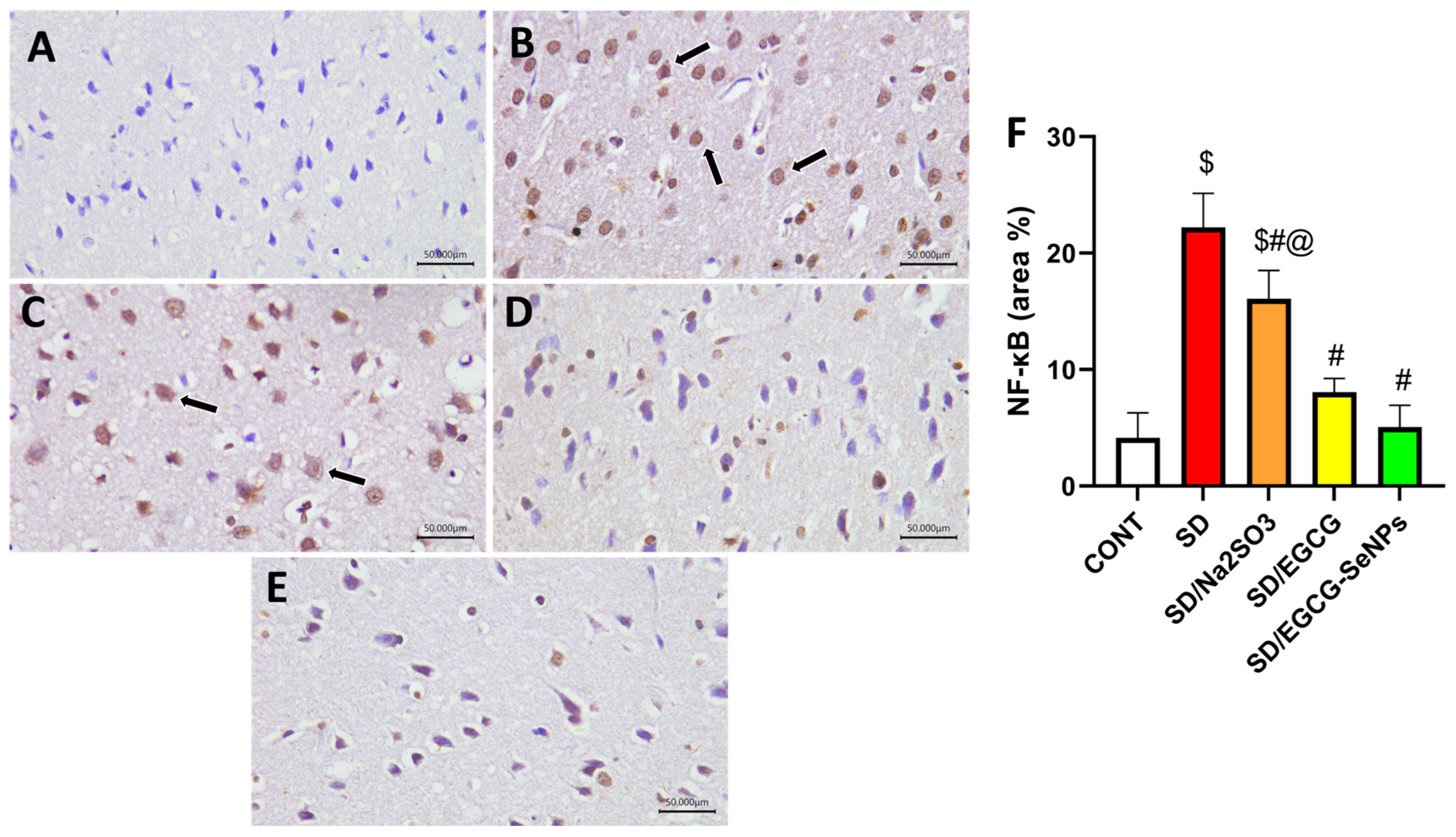
For more accuracy, the transcriptional factor, NF-κB, was estimated by immunohistochemistry. The control rats, EGCG- and EGCG-SeNP-co-treated animals, expressed weak reactivity to NF-κB, contributing to evidence of its anti-inflammatory effect, whereas the sleep deprivation- and sodium selenite-co-treated groups displayed a marked reactivity to NF-κB, indicating inflammatory process. Both EGCG- and EGCG-SeNP-co-treated rats revealed marked decrease in NF-κB compared to the SD group (p < 0.0001). However, both SD and SD/sodium selenite groups showed higher NF-κB expression compared to the control group (p < 0.0001) (Figure 11). Additionally, administration of EGCG-SeNPs to sleep-deprived-rats decreased NF-κB protein expression significantly versus the sodium selenite-co-treated groups.
Figure 11.
Protective effect of EGCG-SeNPs on prefrontal cortex immunoreactivity of NF-κB in SD-evoked cortical damage in rats. Photomicrographs of the prefrontal cortex from all groups. The immunoreactivity of NF-κB were visible in the tissues as a brown color generated by DAB chromogen (arrow) (DAB, X400, Scale bar = 50 µm). The control group (A) exhibited normal cortical structure with weak GFAP staining. SD (B) and SD–sodium-selenite-treated (C) groups revealed intense NF-κB immunostaining. In contrast, the (D) SD-EGCG- and (E) SD-EGCG-SeNP-treated group displayed attenuated NF-κB staining. (F) Quantitative analysis of immunostaining area % for NF-κB was expressed as mean ± S.E.M. (n = 8). Data are represented as mean ± SEM, performed using one way analysis of variance (ANOVA), followed by Tukey’s multiple comparison post hoc test to evaluate intergroup differences. $ Significant to the normal control group; # significant to sleep deprivation group; @ significant to EGCG-SeNPs.
2.8. Selenium Selenite, EGCG, and EGCG-SeNPs Attenuate Apoptosis in the Sleep Deprivation-Subjected Rats
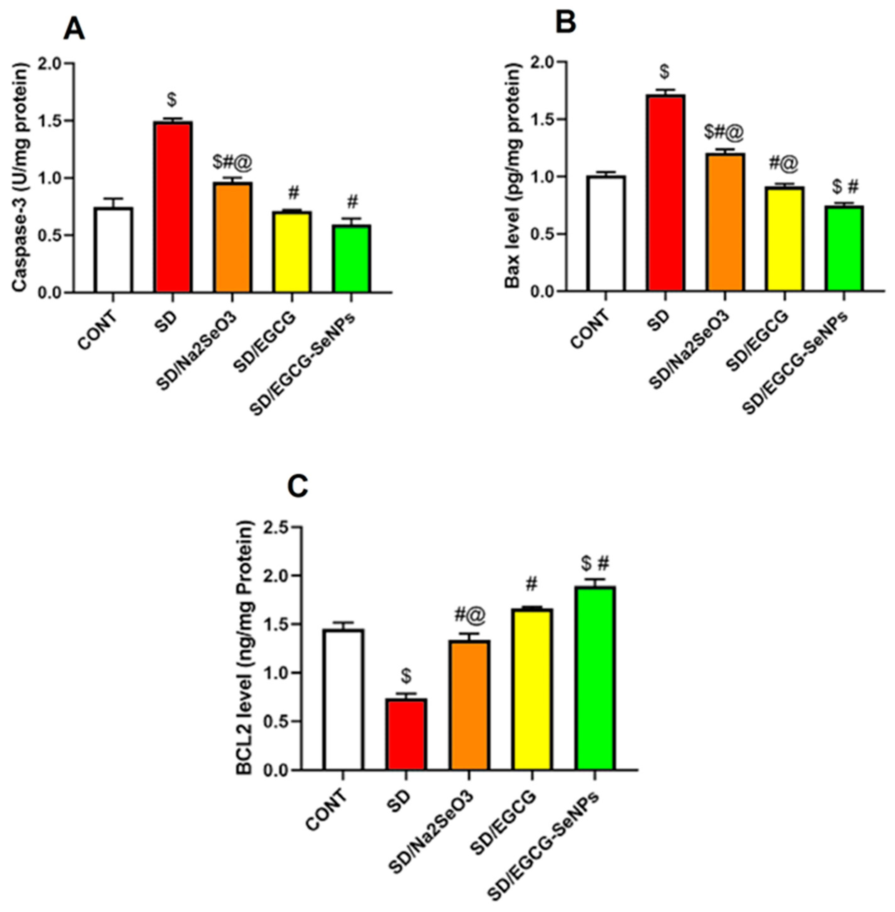
It is believed that cortical apoptotic cell death is correlated with marked degeneration and behavioral deficits in SD-subjected rats. So, the executioner caspase 3, Bax, and Bcl-2 were estimated to determine the cortical apoptosis. As illustrated in Figure 12, it was demonstrated that the rats subjected to sleep deprivation revealed a remarkable elevation in cortical apoptotic markers, caspase-3 activity, and BAX level, together with a considerable decrease in the anti-apoptotic BCL2 level in the prefrontal cortex as compared to the negative control group. On the contrary, sodium selenite-, EGCG-, and EGCG-SeNP-co-treated groups revealed a substantial decline in cortical caspase-3 activity and BAX level, with significant elevation in BCL2 level in the prefrontal cortex versus the sleep deprivation group. Moreover, the EGCG-SeNP-co-treated group markedly decreased in cortical caspase-3 activity and BAX level, with a significant increase in BCL2 level in the prefrontal cortex in comparison with the sodium selenite-co-treated group and considerable decreases in BAX level versus the EGCG-co-treated animals.
Figure 12.
Effect of selenium selenite, EGCG, and EGCG-SeNPs on (A) Caspase 3 activity, (B) BAX level, and (C) BCL2 level in sleep deprivation-subjected rats. In each group, n = 8 and data are represented as mean ± SEM; one way analysis of variance (ANOVA) was performed, followed by Tukey’s multiple comparison post hoc test to evaluate intergroup differences. $ Significant to the normal control group; # significant to sleep deprivation group; @ significant to EGCG-SeNPs.
Also, for the EGCG-SeNP-co-treated group, it causes a remarkable decrease in BAX level and a significant increase in BCL2, in comparison with the normal control group, confirming its anti-apoptotic effect.
So, depending on these results, sodium selenite, EGCG, and EGCG-SeNPs can curtail apoptotic cell death, especially for the EGCG-SeNP-co-treated group, and abrogate the cognitive decline associated with sleep deprivation.
2.9. Selenium Selenite, EGCG, and EGCG-SeNPs Modified the GFAP and BDNF Contents in the Cortical Tissues of the Sleep Deprivation-Subjected Rats
GFAP gene expression has received a lot of interest because its onset is a signal for astrocyte formation and its upregulation indicates reactive gliosis. However, BDNF is the most abundant neurotrophin in the brain, playing a crucial role in the survival, differentiation, synaptic plasticity, and axonal expansion of peripheral and central neurons.
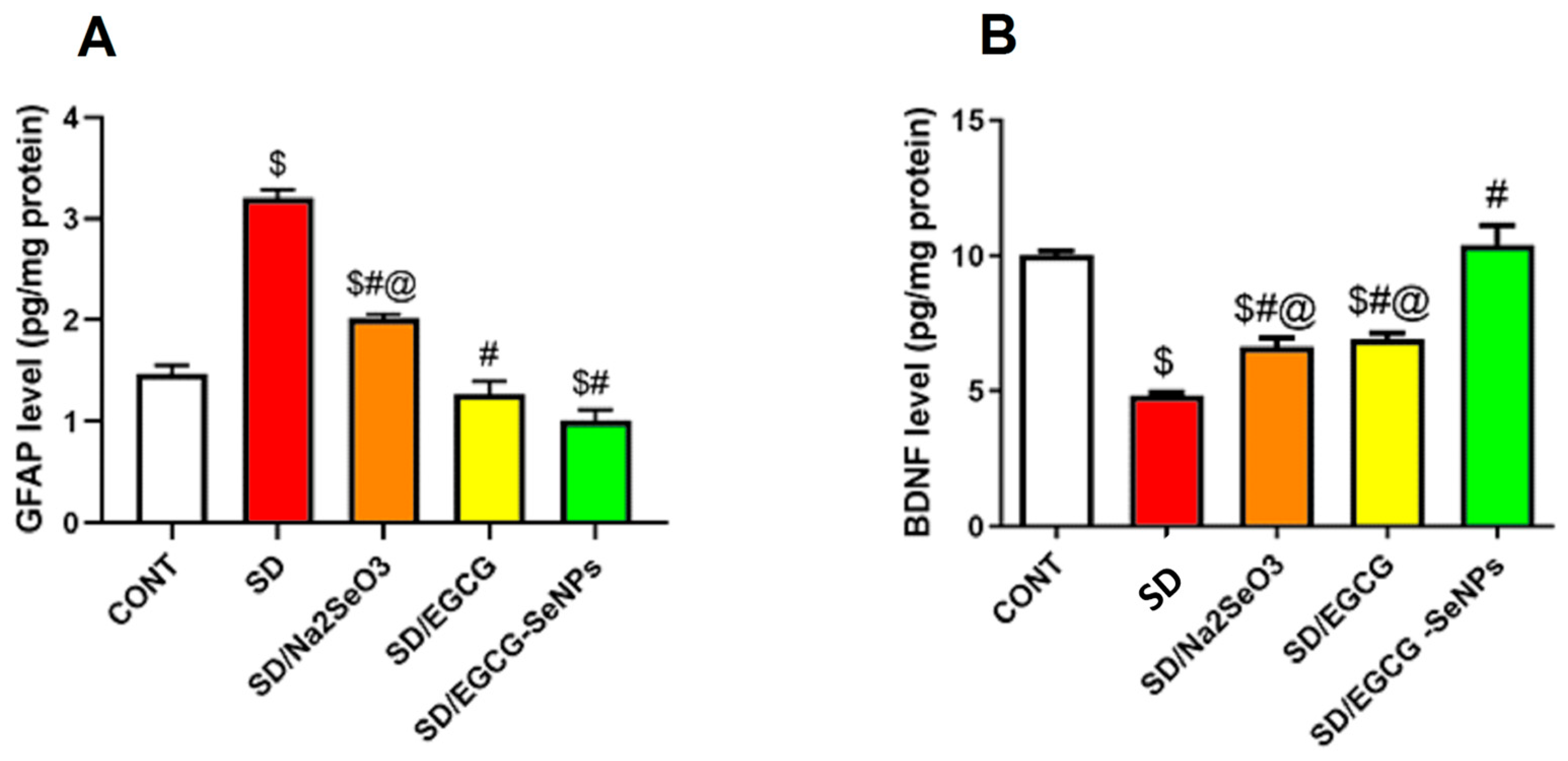
As shown in Figure 13, there is a significant rise in the GFAP (a marker of astrocytosis) with marked decrease in BDNF in the prefrontal cortex in the sleep deprivation-subjected group as opposed to the control one. In contrast, sodium selenite-, EGCG-, and EGCG-SeNP-co-treated groups significantly lowered GFAP and upregulated BDNF in comparison to the sleep deprivation group. In the same context, EGCG-SeNPs produce a more favorable effect regarding BDNF compared to sodium selenite and EGCG, and additionally reduce GFAP more than sodium selenite and normal control groups.
Figure 13.
Effect of selenium selenite, EGCG, and EGCG-SeNPs on (A) GFAP and (B) BDNF levels in sleep deprivation-subjected rats. (n = 8) and data are represented as mean ± SEM; one way analysis of variance (ANOVA) was performed, followed by Tukey’s multiple comparison post hoc test to evaluate intergroup differences. $ Significant to the normal control group; # significant to sleep deprivation group; @ significant to EGCG-SeNPs.
Regarding GFAP immunoreactivity, the control group and EGCG- and EGCG-SeNP-co-treated animals expressed GFAP moderately, which reveals the normal functional glial cellular activity, while the SD group and sodium selenite-co-treated group displayed a marked reactivity to GFAP. Co-administration of EGCG and EGCG-SeNPs produced a considerable reduction in GFAP compared to the SD group (p < 0.0001). In contrast, both SD and SD/sodium selenite groups showed higher GFAP expression compared to the control group (p < 0.0001) (Figure 14).
Figure 14.
Protective effect of EGCG-SeNPs on prefrontal cortex immunoreactivity of GFAP in SD-evoked cortical damage in rats. Photomicrographs of prefrontal cortex from all groups. The immunoreactivity of GFAP was visible in the tissues as a brown color generated by DAB chromogen (arrow) (DAB, X400, Scale bar = 50 µm). The control group (A) exhibited normal cortical structure with moderate GFAP staining. SD- (B) and SD–sodium selenite-treated (C) groups revealed intense GFAP immunostaining. In contrast, SD-EGCG- (D) and SD-EGCG-SeNP-treated (E) group displayed attenuated GFAP staining. (F) Quantitative analysis of immunostaining area % for GFAP was expressed as mean ± S.E.M. (n = 8). Data are represented as mean ± SEM performed using one way analysis of variance (ANOVA), followed by Tukey’s multiple comparison post hoc test to evaluate intergroup differences. $ Significant to the normal control group; # significant to the sleep deprivation group; @ significant to EGCG-SeNPs.
2.10. Histopathological Examination
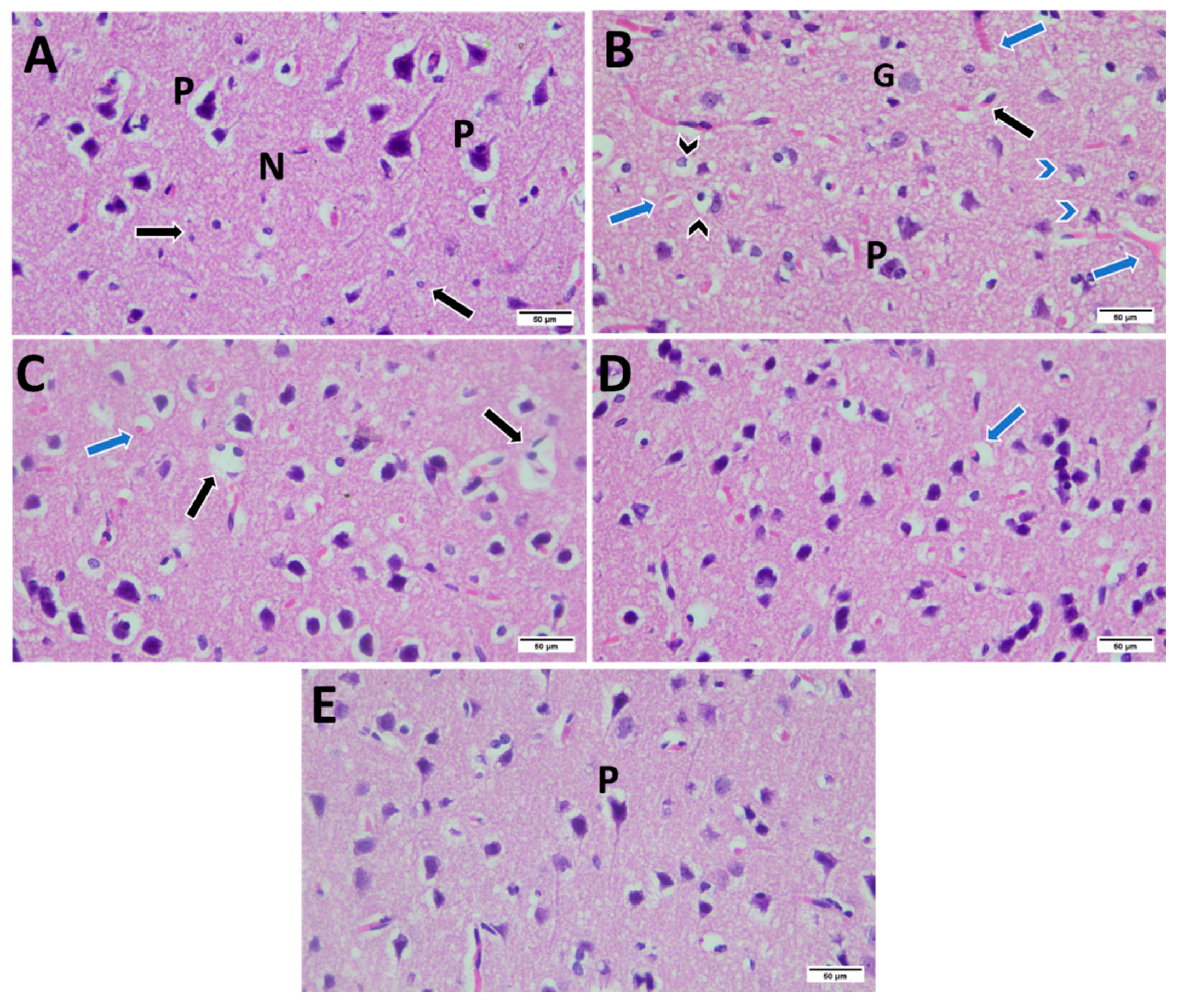
Sections of the prefrontal cortex taken from male control rat displayed normal anatomy. The control group showed granule cells with big, conspicuous nuclei and spherical cell bodies were visible in the outer granular layer. Medium-sized pyramidal cells with basophilic cytoplasm, rounded nuclei, triangular cell bodies, and apical dendrites were visible. The size of the pyramidal cells varied. The neuropil, which was a fibrillar eosinophilic substance, composed of a dense network of branching processes, including glial cells. 24hSD revealed altered architecture of the cerebral cortex, with numerous glial cells visible in the molecular layer. The blood capillaries were dilated, congested, and encircled by perivascular space, degenerated pyramidal cells, and deformed neurons with darkly stained nuclei and surrounded by halos and an increase in the perinuclear space. 24hSD-Na2SeO3 showed degenerated cells with pyknotic nuclei, congested capillaries, and few deformed neurons, as well as few neurons surrounded by clear areas. 24hSD-EGCG exhibited normal neuropil with few shrunken cells with pyknotic nuclei. 24hSD-EGCG-SeNPs showed approximately normal appearance of cortical tissue with normal neurons and neuropil (Figure 15).
Figure 15.
Photomicrographs of male prefrontal cerebral cortex of all groups. (A) Control group showed normal neuropil (N) containing vesicular pyramidal cells (P) and multiple neuroglial cells (black arrow). (B) 24hSD revealed disturbed cortical architecture with congested blood capillaries (blue arrow), deformed neurons with darkly stained nuclei surrounded by halos, and increased perinuclear space (black arrow head), degenerated pyramidal cells (blue arrow head), and increased neuroglial cells (black arrow). (C) 24hSD-Na2SeO3 showed degenerated cells with pyknotic nuclei (black arrow) and congested capillaries (black arrow). (D) 24hSD-EGCG exhibited normal neuropil with few shrunken cells with pyknotic nuclei (arrow). (E) 24hSD-EGCG-SeNPs showed approximately normal appearance of cortical tissue with normal pyramidal cells (P). (H&E, X400, Scale bar = 50 µm).
3. Discussion
EGCG, a major component of green tea, exhibits promising dual inhibitory activity against BCL-2 and MMP-2, as suggested by molecular docking studies [27,28,29,30]. The observed Glide XP G-scores of −4.033 kcal/mol for BCL-2 and −6.749 kcal/mol for MMP-2 suggest a higher binding affinity for MMP-2 [27,31]. These interactions are further stabilized by specific hydrogen bonds formed with key amino acid residues in the active sites of both proteins [32].
The adsorption properties of SeNPs and EGCG–nano-selenium complexes with BCL2 and MMP2 are based on computational data generated using BIOVIA Materials Studio 2024 [33]. Adsorption energy is a critical parameter in assessing the stability and spontaneity of molecular interactions; a more negative value suggests a more favorable binding process [34]. The MMP2-EGCG-SeNP complex shows a total energy of 1.36 × 10⁵ and a significantly more negative adsorption energy of −1.01 × 10⁵, compared to the MMP2-SeNP system, which has a total energy of 3.64 × 10⁴ and an adsorption energy of −7.69 × 10⁴. The deformation energies are −3.21 × 10⁴ and −3.62 × 10⁴ for the EGCG-SeNP complex and SeNPs alone, respectively. This further supports the conclusion that EGCG enhances the binding and stability of SeNP complex with MMP2 [35]. EGCG is a natural polyphenol with known antioxidative and anticancer properties [36,37]. Its ability to enhance the binding affinity of SeNPs can be attributed to several factors. EGCG can stabilize SeNPs, prevent their aggregation, and increase their bioavailability and bioactivity. For instance, starch microgels and EGCG synergistically stabilize SeNPs, improving their properties [36]. EGCG possesses multiple hydroxyl groups that can participate in hydrogen bonding and other polar interactions with the amino acid residues in the binding sites of BCL2 and MMP2 [38,39]. These additional interactions contribute to a more stable complex [40].
Sleep deprivation is increasingly acknowledged as a major contributor to mood disorders, cognitive impairment, and neuronal dysfunction. Experimental models have shown that prolonged wakefulness induces behavioral deficits resembling depression and anxiety, alongside neurochemical, oxidative, and inflammatory changes that compromise brain integrity [41,42]. The current study was designed to systematically investigate these the pathological consequences of sleep deprivation and to evaluate the protective effects of sodium selenite, epigallocatechin gallate, and their nano-formulation.
Rats subjected to 24 h of sleep deprivation exhibited significant behavioral anomalies, including reduced grooming, rearing, and sucrose intake. These results confirm the presence of anhedonia and reduced exploratory activity, which are characteristic features of depression-like behavior. The present study employed a 24 h sleep deprivation protocol as an established model for inducing acute oxidative and neuro-inflammatory responses in cortical tissues. While this paradigm effectively produces measurable neuronal stress, it may not fully reflect the pathophysiological consequences of chronic or recurrent sleep deprivation observed in humans. Future studies using prolonged or repeated sleep deprivation protocols will be essential to extend the translational value of our findings.
These outcomes are consistent with the notion that sleep loss disrupts motivational and reward-related circuits in the brain, particularly within the prefrontal cortex and limbic system. Reduced sucrose preference reflects anhedonia, while decreased grooming and rearing indicate apathy and diminished exploratory drive. Our findings are in agreement with previous studies showing that rodents exposed to sleep deprivation demonstrate decreased sucrose consumption and reduced locomotor activity, which have been linked to monoaminergic dysregulation and HPA axis hyperactivation [43,44]. Sleep deprivation elevates corticosterone, which perturbs dopaminergic and serotonergic signaling, thereby suppressing reward-related behaviors. The resulting imbalance in neurotransmission contributes to depression-like phenotypes. Co-treatment with sodium selenite, EGCG, and EGCG-SeNPs reversed the behavioral deficits. Notably, EGCG-SeNPs restored grooming, rearing, and sucrose intake to levels comparable to normal controls, suggesting near-complete recovery of motivation and exploratory behavior. Sodium selenite likely exerted its effects through antioxidant and stress-modulating actions, while EGCG acted via anti-inflammatory and neurotransmitter-stabilizing properties. The EGCG-SeNPs combined these benefits with enhanced bioavailability, leading to superior normalization of behavioral outcomes. Sleep-deprived rats showed significant reductions in dopamine, serotonin, norepinephrine, and Grin1 expression, along with increased monoamine oxidase activity. This pattern reflects impaired monoaminergic transmission, a key pathological mechanism underlying depressive-like states. The reduction in Grin1, a glutamatergic NMDA receptor subunit, also indicates disrupted excitatory neurotransmission, contributing to cognitive deficits. These results align with studies reporting that sleep deprivation depletes cortical monoamines and enhances monoamine oxidase activity [45,46]. Similar reductions in serotonin and dopamine have been observed in models of sleep loss, correlating with depressive symptoms [47,48]. Sleep deprivation activates stress pathways, increasing corticosterone, which downregulates monoaminergic systems. The concurrent rise in monoamine oxidase accelerates neurotransmitter degradation, exacerbating neurotransmitter deficits. Sodium selenite, EGCG, and EGCG-SeNPs all increased dopamine, serotonin, norepinephrine, and Grin1, while reducing monoamine oxidase activity. EGCG-SeNPs demonstrated the strongest effect, with neurotransmitter levels even surpassing control values in some cases. Sodium selenite likely enhanced monoaminergic stability via antioxidant protection of neuronal terminals. EGCG inhibited monoamine oxidase and preserved neurotransmitter levels. EGCG-SeNPs combined these actions and provided more efficient neuronal uptake, thus potentiating monoaminergic restoration. Sleep-deprived rats exhibited a significant reduction in cortical acetylcholine and GABA, alongside elevated acetylcholinesterase activity. These changes indicate impaired cholinergic and inhibitory signaling, both of which are essential for learning, memory, and emotional regulation. The decline in acetylcholine contributes to cognitive impairment, while reduced GABA promotes hyperexcitability and anxiety-like behavior. Previous studies demonstrated that sleep deprivation leads to cholinergic dysfunction and diminished GABAergic transmission, which are strongly associated with cognitive decline and mood instability [49,50]. Sleep deprivation disrupts neuronal homeostasis, leading to reduced synthesis and accelerated degradation of acetylcholine, together with downregulation of GABAergic tone. Sodium selenite, EGCG, and EGCG-SeNPs restored acetylcholine and GABA, while reducing acetylcholinesterase activity. EGCG-SeNPs produced the strongest effect, elevating neurotransmitter levels beyond normal controls. Sodium selenite likely acted through redox modulation, protecting cholinergic neurons. EGCG improved neurotransmitter homeostasis via its antioxidant and anti-inflammatory roles. EGCG-SeNPs provided enhanced penetration and bioavailability, resulting in superior restoration of the excitatory–inhibitory balance. Sleep deprivation induced pronounced oxidative stress, evidenced by increased MDA and NO, with decreased Nrf2 expression and reduced GSH, GPx, catalase, and SOD activities. This oxidative imbalance reflects excessive production of ROS and reactive nitrogen species (NOS), coupled with suppression of endogenous antioxidant defenses. Such alterations damage cellular macromolecules and exacerbate neuronal vulnerability. Our findings agree with prior reports that sleep loss enhances oxidative damage in the prefrontal cortex and hippocampus [4,51,52]. Suppression of Nrf2 signaling during sleep deprivation has also been described as a pivotal factor in oxidative injury. All three treatments significantly decreased oxidative markers and enhanced antioxidant defenses, with EGCG-SeNPs providing the most robust effect, including overcompensation above normal control values. Sodium selenite contributes as a cofactor for glutathione peroxidase, enhancing antioxidant activity. EGCG directly scavenges free radicals and activates Nrf2. Although our findings suggest that EGCG-SeNPs may exert their neuroprotective actions through modulation of the NOS2/Nrf2 axis, it should be noted that this interpretation is based on correlative expression data rather than direct mechanistic evidence. Future studies employing targeted inhibition or activation of Nrf2 and NOS2, as well as downstream signaling analysis, will be essential to confirm the causal involvement of this axis in mediating the observed effects. EGCG-SeNPs not only preserved EGCG activity but amplified Nrf2 signaling and downstream antioxidants due to improved bioavailability and sustained release. Sleep deprivation markedly upregulated cortical NOS2, TNF-α, IL-1β, and IL-6, alongside elevated corticosterone levels. This demonstrates strong neuroinflammation and hyperactivation of the hypothalamic–pituitary–adrenal axis, which are major drivers of neuronal dysfunction during sleep loss. Previous studies have consistently reported elevated pro-inflammatory cytokines and corticosterone after sleep deprivation, linking them to depressive behavior and impaired cognition [53,54]. Sodium selenite, EGCG, and EGCG-SeNPs attenuated neuroinflammation and reduced corticosterone levels. EGCG-SeNPs showed superior anti-inflammatory and anti-stress actions, suppressing cytokines below normal control levels in some cases. Sodium selenite modulated immune responses through selenoproteins. EGCG suppressed NF-κB activation and cytokine production. EGCG-SeNPs combined these effects with enhanced cortical delivery, enabling more powerful inhibition of neuroinflammation. Sleep deprivation increased cortical apoptosis, as indicated by higher caspase-3 and Bax and lower Bcl-2. This suggests activation of the intrinsic apoptotic pathway, leading to neuronal loss and structural damage. Previous studies confirmed that sleep loss triggers apoptotic cascades in the prefrontal cortex and hippocampus [55,56,57].
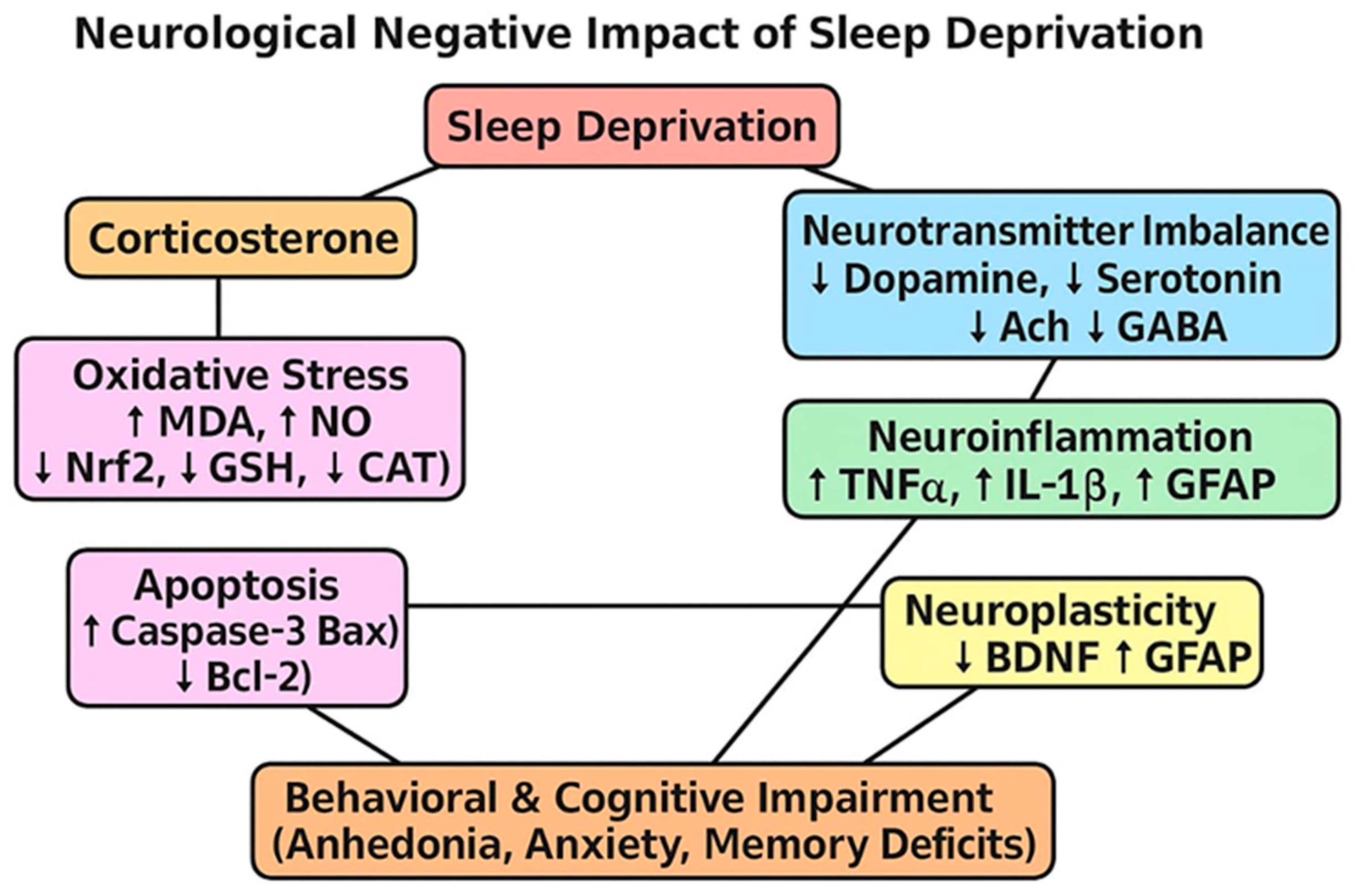
Our findings imply a mechanistic pathway for neuronal cell death. Sleep deprivation induces oxidative stress, provokes inflammation, and leads to the activation of mitochondrial apoptosis, causing upregulation of Caspase-3, Bax, and reducing Bcl-2, which causes neuronal death as shown in Figure 16.
Figure 16.
Neurological negative impact of SD.
Sodium selenite, EGCG, and EGCG-SeNPs suppressed apoptosis, reducing caspase-3 and Bax while restoring Bcl-2. EGCG-SeNPs exerted the most pronounced anti-apoptotic effect, normalizing pro- and anti-apoptotic markers. Sodium selenite protected mitochondrial integrity. EGCG inhibited apoptotic signaling via antioxidant and anti-inflammatory pathways. EGCG-SeNPs provided sustained, enhanced delivery that maximized anti-apoptotic potential. Sleep-deprived rats showed reduced BDNF and elevated GFAP, indicating impaired neuroplasticity and astrogliosis. BDNF is critical for neuronal survival and synaptic plasticity; its decline reflects diminished neurotrophic support. GFAP upregulation indicates reactive gliosis, a pathological marker of neuroinflammation. Our findings are consistent with reports of sleep loss suppressing BDNF and inducing GFAP expression [58,59,60].
Sleep deprivation reduced BDNF, leading to impaired synaptic plasticity and memory, causing increase in GFAP which leads to astrogliosis. All treatments increased BDNF and reduced GFAP, with EGCG-SeNPs again showing the strongest effect. Sodium selenite maintained glial antioxidant capacity, while EGCG upregulated BDNF via CREB activation, and EGCG-SeNPs combined these effects with greater bioavailability, leading to enhanced neuronal plasticity [61]. Although EGCG-SeNPs showed significant neuroprotective efficacy in cortical tissues, the present study did not include direct pharmacokinetic evaluation or biodistribution analysis to confirm nanoparticle penetration into the brain. Future investigations employing fluorescent or radiolabeled EGCG-SeNPs will be essential to delineate their brain uptake, tissue accumulation, and pharmacokinetic profile.
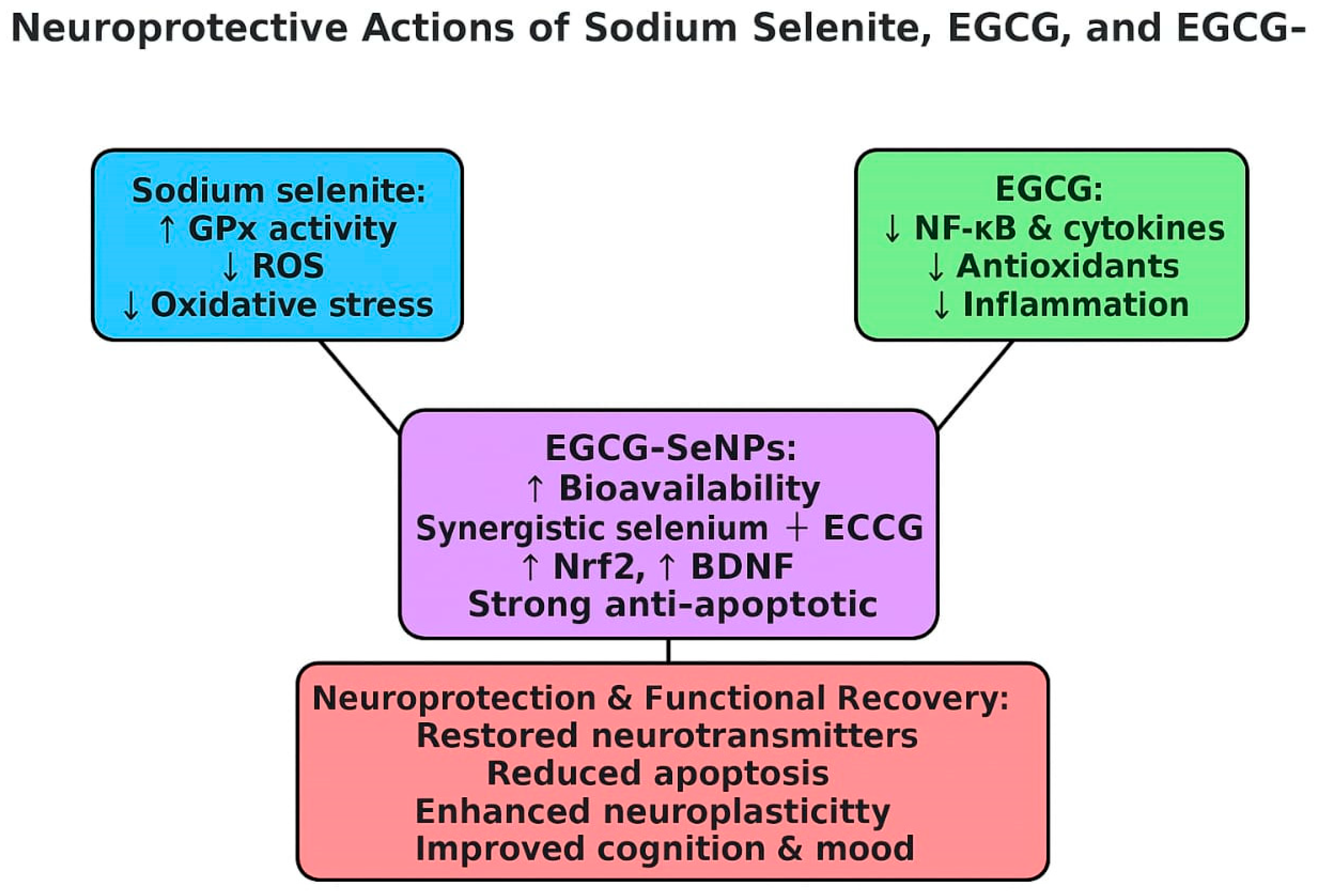
Collectively, these results confirm that sodium selenite, EGCG, and EGCG-SeNPs confer significant neuroprotection against sleep deprivation-induced behavioral, biochemical, and structural changes. Among them, EGCG-SeNPs consistently produced the most robust effects, surpassing both sodium selenite and EGCG. This superiority reflects the synergistic activity of selenium and EGCG, together with the improved bioavailability and sustained action provided by the nanoparticle formulation as shown in Figure 17.
Figure 17.
Neuroprotective actions of sodium selenate, EGCG, and EGCG-SeNPs.
It should be noted that the current study employed an acute (24 h) sleep deprivation paradigm, which primarily reflects short-term neuronal and behavioral consequences. While this model effectively induces measurable cortical oxidative and inflammatory changes, it does not fully replicate the cumulative neurodegenerative impact of chronic sleep loss observed in human conditions. Future studies employing extended or repeated sleep deprivation protocols would be valuable to enhance the translational relevance of these findings. While the current study demonstrated apoptotic involvement through alterations in BAX, Bcl-2, and caspase-3 levels, confirmation via TUNEL staining would further strengthen these findings by directly identifying DNA fragmentation in apoptotic neurons. This will be addressed in future investigations.
Interestingly, sodium selenite, EGCG, and EGCG-SeNP administration mitigated cognitive manifestations and cortical histopathological anomalies in the sleep deprivation-subjected rats. These beneficial effects were elicited by sodium selenite, EGCG, and EGCG-SeNPs and may be related to their ability to counteract corticosterone spike, cortical neurotransmitter decrease, and neurotropic and glial marker alterations, while also inhibiting the inflammatory and apoptotic axis and stimulating Nrf2 antioxidant cascade. The present study was limited to an acute rat model of sleep deprivation, which may not fully capture chronic human sleep loss.
4. Materials and Methods
4.1. In Silico Analysis
4.1.1. Molecular Docking
Protein–ligand docking was executed with Schrödinger Suite 2023-3 Maestro using the Glide module. Human BCL-2 (PDB 4AQ3) and MMP-2 (PDB 1QIB) were prepared with the Protein Preparation Wizard: hydrogens were added, bond orders were assigned, disulfide bonds were created, and water molecules > 5 Å were removed from hetero-atoms; protonation states were predicted by PROPKA at pH 7.4 ± 0.2 and the structures were energy-minimized with the OPLS4 force field (RMSD restraint 0.30 Å). (+)-Epigallocatechin gallate (EGCG) was sketched in 2-D Sketcher, converted to 3-D, desalted, and energetically minimized (LigPrep, OPLS4, 32 stereoisomers, pH 7.0 ± 2.0). Receptor grids (20 Å side length) were centered on the catalytic/active-site clefts. Flexible ligand docking was performed sequentially in Standard Precision (SP) and Extra Precision (XP) modes, retaining up to 10 poses per ligand; final poses were re-scored with Prime MM-GBSA (VSGB 2.0 solvent model). The lowest-energy complexes were inspected for hydrogen bonds, π–π stacking, salt bridges, and metal contacts; Glide XP G-scores are reported.
4.1.2. Absorptivity of Nano-Selenium and EGCG–Nano-Selenium with BCL2 and MMP2
The adsorption properties of nano-selenium and EGCG–nano-selenium with BCL2 and MMP2 structures were investigated using BIOVIA Materials Studio 2024. The lowest-energy poses were initially determined using the Adsorption Locator module, which employs the Forcite and COMPASS III force fields. Subsequently, total energy, adsorption energy, rigid adsorption energy, and deformation energy were calculated for each protein–ligand system to quantify binding stability and conformational changes.
4.2. Drugs
EGCG (CAS No. 989-51-5) was obtained from Sigma-Aldrich (St. Louis, MO, USA) and employed without any further purification. The green synthesis of selenium nanoparticles (SeNPs) functionalized with EGCG (SeNPs-EGCG) was achieved by mixing 10 mL of sodium selenite solution (Na2SeO3, 10 mM) with an equal volume of EGCG solution (3.5 mg/mL). The reaction mixture was maintained under constant magnetic stirring at ambient temperature for 12 h, during which EGCG served simultaneously as a reducing and stabilizing agent to promote nanoparticle formation. The appearance of a characteristic reddish coloration signified successful synthesis of SeNPs-EGCG. The colloidal suspension was subsequently lyophilized using a laboratory freeze-drying system (FreeZone 4.5 L, Labconco, Marshall Scientific, Hampton, NH, USA), and the resulting dry nanoparticle powder was collected for use in downstream experimental applications.
4.2.1. Characterization of Nanoparticle
Zeta Potential and Particle Size Distribution
The physicochemical characteristics of the synthesized selenium nanoparticles (SeNPs) were evaluated using a Zetasizer Nano ZS90 (Malvern Panalytical Ltd., Malvern, Worcestershire, UK) at Helwan University to determine their hydrodynamic size distribution and surface charge. Measurements were performed in ultrapure water at 25 °C using a clear disposable zeta cell, with prior sonication to minimize aggregation [62,63]. Dynamic light scattering (DLS) analysis indicated size distribution, with intensity broad peaks observed at ~18–20 nm and another very narrow peak at ~80–90 nm. The zeta potential analysis revealed a mean surface charge of −48.0 mV, with three distinct peaks at −51.1 mV (55.1%), −28.8 mV (25.0%), and −86.3 mV (19.9%). The strongly negative overall zeta potential reflects good colloidal stability, driven by electrostatic repulsion between particles [64,65].
Transmission Electron Microscopy (TEM)
The morphology and ultrastructural features of the EGCG-mediated SeNPs were further examined by high-resolution-TEM (TEM; JEOL Ltd., Mitaka, Tokyo, Japan) at Al-Azhar University. For sample preparation, a small aliquot of the aqueous nanoparticle suspension was sonicated to disrupt potential aggregates, then drop-cast onto carbon-coated copper grids. Excess liquid was gently blotted with filter paper, and the grids were left to dry under ambient conditions prior to imaging [66,67].
TEM were acquired at optimized accelerating voltages, providing high-contrast visualization of the nanoparticles at nanometer resolution. The images confirmed the nanoscale integrity of the synthesized particles, as well as their shape, size, and spatial distribution within the colloidal system.
UV–Visible Spectroscopic Analysis
The optical properties of the green-synthesized EGCG-mediated selenium nanoparticles (EGCG-SeNPs) were characterized through ultraviolet–visible (UV–Vis) spectroscopy using a Shimadzu spectrophotometer (Shimadzu Corporation, Kyoto, Japan) at Badr University in Cairo, Egypt. The absorption spectra were recorded over the wavelength range of 200–800 nm to monitor the surface plasmon resonance (SPR) signature, confirming the successful synthesis of selenium nanoparticles.
Methods of FTIR Analysis
Fourier transform infrared (FTIR) spectroscopy was employed to identify the characteristic functional groups of pure Epigallocatechin gallate (EGCG) and to investigate its interaction with nano-selenium in the EGCG-SeNP complex. The spectra were recorded in the wavenumber range of 4000 to 400 cm−1. For the analysis, samples were prepared using the conventional KBr pellet method, where a small amount of the sample was thoroughly mixed with dry potassium bromide (KBr) and compressed into a thin, transparent pellet. The instrument was set to a resolution of 4 cm−1, and an average of 32 scans was performed for each spectrum to ensure a high signal-to-noise ratio. The resulting spectra were plotted as transmittance (%) versus wavenumber (cm−1), and the peak positions were determined and assigned to their corresponding molecular vibrations [68].
4.3. Experimental Design
4.3.1. Experimental Animals
Forty healthy adult male albino rats from Badr University animal house weighing between 120 and 130 g were employed for this investigation. Animals were housed in polycarbonate cages with stainless steel wire lids, accommodating no more than five rats per cage, and wood shavings were provided as bedding material. Environmental conditions were carefully regulated, including a 12:12 h light–dark cycle, maintenance of room temperature at 22 ± 3 °C, and controlled relative humidity. Standard pelleted chow and drinking water were supplied ad libitum. Prior to initiation of the experimental procedures, animals were allowed a two-week acclimatization period to adapt to the laboratory environment. All housing, handling, and experimental protocols conformed to internationally accepted guidelines for the care and use of laboratory animals and received prior approval from the Ethics Committee of School of Biotechnology, Badr University in Cairo, Egypt (Approval No. BUC-IACUC/BIOT/009/KM/2025).
4.3.2. Experimental Grouping
It involves 40 animals in five distinct groups, with 8 animals per group subjected to specific conditions and treatments. The animals were grouped as following: (Group 1) (CNT) animals of this group were exposed to standard conditions and injected with saline orally for 8 days contentiously; (Group 2) (24 hrSD) animals of this group were injected with saline orally for 8 days contentiously, and on day 7 the animals were exposed to 24 h of sleep deprivation; (Group 3) (24 hrSD/Na2SeO3) animals of this group was injected orally with Na2SeO3, with 0.5 mg/kg/day for 8 days continuously, and on day 7 animals were exposed to 24 h of sleep deprivation [69]; (Group 4) (24 hrSD/EGCG) animals of this group was injected orally with EGCG, with 100 mg/kg/day for 8 days continuously, and on day 7 animals were exposed to 24 h of sleep deprivation [70,71]; (Group 5) (24 hrSD/EGCG-SeNPs) animals of this group were injected orally with selenium nanoparticles biosynthesized using EGCG, with 0.5 mg/kg/day for 8 days continuously, and on day 7 animals were exposed to 24 h of sleep deprivation [72].
4.4. Sleep Deprivation Induced by Rapid Eye Movement (REM) Sleep Interruption
Paradoxical sleep-deprived groups were subjected to the modified multiple platform method, in which animals were placed on small platforms surrounded by water. In this setup, the natural fear of sleep (REM sleep) and a loss of postural muscle tone (atonia) occurred, causing the animals to lose balance and fall into the water. This interruption effectively prevented the continuation of REM sleep. Throughout the deprivation period, all animals had full access to food and water [73].
4.5. Behavioral Assessments
To investigate behavioral alterations relevant to depression-like states, two well-established paradigms were utilized. The Open-Field Test (OFT) served to evaluate locomotor activity and exploratory behavior, as well as indices of anxiety. Each rat was gently placed at the center of a transparent plexiglass arena, and behavioral parameters including total distance traveled, frequency of rearing, and time spent in the central zone were systematically recorded and analyzed [74,75].
In parallel, the sucrose preference test (SPT) was employed to assess anhedonia, a hallmark behavioral correlate of depressive phenotypes. Animals were initially habituated to two identical drinking bottles, with one containing 1% sucrose solution and the other plain water, to minimize neophobic responses. Subsequently, rats were allowed unrestricted access to both solutions for 24 h. Sucrose preference was represented as the percentage of sucrose solution drank relative to total fluid intake. This provided a quantitative assessment of reward sensitivity and hedonic drive [76]. OFT and SPT were selected to evaluate exploratory behavior, locomotor activity, and anhedonia, which are key indicators of affective and motivational disturbances associated with acute sleep deprivation.
4.6. Scarification and Tissue Collection
Following the completion of all behavioral evaluations, animals were humanely euthanized by rapid decapitation, which was a method selected to minimize procedural stress and ensure immediate preservation of neural tissue integrity. Firstly, as fast as possible, the blood was collected and then centrifuged for 10 min at 5000 rpm to obtain highly clear serum used for hormonal parameters. Also, the brains were carefully excised without delay, gently blotted to remove excess moisture, and handled with caution to prevent mechanical artifacts. Each prefrontal cortex of the brain was subsequently divided into two major portions. One portion was fixed in formalin and long-term preservation, intended for subsequent histopathological assessments. The other portion of the prefrontal cortex of the brain tissues was homogenized in PBs buffer (pH 7.4). The homogenate was centrifuged at 5000 rpm and 4 °C for 10 min, and the supernatant was kept at −80 °C until analysis. This supernatant was utilized to determine oxidative stress, inflammation, apoptosis, gene expression, neurotransmitters, and enzyme characteristics.
4.7. Biochemical Assays
Biochemical investigations were primarily performed on homogenates prepared from the prefrontal cortex, with the exception of corticosterone, which was quantified in serum samples using ELISA and commercially available ELISA kits (Dynatech Microplate Reader Model MR 5000, Santa Monica, CA, USA) according to the manufacturers’ instructions.
4.7.1. Evaluation of Oxidative and Antioxidant Level
Indices of oxidative stress were determined through multiple biochemical endpoints. Lipid peroxidation (LPO) was estimated by measuring MDA content, a reliable indicator of membrane lipid damage [77]. NO concentration was assessed spectrophotometrically at 540 nm [78]. Reduced GSH, representing a key intracellular antioxidant and redox regulator, was quantified using the classical Ellman’s method [79]. To further assess the antioxidant defense system, enzymatic activities of SOD [80], CAT, GPx [81], as well as Nrf2, a transcription factor regulating antioxidant gene expression, were quantified to provide molecular insight into redox homeostasis Table 5.
Table 5.
Primer sequences of target genes analyzed by qRT-PCR, including Nrf2, Nos2, and Grin1, with β-actin used as the internal control.
4.7.2. Inflammatory Mediators
4.7.3. Apoptotic Markers
To evaluate neuronal apoptosis, the expression levels of Bax and caspase-3, as well as Bcl-2, were quantified. These markers collectively provided insight into the balance between pro-death and pro-survival pathways under experimental conditions using specific ELISA kits [84].
4.7.4. Neurotransmitter and Enzymatic Activity
Levels of monoamine neurotransmitters, including serotonin, dopamine, and norepinephrine, were measured in prefrontal cortex homogenates. Cholinergic function was evaluated by quantifying acetylcholine levels alongside acetylcholinesterase (AChE) activity. Monoamine oxidase (MAO) activity, responsible for monoamine degradation, was also determined. Expression of glutamate receptor ionotropic NMDA subunit 1 (Grin1) was analyzed as a molecular marker of excitatory neurotransmission (Table 5) [85].
4.7.5. Neurotrophic and Glial Markers
BDNF was measured as an index of synaptic plasticity and neuronal survival using an ELISA kit sourced from My Bio-Source (Cat. No. MBS355435; San Diego, CA, USA). Assays were performed according to the supplier’s technical instructions, ensuring optimal specificity and sensitivity. Glial fibrillary acidic protein (GFAP) levels were assayed to assess astrocytic activation and gliosis using the commercially available ELISA kit (Cat. No. NS830; Merck, Darmstadt, Germany), following the manufacturer’s validated protocol [86].
4.7.6. Stress Hormone
Serum corticosterone, a key indicator of HPA axis activity, was quantified by ELISA as a biomarker of physiological stress response.
4.8. Gene Expression
RNA was extracted utilizing RNeasy Plus Minikit from tissues from all groups. RevertAid™ H Minus Reverse Transcriptase was used for complementary DNA (cDNA) synthesis. qRT-PCR was performed utilizing the BioRad SYBR green PCR Master Mix kit (Life Technologies, Carlsbad, CA, USA), in which the Rotor-Gene 6000 instrument (QIAGEN, Hilden, Germany) was employed. Nrf2, Nos2, and Grin1, with β-actin used as control were assessed (Table 5). The typical thermal profile is 95 °C for 4 min, followed by 40 cycles of 94 °C for 60 s and 55 °C for 60 s. The fold differences in gene expression between the control and treatment groups by calculation of 2−ΔΔCt was calculated [87,88].
4.9. Immunohistochemical Analysis
We used immunohistochemistry (IHC) to investigate the expression and location of GFAP and NF-κB in the prefrontal cortex sections. As part of these treatments, sections were processed and antigens were retrieved by boiling slides in citrate buffer. Hydrogen peroxide (3%) was administered for 20 min to inhibit tissue endogenous peroxidase enzyme activity. Then, 5% bovine serum albumin was utilized as a blocking reagent to prevent non-specific antibodies from linking to tissue proteins. A total of 4 µm of prefrontal cortex samples were stained with GFAP and NF-κB primary antibodies for 90 min [89]. The obtained specimens were then treated for 30 min with HRP-labeled secondary antibodies. Then, the diaminobenzidine (DAB) kit (ScyTek Laboratories, Inc., Logan, UT, USA) was applied, and hematoxylin was utilized as a counterstain. Images were acquired with a digital imaging equipment coupled to a light microscope (Leica DM 500 with Flexacam i5, Wetzlar, Germany). GFAP and NF-κB immunoreaction area percentages were assessed in all groups at ×400 magnification [90].
4.10. Quantitative Assessment of IHC Staining
This step is an effective tool for studying protein location inside the tissue. Semi-quantitative IHC is conducted using the ImageJ Fiji software, version 1.2. Deconvolution and downstream examination were performed. GFAP and NF-κB immunoreaction area percentage was elucidated at magnification of X 400 for all groups [91].
4.11. Histopathological Examination
Prefrontal cortex specimens fixed in 10% of formalin solution underwent histological examination through staining with hematoxylin and eosin (H&E) [92]. Stained sections were examined under a light microscope to evaluate neuronal morphology, cytoarchitecture, and evidence of degenerative alterations. This assessment facilitated the identification of neuropathological changes induced by sleep deprivation and the extent of neuroprotection afforded by the treatment regimens.
4.12. Statistical Analysis
All data were expressed as mean ± standard error of the mean (SEM). Statistical analyses were performed using one way analysis of variance (ANOVA), followed by Tukey’s multiple comparison post hoc test to evaluate intergroup differences. Analyses were conducted using the GraphPad Prism software (version 9.0; GraphPad Software, San Diego, CA, USA). A probability value of p < 0.05 was considered statistically significant.
5. Conclusions
This study demonstrates that eco-synthesized EGCG-SeNPs effectively counteract the detrimental effects of sleep deprivation by restoring neurotransmitter balance, reinforcing antioxidant defenses, suppressing neuroinflammation, inhibiting apoptosis, and enhancing neurotrophic support. These multifaceted actions translated into marked improvements in behavior and cortical integrity. Importantly, EGCG-SeNPs outperformed sodium selenite and EGCG, highlighting nanotechnology as a powerful strategy for maximizing the therapeutic efficacy of phytochemicals and trace elements.
Author Contributions
Conceptualization, K.M.A.-E., A.M.A. and A.H.I.F.; methodology, R.H.L., A.K., A.A.E., K.M.A.-E., A.H.I.F., M.O.A.H. and M.H.A.G.; software, A.K., A.A.E., K.M.A.-E., M.H.A.G. and N.I.A.; validation, A.K., A.A.E., K.M.A.-E., M.H.A.G., N.I.A. and Z.J.A.D.; formal analysis, K.M.A.-E., M.H.A.G., N.I.A. and A.M.A.; investigation, R.H.L., A.K., A.A.E., K.M.A.-E., A.H.I.F., M.O.A.H. and M.H.A.G.; resources, A.H.I.F., M.O.A.H., N.I.A., Z.J.A.D. and K.M.A.-E.; data curation, A.K., A.A.E., K.M.A.-E. and M.H.A.G.; writing—original draft preparation, N.I.A., A.M.A., A.K., A.A.E., K.M.A.-E. and M.H.A.G.; writing—review and editing, A.K., A.A.E., K.M.A.-E. and M.H.A.G.; visualization, R.H.L., M.O.A.H. and A.H.I.F.; supervision, M.H.A.G. and K.M.A.-E.; project administration, A.M.A.; funding acquisition, A.M.A. All authors have read and agreed to the published version of the manuscript.
Funding
This research was funded by Umm Al-Qura University, Saudi Arabia through grant number 25UQU4310007GSSR02.
Institutional Review Board Statement
The animal study protocol was approved by the Ethics Committee of School of Biotechnology, Badr University in Cairo, Egypt (protocol code BUC-IACUC/BIOT/009/KM/2025 and 9 February 2025 of approval). Animal pain or suffering was minimized as much as possible during experimentation. All processes of animal experimentation were executed as stated in the ARRIVE guidelines and in accordance with the UK Animals Act, 1986 and approved by the Research Ethics Committee. We ensure that all aspects of the research comply with the guidelines of the ethical review committee and international ethical research standards.
Informed Consent Statement
Not applicable.
Data Availability Statement
The original contributions presented in this study are included in the article. Further inquiries can be directed to the corresponding authors.
Acknowledgments
The authors extend their appreciation to Umm Al-Qura University, Saudi Arabia for funding this research work through grant number 25UQU4310007GSSR02.
Conflicts of Interest
The authors declare that they have no known competing financial interests or personal relationships that could have appeared to influence the work reported in this paper.
Abbreviations
The following abbreviations are used in this manuscript:
| SD | Sleep deprivation |
| EGCG | Epigallocatechin-3-gallate |
| Bcl2 | B-cell lymphoma-2 |
| MMP2 | Matrix metallopeptidase 2 |
| SeNPs | Selenium nanoparticles |
| Nrf2 | Nuclear factor erythroid 2-related factor 2 |
| NOS2 | Nitric Oxide Synthase 2 |
| MDA | Malondialdehyde |
| LPO | Lipid peroxidation |
| NO | Nitroxide |
| GSH | Glutathione synthetase |
| SOD | Superoxide dismutase |
| CAT | Catalase |
| TNF-α | Tumor necrosis factor alpha |
| IL-6 | Interlukin-6 |
| IL-1β | Interlukin-1β |
| Bax | Bcl-2 Associated X-protein |
| 5-HT | Serotonin |
| DA | Dopamine |
| NE | Norepinephrine |
| Ach | Acetylcholine |
| AChE | Acetylcholinesterase |
| MAO | Monoamine Oxidase |
| BDNF | Brain-derived neurotrophic factor |
| GFAP | Glial fibrillary acidic protein |
| HPA | Hypothalamic–pituitary–adrenal |
| TEM | Transmission electron microscopy |
| GABA | γ-aminobutyric acid |
| SEM | Standard error of the mean |
| GPx | Glutathione peroxidase |
| Grin1 | Glutamate receptor ionotropic NMDA subunit 1 |
| NMDA | N-methyl-D-aspartate |
| PDB | Protein data bank |
| OFT | Open-Field Test |
| SPT | Sucrose Preference Test |
| cDNA | Complementary DNA |
References
- Mayeli, A.; Sanguineti, C.; Ferrarelli, F. Recent Evidence of Non-Rapid Eye Movement Sleep Oscillation Abnormalities in Psychiatric Disorders. Curr. Psychiatry Rep. 2024, 27, 765–781. [Google Scholar] [CrossRef]
- Zielinski, M.R.; Gibbons, A.J. Neuroinflammation, Sleep, and Circadian Rhythms. Front. Cell. Infect. Microbiol. 2022, 12, 853096. [Google Scholar] [CrossRef]
- Kim, D.Y.; Kim, S.-M.; Han, I.-O. Chronic rapid eye movement sleep deprivation aggravates the pathogenesis of Alzheimer’s disease by decreasing brain O-GlcNAc cycling in mice. J. Neuroinflamm. 2024, 21, 180. [Google Scholar] [CrossRef] [PubMed]
- Beiyu, Z.; Rong, Z.; Yi, Z.; Shan, W.; Peng, L.; Meng, W.; Wei, P.; Ye, Y.; Qiumin, Q. Oxidative stress is associated with Aβ accumulation in chronic sleep deprivation model. Brain Res. 2024, 1829, 148776. [Google Scholar] [CrossRef] [PubMed]
- Theodosis-Nobelos, P.; Varra, F.-N.; Varras, M.; Rekka, E.A. The Effects of Antioxidant Approved Drugs and Under Investigation Compounds with Potential of Improving Sleep Disorders and their Associated Comorbidities Associated with Oxidative Stress and Inflammation. Mini-Rev. Med. Chem. 2025, 25, 795–815. [Google Scholar] [CrossRef] [PubMed]
- Davinelli, S.; Medoro, A.; Savino, R.; Scapagnini, G. Sleep and Oxidative Stress: Current Perspectives on the Role of NRF2. Cell. Mol. Neurobiol. 2024, 44, 52. [Google Scholar] [CrossRef]
- Yuan, X.-L.; Wang, C.-Y. Sleep deprivation-induced cognitive impairment: Unraveling the role of neuroinflammation. Exp. Neurol. 2025, 394, 115419. [Google Scholar] [CrossRef]
- Valencia-Sanchez, S.; Davis, M.; Martensen, J.; Hoeffer, C.; Link, C.; Opp, M.R. Sleep-wake behavior and responses to sleep deprivation and immune challenge of protein kinase RNA-activated knockout mice. Brain. Behav. Immun. 2024, 121, 74–86. [Google Scholar] [CrossRef]
- Gamal, S.M.; Shamseldeen, A.M.; Hosny, S.A.; Fawzy, A. Impact of Sleep Deprivation Combined with Cell Phone Radiation on Behavior, Cognitive Function, Neuroinflammation and Apoptosis in Rat Model: A Functional and Histological Study. Med. J. Cairo Univ. 2024, 91, 1499–1511. [Google Scholar] [CrossRef]
- Weiss, J.T.; Blundell, M.Z.; Singh, P.; Donlea, J.M. Sleep deprivation drives brain-wide changes in cholinergic presynapse abundance in Drosophila melanogaster. Proc. Natl. Acad. Sci. USA 2024, 121, e2312664121. [Google Scholar] [CrossRef]
- Zhang, R.; Demiral, S.B.; Tomasi, D.; Yan, W.; Manza, P.; Wang, G.-J.; Volkow, N.D. Sleep Deprivation Effects on Brain State Dynamics Are Associated With Dopamine D2 Receptor Availability Via Network Control Theory. Biol. Psychiatry 2025, 97, 89–96. [Google Scholar] [CrossRef]
- Kubota, K.; Hiraide, T.; Nakama, M.; Adachi, M.; Nakashima, M.; Saitsu, H.; Ohnishi, H. GRIN2A-related disorder causes profound developmental delay and a disorder affecting movement. Brain Dev. Case Rep. 2024, 2, 100034. [Google Scholar] [CrossRef]
- Sato, T.; Yamaguchi, A.; Onishi, M.; Abe, Y.; Shiga, T.; Ishikawa, K.; Baba, K.; Akamatsu, W. Comprehensive Gene Expression Analysis Using Human Induced Pluripotent Stem Cells Derived from Patients with Sleep Bruxism: A Preliminary In Vitro Study. Int. J. Mol. Sci. 2024, 25, 13141. [Google Scholar] [CrossRef]
- Azizi, P.; Najafi, A.; Samizadeh, M.; Mohamadian, M.; Jabbari, A.; Rezaie, M.; Vaseghi, S. The Effect of CB1r Hippocampal Activation on Behavioral Changes and BDNF Levels in Rapid-Eye Movement Sleep-Deprived Rats. Eur. J. Neurosci. 2025, 62, e70216. [Google Scholar] [CrossRef]
- Lyckenvik, T.; Olsson, M.; Forsberg, M.; Wasling, P.; Zetterberg, H.; Hedner, J.; Hanse, E. Sleep reduces CSF concentrations of beta-amyloid and tau: A randomized crossover study in healthy adults. Fluids Barriers CNS 2025, 22, 84. [Google Scholar] [CrossRef]
- Bastawy, E.M.; Eraslan, I.M.; Voglsanger, L.; Suphioglu, C.; Walker, A.J.; Dean, O.M.; Read, J.L.; Ziemann, M.; Smith, C.M. Novel Insights into Changes in Gene Expression within the Hypothalamus in Two Asthma Mouse Models: A Transcriptomic Lung–Brain Axis Study. Int. J. Mol. Sci. 2024, 25, 7391. [Google Scholar] [CrossRef] [PubMed]
- Geerlings, M.I.; Gerritsen, L. Late-Life Depression, Hippocampal Volumes, and Hypothalamic-Pituitary-Adrenal Axis Regulation: A Systematic Review and Meta-analysis. Biol. Psychiatry 2017, 82, 339–350. [Google Scholar] [CrossRef] [PubMed]
- Kennis, M.; Gerritsen, L.; Van Dalen, M.; Williams, A.; Cuijpers, P.; Bockting, C. Prospective biomarkers of major depressive disorder: A systematic review and meta-analysis. Mol. Psychiatry 2020, 25, 321–338. [Google Scholar] [CrossRef] [PubMed]
- Natsubori, A.; Hirai, S.; Kwon, S.; Ono, D.; Deng, F.; Wan, J.; Miyazawa, M.; Kojima, T.; Okado, H.; Karashima, A.; et al. Serotonergic neurons control cortical neuronal intracellular energy dynamics by modulating astrocyte-neuron lactate shuttle. iScience 2023, 26, 105830. [Google Scholar] [CrossRef]
- Mokra, D.; Adamcakova, J.; Mokry, J. Green Tea Polyphenol (-)-Epigallocatechin-3-Gallate (EGCG): A Time for a New Player in the Treatment of Respiratory Diseases? Antioxidants 2022, 11, 1566. [Google Scholar] [CrossRef]
- Abdelmeguid, N.E.; Hammad, T.M.; Abdel-Moneim, A.M.; Salam, S.A. Effect of Epigallocatechin-3-gallate on Stress-Induced Depression in a Mouse Model: Role of Interleukin-1β and Brain-Derived Neurotrophic Factor. Neurochem. Res. 2022, 47, 3464–3475. [Google Scholar] [CrossRef] [PubMed]
- Babaei, F.G.; Saburi, E.; Forouzanfar, F.; Asgari, M.; Keshavarzi, Z.; Hajali, V. Effect of epigallocatechin-3-gallate (EGCG) on cognitive functioning and the expression of APP and BDNF in the hippocampus of rats with streptozotocin -induced Alzheimer-like disease. Biochem. Biophys. Rep. 2025, 41, 101930. [Google Scholar] [CrossRef] [PubMed]
- Ansari, J.A.; Malik, J.A.; Ahmed, S.; Manzoor, M.; Ahemad, N.; Anwar, S. Recent advances in the therapeutic applications of selenium nanoparticles. Mol. Biol. Rep. 2024, 51, 688. [Google Scholar] [CrossRef]
- Hsu, L.-S.; Lin, C.-L.; Pan, M.-H.; Chen, W.-J. Intervention of a Communication Between PI3K/Akt and β-Catenin by (−)-Epigallocatechin-3-Gallate Suppresses TGF-β1-Promoted Epithelial-Mesenchymal Transition and Invasive Phenotype of NSCLC Cells. Environ. Toxicol. 2025, 40, 848–859. [Google Scholar] [CrossRef] [PubMed]
- Al Omairi, N.E.; Albrakati, A.; Alsharif, K.F.; Almalki, A.S.; Alsanie, W.; Abd Elmageed, Z.Y.; Zaafar, D.; Lokman, M.S.; Bauomy, A.A.; Belal, S.K.; et al. Selenium Nanoparticles with Prodigiosin Rescue Hippocampal Damage Associated with Epileptic Seizures Induced by Pentylenetetrazole in Rats. Biology 2022, 11, 354. [Google Scholar] [CrossRef]
- Elfakharany, S.A.; Eskaros, S.S.; Azhary, N.M.E.; Abdelmonsif, D.A.; Zeitoun, T.M.; Ammar, G.A.G.; Hatem, Y.A. Neuroprotective Role of Selenium Nanoparticles Against Behavioral, Neurobiochemical and Histological Alterations in Rats Subjected to Chronic Restraint Stress. Mol. Neurobiol. 2024, 61, 10159–10181. [Google Scholar] [CrossRef]
- Olotu, F.A.; Agoni, C.; Adeniji, E.; Abdullahi, M.; Soliman, M.E. Probing Gallate-Mediated Selectivity and High-Affinity Binding of Epigallocatechin Gallate: A Way-Forward in the Design of Selective Inhibitors for Anti-apoptotic Bcl-2 Proteins. Appl. Biochem. Biotechnol. 2019, 187, 1061–1080. [Google Scholar] [CrossRef]
- Cheng, X.W.; Kuzuya, M.; Kanda, S.; Maeda, K.; Sasaki, T.; Wang, Q.L.; Tamaya-Mori, N.; Shibata, T.; Iguchi, A. Epigallocatechin-3-gallate binding to MMP-2 inhibits gelatinolytic activity without influencing the attachment to extracellular matrix proteins but enhances MMP-2 binding to TIMP-2. Arch. Biochem. Biophys. 2003, 415, 126–132. [Google Scholar] [CrossRef]
- Chowdhury, A.; Nandy, S.K.; Sarkar, J.; Chakraborti, T.; Chakraborti, S. Inhibition of pro-/active MMP-2 by green tea catechins and prediction of their interaction by molecular docking studies. Mol. Cell. Biochem. 2017, 427, 111–122. [Google Scholar] [CrossRef]
- Sen, T.; Moulik, S.; Dutta, A.; Choudhury, P.R.; Banerji, A.; Das, S.; Roy, M.; Chatterjee, A. Multifunctional effect of epigallocatechin-3-gallate (EGCG) in downregulation of gelatinase-A (MMP-2) in human breast cancer cell line MCF-7. Life Sci. 2009, 84, 194–204. [Google Scholar] [CrossRef]
- Zhao, H.; Sun, L.; Zhang, D.; Hu, X.; Deng, W. Molecular docking with conformer-dependent charges. Phys. Chem. Chem. Phys. 2024, 26, 22598–22610. [Google Scholar] [CrossRef] [PubMed]
- Yadav, R.; Hasan, S.; Mahato, S.; Celik, I.; Mary, Y.S.; Kumar, A.; Dhamija, P.; Sharma, A.; Choudhary, N.; Chaudhary, P.K.; et al. Molecular docking, DFT analysis, and dynamics simulation of natural bioactive compounds targeting ACE2 and TMPRSS2 dual binding sites of spike protein of SARS-CoV-2. J. Mol. Liq. 2021, 342, 116942. [Google Scholar] [CrossRef]
- Abdelsattar, A.S.; Dawoud, A.; Helal, M.A. Interaction of nanoparticles with biological macromolecules: A review of molecular docking studies. Nanotoxicology 2021, 15, 66–95. [Google Scholar] [CrossRef] [PubMed]
- Okita, K.; Maruyama, Y.; Kasahara, K.; Matubayasi, N. Flexible framework of computing binding free energy using the energy representation theory of solution. J. Chem. Phys. 2025, 162, 034103. [Google Scholar] [CrossRef]
- Jang, Y.-H.; Jin, X.; Shankar, P.; Lee, J.H.; Jo, K.; Lim, K.-I. Molecular-Level Interactions between Engineered Materials and Cells. Int. J. Mol. Sci. 2019, 20, 4142. [Google Scholar] [CrossRef] [PubMed]
- Zhou, J.; Liu, Y.; Hu, Y.; Zhang, D.; Xu, W.; Chen, L.; He, J.; Cheng, S.; Cai, J. Selenium Nanoparticles Synergistically Stabilized by Starch Microgel and EGCG: Synthesis, Characterization, and Bioactivity. Foods 2022, 12, 13. [Google Scholar] [CrossRef]
- Wang, M.; Yu, A.; Han, W.; Chen, J.; Lu, C.; Tu, X. Self-assembled metal-phenolic nanocomplexes comprised of green tea catechin for tumor-specific ferroptosis. Mater. Today Bio 2024, 26, 101040. [Google Scholar] [CrossRef]
- Du, X.; Niu, W.; Xin, Y.; Lou, T. Effect of ultrasound treatment on the structure, physicochemical properties, and functional characteristics of rice-peanut protein/EGCG ternary composite nanoparticles. Int. J. Biol. Macromol. 2025, 312, 144128. [Google Scholar] [CrossRef]
- Zhou, S.-D.; Huang, L.; Meng, L.; Lin, Y.-F.; Xu, X.; Dong, M.-S. Soy protein isolate -(-)-epigallocatechin gallate conjugate: Covalent binding sites identification and IgE binding ability evaluation. Food Chem. 2020, 333, 127400. [Google Scholar] [CrossRef]
- Cao, B.; Wang, C.; Zhou, Z. Insights into the interactions between cellulose and biological molecules. Carbohydr. Res. 2023, 523, 108738. [Google Scholar] [CrossRef]
- Bishir, M.; Bhat, A.; Essa, M.M.; Ekpo, O.; Ihunwo, A.O.; Veeraraghavan, V.P.; Mohan, S.K.; Mahalakshmi, A.M.; Ray, B.; Tuladhar, S.; et al. Sleep Deprivation and Neurological Disorders. BioMed Res. Int. 2020, 2020, 5764017. [Google Scholar] [CrossRef]
- Palagini, L.; Geoffroy, P.A.; Miniati, M.; Perugi, G.; Biggio, G.; Marazziti, D.; Riemann, D. Insomnia, sleep loss, and circadian sleep disturbances in mood disorders: A pathway toward neurodegeneration and neuroprogression? A theoretical review. CNS Spectr. 2022, 27, 298–308. [Google Scholar] [CrossRef] [PubMed]
- Ahmed, A.; Hussain, M.; Ahmed, N.; Ali, A.; Norouziyan, F.; Umar, M.; Moradikor, N. Sleep Deprivation and Cognitive-Behavioral Impairment in Neurodegenerative Disorders. In Integrating Neuroimaging, Computational Neuroscience, and Artificial Intelligence; CRC Press: Boca Raton, FL, USA, 2024; ISBN 978-1-032-71110-2. [Google Scholar]
- Cao, Q.; Xiang, H.; Wang, Y.; Liu, F.; Weng, X.; Xu, F. Negative impact of insufficient sleep on the brain. Brain-Appar. Commun. J. Bacomics 2025, 4, 2465538. [Google Scholar] [CrossRef]
- Salih, S.K.; Namiq, H.S.; Othman, H.H. Impact of Sleep Deprivation on the Central Nervous System Neurotransmitters and Immune Function in Male Albino Rats. Al-Rafidain J. Med. Sci. 2025, 9, 28–36. [Google Scholar] [CrossRef]
- Saggu, S.; Pless, A.; Dew, E.; Ware, D.; Jiao, K.; Wang, Q. Monoamine signaling and neuroinflammation: Mechanistic connections and implications for neuropsychiatric disorders. Front. Immunol. 2025, 16, 1543730. [Google Scholar] [CrossRef] [PubMed]
- Zhang, X.; Liu, C.; Sheng, X.; Jiang, Y.; Zhang, S.; Mo, X.; Yang, Y.; Ding, F. Sleep deprivation affects memory function, depression, and anxiety-like behaviours in rats and mice: A systematic review and meta-analysis. Brain Commun. 2025, 7, fcaf309. [Google Scholar] [CrossRef]
- Yasugaki, S.; Okamura, H.; Kaneko, A.; Hayashi, Y. Bidirectional relationship between sleep and depression. Neurosci. Res. 2025, 211, 57–64. [Google Scholar] [CrossRef]
- Kaczmarski, P.; Sochal, M.; Strzelecki, D.; Białasiewicz, P.; Gabryelska, A. Influence of glutamatergic and GABAergic neurotransmission on obstructive sleep apnea. Front. Neurosci. 2023, 17, 1213971. [Google Scholar] [CrossRef]
- Varinthra, P.; Anwar, S.N.M.N.; Shih, S.-C.; Liu, I.Y. The role of the GABAergic system on insomnia. Tzu Chi Med. J. 2024, 36, 103–109. [Google Scholar] [CrossRef]
- Lasisi, T.J.; Shittu, S.-T.T.; Abeje, J.I.; Ogunremi, K.J.; Shittu, S.A. Paradoxical sleep deprivation induces oxidative stress in the submandibular glands of Wistar rats. J. Basic Clin. Physiol. Pharmacol. 2021, 33, 399–408. [Google Scholar] [CrossRef]
- Neculicioiu, V.S.; Colosi, I.A.; Costache, C.; Toc, D.A.; Sevastre-Berghian, A.; Colosi, H.A.; Clichici, S. Sleep Deprivation-Induced Oxidative Stress in Rat Models: A Scoping Systematic Review. Antioxidants 2023, 12, 1600. [Google Scholar] [CrossRef]
- Zhang, Y.-M.; Wei, R.-M.; Feng, Y.-Z.; Zhang, K.-X.; Ge, Y.-J.; Kong, X.-Y.; Li, X.-Y.; Chen, G.-H. Sleep deprivation aggravates lipopolysaccharide-induced anxiety, depression and cognitive impairment: The role of pro-inflammatory cytokines and synaptic plasticity-associated proteins. J. Neuroimmunol. 2024, 386, 578252. [Google Scholar] [CrossRef]
- Zhang, Y.-M.; Wei, R.-M.; Zhang, J.-Y.; Liu, S.; Zhang, K.-X.; Kong, X.-Y.; Ge, Y.-J.; Li, X.-Y.; Chen, G.-H. Resveratrol prevents cognitive deficits induced by sleep deprivation via modulating sirtuin 1 associated pathways in the hippocampus. J. Biochem. Mol. Toxicol. 2024, 38, e23698. [Google Scholar] [CrossRef]
- Zhang, Y.; Su, M.; Liu, G.; Wu, X.; Feng, X.; Tang, D.; Jiang, H.; Zhang, X. Chronic sleep deprivation induces erectile dysfunction through increased oxidative stress, apoptosis, endothelial dysfunction, and corporal fibrosis in a rat model. J. Sex. Med. 2024, 21, 1098–1110. [Google Scholar] [CrossRef]
- Chen, S.; Xie, Y.; Liang, Z.; Liu, J.; Wang, J.; Mao, Y.; Xing, F.; Wei, X.; Wang, Z.; Yang, J.; et al. Sleep deprivation affects pain sensitivity by increasing oxidative stress and apoptosis in the medial prefrontal cortex of rats via the HDAC2-NRF2 pathway. Biomed. J. 2025, 100826. [Google Scholar] [CrossRef] [PubMed]
- Li, L.-Y.; Liu, S.-Z.; Yu, X.; Shi, X.; You, H.; Liu, P.; Wang, F.; Wang, P.; Chen, L.-L. Liuwei Anshen Capsule alleviates cognitive impairment induced by sleep deprivation by reducing neuroapoptosis and inflammation. J. Ethnopharmacol. 2025, 341, 119311. [Google Scholar] [CrossRef]
- Vaseghi, S.; Mostafavijabbari, A.; Alizadeh, M.-S.; Ghaffarzadegan, R.; Kholghi, G.; Zarrindast, M.-R. Intricate role of sleep deprivation in modulating depression: Focusing on BDNF, VEGF, serotonin, cortisol, and TNF-α. Metab. Brain Dis. 2023, 38, 195–219. [Google Scholar] [CrossRef]
- Zhang, S.; Zhang, Y.; Zheng, Y.; Zhu, S.; Sun, J.; Deng, Y.; Wang, Q.; Zhai, Q. Dexmedetomidine attenuates sleep deprivation-induced inhibition of hippocampal neurogenesis via VEGF-VEGFR2 signaling and inhibits neuroinflammation. Biomed. Pharmacother. 2023, 165, 115085. [Google Scholar] [CrossRef] [PubMed]
- Abbasi, N.; Mirabzadeh, Y.; Khesali, G.; Ebrahimkhani, Z.; Karimi, H.; Vaseghi, S. Chronic REM sleep deprivation leads to manic- and OCD-related behaviors, and decreases hippocampal BDNF expression in female rats. Psychopharmacology 2024, 241, 1345–1363. [Google Scholar] [CrossRef] [PubMed]
- Gupta, A.K.; Gupta, S.; Mehan, S.; Khan, Z.; Das Gupta, G.; Narula, A.S. Exploring the Connection Between BDNF/TrkB and AC/cAMP/PKA/CREB Signaling Pathways: Potential for Neuroprotection and Therapeutic Targets for Neurological Disorders. Mol. Neurobiol. 2025, 62, 14627–14659. [Google Scholar] [CrossRef]
- Clogston, J.D.; Patri, A.K. Zeta Potential Measurement. In Characterization of Nanoparticles Intended for Drug Delivery; McNeil, S.E., Ed.; Humana Press: Totowa, NJ, USA, 2011; pp. 63–70. ISBN 978-1-60327-198-1. [Google Scholar]
- Dougherty, G.M.; Rose, K.A.; Tok, J.B.-H.; Pannu, S.S.; Chuang, F.Y.S.; Sha, M.Y.; Chakarova, G.; Penn, S.G. The zeta potential of surface-functionalized metallic nanorod particles in aqueous solution. Electrophoresis 2008, 29, 1131–1139. [Google Scholar] [CrossRef]
- Bhattacharjee, S. DLS and zeta potential—What they are and what they are not? J. Control. Release 2016, 235, 337–351. [Google Scholar] [CrossRef]
- Kheirollahi-Kouhestani, M.; Razavi, S.; Tavalaee, M.; Deemeh, M.R.; Mardani, M.; Moshtaghian, J.; Nasr-Esfahani, M.H. Selection of sperm based on combined density gradient and Zeta method may improve ICSI outcome. Hum. Reprod. 2009, 24, 2409–2416. [Google Scholar] [CrossRef]
- Van Erum, J.; Van Dam, D.; De Deyn, P.P. PTZ-induced seizures in mice require a revised Racine scale. Epilepsy Behav. 2019, 95, 51–55. [Google Scholar] [CrossRef]
- Abdel-Wahhab, K.G.; Ashry, M.; Hassan, L.K.; Gadelmawla, M.H.A.; Elqattan, G.M.; El-Fakharany, E.M.; Mannaaa, F.A. Nano-chitosan/bovine lactoperoxidase and lactoferrin formulation modulates the hepatic deterioration induced by 7,12-dimethylbenz[a]anthracene. Comp. Clin. Pathol. 2023, 32, 981–991. [Google Scholar] [CrossRef]
- Saad, S.S.; El-Sakka, S.S.A.; Soliman, M.H.A.; Gadelmawla, M.H.A.; Mahmoud, N.M. Thiazolidin-4-one derivatives as antitumor agents against (Caco-2) cell line: Synthesis, characterization, in silico and in vitro studies. Phosphorus Sulfur Silicon Relat. Elem. 2024, 199, 732–745. [Google Scholar] [CrossRef]
- Abdullah, A.M.; Mohamed, E.-B.; AL Badawi, M.H.; Abdelhady, A.A. The Protective Role of Selenium Against Acrylamide-Induced Cerebellar Neurotoxicity in Adult Male Albino Rats. Egypt. J. Histol. 2024, 47, 13. [Google Scholar] [CrossRef]
- Dahiya, S.; Rani, R.; Dilbaghi, N.; Dhingra, D.; Lal, S.; Verma, J. Evaluation of the anti-depressant potential of EGCG-loaded nanoparticles in unstressed and stressed mice. RSC Pharm. 2024, 1, 344–356. [Google Scholar] [CrossRef]
- Ferrari, E.; Bettuzzi, S.; Naponelli, V. The Potential of Epigallocatechin Gallate (EGCG) in Targeting Autophagy for Cancer Treatment: A Narrative Review. Int. J. Mol. Sci. 2022, 23, 6075. [Google Scholar] [CrossRef]
- Alrashdi, B.M.; Fehaid, A.; Kassab, R.B.; Rizk, S.; Habotta, O.A.; Abdel Moneim, A.E. Biosynthesized Selenium Nanoparticles Using Epigallocatechin Gallate Protect against Pentylenetetrazole-Induced Acute Epileptic Seizures in Mice via Antioxidative, Anti-Inflammatory, and Anti-Apoptotic Activities. Biomedicines 2023, 11, 1955. [Google Scholar] [CrossRef] [PubMed]
- Yeh, W.-C.; Lai, C.-L.; Wu, M.-N.; Lin, H.-C.; Lee, K.-W.; Li, Y.-S.; Hsu, C.-Y. Rapid eye movement sleep disturbance in patients with refractory epilepsy: A polysomnographic study. Sleep Med. 2021, 81, 101–108. [Google Scholar] [CrossRef] [PubMed]
- Georgiou, P.; Farmer, C.A.; Medeiros, G.C.; Yuan, P.; Johnston, J.; Kadriu, B.; Gould, T.D.; Zarate, C.A. Associations between hypothalamic-pituitary-adrenal (HPA) axis hormone levels, major depression features and antidepressant effects of ketamine. J. Affect. Disord. 2025, 373, 126–132. [Google Scholar] [CrossRef]
- Ji, J.; Chao, H.; Chen, H.; Liao, J.; Shi, W.; Ye, Y.; Wang, T.; You, Y.; Liu, N.; Ji, J.; et al. Decoding frontotemporal and cell-type-specific vulnerabilities to neuropsychiatric disorders and psychoactive drugs. Open Biol. 2024, 14, 240063. [Google Scholar] [CrossRef] [PubMed]
- Markov, D.D. Sucrose Preference Test as a Measure of Anhedonic Behavior in a Chronic Unpredictable Mild Stress Model of Depression: Outstanding Issues. Brain Sci. 2022, 12, 1287. [Google Scholar] [CrossRef]
- Ohkawa, H.; Ohishi, N.; Yagi, K. Assay for lipid peroxides in animal tissues by thiobarbituric acid reaction. Anal. Biochem. 1979, 95, 351–358. [Google Scholar] [CrossRef] [PubMed]
- Green, L.C.; Wagner, D.A.; Glogowski, J.; Skipper, P.L.; Wishnok, J.S.; Tannenbaum, S.R. Analysis of nitrate, nitrite, and [15N]nitrate in biological fluids. Anal. Biochem. 1982, 126, 131–138. [Google Scholar] [CrossRef]
- Ellman, G.L.; Courtney, K.D.; Andres, V., Jr.; Featherstone, R.M. A new and rapid colorimetric determination of acetylcholinesterase activity. Biochem. Pharmacol. 1961, 7, 88–95. [Google Scholar] [CrossRef]
- Nishikimi, M.; Appaji Rao, N.; Yagi, K. The occurrence of superoxide anion in the reaction of reduced phenazine methosulfate and molecular oxygen. Biochem. Biophys. Res. Commun. 1972, 46, 849–854. [Google Scholar] [CrossRef]
- Paglia, D.E.; Valentine, W.N. Studies on the quantitative and qualitative characterization of erythrocyte glutathione peroxidase. J. Lab. Clin. Med. 1967, 70, 158–169. [Google Scholar]
- Elhemiely, A.A.; El-Fayoumi, S.H.; Gadelmawla, M.H.A.; Mahran, N.A.; Gad, A.M. Hesperidin Reduces Hepatic Injury Induced by Doxorubicin in Rat Model Through Its Antioxidative and Anti-Inflammatory Effects, Focusing on SIRT-1/NRF-2 Pathways. J. Biochem. Mol. Toxicol. 2025, 39, e70465. [Google Scholar] [CrossRef]
- Khames, A.; Gad, A.M.; Abd El-raouf, O.M.; Kandeil, M.A.; Khalaf, M.M. Sodium thiosulphate shows promising anti-inflammatory role against doxorubicin-induced renal injury depending on tlr4 pathway inhibition. Plant Arch. 2020, 20, 2948–2958. [Google Scholar]
- Alrashdi, B.M.; Ashry, M.; Germoush, M.O.; Fouda, M.; Abdel-Farid, I.; Massoud, D.; Shaldoum, F.; Abdel Moneim, A.E.; Gadel-Rab, A.G.; Mahrous, M.; et al. Anti-nephrotoxic, antioxidant and anti-inflammatory efficiency of Nigella sativa ethanolic extract against CCl4-induced nephrotoxicity in rats. Open Vet. J. 2025, 15, 402–415. [Google Scholar] [CrossRef]
- Pagel, P.; Blome, J.; Wolf, H.U. High-performance liquid chromatographic separation and measurement of various biogenic compounds possibly involved in the pathomechanism of Parkinson’s disease. J. Chromatogr. B Biomed. Sci. App. 2000, 746, 297–304. [Google Scholar] [CrossRef]
- Towers, P.R. The Role of Glial Cell Line-Derived Neurotrophic Factor Signalling Pathway in Mammalian Organogenesis; University of London: London, UK, 1998; ISBN 1-339-30875-4. [Google Scholar]
- Gadelmawla, M.H.A.; Alazzouni, A.S.; Farag, A.H.; Gabri, M.S.; Hassan, B.N. Enhanced effects of ferulic acid against the harmful side effects of chemotherapy in colon cancer: Docking and in vivo study. J. Basic Appl. Zool. 2022, 83, 28. [Google Scholar] [CrossRef]
- Livak, K.J.; Schmittgen, T.D. Analysis of Relative Gene Expression Data Using Real-Time Quantitative PCR and the 2−ΔΔCT Method. Methods 2001, 25, 402–408. [Google Scholar] [CrossRef] [PubMed]
- Arab, H.H.; Alsufyani, S.E.; Ashour, A.M.; Gad, A.M.; Elhemiely, A.A.; Gadelmawla, M.H.A.; Mahmoud, M.A.; Khames, A. Targeting JAK2/STAT3, NLRP3/Caspase-1, and PK2/PKR2 Pathways with Arbutin Ameliorates Lead Acetate-Induced Testicular Injury in Rats. Pharmaceuticals 2024, 17, 909. [Google Scholar] [CrossRef]
- Abdel-Wahhab, K.G.; Ashry, M.; Hassan, L.K.; El-Azma, M.H.; Elqattan, G.M.; Gadelmawla, M.H.A.; Mannaa, F.A. Hepatic and immune modulatory effectiveness of lactoferrin loaded Selenium nanoparticles on bleomycin induced hepatic injury. Sci. Rep. 2024, 14, 21066. [Google Scholar] [CrossRef]
- Alrashdi, B.; Askar, H.; Germoush, M.; Fouda, M.; Abdel-Farid, I.; Massoud, D.; Alzwain, S.; Gadelmawla, M.; Ashry, M. Evaluation of the anti-diabetic and anti-inflammatory potentials of curcumin nanoparticle in diabetic rat induced by streptozotocin. Open Vet. J. 2024, 14, 3375. [Google Scholar] [CrossRef]
- Alrashdi, B.M.; Askar, H.; Germoush, M.O.; Fouda, M.; Massoud, D.; Alzwain, S.; Abdelsater, N.; Salim, L.M.S.; Gadelmawla, M.H.A.; Ashry, M. Cardioprotective, anti-inflammatory, and antioxidative outcome of costus against bleomycin-induced cardiotoxicity in rat model. J. Genet. Eng. Biotechnol. 2025, 23, 100466. [Google Scholar] [CrossRef] [PubMed]
Disclaimer/Publisher’s Note: The statements, opinions and data contained in all publications are solely those of the individual author(s) and contributor(s) and not of MDPI and/or the editor(s). MDPI and/or the editor(s) disclaim responsibility for any injury to people or property resulting from any ideas, methods, instructions or products referred to in the content. |
© 2025 by the authors. Licensee MDPI, Basel, Switzerland. This article is an open access article distributed under the terms and conditions of the Creative Commons Attribution (CC BY) license (https://creativecommons.org/licenses/by/4.0/).