Can Molecular Attributes of Mammalian Granulosa Cells and Ovarian Putative Stem Cells Predestine Them to Be a Promising Tool for Tissue Engineering and Regenerative Medicine?
Abstract
1. Introduction
2. Granulosa Cells: More than Just Endocrine Cells
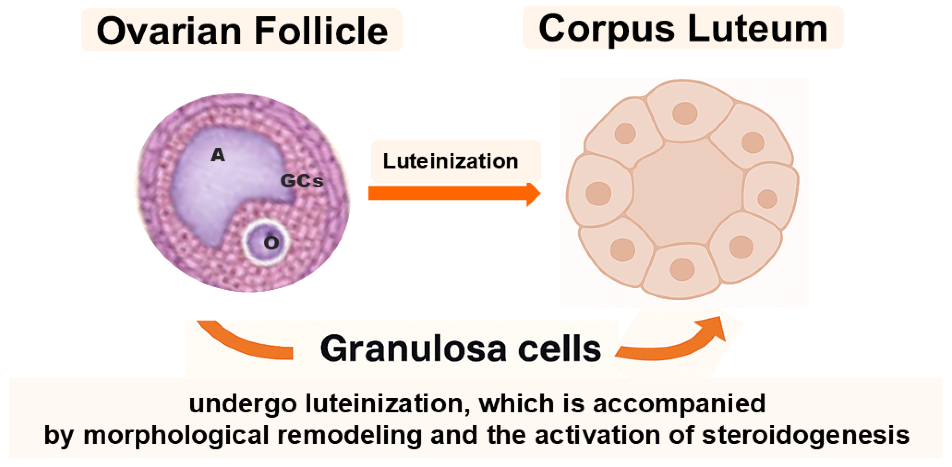
2.1. Cellular and Functional Identity
2.2. Molecular Signature and Stemness-Related Markers
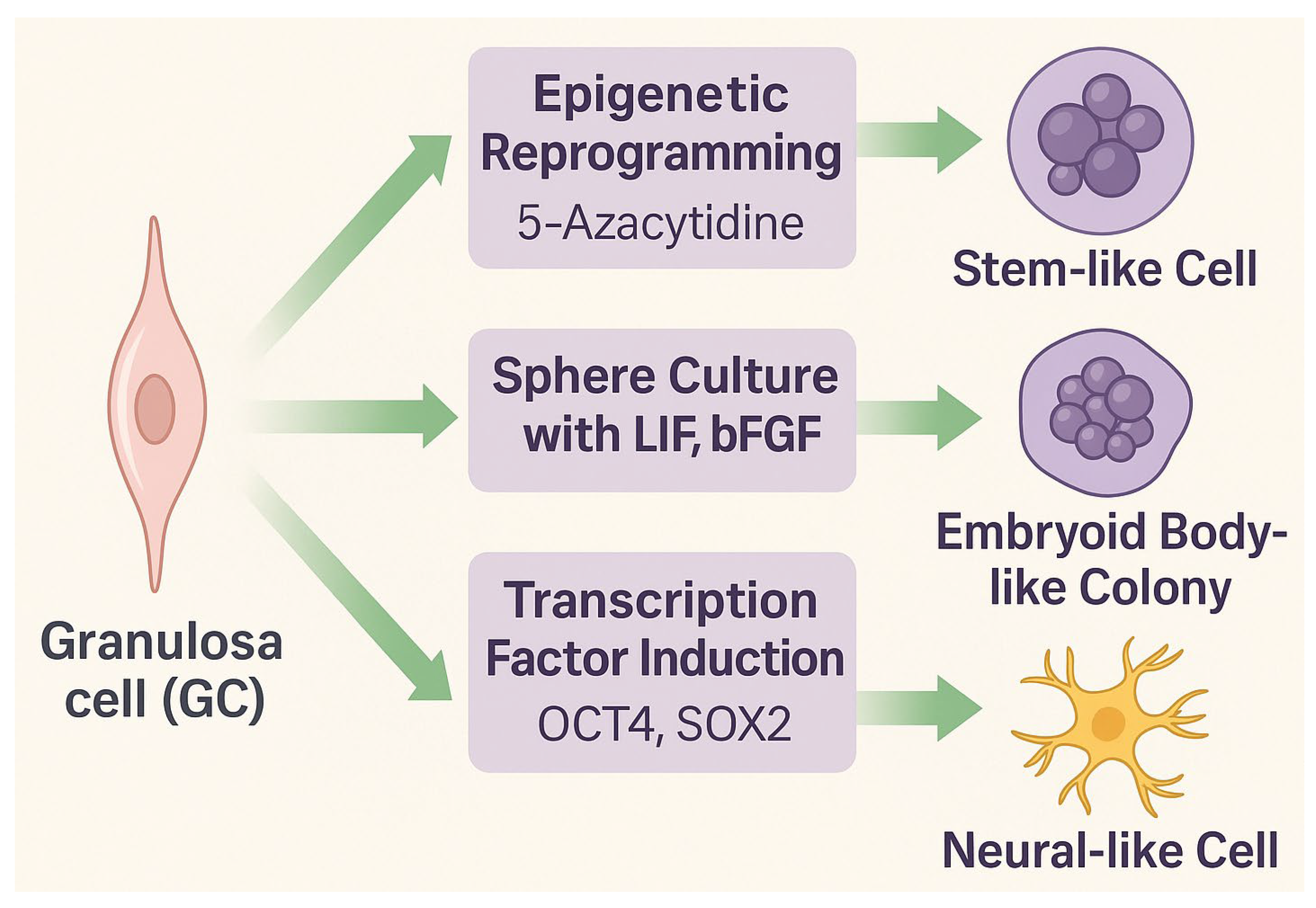
2.3. Molecular Plasticity, Epigenetic Reprogrammability, and Transdifferentiation Potential
3. Ovarian Putative Stem Cells (oPSCs)
3.1. Isolation and Phenotypic Features
3.2. Differentiation Capacity
3.3. Functional Integration and Electrophysiological Properties
4. Epigenetic Reprogramming of Granulosa Cells
4.1. Mechanisms of Epigenetic Plasticity
4.2. Experimental Approaches
4.3. Advantages and Limitations
- Low cost and procedural simplicity of small-molecule use.
- Scalability for preclinical applications.
- Compatibility with 3D culture systems, enhancing differentiation outcomes.
- Reprogramming efficiency remains variable and typically low, depending on donor origin, passage number, and hormonal context.
- Residual epigenetic memory may bias lineage fate, limiting full reprogramming and long-term stability [69].
- Incomplete chromatin remodeling may restrict activation of lineage-specific programs.
- Functional validation in vivo is still lacking: the ability of reprogrammed GCs to integrate, survive, and function after transplantation must be tested in preclinical models [70].
5. Comparative Perspective: GCs vs. oPSCs
6. Applications in Regenerative Medicine
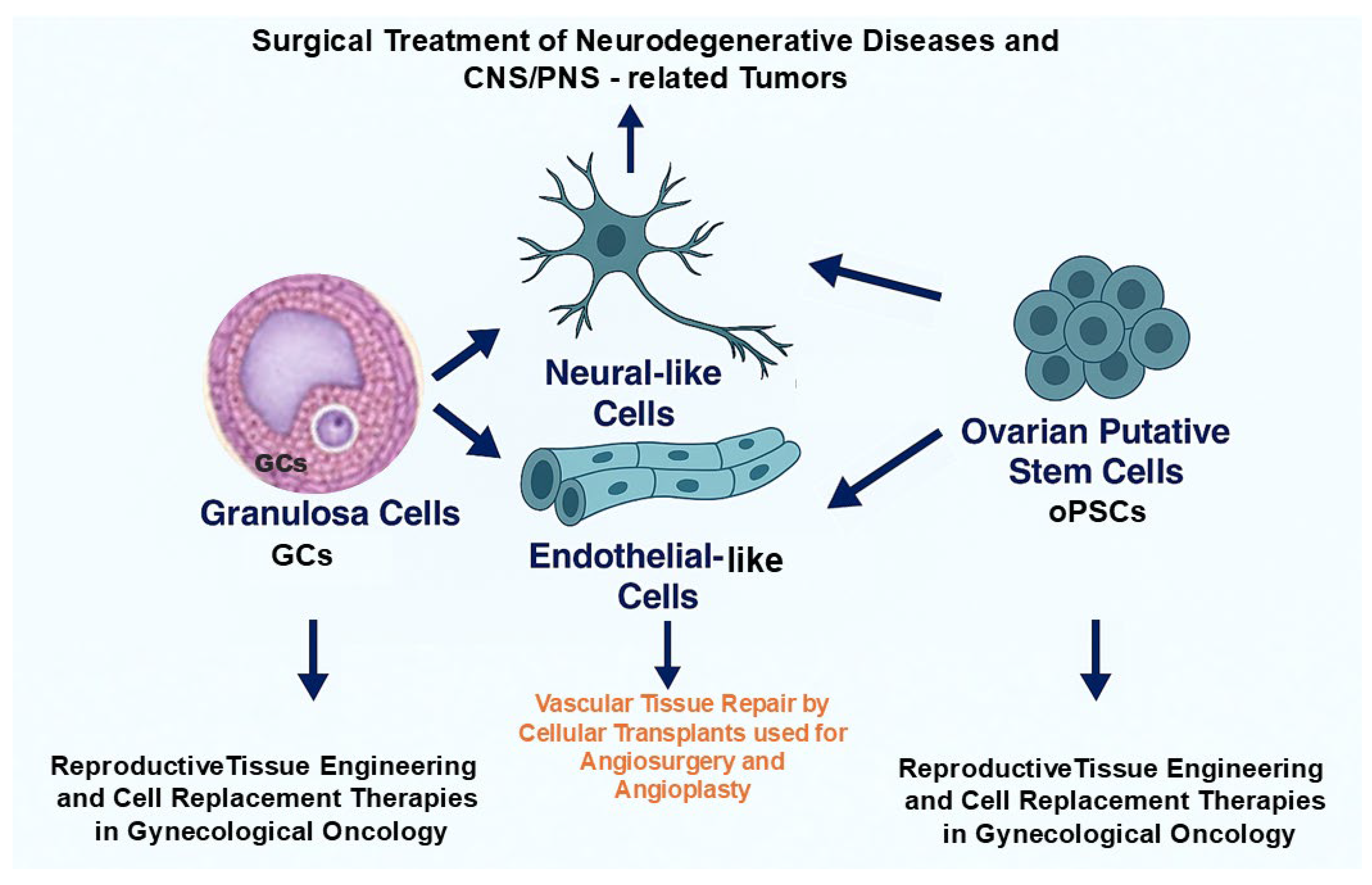
6.1. Neurological Applications
- Modeling neurodegenerative diseases such as Alzheimer’s and Parkinson’s;
- Drug screening and neurotoxicity testing using patient-derived lines;
- Autologous neural cell therapy—especially in women—providing sex-matched compatibility [78].
6.2. Vascular Tissue Engineering
- Therapeutic revascularization and angiogenesis in ischemic diseases (e.g., peripheral artery disease, myocardial infarction);
- Pre-vascularization of engineered grafts, improving perfusion, survival, and tissue integration post-implantation;
- Restoration of microvascular networks damaged by chemo- or radiotherapy, particularly:
- -
- In the ovary, supporting oncofertility restoration after gynecological cancer treatment;
- -
- In the CNS, aiding vascular repair and potentially alleviating neurodegenerative or tumor-related pathologies.
6.3. Reproductive Tissue Engineering
- In vitro folliculogenesis: Both cell types can support follicle formation and maturation, enabling fertility preservation in cancer survivors [81];
- Three-dimensional-printed ovarian scaffolds: GCs embedded in biocompatible hydrogels restore partial endocrine function and support oocyte maturation in animal models [82].
- Bioartificial ovarian constructs: Integration of oPSCs enhances vascularization, organization, and graft longevity, providing a potential replacement for damaged ovarian tissue [83].
- Endocrine restoration: Autologous transplantation of steroidogenic GCs or oPSC-derived cells could serve as a physiological alternative to hormone replacement therapy, especially in women with premature ovarian insufficiency (POI) or polycystic ovary syndrome (PCOS) [84].
7. Recent Challenges and Future Targets
7.1. Standardization of Isolation and Differentiation Protocols
7.2. Genomic Stability and Epigenetic Memory
7.3. Functional Validation In Vivo
7.4. Immunological and Ethical Considerations
7.5. Regulatory Framework and Clinical Translation
- Reprogrammed GCs and oPSCs must meet rigorous safety, genomic integrity, and non-tumorigenicity standards.
- MA and FDA regulations currently lack clear classification criteria for stem cell-like products derived from reproductive tissues, complicating approval pathways.
- To date, no GC- or oPSC-based therapy has advanced beyond early preclinical studies; this statement should be verified prior to publication.
- Successful translation will depend on close collaboration among molecular biologists, clinicians, toxicologists, and regulatory authorities to ensure both efficacy and ethical compliance [90].
8. Applicability of GCs and oPSCs to Somatic Cell Cloning-Based Assisted Reproductive Technologies in Mammals
- Enhancement of genetic merit and quantitative trait locus (QTL) expression in cloned livestock [100];
- Genetic rescue programs aimed at endangered species and rare livestock breeds [103];
- Fundamental studies exploring the cellular and molecular determinants of epigenetic reprogramming that govern the developmental competence of cloned embryos reconstructed with somatic cell nuclei [99].
9. Comprehensive Summary and Paramount Conclusions
- Protocol variability in isolation and differentiation impedes reproducibility;
- Incomplete epigenetic reprogramming and potential genomic instability raise safety concerns;
- Functional in vivo validation of graft survival and integration remains scarce;
- Ethical and regulatory frameworks for reproductive tissue-derived cells are still evolving.
10. Concluding Remarks and Future Perspectives
Author Contributions
Funding
Institutional Review Board Statement
Informed Consent Statement
Data Availability Statement
Conflicts of Interest
Abbreviations
| 5-azaC | 5-Azacytidine; DNA methyltransferase inhibitor |
| 5-aza-dC | 5-Aza-2′-deoxycytidine (decitabine); DNA methyltransferase inhibitor |
| AKT | Serine/threonine protein kinase involved in cell survival signaling |
| ALB | Albumin |
| AMPAR | Ionotropic glutamate receptor mediating fast excitatory transmission |
| ARTs | Assisted reproductive technologies |
| ATAC-seq | Assay for transposase-accessible chromatin with sequencing |
| bFGF | Basic fibroblast growth factor |
| CACNA1C | Gene encoding L-type voltage-gated calcium channel subunit α1C |
| CD | Cluster of differentiation |
| c-KIT | Proto-oncogene encoding receptor tyrosine kinase CD117 |
| CpG | DNA region rich in cytosine–guanine dinucleotides |
| CREB | cAMP response element-binding protein (transcription factor) |
| CYP3A4 | Cytochrome P450 enzyme involved in drug metabolism |
| c-MYC | Proto-oncogene encoding transcription factor regulating proliferation |
| CNS | Central nervous system |
| DAZL | Deleted in azoospermia-like; germ cell RNA-binding protein |
| DDX4 (VASA) | DEAD-box RNA helicase; germline marker |
| DNMT | DNA methyltransferase |
| DNMTi | DNA methyltransferase inhibitor |
| DPPA3 (STELLA) | Maternal factor regulating DNA methylation in germ cells |
| ECM | Extracellular matrix |
| EPCs | Endothelial progenitor cells |
| ESCs | Embryonic stem cells |
| FABP4 | Fatty acid-binding protein 4 |
| FACS | Fluorescence-activated cell sorting |
| FGSCs (PGCs) | Female germline stem cells (primordial germ cells) |
| Fluo-4 AM | Calcium-sensitive fluorescent indicator dye |
| GCs | Granulosa cells |
| GFAP | Glial fibrillary acidic protein |
| HAT | Histone acetyltransferase |
| HDAC | Histone deacetylase |
| HGF | Hepatocyte growth factor |
| HRT | Hormone replacement therapy |
| ICSI | Intracytoplasmic sperm injection |
| IFITM3 (FRAGILIS) | Interferon-induced transmembrane protein 3; germline marker |
| iPSCs | Induced pluripotent stem cells |
| IVF | In vitro fertilization |
| KCNQ2 | Voltage-gated potassium channel subunit regulating excitability |
| KLF4 | Krüppel-like factor 4; zinc finger transcription factor |
| LIF | Leukemia inhibitory factor |
| MACS | Magnetic-activated cell sorting |
| MAP2 | Microtubule-associated protein 2 |
| MAPK/ERK | Mitogen-activated protein kinase/extracellular signal-regulated kinase |
| MSCs | Mesenchymal stem cells |
| NANOG | Homeobox transcription factor maintaining pluripotency |
| NeuN | Neuronal nuclei antigen; marker of mature neurons |
| NMDAR | N-methyl-D-aspartate receptor; ionotropic glutamate receptor |
| NPY | Neuropeptide Y |
| NR1 | NMDA receptor subunit 1 |
| OCT4 (POU5F1) | Octamer-binding transcription factor 4; pluripotency regulator |
| oPSCs | Ovarian putative stem cells |
| OSCs | Oogonial stem cells |
| PECAM-1 | Platelet–endothelial cell adhesion molecule-1 (CD31) |
| PI3K | Phosphatidylinositol 3-kinase |
| PNS | Peripheral nervous system |
| PCOS | Polycystic ovary syndrome |
| POI/POF | Primary ovarian insufficiency/premature ovarian failure |
| PPARγ | Peroxisome proliferator-activated receptor gamma |
| PSD-95 | Postsynaptic density protein 95 |
| qRT-PCR | Quantitative real-time polymerase chain reaction |
| QTLs | Quantitative trait loci |
| RA | Retinoic acid |
| RUNX2 | Runt-related transcription factor 2 |
| SCNT | Somatic cell nuclear transfer |
| SCN1A | Sodium voltage-gated channel α subunit 1 |
| SOX2 | SRY-box transcription factor 2 |
| SOX9 | SRY-box transcription factor 9 |
| SSEA-4 | Stage-specific embryonic antigen 4 |
| STAT3 | Signal transducer and activator of transcription 3 |
| TRA-1-81 | Surface antigen of pluripotent stem cells |
| TUBB3 | Class III β-tubulin |
| VSELs | Very small embryonic-like stem cells |
| VEGF | Vascular endothelial growth factor |
| VEGFR2 | Vascular endothelial growth factor receptor 2 |
| VEGFR3 | Vascular endothelial growth factor receptor 3 |
| VGCCs | Voltage-gated calcium channels |
| vWF | Von Willebrand factor |
| Wnt | Family of signaling proteins regulating cell fate and development |
References
- Kossowska-Tomaszczuk, K.; De Geyter, C. Cells with stem cell characteristics in somatic compartments of the ovary. Biomed Res. Int. 2013, 2013, 310859. [Google Scholar] [CrossRef] [PubMed]
- Bhartiya, D.; Patel, H.; Parte, S. Improved understanding of very small embryonic-like stem cells in adult mammalian ovary. Hum. Reprod. 2018, 33, 978–979. [Google Scholar] [CrossRef]
- White, Y.A.; Woods, D.C.; Takai, Y.; Ishihara, O.; Seki, H.; Tilly, J.L. Oocyte Formation by Mitotically Active Germ Cells Purified from Ovaries of Reproductive-Age Women. Nat. Med. 2012, 18, 413–421. [Google Scholar] [CrossRef] [PubMed]
- Zhang, H.; Panula, S.; Petropoulos, S.; Edsgärd, D.; Busayavalasa, K.; Liu, L.; Li, X.; Risal, S.; Shen, Y.; Shao, J.; et al. Adult Human and Mouse Ovaries Lack DDX4-Expressing Functional Oogonial Stem Cells. Nat. Med. 2015, 21, 1116–1118. [Google Scholar] [CrossRef] [PubMed]
- Fontana, J.; Martínková, S.; Petr, J.; Žalmanová, T.; Trnka, J. Metabolic Cooperation in the Ovarian Follicle. Physiol. Res. 2020, 69, 33–48. [Google Scholar] [CrossRef]
- Virant-Klun, I.; Stimpfel, M.; Skutella, T. Stem Cells in Adult Human Ovaries: From Female Fertility to Ovarian Cancer. Curr. Pharm. Des. 2012, 18, 283–292. [Google Scholar] [CrossRef]
- Goldberg, A.D.; Allis, C.D.; Bernstein, E. Epigenetics: A Landscape Takes Shape. Cell 2007, 128, 635–638. [Google Scholar] [CrossRef]
- Reik, W.; Dean, W.; Walter, J. Epigenetic Reprogramming in Mammalian Development. Science 2001, 293, 1089–1093. [Google Scholar] [CrossRef]
- Slotkin, R.K.; Martienssen, R. Transposable Elements and the Epigenetic Regulation of the Genome. Nat. Rev. Genet. 2007, 8, 272–285. [Google Scholar] [CrossRef]
- Kelly, R.D.W.; Stengel, K.R.; Chandru, A.; Johnson, L.C.; Hiebert, S.W.; Cowley, S.M. Histone Deacetylases Maintain Expression of the Pluripotent Gene Network via Recruitment of RNA Polymerase II to Coding and Noncoding Loci. Genome Res. 2024, 34, 34–46. [Google Scholar] [CrossRef]
- Plath, K.; Lowry, W.E. Progress in Understanding Reprogramming to the Induced Pluripotent State. Nat. Rev. Genet. 2011, 12, 253–265. [Google Scholar] [CrossRef] [PubMed]
- Talaei-Khozani, T.; Kharazinejad, E.; Rohani, L.; Vojdani, Z.; Mostafavi Pour, Z.; Tabei, S.Z. Expression of Pluripotency Markers in Human Granulosa Cells after Embryonic Stem Cell Extract Exposure and Epigenetic Modification. Iran J. Reprod. Med. 2012, 10, 193–200. [Google Scholar] [PubMed]
- Hoang, S.N.; Ho, C.N.Q.; Nguyen, T.T.P.; Doan, C.C.; Tran, D.H.; Le, L.T. Evaluation of Stemness Marker Expression in Bovine Ovarian Granulosa Cells and Their In Vitro Expanded Culture. Anim. Reprod. 2019, 16, 277–281. [Google Scholar] [CrossRef] [PubMed]
- Dompe, C.; Kulus, M.; Stefańska, K.; Kranc, W.; Chermuła, B.; Bryl, R.; Pieńkowski, W.; Nawrocki, M.J.; Petitte, J.N.; Stelmach, B.; et al. Human Granulosa Cells—Stemness Properties, Molecular Cross-Talk and Follicular Angiogenesis. Cells 2021, 10, 1396. [Google Scholar] [CrossRef]
- Dzafic, E.; Stimpfel, M.; Virant-Klun, I. Plasticity of Granulosa Cells: On the Crossroad of Stemness and Transdifferentiation Potential. J. Assist. Reprod. Genet. 2013, 30, 1255–1261. [Google Scholar] [CrossRef]
- Giorgetti, A.; Montserrat, N.; Aasen, T.; Gonzalez, F.; Rodríguez-Pizà, I.; Vassena, R.; Raya, A.; Boué, S.; Barrero, M.J.; Corbella, B.A.; et al. Generation of Induced Pluripotent Stem Cells from Human Cord Blood Using OCT4 and SOX2. Cell Stem Cell 2009, 5, 353–357. [Google Scholar] [CrossRef]
- Brevini, T.A.L.; Pennarossa, G.; Rahman, M.M.; Paffoni, A.; Antonini, S.; Ragni, G.; de Eguileor, M.; Tettamanti, G.; Gandolfi, F. Morphological and Molecular Changes of Human Granulosa Cells Exposed to 5-Azacytidine and Addressed Toward Muscular Differentiation. Stem Cell Rev. Rep. 2014, 10, 633–642. [Google Scholar] [CrossRef]
- Hryciuk, M.M.; Schröter, F.; Hennicke, L.; Braun, B.C. Spheroid Formation and Luteinization of Granulosa Cells of Felids in a Long-Term 3D Culture. Differentiation 2023, 131, 38–48. [Google Scholar] [CrossRef]
- Kranc, W.; Brązert, M.; Budna, J.; Celichowski, P.; Bryja, A.; Nawrocki, M.J.; Ożegowska, K.; Jankowski, M.; Chermuła, B.; Dyszkiewicz-Konwińska, M.; et al. Genes Responsible for Proliferation, Differentiation, and Junction Adhesion Are Significantly Up-Regulated in Human Ovarian Granulosa Cells during a Long-Term Primary In Vitro Culture. Histochem. Cell Biol. 2019, 151, 125–143. [Google Scholar] [CrossRef]
- Guo, Z.; Yu, Q. Role of mTOR Signaling in Female Reproduction. Front. Endocrinol. 2019, 9, 692. [Google Scholar] [CrossRef]
- Anchan, R.M.; Gerami-Naini, B.; Lindsey, J.; Lipskind, S.; Williams, S.Z.; Kearns, W.G. Ovarian Granulosa Cells Are a Viable Source of iPSCs with the Capacity for Epigenetically-Biased Differentiation into Steroidogenic Tissue of the Ovary. Fertil. Steril. 2013, 100, S454. [Google Scholar] [CrossRef]
- Chuang, C.Y.; Huang, M.C.; Chen, H.F.; Tseng, L.H.; Yu, C.Y.; Stone, L.; Huang, H.P.; Ho, H.N.; Kuo, H.C. Granulosa Cell-Derived Induced Pluripotent Stem Cells Exhibit Pro-Trophoblastic Differentiation Potential. Stem Cell Res. Ther. 2015, 6, 14. [Google Scholar] [CrossRef] [PubMed]
- Anchan, R.; Gerami-Naini, B.; Lindsey, J.S.; Ho, J.W.K.; Kiezun, A.; Lipskind, S.; Ng, N.; LiCausi, J.A.; Kim, C.S.; Brezina, P.; et al. Efficient Differentiation of Steroidogenic and Germ-Like Cells from Epigenetically-Related iPSCs Derived from Ovarian Granulosa Cells. PLoS ONE 2015, 10, e0119275. [Google Scholar] [CrossRef] [PubMed]
- Heng, D.; Wang, Q.; Ma, X.; Tian, Y.; Xu, K.; Weng, X.; Hu, X.; Liu, W.; Zhang, C. Role of OCT4 in the Regulation of FSH-Induced Granulosa Cells Growth in Female Mice. Front. Endocrinol. 2020, 10, 915. [Google Scholar] [CrossRef]
- Hummitzsch, K.; Ricken, A.M.; Kloss, D.; Erdmann, S.; Nowicki, M.S.; Rothermel, A.; Robitzki, A.A.; Spanel-Borowski, K. Spheroids of Granulosa Cells Provide an In Vitro Model for Programmed Cell Death Coupled to Steroidogenesis. Differentiation 2009, 77, 60–69. [Google Scholar] [CrossRef]
- Bazley, F.A.; Liu, C.F.; Yuan, X.; Hao, H.; All, A.H.; De Los Angeles, A.; Zambidis, E.T.; Gearhart, J.D.; Kerr, C.L. Direct Reprogramming of Human Primordial Germ Cells into Induced Pluripotent Stem Cells: Efficient Generation of Genetically Engineered Germ Cells. Stem Cells Dev. 2015, 24, 2634–2648. [Google Scholar] [CrossRef]
- Wu, H.; Zhu, R.; Zheng, B.; Liao, G.; Wang, F.; Ding, J.; Li, H.; Li, M. Single-Cell Sequencing Reveals an Intrinsic Heterogeneity of the Preovulatory Follicular Microenvironment. Biomolecules 2022, 12, 231. [Google Scholar] [CrossRef]
- Fiorentino, G.; Cimadomo, D.; Innocenti, F.; Soscia, D.; Vaiarelli, A.; Ubaldi, F.M.; Gennarelli, G.; Garagna, S.; Rienzi, L.; Zuccotti, M. Biomechanical Forces and Signals Operating in the Ovary during Folliculogenesis and Their Dysregulation: Implications for Fertility. Hum. Reprod. Update 2023, 29, 1–23. [Google Scholar] [CrossRef]
- Dereń, J. Epigenetic Conversion of Granulosa Cells Isolated from Porcine Ovarian Follicles into Nervous System Cells. Master’s Thesis, Jagiellonian University, Kraków, Poland, 2022. Available online: https://ruj.uj.edu.pl/xmlui/handle/item/294409 (accessed on 10 September 2025).
- Brązert, M.; Kranc, W.; Celichowski, P.; Jankowski, M.; Piotrowska-Kempisty, H.; Pawelczyk, L.; Bruska, M.; Zabel, M.; Nowicki, M.; Kempisty, B. Expression of Genes Involved in Neurogenesis and Neuronal Precursor Cell Proliferation and Development: Novel Pathways of Human Ovarian Granulosa Cell Differentiation and Transdifferentiation Capability In Vitro. Mol. Med. Rep. 2020, 21, 1749–1760. [Google Scholar] [CrossRef]
- Mattioli, M.; Gloria, A.; Turriani, M.; Berardinelli, P.; Russo, V.; Nardinocchi, D.; Curini, V.; Baratta, M.; Martignani, E.; Barboni, B. Osteo-Regenerative Potential of Ovarian Granulosa Cells: An In Vitro and In Vivo Study. Theriogenology 2012, 77, 1425–1437. [Google Scholar] [CrossRef]
- De La Fuente, R.; Eppig, J.J. Transcriptional Activity of the Mouse Oocyte Genome: Companion Granulosa Cells Modulate Transcription and Chromatin Remodeling. Dev. Biol. 2001, 229, 224–236. [Google Scholar] [CrossRef] [PubMed]
- Li, D.; Ning, C.; Zhang, J.; Wang, Y.; Tang, Q.; Kui, H.; Wang, T.; He, M.; Jin, L.; Li, J.; et al. Dynamic Transcriptome and Chromatin Architecture in Granulosa Cells during Chicken Folliculogenesis. Nat. Commun. 2022, 13, 131. [Google Scholar] [CrossRef] [PubMed]
- Bhartiya, D.; Unni, S.; Parte, S.; Anand, S. Very Small Embryonic-like Stem Cells: Implications in Reproductive Biology. Biomed Res. Int. 2013, 2013, 682326. [Google Scholar] [CrossRef] [PubMed]
- Zhang, H.; Zheng, W.; Shen, Y.; Adhikari, D.; Ueno, H.; Liu, K. Experimental Evidence Showing that DDX4-Positive Cells Sorted from Adult Human Ovaries Are Oogonial Stem Cells. Reprod. Sci. 2015, 22, 30–38. [Google Scholar] [CrossRef]
- Bhartiya, D.; Sriraman, K.; Parte, S.; Patel, H. Ovarian Stem Cells: Absence of Evidence Is Not Evidence of Absence. J. Ovarian Res. 2013, 6, 65. [Google Scholar] [CrossRef]
- Song, S.H.; Kumar, B.M.; Kang, E.J.; Lee, Y.M.; Kim, T.H.; Ock, S.A.; Lee, S.L.; Jeon, B.G.; Rho, G.J. Characterization of Porcine Multipotent Stem/Stromal Cells Derived from Skin, Adipose, and Ovarian Tissues and Their Differentiation In Vitro into Putative Oocyte-Like Cells. Stem Cells Dev. 2011, 20, 1359–1370. [Google Scholar] [CrossRef]
- Niikura, Y.; Niikura, T.; Tilly, J.L. Aged Mouse Ovaries Possess Rare Premeiotic Germ Cells That Can Generate Oocytes Following Transplantation into a Young Host Environment. Aging 2009, 1, 971–978. [Google Scholar] [CrossRef]
- Lei, L.; Spradling, A.C. Mouse Primordial Germ Cells Produce Cysts That Partially Fragment Prior to Meiosis. Development 2013, 140, 2075–2081. [Google Scholar] [CrossRef]
- Bhartiya, D.; Jha, N.; Tripathi, A.; Tripathi, A. Very Small Embryonic-Like Stem Cells Have the Potential to Win the Three-Front War on Tissue Damage, Cancer, and Aging. Front. Cell Dev. Biol. 2023, 10, 1061022. [Google Scholar] [CrossRef]
- Li, X.; Yu, Y.; Wei, R.; Li, Y.; Lv, J.; Liu, Z.; Zhang, Y. In Vitro and In Vivo Study on Angiogenesis of Porcine Induced Pluripotent Stem Cell-Derived Endothelial Cells. Differentiation 2021, 120, 10–18. [Google Scholar] [CrossRef]
- Bukovsky, A.; Caudle, M.R.; Svetlikova, M.; Upadhyaya, N.B. Oogenesis in Cultures Derived from Adult Human Ovaries: Bipotential Source of Oocytes and Granulosa Cells. Reprod. Biol. Endocrinol. 2005, 3, 17. [Google Scholar] [CrossRef] [PubMed]
- Virant-Klun, I.; Rozman, P.; Cvjeticanin, B.; Vrtacnik-Bokal, E.; Novakovic, S.; Rülicke, T.; Dovc, P.; Meden-Vrtovec, H. Parthenogenetic Embryo-Like Structures in the Human Ovarian Surface Epithelium Cell Culture in Postmenopausal Women with No Naturally Present Follicles and Oocytes. Stem Cells Dev. 2009, 18, 137–149. [Google Scholar] [CrossRef] [PubMed]
- Delaspre, F.; Massumi, M.; Salido, M.; Soria, B.; Ravassard, P.; Savatier, P.; Skoudy, A. Directed Pancreatic Acinar Differentiation of Mouse Embryonic Stem Cells via Embryonic Signaling Molecules and Exocrine Transcription Factors. PLoS ONE 2013, 8, e54243. [Google Scholar] [CrossRef]
- Zhang, Y.; Sun, H.; Yang, Y.; Xie, C.; Wang, Q.; Ma, R.; Zhang, W.; Lu, X.; Wang, Y.; Li, S.; et al. Epigenetic Profiling of Ovarian Stem-Like Cells Reveals Chromatin Plasticity Linked to Multilineage Potential. Stem Cell Rep. 2020, 15, 1223–1236. [Google Scholar] [CrossRef]
- Virant-Klun, I.; Skerl, P.; Novakovic, S.; Vrtacnik-Bokal, E.; Smrkolj, S. Similar Population of CD133+ and DDX4+ VSEL-Like Stem Cells Sorted from Human Embryonic Stem Cell, Ovarian, and Ovarian Cancer Ascites Cell Cultures: The Real Embryonic Stem Cells? Cells 2019, 8, 706. [Google Scholar] [CrossRef]
- Kalia, A.K.; Rösseler, C.; Granja-Vazquez, R.; Ahmad, A.; Pancrazio, J.J.; Neureiter, A.; Zhang, M.; Sauter, D.; Vetter, I.; Andersson, A.; et al. How to Differentiate Induced Pluripotent Stem Cells into Sensory Neurons for Disease Modelling: A Functional Assessment. Stem Cell Res. Ther. 2024, 15, 99. [Google Scholar] [CrossRef]
- de Melo Reis, R.A.; Freitas, H.R.; de Mello, F.G. Cell Calcium Imaging as a Reliable Method to Study Neuron–Glial Circuits. Front. Neurosci. 2020, 14, 569361. [Google Scholar] [CrossRef]
- Sun, Z.; Südhof, T.C. A Simple Ca2+-Imaging Approach to Neural Network Analyses in Cultured Neurons. J. Neurosci. Methods 2021, 350, 109041. [Google Scholar] [CrossRef]
- Stimpfel, M.; Cerkovnik, P.; Novakovic, S.; Maver, A.; Virant-Klun, I. Putative Mesenchymal Stem Cells Isolated from Adult Human Ovaries. J. Assist. Reprod. Genet. 2014, 31, 959–974. [Google Scholar] [CrossRef]
- Chandrakanthan, V.; Yeola, A.; Kwan, J.C.; Oliver, R.A.; Qiao, Q.; Kang, Y.C.; Zarzour, P.; Beck, D.; Boelen, L.; Unnikrishnan, A.; et al. PDGF-AB and 5-Azacytidine Induce Conversion of Somatic Cells into Tissue-Regenerative Multipotent Stem Cells. Proc. Natl. Acad. Sci. USA 2016, 113, E2306–E2315. [Google Scholar] [CrossRef]
- Lee, L.; Asada, H.; Kizuka, F.; Tamura, I.; Maekawa, R.; Taketani, T.; Sato, S.; Yamagata, Y.; Tamura, H.; Sugino, N. Changes in Histone Modification and DNA Methylation of the StAR and Cyp19a1 Promoter Regions in Granulosa Cells Undergoing Luteinization during Ovulation in Rats. Endocrinology 2013, 154, 458–470. [Google Scholar] [CrossRef] [PubMed]
- Gupta, M.K.; Heo, Y.T.; Kim, D.K.; Lee, H.T.; Uhm, S.J. 5-Azacytidine Improves the Meiotic Maturation and Subsequent In Vitro Development of Pig Oocytes. Anim. Reprod. Sci. 2019, 208, 106118. [Google Scholar] [CrossRef] [PubMed]
- Shi, Y.; Do, J.T.; Desponts, C.; Hahm, H.S.; Schöler, H.R.; Ding, S. Induction of Pluripotent Stem Cells from Mouse Embryonic Fibroblasts by Oct4 and Klf4 with Small-Molecule Compounds. Cell Stem Cell 2008, 3, 568–574. [Google Scholar] [CrossRef] [PubMed]
- Pennarossa, G.; Zenobi, A.; Gandolfi, C.E.; Manzoni, E.F.; Gandolfi, F.; Brevini, T.A. Erase and Rewind: Epigenetic Conversion of Cell Fate. Stem Cell Rev. Rep. 2016, 12, 163–170. [Google Scholar] [CrossRef]
- Dinh, D.T.; Breen, J.; Nicol, B.; Smith, K.M.; Nicholls, M.; Emery, A.; Wong, Y.Y.; Barry, S.C.; Yao, H.H.-C.; Robker, R.L.; et al. Progesterone Receptor Mediates Ovulatory Transcriptional Regulation through Enhancer–Promoter Chromatin Accessibility. Nucleic Acids Res. 2023, 51, 5981–6000. [Google Scholar] [CrossRef]
- Xu, N.; Papagiannakopoulos, T.; Pan, G.; Thomson, J.A.; Kosik, K.S. MicroRNA-145 Regulates OCT4, SOX2, and KLF4 and Represses Pluripotency in Human Embryonic Stem Cells. Cell 2009, 137, 647–658. [Google Scholar] [CrossRef]
- Subramanyam, D.; Lamouille, S.; Judson, R.L.; Liu, J.Y.; Bucay, N.; Derynck, R.; Blelloch, R. Multiple Targets of miR-302 and miR-372 Promote Reprogramming of Human Fibroblasts to Induced Pluripotent Stem Cells. Nat. Biotechnol. 2011, 29, 443–448. [Google Scholar] [CrossRef]
- Bahmanpour, S.; Keshavarz, A.; Zarei Fard, N. Effect of Different Concentrations of Forskolin Along with Mature Granulosa Cell Co-Culturing on Mouse Embryonic Stem Cell Differentiation into Germ-Like Cells. Iran Biomed. J. 2020, 24, 30–38. [Google Scholar] [CrossRef]
- Manzoni, E.F.M.; Brevini, T.A.L.; Bovolin, P.; Corradetti, B.; Gandolfi, F. 5-Azacytidine Affects TET2 and Histone Transcription and Morphology of Human Fibroblasts—Exemplifying a Cell Reprogramming Stage. Sci. Rep. 2016, 6, 37891. [Google Scholar] [CrossRef]
- Bicer, M.; Sadeghi, S.; Caballero, D. Impact of 3D Cell Culture on Bone Regeneration Potential of Mesenchymal Stromal Cells. Stem Cell Res. Ther. 2021, 12, 428. [Google Scholar] [CrossRef]
- Engler, A.J.; Sen, S.; Sweeney, H.L.; Discher, D.E. Matrix Elasticity Directs Stem Cell Lineage Specification. Cell 2006, 126, 677–689. [Google Scholar] [CrossRef] [PubMed]
- Sood, D.; Cairns, D.M.; Dabbi, J.M.; Ramakrishnan, C.; Deisseroth, K.; Black, L.D., 3rd; Santaniello, S.; Kaplan, D.L. Functional Maturation of Human Neural Stem Cells in a 3D Bioengineered Brain Model Enriched with Fetal Brain-Derived Matrix. Sci. Rep. 2019, 9, 17874. [Google Scholar] [CrossRef] [PubMed]
- Liu, X.; Duan, C.; Yang, P.; Long, Q.; Feng, L.; Li, Y.; He, J.; Zhang, C. Biomaterial Scaffolds for Reproductive Tissue Engineering. Ann. Biomed. Eng. 2017, 45, 1592–1607. [Google Scholar] [CrossRef]
- Zhou, H.; Wu, S.; Joo, J.Y.; Zhu, S.; Han, D.W.; Lin, T.; Trauger, S.; Bien, G.; Yao, S.; Zhu, Y.; et al. Generation of Induced Pluripotent Stem Cells Using Recombinant Proteins. Cell Stem Cell 2009, 4, 381–384. [Google Scholar] [CrossRef]
- Pradella, D.; Naro, C.; Sette, C.; Ghigna, C. EMT and Stemness: Flexible Processes Tuned by Alternative Splicing in Development and Cancer Progression. Mol. Cancer 2017, 16, 8. [Google Scholar] [CrossRef]
- Kim, K.; Doi, A.; Wen, B.; Ng, K.; Zhao, R.; Cahan, P.; Kim, J.; Aryee, M.J.; Ji, H.; Ehrlich, L.I.; et al. Epigenetic Memory in Induced Pluripotent Stem Cells. Nature 2010, 467, 285–290. [Google Scholar] [CrossRef]
- Bhat, G.R.; Sethi, I.; Sadida, H.Q.; Subramanian, R.; Singh, S.; Chauhan, S.S.; Yallapu, M.M.; Chauhan, S.C. Cancer Cell Plasticity: From Cellular, Molecular, and Genetic Mechanisms to Tumor Heterogeneity and Drug Resistance. Cancer Metastasis Rev. 2024, 43, 197–228. [Google Scholar] [CrossRef]
- Bukovsky, A.; Caudle, M.R.; Svetlikova, M.; Wimalasena, J.; Ayala, M.E.; Dominguez, R. Oogenesis in Adult Mammals, Including Humans: A Review. Endocrine 2005, 26, 301–316. [Google Scholar] [CrossRef]
- Virant-Klun, I.; Skutella, T.; Hren, M.; Gruden, K.; Cvjeticanin, B.; Vogler, A.; Sinkovec, J. Isolation of Small SSEA-4-Positive Putative Stem Cells from the Ovarian Surface Epithelium of Adult Human Ovaries by Two Different Methods. Biomed. Res. Int. 2013, 2013, 690415. [Google Scholar] [CrossRef]
- Bui, H.T.; Van Thuan, N.; Kwon, D.N.; Choi, Y.J.; Kang, M.H.; Han, J.W.; Kim, T.; Kim, J.H. Identification and characterization of putative stem cells in the adult pig ovary. Development 2014, 141, 2235–2244. [Google Scholar] [CrossRef][Green Version]
- Sharma, D.; Bhartiya, D. Aged mice ovaries harbor stem cells and germ cell nests but fail to form follicles. J. Ovarian Res. 2022, 15, 37, Erratum in J. Ovarian Res. 2023, 16, 165. https://doi.org/10.1186/s13048-023-01256-5. [Google Scholar] [CrossRef]
- Ozakpinar, O.B.; Maurer, A.M.; Ozsavci, D. Ovarian Stem Cells: From Basic to Clinical Applications. World J. Stem Cells 2015, 7, 757–768. [Google Scholar] [CrossRef]
- Elias, K.M.; Ng, N.W.; Dam, K.U.; Milne, A.; Disler, E.R.; Gockley, A.; Holub, N.; Seshan, M.L.; Church, G.M.; Ginsburg, E.S.; et al. Fertility Restoration in Mice with Chemotherapy-Induced Ovarian Failure Using Differentiated iPSCs. EBioMedicine 2023, 94, 104715. [Google Scholar] [CrossRef]
- Bharti, D.; Jang, S.J.; Lee, S.Y.; Lee, S.L.; Rho, G.J. In Vitro Generation of Oocyte-like Cells and Their In Vivo Efficacy: How Far We Have Been Succeeded. Cells 2020, 9, 557, Erratum in Cells 2020, 9, 1262. https://doi.org/10.3390/cells9030557. [Google Scholar] [CrossRef]
- Ladanyi, C.; Mor, A.; Christianson, M.S.; Dhillon, N.; Segars, J.H. Recent Advances in the Field of Ovarian Tissue Cryopreservation and Opportunities for Research. J. Assist. Reprod. Genet. 2017, 34, 709–722. [Google Scholar] [CrossRef] [PubMed]
- Moradi-Gharibvand, N.; Hashemibeni, B. The Effect of Stem Cells and Vascular Endothelial Growth Factor on Cancer Angiogenesis. Adv. Biomed. Res. 2023, 12, 124. [Google Scholar] [CrossRef] [PubMed]
- Sriraman, K.; Bhartiya, D.; Anand, S.; Bhutda, S. Mouse Ovarian Very Small Embryonic-Like Stem Cells Resist Chemotherapy and Retain Ability to Initiate Oocyte-Specific Differentiation. Reprod. Sci. 2015, 22, 884–903. [Google Scholar] [CrossRef] [PubMed]
- Pierson Smela, M.D.; Kramme, C.C.; Fortuna, P.R.J.; Adams, J.L.; Su, R.; Dong, E.; Kobayashi, M.; Brixi, G.; Kavirayuni, V.S.; Tysinger, E.; et al. Directed differentiation of human iPSCs to functional ovarian granulosa-like cells via transcription factor overexpression. eLife 2023, 12, e83291, Erratum in eLife 2023, 12, e87987. https://doi.org/10.7554/eLife.87987. [Google Scholar] [CrossRef]
- Martin, J.J.; Woods, D.C.; Tilly, J.L. Implications and Current Limitations of Oogenesis from Female Germline or Oogonial Stem Cells in Adult Mammalian Ovaries. Cells 2019, 8, 93. [Google Scholar] [CrossRef]
- Lin, H.C.; Makhlouf, A.; Vazquez Echegaray, C.; Zawada, D.; Simões, F. Programming Human Cell Fate: Overcoming Challenges and Unlocking Potential through Technological Breakthroughs. Development 2023, 150, dev202300. [Google Scholar] [CrossRef]
- Nair, R.; Kasturi, M.; Mathur, V.; Seetharam, R.N.; Vasanthan, K.S. Strategies for Developing 3D Printed Ovarian Models for Restoring Fertility. Clin. Transl. Sci. 2024, 17, e13863. [Google Scholar] [CrossRef] [PubMed]
- Zhang, T.; Zhang, M.; Zhang, S.; Wang, S. Research Advances in the Construction of Stem Cell-Derived Ovarian Organoids. Stem Cell Res. Ther. 2024, 15, 505. [Google Scholar] [CrossRef]
- Kim, H.K.; Kim, T.J. Current Status and Future Prospects of Stem Cell Therapy for Infertile Patients with Premature Ovarian Insufficiency. Biomolecules 2024, 14, 242. [Google Scholar] [CrossRef]
- Jozkowiak, M.; Hutchings, G.; Jankowski, M.; Kulcenty, K.; Mozdziak, P.; Kempisty, B.; Spaczynski, R.Z.; Piotrowska-Kempisty, H. The Stemness of Human Ovarian Granulosa Cells and the Role of Resveratrol in the Differentiation of MSCs—A Review Based on Cellular and Molecular Knowledge. Cells 2020, 9, 1418. [Google Scholar] [CrossRef]
- Sriraman, A.; Debnath, T.K.; Xhemalce, B.; Miller, K.M. Making It or Breaking It: DNA Methylation and Genome Integrity. Essays Biochem. 2020, 64, 687–703. [Google Scholar] [CrossRef]
- Lindvall, O.; Kokaia, Z.; Martinez-Serrano, A. Stem Cell Therapy for Human Neurodegenerative Disorders—How to Make It Work. Nat. Med. 2004, 10 (Suppl. S7), S42–S50. [Google Scholar] [CrossRef]
- Wartalski, K.; Gorczyca, G.; Wiater, J.; Tabarowski, Z.; Duda, M. Porcine Ovarian Cortex-Derived Putative Stem Cells Can Differentiate into Endothelial Cells In Vitro. Histochem. Cell Biol. 2021, 156, 349–362. [Google Scholar] [CrossRef]
- Canosa, S.; Silvestris, E.; Carosso, A.R.; Ruffa, A.; Evangelisti, B.; Gennarelli, G.; Cormio, G.; Loizzi, V.; Rolfo, A.; Benedetto, C.; et al. Ovarian Stem Cells: Will the Dream of Neo-Folliculogenesis after Birth Become Real? Obstet. Gynecol. Surv. 2025, 80, 112–120. [Google Scholar] [CrossRef]
- El-Tanani, M.; Rabbani, S.A.; El-Tanani, Y.; Matalka, I.I.; Khalil, I.A. Bridging the Gap: From Petri Dish to Patient—Advancements in Translational Drug Discovery. Heliyon 2024, 11, e41317. [Google Scholar] [CrossRef]
- Samiec, M. Molecular Mechanisms of Somatic Cell Cloning and Other Assisted Reproductive Technologies in Mammals: Which Determinants Have Been Unraveled Thus Far? Int. J. Mol. Sci. 2024, 25, 13675. [Google Scholar] [CrossRef]
- Moura, M.T. Cloning by SCNT: Integrating Technical and Biology-Driven Advances. Methods Mol. Biol. 2023, 2647, 1–35. [Google Scholar] [CrossRef] [PubMed]
- Srirattana, K.; Kaneda, M.; Parnpai, R. Strategies to Improve the Efficiency of Somatic Cell Nuclear Transfer. Int. J. Mol. Sci. 2022, 23, 1969. [Google Scholar] [CrossRef] [PubMed]
- Keefer, C.L. Artificial Cloning of Domestic Animals. Proc. Natl. Acad. Sci. USA 2015, 112, 8874–8878. [Google Scholar] [CrossRef]
- Hirao, Y.; Naruse, K.; Kaneda, M.; Somfai, T.; Iga, K.; Shimizu, M.; Akagi, S.; Cao, F.; Kono, T.; Nagai, T.; et al. Production of fertile offspring from oocytes grown in vitro by nuclear transfer in cattle. Biol. Reprod. 2013, 89, 57. [Google Scholar] [CrossRef]
- Wakayama, S.; Kohda, T.; Obokata, H.; Tokoro, M.; Li, C.; Terashita, Y.; Mizutani, E.; Nguyen, V.T.; Kishigami, S.; Ishino, F.; et al. Successful serial recloning in the mouse over multiple generations. Cell Stem Cell 2013, 12, 293–297. [Google Scholar] [CrossRef]
- Wani, N.A.; Wernery, U.; Hassan, F.A.; Wernery, R.; Skidmore, J.A. Production of the first cloned camel by somatic cell nuclear transfer. Biol. Reprod. 2010, 82, 373–379. [Google Scholar] [CrossRef]
- Liu, Z.; Cai, Y.; Wang, Y.; Nie, Y.; Zhang, C.; Xu, Y.; Zhang, X.; Lu, Y.; Wang, Z.; Poo, M.; et al. Cloning of Macaque Monkeys by Somatic Cell Nuclear Transfer. Cell 2018, 172, 881–887.e7. [Google Scholar] [CrossRef]
- Samiec, M.; Trzcińska, M. From genome to epigenome: Who is a predominant player in the molecular hallmarks determining epigenetic mechanisms underlying ontogenesis? Reprod. Biol. 2024, 24, 100965. [Google Scholar] [CrossRef]
- Galli, C.; Lazzari, G. 25th ANNIVERSARY OF CLONING BY SOMATIC-CELL NUCLEAR TRANSFER: Current applications of SCNT in advanced breeding and genome editing in livestock. Reproduction 2021, 162, F23–F32. [Google Scholar] [CrossRef]
- Lee, K.; Uh, K.; Farrell, K. Current progress of genome editing in livestock. Theriogenology 2020, 150, 229–235. [Google Scholar] [CrossRef]
- Yamashita, M.S.; Melo, E.O. Animal Transgenesis and Cloning: Combined Development and Future Perspectives. Methods Mol. Biol. 2023, 2647, 121–149. [Google Scholar] [CrossRef] [PubMed]
- Novak, B.J.; Brand, S.; Phelan, R.; Plichta, S.; Ryder, O.A.; Wiese, R.J. Towards Practical Conservation Cloning: Understanding the Dichotomy Between the Histories of Commercial and Conservation Cloning. Animals 2025, 15, 989. [Google Scholar] [CrossRef]


| Advantages | Limitations |
|---|---|
| Non-integrative method: Avoids genome modification, reducing oncogenic risk. | Low and variable efficiency: Responses to chemical agents depend on cell context and the protocol used. |
| Transgene-free: Does not require viral vectors or exogenous gene delivery. | Epigenetic memory: Residual somatic signature may bias differentiation outcomes. |
| Safer for clinical translation: Reduced mutagenic potential. | Incomplete reprogramming: Limited chromatin remodeling may impair full lineage commitment. |
| Cost-effective and scalable: Small molecules are easy to produce and apply. | Limited in vivo validation: Functional integration and long-term behavior are still unproven. |
| GCs are highly responsive: Naturally plastic due to the hormonal environment. | Batch-to-batch variability: Different donor sources or ovarian stages may impact reproducibility. |
| Compatible with 3D and niche-mimicking systems. | Need for prolonged culture or complex combinations of agents. |
| Parameter | Granulosa Cells (GCs) | Ovarian Putative Stem Cells (oPSCs) |
|---|---|---|
| Origin | Follicular granulosa layer (coelomic epithelium-derived). | Ovarian cortex (tunica albuginea and stromal niche). |
| Accessibility | High (e.g., denudation of oocytes from cumulus and mural granulosa cells during IVF, ICSI, and SCNT-based cloning procedures; postmortem retrieval from ovarian follicle-derived aspirates). | Low (require FACS/MACS; rare population). |
| Marker expression | Inducible expression of OCT4, SOX2, and NANOG under in vitro reprogramming. | Endogenous expressions of OCT4, NANOG, VASA, DDX4, CD90, and CD105. |
| Plasticity mechanism | Requires chemically assisted reprogramming (5-azaC, RA, LIF, and bFGF). | Innate multipotency; spontaneous or induced differentiation. |
| Differentiation capacity | Neural, glial, and endothelial (after reprogramming). | Ectodermal (neurogenic), endodermal (endotheliogenic), mesodermal (adipogenic, osteogenic), and possibly gametogenic (germline-specific). |
| Stability | Genetically stable; matched to donor genome. | Stability under debate; heterogeneous population suspected. |
| Immunogenicity | Low (autologous source). | Unknown; more studies needed. |
| Clinical potential (possible applicability) | Toxicology; disease modeling; regenerative medicine strategies based on undifferentiated GC-mediated repair of ovarian tissues or focused on the potential usefulness of transdifferentiated GC culture engineering (e.g., therapeutic approaches either with the aid of auto-/allo-/xenogenetically grafted GC-descended neuronal and glial cells suitable for surgical treatment of neurodegenerative diseases or with the aid of GC-derived endothelial cell transplants relevant to angiology, angiosurgery, and angioplasty). | Regenerative therapies based on auto-/allo-/xenogenetically grafting either oPSCs or their transdifferentiated cell derivatives, potentially applicable to tissue repair of: (1) female gonads and other compartments of reproductive system, or (2) target cardiac/pulmonary/cerebral/peripheral vascular bed, or (3) central nervous system (CNS) and peripheral nervous system (PNS); reconstructive modalities of cell/tissue engineering potentially applicable to gynecological oncology and preservation of fertility or oncofertility; all the above-indicated therapies/modalities remain largely preclinical and have not yet advanced to clinical trials. |
Disclaimer/Publisher’s Note: The statements, opinions and data contained in all publications are solely those of the individual author(s) and contributor(s) and not of MDPI and/or the editor(s). MDPI and/or the editor(s) disclaim responsibility for any injury to people or property resulting from any ideas, methods, instructions or products referred to in the content. |
© 2025 by the authors. Licensee MDPI, Basel, Switzerland. This article is an open access article distributed under the terms and conditions of the Creative Commons Attribution (CC BY) license (https://creativecommons.org/licenses/by/4.0/).
Share and Cite
Duda, M.; Samiec, M. Can Molecular Attributes of Mammalian Granulosa Cells and Ovarian Putative Stem Cells Predestine Them to Be a Promising Tool for Tissue Engineering and Regenerative Medicine? Int. J. Mol. Sci. 2025, 26, 10667. https://doi.org/10.3390/ijms262110667
Duda M, Samiec M. Can Molecular Attributes of Mammalian Granulosa Cells and Ovarian Putative Stem Cells Predestine Them to Be a Promising Tool for Tissue Engineering and Regenerative Medicine? International Journal of Molecular Sciences. 2025; 26(21):10667. https://doi.org/10.3390/ijms262110667
Chicago/Turabian StyleDuda, Małgorzata, and Marcin Samiec. 2025. "Can Molecular Attributes of Mammalian Granulosa Cells and Ovarian Putative Stem Cells Predestine Them to Be a Promising Tool for Tissue Engineering and Regenerative Medicine?" International Journal of Molecular Sciences 26, no. 21: 10667. https://doi.org/10.3390/ijms262110667
APA StyleDuda, M., & Samiec, M. (2025). Can Molecular Attributes of Mammalian Granulosa Cells and Ovarian Putative Stem Cells Predestine Them to Be a Promising Tool for Tissue Engineering and Regenerative Medicine? International Journal of Molecular Sciences, 26(21), 10667. https://doi.org/10.3390/ijms262110667


