Article

Efficient Hydrolysis of Dichlorvos in Water by Stenotrophomonas acidaminiphila G1 and Methyl Parathion Hydrolase

1
Anhui Provincial Key Laboratory of Hazardous Factors and Risk Control of Agri-Food Quality Safety, College of Resources and Environment, Anhui Agricultural University, Hefei 230036, China
2
Institute of Ecological Environmental Protection and Pollution Remediation Engineering, Anhui Agricultural University, Hefei 230036, China
3
Joint Research Center for Food Nutrition and Health of IHM, Anhui Agricultural University, Hefei 230036, China
*
Author to whom correspondence should be addressed.
Int. J. Mol. Sci. 2025, 26(19), 9572; https://doi.org/10.3390/ijms26199572
Submission received: 6 August 2025 / Revised: 18 September 2025 / Accepted: 24 September 2025 / Published: 30 September 2025

Abstract

Dichlorvos (DDVP) has been used in the management of agricultural pests for a long time. DDVP can cause DNA damage in mammals, and its residues in the environment and food have attracted attention. In this study, we reported a DDVP-degrading strain, Stenotrophomonas acidaminiphila G1, which could degrade DDVP to 20 mg/L with a DT50 of 3.81 min at 37 °C, a pH of 7.0, and a concentration of 1.18 × 10¹⁰ colony-forming units (CFUs)/mL. Strain G1’s DDVP degradation products were determined by comparison with standard substances and UPLC-MS/MS analysis. The results showed that dimethyl phosphate (DMPP) was the main metabolite of DDVP, and its toxicity to non-target organisms was significantly lower than that of the parent compound. Furthermore, the key genes for the degradation of DDVP by strain G1 were analyzed using whole-genome sequencing. A methyl parathion hydrolase gene, mpd, was identified, and its activity was verified through prokaryotic expression and enzyme kinetics. The purified enzyme MPD could entirely degrade 20 mg/L DDVP within 1 min. These results not only provide biological resources for the rapid degradation of organophosphorus pesticides but also offer a theoretical basis for the efficient remediation of pesticide residues.

Graphical Abstract

1. Introduction

Organophosphorus pesticides (OPs) are one of the most widely used insecticides worldwide [1]. Due to their high toxicity and the detection rate in environmental and agricultural product samples, they have attracted extensive attention [2]. Although some highly toxic OP varieties have been banned in developed countries, they are still widely used in developing countries. For example, dichlorvos (DDVP) was classified as a class 1B “highly hazardous” pesticide by the World Health Organization (WHO) in 1992, and the U.S. Environmental Protection Agency (EPA) also classified it as a highly toxic compound [3,4]. However, it is still one of the main OP varieties for controlling household sanitary pests and agricultural pests in developing countries today [5,6]. Furthermore, DDVP residues pose a high risk to the environment and agricultural products. A study detected pesticide residues in 29 samples from seven major coix seed-producing areas in China, among which DDVP had the highest residual risk [7]. Studies determined the pesticide residues in water samples from the Llobregat and Ter basins in Catalonia, Spain. Among them, the residue of DDVP exceeded the limit standard with high risk (hazard quotient > 10) [8]. DDVP residues not only harm the environment but also pose a potential threat to human health.
DDVP mainly causes the death of target insects by inhibiting the activity of acetylcholinesterase [9]. At the same time, this effect is also harmful to mammals. It can be ingested, inhaled, and absorbed through the skin, and can penetrate biological barriers and accumulate in the cytoplasm [10]. Studies have found that in addition to neurotoxicity, DDVP also has hepatotoxicity, nephrotoxicity, and genotoxicity [11,12]. Epidemiological studies showed that exposure to DDVP can increase the incidence of liver diseases. In vivo and in vitro model experiments on rats also found that DDVP can affect the ABC transporters and amino acid biosynthesis in rat liver cells [13]. Furthermore, DDVP can reduce sperm cells in male rats through oxidative stress and cause testicular tissue lesions [14]. Therefore, finding a rapid degradation method for DDVP residues is of great significance for environmental and ecological risks and human health.
Photolysis and biodegradation are the main ways to remove DDVP residues in the environment [15]. However, in actual remediation, to improve the efficiency of photodegradation, additional catalysts are often required, which may cause secondary pollution [16]. In contrast, degrading functional microorganisms screened from environmental samples have become a better choice [17]. In recent years, some microorganisms with the ability to degrade DDVP have been screened and used for the actual remediation of environmental pollution and detoxification after DDVP exposure poisoning. Pseudomonas stutzeri smk can grow with DDVP as the sole carbon source, and it can degrade 80% of DDVP at 30 °C within 7 d [18]. The degradation activity of Trichoderma atroviride T23 is affected by the initial concentration of DDVP and the type of culture medium. The intracellular enzymes produced in strain T23 on a PDA medium can degrade more than 90% of 100 μg/mL DDVP within 72 h [19]. Feeding with Lactobacillus plantarum CCFM8661 can significantly alleviate the oxidative stress and inflammatory response caused by DDVP poisoning in mice [20]. However, the degradation activities of these degrading strains are limited, especially in the actual complex environment. Meanwhile, more studies have focused on the microbial degradation activity of DDVP and the determination of its metabolites, while relatively few studies have been conducted on the degradation functional genes of DDVP and the catalytic molecular mechanism of degradation enzymes [21]. This further restricts the optimization of the structure of the degrading enzyme to obtain a DDVP-degrading enzyme with higher catalytic activity and stronger environmental adaptability.
Therefore, we isolated a DDVP-degrading strain, Stenotrophomonas acidaminiphila G1. The degradation half-life of DDVP by strain G1 is only 3.81 min, which is the fastest reported DDVP-degrading strain so far. On this basis, we systematically studied: (1) the degradation kinetics and characteristics of DDVP by strain G1; (2) analyzed the biodegradation products of DDVP; (3) identified the key genes involved in DDVP degradation by strain G1 using genomics; and (4) expressed and purified the degrading enzyme and analyzed the catalytic mechanism of the degrading enzyme for DDVP hydrolysis. The results provide a new solution strategy for the rapid removal of DDVP residues and pollution remediation in environmental and agricultural product samples.

2. Results

2.1. Metabolism of DDVP by Strain G1

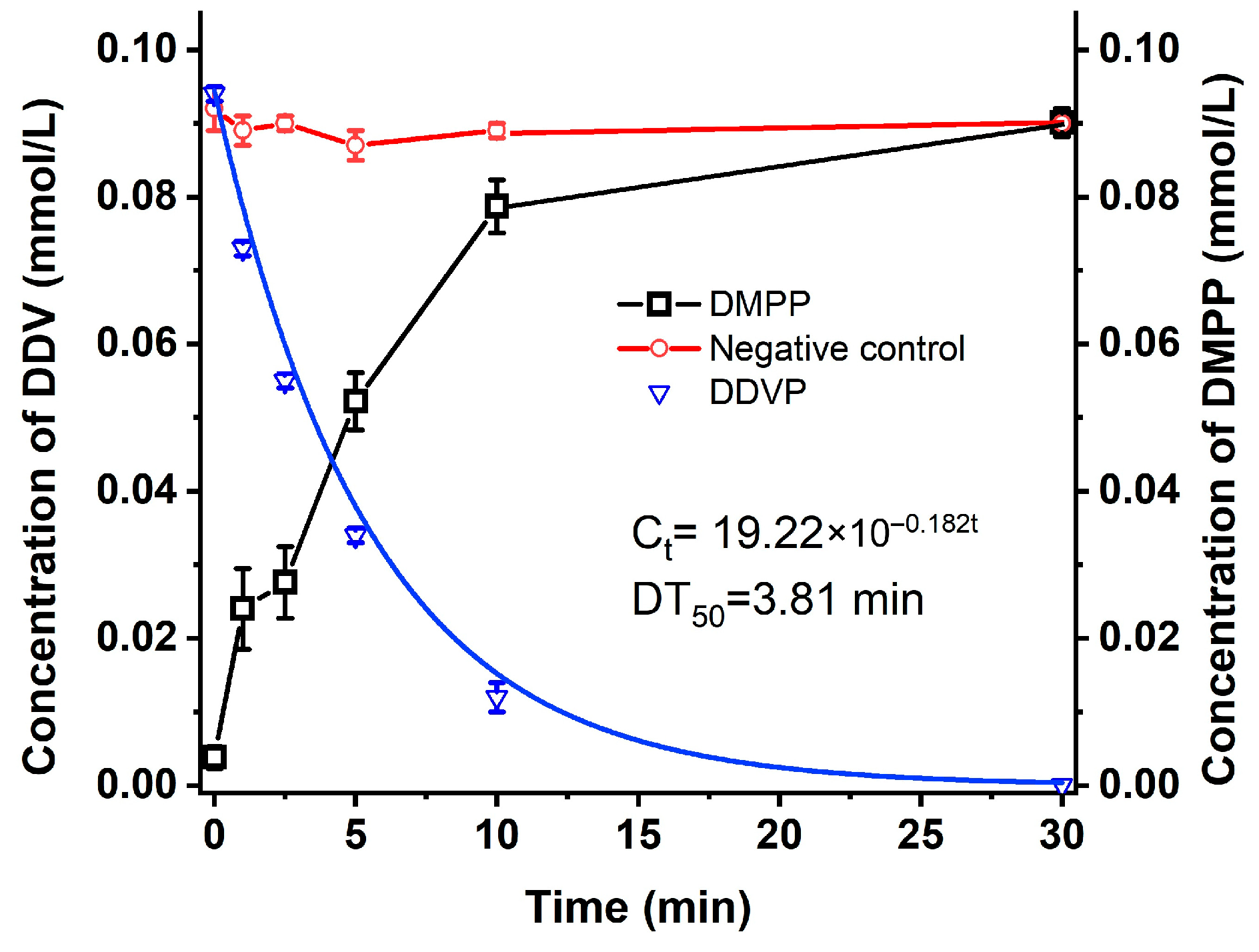
A total of 20 mg/L of DDVP could be degraded by strain G1 at a pH of 7 and a temperature of 37 °C. The degradation curve was well fitted with the first-order kinetic equation (R2 > 0.95). The DT50 of DDVP was only 3.81 min. We further determined the influencing factors of DDVP degradation by strain G1. The degradation rates of DDVP were 13.3%, 51.6%, 54.6%, and 54.6% at pH levels of 5, 6, 7, and 8, respectively, after 5 min (Figure 1A). The optimum pH for DDVP degradation was between 7 and 8. Figure 1B shows the degradation of DDVP by strain G1 at different temperatures (16, 25, 30, 37, and 42 °C), with degradation rates of 42.6%, 46.3%, 48.3%, 50.9%, and 52.1% at 5 min, respectively. The optimum temperature for DDVP degradation by strain G1 was between 25 and 42 °C. Figure 1C showed a proportional relationship between the degradation rates and the G1 inoculation amounts at pH 7 for 5 min.
The metabolite of DDVP, produced by strain G1, was also detected by UPLC, and a metabolite, DMPP, was measured and identified (Figure S1). Strain G1 could metabolize DDVP to DMPP as the sole metabolite but was unable to degrade its metabolite, DMPP (Figure 2).

2.2. Genome Features of Strain G1 and Degradation Gene in Strain G1

To analyze the molecular mechanism of DDVP degradation by strain G1, we determined the whole-genome sequence of strain G1 through genome sequencing. A total of 153,699 reads were obtained and sequenced. The mean read length and N50 read length were 11,725 bp and 14,657 bp, respectively. The sequencing depth and coverage were 450× and 100%. After assembly, the full length of the genome was 4,016,985 bp, and it consisted of only one circular chromosome with a G+C content of 69% (Table S1). A total of 3674 coding sequences (CDSs) were identified within the genome, and 2331 genes were assigned functional annotations, of which 1236 genes exhibited enzymatic catalytic activity in the GO database (Figure S2).
In strain G1, DDVP was primarily metabolized through the hydrolysis of the P-O bond, which should be catalyzed by phosphate ester hydrolase. We screened genes related to phosphatase activity in the annotation of strain G1 genome, and 21 phosphatase genes were identified in the KEGG database, including phosphotriesterase and phosphodiesterase (Table 1). Among these genes, a methyl-parathion hydrolase gene (mpd, G1_GM001006) has attracted attention, as it has been reported to have the ability to degrade a variety of organophosphorus insecticides.
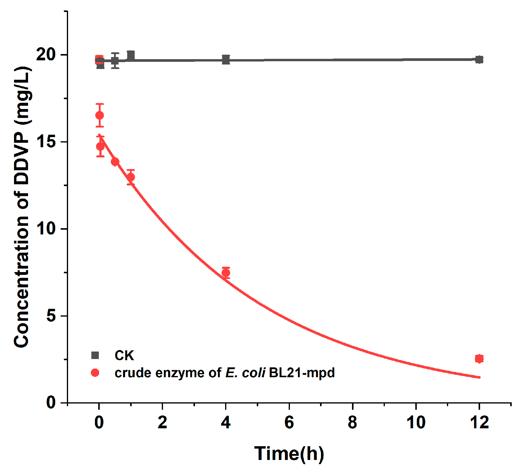
To verify the functional characteristics and catalytic properties of the mpd gene, we generated a recombinant strain to express the enzyme and assessed its capability to degrade DDVP. The mpd gene was cloned into the pMD19-T vector and transferred to the E. coli DH5α competent cells. The positive clone was isolated, and its degradation ability was measured. The results were shown in Figure 3. DDVP could be degraded by mpd-positive clone cells, and the DT50 of DDVP was only 7.2 min. The results indicated that the mpd gene was the key gene for DDVP degradation in strain G1.

2.3. Degradation Kinetics of Purified Enzyme MPD for DDVP

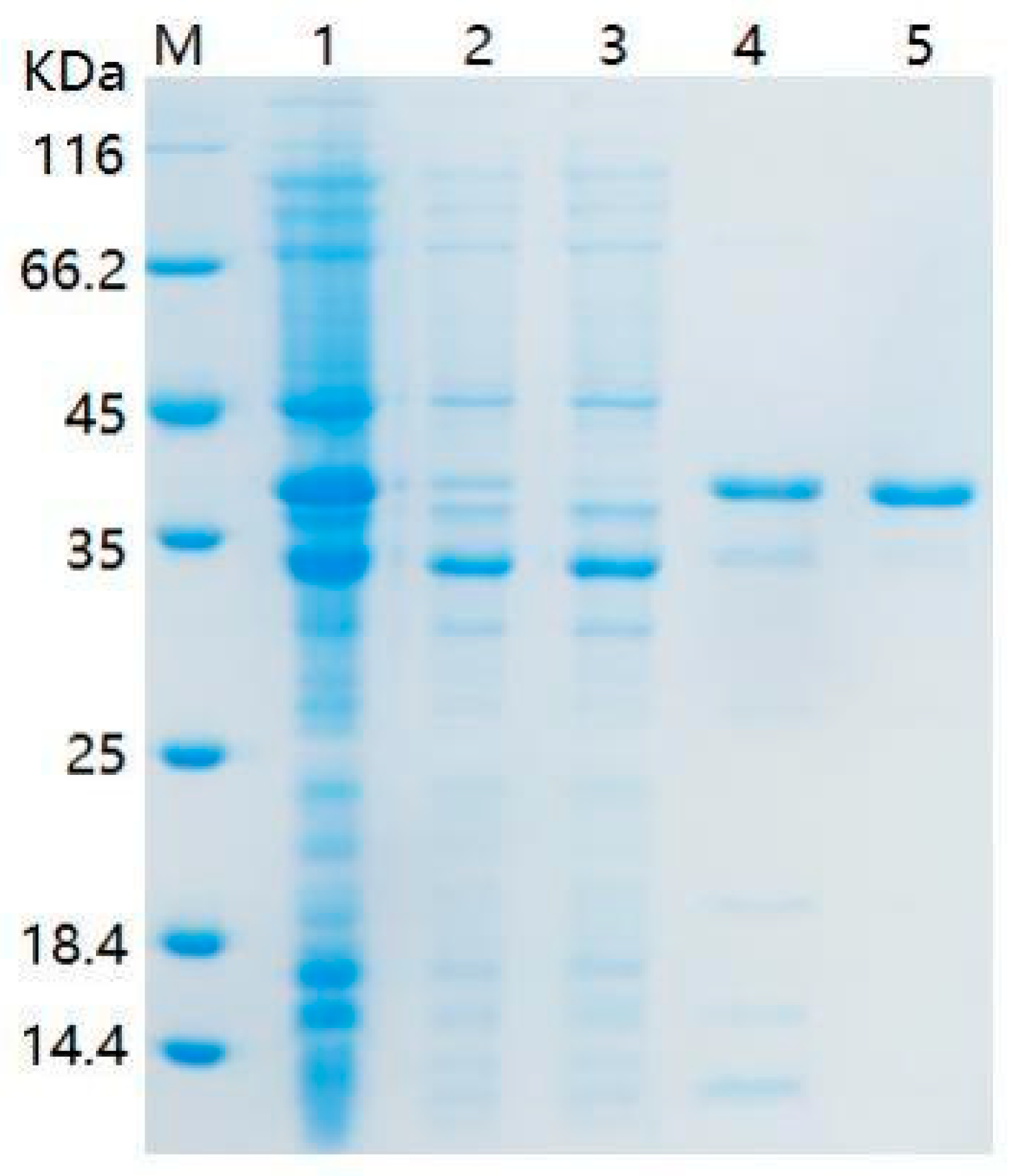
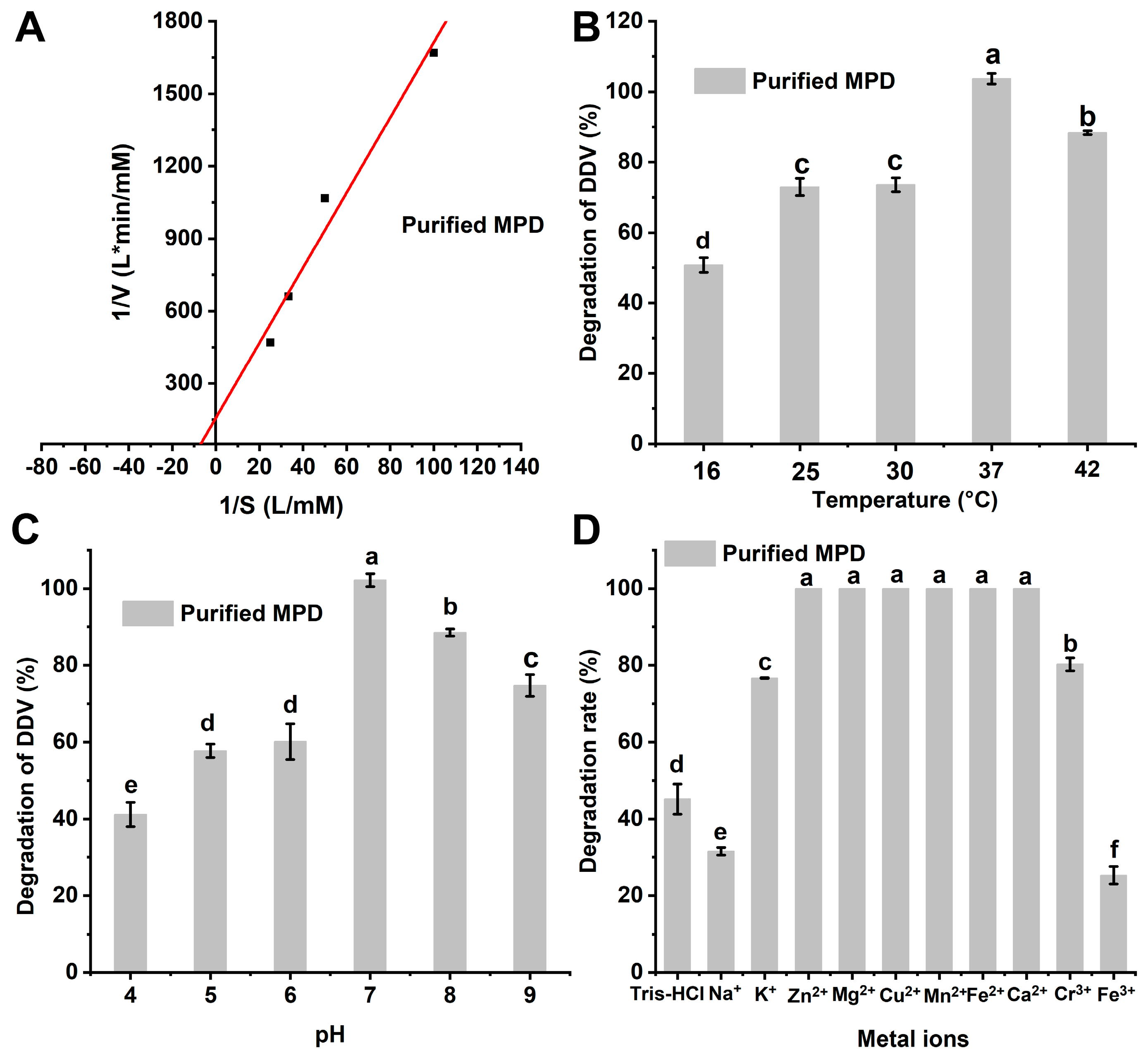
After determining the activity of the mpd gene, we constructed an expression vector and purified the expressed degradation enzyme MPD using a Ni-affinity chromatography column (Figure 4). The degradation kinetic constants of enzyme MPD were also measured. The results showed that 150 mg/L purified MPD could entirely degrade 20 mg/L DDVP within 1 min. The data were well fitted to the Michaelis–Menten equation (R2 = 0.9563). The Vmax and Km of purified MPD were 6.37 × 10⁻³ mmol/L·min and 0.0990 mmol/L, respectively (Figure 5A).
The degradation characteristics of purified MPD under different temperatures, pH levels, and metal ion conditions were investigated in Figure 5B–D. The degradation rates of DDVP by MPD at 16, 25, 30, 37, and 42 °C were 50.8%, 73.0%, 73.6%, 100%, and 88.3%, respectively, indicating that the optimal temperature for MPD was 37 °C. At different pH values, the catalysis efficiency of purified MPD varied significantly (p < 0.05). MPD exhibited the highest degradation activity at a pH level of 7.0, with a degradation rate of 92.9% for 20 mg/L DDVP in 1 min.
The degradation enzyme MPD also displayed great differences under varying valence metal ions. As shown in Figure 5D, divalent metal cations (Zn2+, Mg2+, Cu2+, Mn2+, Fe2+, and Ca2+) promoted the degradation of purified MPD. A noticeable difference was seen (p < 0.05) under the conditions of K+ and Na+ during the DDVP degradation process. K+ promoted the activity of MPD, while Na+ inhibited it. Cr3+ promoted the activity of MPD, whereas Fe3+ inhibited it.

2.4. Catalytic Mechanism of Degrading Enzyme MPD on DDVP

To analyze the degradation and catalytic mechanism of the degrading enzyme MPD on DDVP, we used the AlphaFold2 model to construct the 3D structure of enzyme MPD and determined the binding sites through molecular docking. The results showed that the binding energy between DDVP and MPD was −5.24 kcal/mol (Table 2). DDVP could interact with the active pocket (comprising Ser87, Ala261, Phe264, Asp265, Tyr318, Arg319, and Phe320) in MPD through hydrogen bond and halogen bond (Figure 6). Phe320 and Arg319 formed hydrogen bonds with the O (on the phosphate group) of DDVP, respectively. Try318 formed halogen bonds with the Cl (on the phosphate group) of DDVP.
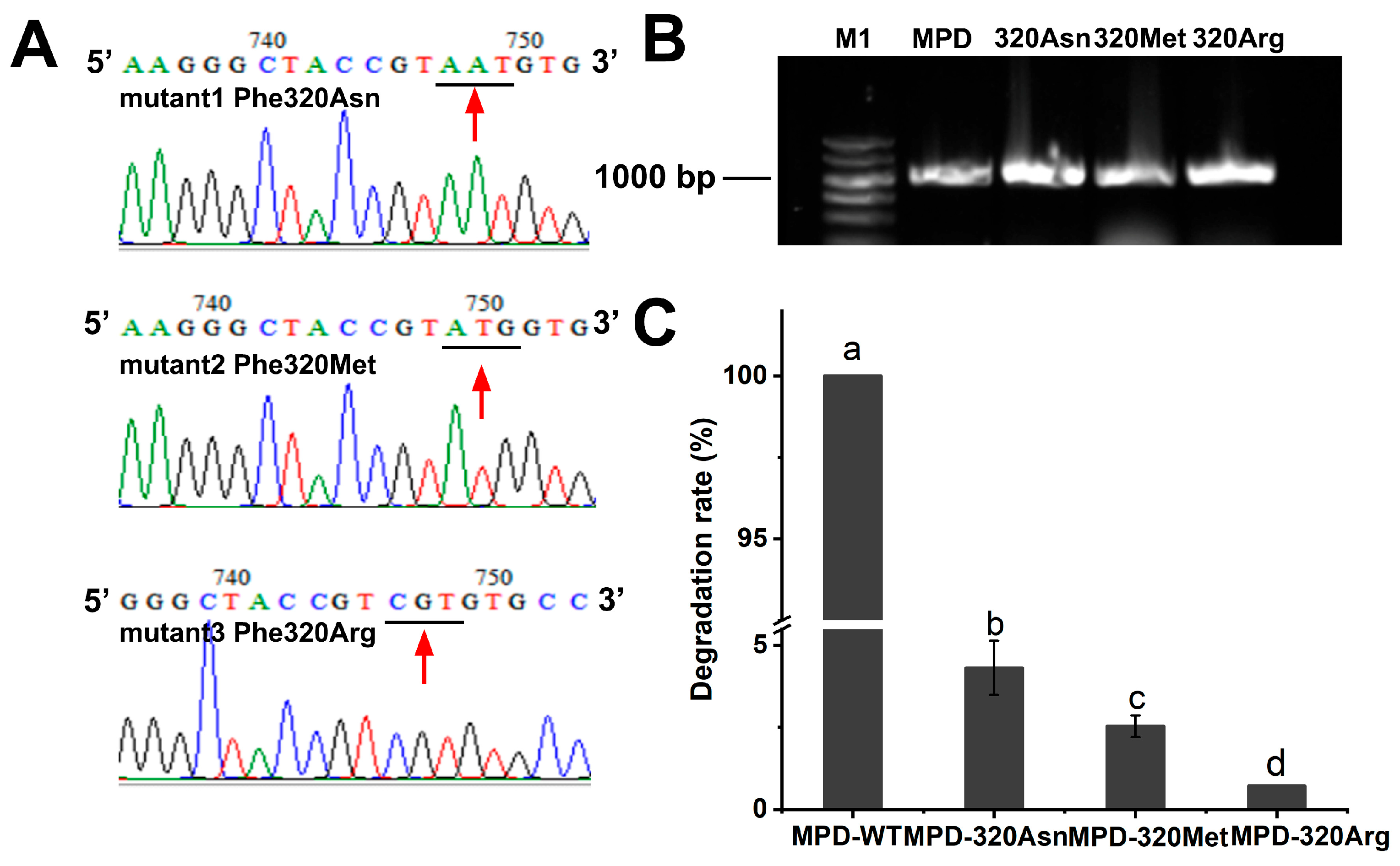
The molecular docking analysis revealed that the ester bond served as a hydrogen bonding site for Phe320, indicating its potential function as a key interaction site. The finding suggested that Phe320 may play a crucial structural or catalytic role in the molecular recognition process; therefore, we mutated Phe to Asn, Met, and Arg and compared the degradation ability of DDVP by the wild-type MPD and the mutants Phe320Asn, Phe320Met, and Phe320Arg (Figure 7). The wild-type MPD could completely degrade 20 mg/L DDVP within 5 min, whereas the mutants Phe320Asn, Phe320Met, and Phe320Arg were unable to degrade DDVP. These results indicated that Phe320 was the key amino acid site for MPD to catalyze the breakage of the phosphate ester linkage of DDVP.

3. Discussion

DDVP is an organophosphorus phosphate insecticide with high water solubility, which has been widely used in agriculture and poses a threat to fish, mammals, and humans [22]. The toxicity is mainly caused by inhibition of acetylcholine esterase, an enzyme crucial for the nervous system of organisms [18]. The DT50 of DDVP ranges from 10.5 to 19.8 d in soil [23]. Microbial degradation is recognized as an effective, low-cost, and high-efficiency method for eliminating DDVP pollution [18,19,24]. We found that strain G1 was identified as an efficient strain in the degradation of DDVP; pH level, temperature, and incubation time were found to affect the degradation by strain G1.
Strain G1 showed high efficiency when it was compared with other DDVP degradation strains (Table 3). Pseudomonas AUG12 could grow in 1500 mg/L of DDVP and degrade 100 mg/L of DDVP in 140 h; the degradation rate was 1.435 mg/L·h [25]. Ochrobactrum sp. DDV-1 could completely degrade 100 mg/L DDVP within 24 h [26]. Strain G1 was the most efficient bacterium for DDVP degradation, with a DT50 of 3.81 min. The DT50 of Flavobacterium YD-4 and halophilic bacteria T10/1 were 245 and 69 times greater than that of strain G1 [24,27]. Even under high-concentration conditions, strain G1 can still maintain high degradation activity. We further determined the degradation kinetics of strain G1 for 200 mg/L and 500 mg/L DDVP. The DT50 of 200 mg/L and 500 mg/L DDVP by strain G1 were only 18.24 min and 46.20 min, respectively (Table S3).
In addition, some bacterial strains could degrade DDVP to other toxic metabolites. Trichoderma atroviride T23 could degrade DDVP to 2,2-dichloroethanol and 2,2-dichloroacetate [31]. Stutzeri smk could produce degradation intermediates, including dimethyl vinyl phosphate, 2-chloroacrylyl dimethyl phosphate, dimethyl phosphate, and methyl phosphate, during the biodegradation process [18]. d-DDVP was produced, which proved to be more toxic than DMPP [32]. Dichloropropanol residues were found among the degradation products of DDVP [33]. Except for DMPP, dimethyl phosphate and trimethyl phosphate were also identified among the products [34].
Unlike previous studies that generated persistent toxins, strain G1 could degrade DDVP to the sole metabolite DMPP (LC50 for mice is 8714 mg/kg) that was not toxic to non-target organisms [35]. The strain exhibited a “clean” metabolic profile, minimizing secondary pollution risks. We observed that no other products were produced except for DMPP during DDVP degradation by strain G1. This is of great benefit in practical applications, suggesting the environmental safety of this bacterial strain and the potential for eco-friendly applications. Effective detoxification could be achieved through the biodegradation of strain G1, thereby reducing environmental residual risks. While current results demonstrated an absence of harmful byproducts, large-scale field trials are needed to rule out context-dependent metabolite generation. Meanwhile, we also measured the degradation activity of strain G1 against various pesticides. The results showed that, in addition to DDVP, strain G1 could also efficiently degrade methyl parathion, parathion, chlorpyrifos, fenitrothion, phoxim, triazophos, thiram, and profenofos (Table S4). This characteristic enables strain G1 to be applied in the remediation of environments contaminated with various pesticide residues.
Enzymes primarily catalyze the biodegradation of contaminants. Some genes were responsible for the degradation of DDVP, including mpd, opd, hph [35], cytochrome P450 [31], Tapon1-like [36], and aph [37]. However, the gene responsible for DDVP degradation in strain G1 was not clear. We searched for the related degradation genes of DDVP by sequencing and analyzing the genome of strain G1. Genomic analysis revealed that the strain G1 utilizes an enzyme, MPD, which can degrade DDVP into DMPP, thereby preventing the accumulation of other hazardous metabolites. The strain’s MPD enzymes ensured the breakdown of DDVP into non-toxic DMPP molecules, thereby preventing the formation of harmful intermediates. In addition, by comparing the reported DDVP-degrading genes in the NCBI database, we found that the key degrading gene of strain G1 had the highest homology with the mpd gene from the strain WBC-3. Instead of being on a plasmid, the mpd gene was located on the chromosome, indicating that the degradation gene was conserved in strain G1. Additionally, there were two regulatory factors, arsR and arsC, downstream of the mpd gene. According to the literature reported, transcription regulators can regulate the expression of degradative genes for exogenous substances such as aromatic compounds. After the induction of exogenous substances, transcription regulators can enhance the expression of genes encoding degrading enzymes [38]. In addition, strain G1 exhibited robust xenobiotic metabolic capabilities. Whole-genome analysis revealed 17 CDSs associated with aromatic compound metabolism, along with functional genes for heavy metal degradation, highlighting its potential for bioremediation in complex polluted environments.
According to the genome sequence, 21 phosphate genes were annotated in strain G1; the mpd gene was identified as the key degradation gene of DDVP. There were conserved sequences with higher homology in MPD compared with MPH (1P9E_A) from Pseudomonas WBC and MBL (WP_080708177). MPD contained a serine residue, a typical catalytic active site for metalloenzymes, and a Ser-His-Asp/Glu triad, which is a hallmark of α/β hydrolases. Conserved amino acids were found in the sequences of these enzymes. They all contained four residues in the H-X-H-X-DH sequences, which were involved in metal coordination (residues 157 to 162) (Figure S3).
MPD had the highest similarity with the metal folding enzyme MBL from Pseudomonas and methyl parathion hydrolase (MPD, EC 3.1.8.1) from Pseudomonas WBC-3, with a similarity of 100%. MPD, a Zn2+-containing enzyme, catalyzes the degradation of the organophosphorus pesticide methyl parathion. The results of crystal structure analysis showed that MPD has a dimeric structure with 331 amino acids per monomer [39]. Class B beta-lactamases have broad-spectrum substrate characteristics, which are associated with bacterial resistance to penicillin. The amino acid sequence comparison of MPD with BBLs showed that the amino acid sequence similarity between BBLs and MPD was less than 30%, but five of the six potential zinc-binding residues were conserved [40]. Zn2+, Mg2+, Cu2+, Mn2+, Fe2+, Ca2+ and Cr3+ could enhance the activity of MPD, while Na+ and Fe3+ inhibited its activity. The crystal structure showed that the presence of two metal cations in the active center significantly affected the degradation efficiency. The activities of the enzyme and substrate rely on the interaction between the substrate and divalent cations [41]. The optimum temperature, pH level, and conditions of OPHC2 with methyl parathion were 65 °C. As MPD was a metalloenzyme with a bivalent metal ion at its active site, the influence of exogenous metals in the environment on their enzymatic activity and the practical efficacy of the bacterial strain required further investigation.
Studies were conducted to clarify the catalytic mechanism of MPD when it interacted with the substrate. In the connection between MPD and methyl parathion, the histidine and tyrosine in MPD promote substrate binding through π-π stacking interactions [42]. The active pocket (Ser87, Ala261, Phe264, Asp265, Tyr318, Arg319, and Phe320) for DDVP degradation was adjacent to the MPD binding site. DDVP interacted with MPD through hydrogen bonding and halogen bonding interactions. The free binding energies between MPD and DDVP were exergonic, indicating that the interaction was spontaneous [43]. Molecular docking simulations revealed that MPD formed a stable complex with DDVP through hydrogen bonding and halogen bonding, with a calculated binding energy of −5.24 kcal/mol, indicating a high binding affinity. DDVP’s phosphate group formed hydrogen bonds with Phe320 (distance: 3.0 Å) and Arg319 (distance: 2.6 Å), while its Cl interacted with Try318 via halogen bonding. The docked conformation positioned DDVP’s reactive group within the catalytic amino acid, facilitating subsequent hydrolysis.
Previous studies have shown that Phe was involved in the binding process of DDVP in the degradation of methyl parathion. We found that during the binding process of DDVP and MPD, Phe, as a conserved hydrophobic residue, formed a hydrophobic cavity with DDVP. Therefore, we designed a targeted mutation at Phe to verify its key role in the degradation process of DDVP. Site-directed mutagenesis of Phe320 to Asn, Met, and Arg reduced the degradation efficiency of DDVP, validating its role in docking predicted by our model. Engineering the pocket (e.g., Phe320Arg mutation) could change the size, shape, or charge distribution of the binding pocket, which affects the entry or orientation of the substrate, thus changing the catalytic ability of enzymes. Future work should integrate QM/MM calculations to precisely model the electron transfer processes during the catalytic step. Targeted amino acid substitutions allow for the tailored optimization of enzyme activity, facilitating the design of high-performance microbial biocatalysts for environmental remediation.

4. Materials and Methods

4.1. Chemicals and Materials

DDVP (purity > 95%) was purchased from Dr. Ehrenstorfer (Augsburg, Germany); dimethyl phosphate (purity > 95%) was purchased from AccuStandard (New Haven, CT, USA). Restriction endonucleases (EcoR I and Hind III), T4 DNA ligase, and Taq DNA polymerase were purchased from Takara Biotech Company (Dalian, China).

4.2. Strains, Plasmids, and Growth Conditions

Strain G1 was isolated from activated sludge of an insecticide wastewater treatment facility in Jiangsu, China. Optimal growth conditions were 37 °C and a pH of 7 in a Luria–Bertani (LB) medium. E. coli BL21(DE3) and E. coli DH5α competent cells, pMD19-T, and the pET28a vector for degradation gene cloning and expression were purchased from Takara Biotech Co. (Dalian, China).

4.3. Degradation of DDVP by Strain G1

Strain G1 was activated and incubated in LB medium to an optical density at 600 nm (OD600) of 1.0 (~5.9 × 10¹⁰ CFU/mL). To evaluate the degradation capability of strain G1, MSM was supplemented with 20 mg/L DDVP as the sole source of carbon and energy. A 1.0 aliquot of strain G1 suspension was added to 4 mL of MSM in a 20 mL test tube. All samples in triplicate were incubated at 37 °C and 150 rpm. MSM inoculated without strain G1 was set as the negative control. To terminate the reaction and extract residual DDVP, a 5 mL aliquot of pure acetonitrile was added to each tube. All samples were sampled and analyzed to measure DDVP and its metabolites.
To evaluate the degradation of DDVP by strain G1, the effects of inoculum amount, pH, and temperature were examined. We determined inoculation amount levels of 10%, 20%, 50%, and 100% under ambient conditions of 37 °C and pH 7. The pH was adjusted to values of 5, 6, 7, 8, and 9 under the 20% inoculation amount and 37 °C. The effect of temperature was assessed at 16, 25, 30, 37, and 42 °C, with a pH of 7 and a 20% inoculation rate.

4.4. Complete Genome Sequencing

We used the TIANamp Bacteria DNA Kit (Tiangen Biotech Company, Beijing, China) to extract the genomic DNA of strain G1. The total DNA content was assessed through agarose gel electrophoresis and a nucleic acid protein detector. Large-scale parallel massive parallel sequencing (MPS) was conducted on samples that met the necessary sequencing criteria, employing the Illumina sequencing platform. During the sequencing process, two types of libraries were constructed: a conventional 500 bp small fragment library and a 5 kb large fragment library. Qualified DNA samples were randomly fragmented using ultrasonic treatment to generate DNA fragments of the requisite size, and an A tail was appended to the 3′ end of the fragments to complement the T base at that terminus. Subsequently, libraries of sufficient quality were constructed for cluster preparation and sequencing. Low-quality read fragments in the clean data generated from sequencing were filtered and assembled using SOAPdenovo (version 2.04) to produce a refined genomic map; however, gaps remained that required further filling. Primers were designed to perform polymerase chain reaction (PCR) using known highly homologous bacterial genome sequences and contigs derived from de novo sequencing. The specific bands obtained were sequenced, allowing for the resolution of gaps in the refined genomic maps, ultimately yielding a complete genomic representation of strain G1.

4.5. Prediction and Annotation of DDVP Degradation Genes

We used six databases to analyze the complete genome sequence of strain G1. Functional annotation was conducted using GO, Swiss-Prot, NR, KEGG, COG, and TCDB. Bioinformatics-based comparative annotation analysis was carried out to identify genes involved in DDVP degradation.

4.6. Cloning, Expression of the mpd Gene, and Purification of Enzyme MPD

The mpd gene was cloned from strain G1 genomic DNA by the PCR method with the primer pairs mpd-F and mpd-R. The primers were as follows: cgcggatccGAATTCATGCCCCTGAAGAACCGCTTGCTG (mpd-F) and gcggccgcAAGCTTgTCACTTGGGGTTGACGACCGAGTAG (mpd-R). EcoR I and Hind III restriction sites were added to primer pairs. The PCR procedure was as follows: polymerase was activated at 98 °C for 3 min, followed by 98 °C for 15 s, 58 °C for 15 s, and 34 cycles at 72 °C for 60 s. Then, the PCR products were ligated to the T-vector to construct pMD-mpd and transferred to E. coli BL21 competent cells. After sequencing, the plasmids pMD-mpd were then gel-purified and double-digested with EcoR I and Hind III restriction endonuclease. The fragments were ligated into the pET-28a (+) vector, and the expression vector pET28a-mpd was obtained. The constructed expression vector pET28a-mpd was introduced into E. coli BL21(DE3) competent cells, and positive clones, designated E. coli BL21-mpd, were screened on plates containing 100 mg/L kanamycin. After sequencing verification, the positive clone was cultured in 100 mL of LB medium at 37 °C until the optical density at 600 nm (OD600) reached 0.8. Subsequently, 1 mM IPTG was added to the medium, and the cells were further incubated at 37 °C in a rotary shaker set at 220 rpm for the overexpression of MPD. After 4 h of incubation, the cells were harvested and resuspended in Tris-NaCl buffer (20 mM Tris-HCl, 500 mM NaCl, pH 8.0). The cells were then disrupted using an ultrasonic homogenizer (Scientz-IID, Ningbo Scientz Biotech Co., Ltd., Ningbo, China) and centrifuged at 10,000× g for 10 min at 4 °C. The precipitated fraction was washed, collected, and re-dissolved in denaturing buffer (20 mM Tris-HCl, 8 M urea, pH 8.0). MPD in solution was then bound to a Ni-NTA resin column and washed with wash buffer (20 mM Tris-HCl, 8 M urea, 20 mM imidazole, pH 8.0). Finally, the purified MPD was eluted using elution buffer (20 mM Tris, 8 M urea, 500 mM imidazole, pH 8.0) and subsequently renatured in Tris-NaCl buffer via dialysis.

4.7. Determination of Enzyme Activity

We used the Linewear–Burk method to measure the Michaelis constant (Km) of MPD with the DDVP concentrations from 0.01 to 0.04 mM, and we determined the enzyme kinetic parameters by nonlinear curve fitting to the Michaelis–Menten equation. The reaction solution contained 50 µL of purified MPD (150 mg/L) and concentrations of DDVP of 0.01, 0.02, 0.03, and 0.04 mM, respectively. After 1 min incubation at 37 °C, the solution was sampled. Then, the DDVP concentrations were measured, and the results were fitted with the Michaelis–Menten equation to calculate the Km. Each treatment had three replicates.

4.8. Degradation Characteristics of DDVP by Purified Enzyme

Under varying temperatures (16–42 °C), pH levels (4–9), and metal ions (Ag+, K+, Na+, Ca2+, Mg2+, Mn2+, Zn2+, Cu2+, Fe2+, Cr3+, and Fe3+), the degradation of DDVP by purified MPD was assessed. A total of 50 μL of pure MPD enzyme, with a final concentration of 150 mg/L, was sequentially added to the standard solution of DDVP to achieve a concentration of 10 mg/L. Subsequently, Tris-HCl (pH = 7.0) was added to achieve a final volume of 5 mL. Samples lacking MPD served as negative controls. After incubation at 37 °C, samples were collected at 0 min and 1 min, and then 5 mL of acetonitrile was added to halt the reaction and extract the DDVP. The residual DDVP was subsequently analyzed using ultraperformance liquid chromatography (UPLC).

4.9. Analytical Methods

Metabolites of DDVP were separated by an Acquity UPLC BEH C18 column (1.7 μm, 2.1 mm × 100 mm) with the injection volume 10 μL and the flow rate 0.25 mL/min. The mobile phase consisted of solvent A (5‰ phosphoric acid in water) and solvent B (0.1% methanol). The separation was achieved using the liquid chromatography gradient program at 40 °C: 0–2.00 min, 5% B; 2–15.00 min, from 5% to 20% B; 15–18 min, from 20% to 95% B; 18.00–18.10 min, from 95% to 5% B; 18.10–20 min, 5% B. The capillary and cone voltages were 1500 V and 20 V, respectively. The mass spectrometer was operated in electrospray ionization (ESI) mode, with scans ranging from m/z 30 to 225. The mobile phase employed a gradient elution method, collecting both positive and negative ions.
DDVP and DMPP were quantitatively analyzed as shown in Table S2. The parent ion of DMPP was detected at m/z 221.00, with one of its daughter ions at m/z 109.00, having a dwell time of 0.05 s and cone and capillary voltages of 28 V and 22 V, respectively. Additionally, another daughter ion was detected at m/z 79.00, with the same dwell time and cone/capillary voltages of 28/34 V, respectively. The parent ion of DDVP was recorded at m/z 125.03, with daughter ions at m/z 109.00 and 62.95, with cone and capillary voltages set to 22 V and 14 V, respectively, and a dwell time of 0.05 s.

4.10. Homology Modeling and Molecular Docking of DDVP with MPD

The structure of the enzyme MPD was constructed via the AlphaFold2 model. Initially, hydrogenation was performed using UCSF Chimera (version 1.19), followed by the calculation of the atomic charge of the protein using the AMBER14SB force field. Configuration optimization was achieved utilizing the MMFF94 force field, resulting in the selection of the low-energy conformation. Local charge assignments were made using the AM1-BCC method within UCSF Chimera.

4.11. Point Mutation

To ensure the active sites, point mutations were performed by modifying the amino acid residues in the binding pocket according to the results of molecular docking. Site-specific mutations were generated using the overlap PCR method. Phe320 of MPD was replaced by Asn, Met, and Arg through site-directed mutagenesis by PCR with different primers (Table S2). Phe320Asn, Phe320Met, and Phe320Arg were generated with the primers Asn-F/Asn-R, Met-F/Met-R, and Arg-F/Arg-R by conventional PCR. We used the pET-28a-MPD plasmid as the template and performed PCR under the following conditions: 95 °C for 3 min, followed by 30 cycles of 95 °C for 15 s, 58 °C for 15 s, and 72 °C for 18 s, with a final extension step of 6.5 min at 72 °C. Subsequently, the enzyme activity of the mutant variants was determined using the same method in Section 4.6.

5. Conclusions

In conclusion, strain G1 could efficiently degrade DDVP in water. The sole detoxification metabolite DMPP was formed during the degradation process. The degradation enzyme MPD, encoded by the mpd gene, was responsible for catalyzing the degradation of DDVP. Purified MPD could degrade DDVP. DDVP was stably bound within the MPD cavity primarily through hydrogen bonding and halogen bonding interactions. Therefore, the strain G1 is an efficient candidate for the remediation of DDVP pollution in water.

Supplementary Materials

The supporting information can be downloaded at: https://www.mdpi.com/article/10.3390/ijms26199572/s1.

Author Contributions

Conceptualization: Q.M. and R.H.; methodology: Q.M.; software: Q.M.; validation: Q.M.; writing—original draft preparation: Q.M.; writing—review and editing: R.H.; visualization: Q.M.; supervision: R.H.; project administration: R.H.; funding acquisition: R.H. All authors have read and agreed to the published version of the manuscript.

Funding

This work was supported in part by grants from the National Natural Science Foundation of China (31471792), Collaborative Innovation Projects of Anhui Province (GXXT-2019-034), and Research Funds of the Joint Research Center for Food Nutrition and Health of IHM (2024SJY04).

Data Availability Statement

Data will be made available on request.

Conflicts of Interest

The authors declare no conflicts of interest.

Abbreviations

The following abbreviations are used in this manuscript:
DDVPDichlorvos
CFUColony-forming units
DMPPDimethyl phosphate
OPsOrganophosphorus pesticides
WHOWorld Health Organization
EPAEnvironmental Protection Agency
LBLuria–Bertani
MPSMassive parallel sequencing
PCRPolymerase chain reaction
UPLCUltraperformance liquid chromatography
MPDMethyl parathion hydrolase

References

  1. Jiang, J.F.; Deng, K.Q.; Duan, R.; An, C.; Dao, F.; Huang, J.L. Iron/manganese-zeolitic imidazolate framework (Fe/Mn-ZIF) nanozyme combined with acetylcholinesterase for colorimetric rapid detection of organophosphorus pesticides. Food Chem. 2025, 473, 143090. [Google Scholar] [CrossRef]
  2. Zhang, Y.J.; Zhou, B.H.; Chen, H.L.; Yuan, R.F. Heterogeneous photocatalytic oxidation for the removal of organophosphorus pollutants from aqueous solutions: A review. Sci. Total Environ. 2023, 856, 159048. [Google Scholar]
  3. Mishra, A.; Kumar, J.; Melo, J.S.; Sandaka, B.P. Progressive development in biosensors for detection of dichlorvos pesticide: A review. J. Environ. Chem. Eng. 2021, 9, 105067. [Google Scholar] [CrossRef]
  4. Quintino-Ottonicar, G.G.; da Silva, L.R.; Maria, V.L.R.d.S.; Pizzo, E.M.; de Santana, A.C.P.; Lenharo, N.R.; Pinho, C.F.; Pereira, S. Exposure to Dichlorvos pesticide alters the morphology of and lipid metabolism in the ventral prostate of rats. Front. Toxicol. 2023, 5, 1207612. [Google Scholar] [CrossRef]
  5. Fagbohun, I.K.; Idowu, E.T.; Onafuwa, A.O.; Adeneye, A.K.; Adeogun, A.O.; Adetoro, O.O. Knowledge, attitudes and perception of communities on mosquitoes and its control practices in Lagos State, Nigeria. Pan Afr. Med. J. 2021, 38, 44. [Google Scholar] [CrossRef]
  6. Nteziyaremye, P.; Cherutoi, J.; Makatiani, J.; Muhizi, T. Insecticidal potential of essential oils from Cupressus lusitanica growing in ecological zones of Rwanda against adult housefly, Musca domestica L. Int. J. Trop. Insect Sci. 2023, 43, 895–907. [Google Scholar] [CrossRef]
  7. Yu, J.X.; Wang, X.G.; Yao, X.L.; Wu, X.M. Safety Evaluation of Heavy Metal Contamination and Pesticide Residues in Coix Seeds in Guizhou Province, China. Foods 2022, 11, 2286. [Google Scholar] [CrossRef]
  8. Barbieri, M.V.; Monllor-Alcaraz, L.S.; Postigo, C.; de Alda, M.L. Improved fully automated method for the determination of medium to highly polar pesticides in surface and groundwater and application in two distinct agriculture-impacted areas. Sci. Total Environ. 2020, 745, 140650. [Google Scholar] [CrossRef]
  9. Alhassan, A.B.; Aljahdali, M.O. Behavioural and Biochemical Responses of Freshwater Bivalve Anodonta marginata Exposed to Dichlorvos. Water 2024, 16, 3572. [Google Scholar] [CrossRef]
  10. Ben Salem, I.; Boussabbeh, M.; Kantaoui, H.; Bacha, H.; Abid-Essefi, S. Crocin, the main active saffron constituent, mitigates dichlorvos-induced oxidative stress and apoptosis in HCT-116 cells. Biomed. Pharmacother. 2016, 82, 65–71. [Google Scholar] [CrossRef]
  11. Saravanakumar, K.; Park, S.; Vijayasarathy, S.; Swaminathan, A.; Sivasantosh, S.; Kim, Y.; Yoo, G.; Madhumitha, H.; Mubarakali, D.; Cho, N.M. Cellular metabolism and health impacts of dichlorvos: Occurrence, detection, prevention, and remedial strategies—A review. Environ. Res. 2024, 242, 117600. [Google Scholar] [CrossRef]
  12. Ben Salem, I.; Boussabbeh, M.; Bacha, H.; Abid, S. Dichlorvos-induced toxicity in HCT116 cells: Involvement of oxidative stress and apoptosis. Pestic. Biochem. Physiol. 2015, 119, 62–66. [Google Scholar] [CrossRef]
  13. Zhang, P.C.; Zhou, Z.X.; Yao, J.Q.; Jiang, Y.H.; Lei, H.; Xie, Z.J.; Li, J.; Zhao, X.L.; Zhu, L.; Wan, M.H.; et al. Effects of pesticide dichlorvos on liver injury in rats and related toxicity mechanisms. Ecotoxicol. Environ. Saf. 2025, 290, 117747. [Google Scholar] [CrossRef]
  14. Saka, W.A.; Adeogun, A.E.; Adisa, V.I.; Olayioye, A.; Igbayilola, Y.D.; Akhigbe, R.E. L-arginine attenuates dichlorvos-induced testicular toxicity in male Wistar rats by suppressing oxidative stress-dependent activation of caspase 3-mediated apoptosis. Biomed. Pharmacother. 2024, 178, 117136. [Google Scholar] [CrossRef]
  15. Li, D.Y.; Ma, X.Y.; Zhang, S.Y.; Wang, Y.K.; Han, Y.N.; Chen, R.; Wang, X.C.; Ngo, H.H. Aquatic photolysis of high-risk chemicals of emerging concern from secondary effluent mediated by sunlight irradiation for ecological safety and the enhanced methods. Water Res. 2023, 238, 120002. [Google Scholar] [CrossRef]
  16. Kumar, R.; George, L.; Jun, Z.; Mukherji, S. Photocatalytic activity of graphene oxide-TiO2 nanocomposite on dichlorvos and malathion and assessment of toxicity changes due to photodegradation. Chemosphere 2022, 308, 136402. [Google Scholar] [CrossRef]
  17. Asemoloye, M.D.; Jonathan, S.G.; Ahmad, R. Degradation of 2, 2-Dichlorovinyl dimethyl phosphate (dichlorvos) through the rhizosphere interaction between Panicum maximum Jacq and some selected fungi. Chemosphere 2019, 221, 403–411. [Google Scholar] [CrossRef]
  18. Parte, S.G.; Mohekar, A.D.; Kharat, A.S. Aerobic dichlorvos degradation by Pseudomonas stutzeri smk: Complete pathway and implications for toxicity in Mus musculus. Iran. J. Microbiol. 2020, 12, 138–147. [Google Scholar] [CrossRef]
  19. Sun, J.N.; Si, G.Y.; Liu, H.Y.; Li, Y.Q.; Wang, X.H.; Chen, J. Degradation effects on dichlorvos by a biocontrol strain, Trichoderma atroviride T23. J. Integr. Agric. 2023, 22, 2746–2758. [Google Scholar] [CrossRef]
  20. Ma, W.W.; Zhao, Y.Y.; Sun, H.; Zhang, Z.W.; Huang, L.L. Oral Administration of Lactiplantibacillus plantarum CCFM8661 Alleviates Dichlorvos-Induced Toxicity in Mice. Foods 2024, 13, 3211. [Google Scholar] [CrossRef]
  21. Zhang, Y.M.; Zhang, W.P.; Li, J.Y.; Pang, S.M.; Mishra, S.; Bhatt, P.; Zeng, D.X.; Chen, S.H. Emerging Technologies for Degradation of Dichlorvos: A Review. Int. J. Environ. Res. Public Health 2021, 18, 5789. [Google Scholar]
  22. Malakootian, M.; Shahesmaeili, A.; Faraji, M.; Amiri, H.; Silva Martinez, S. Advanced oxidation processes for the removal of organophosphorus pesticides in aqueous matrices: A systematic review and meta-analysis. Process Saf. Environ. Prot. 2020, 134, 292–307. [Google Scholar] [CrossRef]
  23. Youfeng, Z.; Dongdong, Z.; Ling, H. Enantioselective biodegradation and enantiomerization of dichlorprop in soils. Chemosphere 2020, 258, 127322. [Google Scholar] [CrossRef]
  24. Ning, J.; Gang, G.; Bai, Z.; Hu, Q.; Qi, H.; Ma, A.; Zhuan, X.; Zhuang, G. In situ enhanced bioremediation of dichlorvos by a phyllosphere Flavobacterium strain. Front. Environ. Sci. Eng. 2012, 6, 231–237. [Google Scholar] [CrossRef]
  25. Yadav, S.; Verma, S.K.; Chaudhary, H.S. Isolation and Characterization of Organophosphate Pesticides Degrading Bacteria from Contaminated Agricultural Soil. OnLine J. Biol. Sci. 2015, 15, 113–125. [Google Scholar] [CrossRef]
  26. Zhang, X.-H.; Zhang, G.-S.; Zhang, Z.-H.; Xu, J.-H.; Li, S.-P. Isolation and Characterization of a Dichlorvos-Degrading Strain DDV-1 of Ochrobactrum sp. Pedosphere 2006, 16, 64–71. [Google Scholar] [CrossRef]
  27. Oncescu, T.; Oancea, P.; Enache, M.; Popescu, G.; Dumitru, L.; Kamekura, M. Halophilic bacteria are able to decontaminate dichlorvos, a pesticide, from saline environments. Open Life Sci. 2007, 2, 563–573. [Google Scholar] [CrossRef]
  28. Yuan, S.; Li, C.; Yu, H.; Xie, Y.; Guo, Y.; Yao, W. Selective uptake determines the variation in degradation of organophosphorus pesticides by Lactobacillus plantarum. Food Chem. 2021, 360, 130106. [Google Scholar] [CrossRef]
  29. Zhao, K.; Yu, Y.; Jiang, D.; Wang, D.; Li, Z.-M.; Huang, G.-Z.; Bai, Z.-H.; Bai, Z.H. Degradation of Dichlorvos by Rhodobacter sphaeroides. Environ. Sci. 2009, 4, 1200–1204. [Google Scholar]
  30. Sukirtha, T.; Usharani, M. Production and Qualitative Analysis of Biosurfactant and Biodegradation of the Organophosphate by Nocardia mediterranie. J. Bioremediat. Biodegrad. 2013, 4, 198–205. [Google Scholar]
  31. Sun, J.; Karuppiah, V.; Li, Y.; Pandian, S.; Kumaran, S.; Chen, J. Role of cytochrome P450 genes of Trichoderma atroviride T23 on the resistance and degradation of dichlorvos. Chemosphere 2022, 290, 133173. [Google Scholar] [CrossRef]
  32. Cruz-Alcalde, A.; Sans, C.; Esplugas, S. Priority pesticide dichlorvos removal from water by ozonation process: Reactivity, transformation products and associated toxicity. Sep. Purif. Technol. 2018, 192, 123–129. [Google Scholar] [CrossRef]
  33. Richardson, D.D.; Caruso, J.A. Screening organophosphorus nerve agent degradation products in pesticide mixtures by GC-ICPMS. Anal. Bioanal. Chem. 2007, 389, 679–682. [Google Scholar] [CrossRef]
  34. Magny, R.; Lejeune, M.; Mégarbane, B.; Houzé, P.; Labat, L. Cas d’intoxication au dichlorvos: Identification de métabolites et de produits de dégradation par une approche utilisant les réseaux moléculaires. Toxicol. Anal. Clin. 2022, 34, 207–208. [Google Scholar] [CrossRef]
  35. Abe, K.; Yoshida, S.; Suzuki, Y.; Mori, J.; Doi, Y.; Takahashi, S.; Kera, Y. Haloalkylphosphorus Hydrolases Purified from Sphingomonas sp. Strain TDK1 and Sphingobium sp. Strain TCM1. Appl. Environ. Microbiol. 2014, 80, 5866–5873. [Google Scholar] [CrossRef] [PubMed]
  36. Sun, J.; Yuan, X.; Li, Y.; Wang, X.; Chen, J. The pathway of 2,2-dichlorovinyl dimethyl phosphate (DDVP) degradation by Trichoderma atroviride strain T23 and characterization of a paraoxonase-like enzyme. Appl. Microbiol. Biotechnol. 2019, 103, 8947–8962. [Google Scholar] [CrossRef] [PubMed]
  37. Jin, H.; Zhou, Z.; Wang, D.; Guan, S.; Han, W. Molecular Dynamics Simulations of Acylpeptide Hydrolase Bound to Chlorpyrifosmethyl Oxon and Dichlorvos. Int. J. Mol. Sci. 2015, 16, 6217–6234. [Google Scholar] [CrossRef]
  38. Okoroiwu, H.U.; Iwara, I.A. Dichlorvos toxicity: A public health perspective. Interdiscip. Toxicol. 2019, 11, 129–137. [Google Scholar] [CrossRef]
  39. Dong, Y.; Bartlam, M.; Sun, L.; Zhou, Y.; Zhang, Z.-P.; Zhang, C.-G.; Rao, Z.; Zhang, X.E. Crystal structure of methyl parathion hydrolase from Pseudomonas sp. WBC-3. J. Mol. Biol. 2005, 353, 655–663. [Google Scholar] [CrossRef]
  40. Liu, H.; Zhang, J.-J.; Wang, S.-J.; Zhang, X.-E.; Zhou, N.-Y. Plasmid-borne catabolism of methyl parathion and p-nitrophenol in Pseudomonas sp. strain WBC-3. Biochem. Biophys. Res. Commun. 2005, 334, 1107–1114. [Google Scholar] [CrossRef]
  41. Schenk, G.; Mateen, I.; Ng, T.-K.; Pedroso, M.M.; Mitić, N.; Jafelicci, M.; Marques, R.F.; Gahan, L.R.; Ollis, D.L. Organophosphate-degrading metallohydrolases: Structure and function of potent catalysts for applications in bioremediation. Coord. Chem. Rev. 2016, 317, 122–131. [Google Scholar] [CrossRef]
  42. Li, Y.; Yang, H.; Xu, F. Identifying and engineering a critical amino acid residue to enhance the catalytic efficiency of Pseudomonas sp. methyl parathion hydrolase. Appl. Microbiol. Biotechnol. 2018, 102, 6537–6545. [Google Scholar] [CrossRef]
  43. Moradi, S.; Khani, S.; Ansari, M.; Shahlaei, M. Atomistic details on the mechanism of organophosphates resistance in insects: Insights from homology modeling, docking and molecular dynamic simulation. J. Mol. Liq. 2019, 276, 59–66. [Google Scholar] [CrossRef]
Figure 1. Degradation rate of 20 mg/L DDVP by strain G1 under different pH levels (A), temperatures (B), and inoculation amounts (C) within 5 min. (The data are represented as the mean ± standard deviation for triplicate. A–d letters are marked based on difference significance analysis).
Ijms 26 09572 g001
Figure 2. Degradation kinetics of DDVP and DMPP by strain G1. (pH 7, 37 °C, incubation amount 20% (OD600 = 1.0), initial concentration 20 mg/L); red line: 20 mg/L DDVP in MSM medium without strain G1; black line: DMPP generated from 20 mg/L DDVP in MSM medium with strain G1; blue line: DDVP residual in MSM medium with strain G1.
Ijms 26 09572 g002
Figure 3. Degradation of 20 mg/L DDVP by crude enzyme from an engineered strain.
Ijms 26 09572 g003
Figure 4. The SDS-Page of crude enzyme and purified MPD. Line M: Marker; Line 1: IPTG induced; Line 2: supernatant; Line 3: effluent; Line 4: 50 mM imidazole eluent; Line 5: 500 mM imidazole eluent.
Ijms 26 09572 g004
Figure 5. Michaelis constant of enzyme MPD (A); degradation characteristics of enzyme MPD under different temperatures (B), pH levels (C), and metal ions (D). (The data are represented as the mean ± standard deviation for triplicate. a–f letters are marked based on difference significance analysis).
Ijms 26 09572 g005
Figure 6. Molecular docking of DDVP with enzyme MPD (A); the binding mode diagram of DDVP and MPD (B). (The yellow and purple lines represent hydrogen bonds and halogen bonds respectively, and the numbers on the lines indicate the bond lengths).
Ijms 26 09572 g006
Figure 7. (A) Sequencing chromatogram of the mutants; (B) gel electrophoresis of the mutant Phe320Asn, Phe320Met, and Phe320Arg; and (C) degradation of DDVP via wild-type MPD and mutants. (The data are represented as the mean ± standard deviation for triplicate. a–d letters are marked based on difference significance analysis).
Ijms 26 09572 g007
Table 1. Phosphatase genes in the genome of strain G1. (“+” and “−” indicates the sense and antisense strand, respectively.)
Gene NumberLocusAnnotation
G1_GM000376Chr1:451815:452579:+c-di-GMP-specific phosphodiesterase class I
G1_GM000403Chr1:479899:482832:+Sensor domain-containing phosphodiesterase
G1_GM000617Chr1:725113:726333:−2′3′-cyclic phosphodiesterase
G1_GM000878Chr1:1012855:1014471:−Phosphodiesterase
G1_GM001006Chr1:1174990:1175985:−Methyl-parathion hydrolase mpd
G1_GM001037Chr1:1214103:1214729:−c-di-GMP phosphodiesterase class II
G1_GM001072Chr1:1257629:1259686:−CdpA cyclic di-GMP phosphodiesterase
G1_GM001177Chr1:1376461:1377048:+RNA2′,3′-cyclic phosphodiesterase
G1_GM001721Chr1:1922141:1923490:+Phosphodiesterase
G1_GM001738Chr1:1942976:1945048:−c-di-GMP-specific phosphodiesterase class I
G1_GM001887Chr1:2112072:2113784:+c-di-GMP phosphodiesterase
G1_GM001963Chr1:2197887:2200340:+c-di-GMP phosphodiesterase A
G1_GM002029Chr1:2274043:2275569:−3′,5′-cyclic AMP phosphodiesterase CpdA T
G1_GM002209Chr1:2470901:2473153:+Phosphodiesterase
G1_GM002415Chr1:2714768:2716021:+Phosphodiesterase
G1_GM003033Chr1:3405300:3405869:+Type I phosphodiesterase
G1_GM003125Chr1:3504090:3505742:+Type I phosphodiesterase
G1_GM003196Chr1:3576332:3577663:−Diguanylate phosphodiesterase
G1_GM003204Chr1:3587564:3588784:+c-di-GMP-specific phosphodiesterase class I
G1_GM003579Chr1:4007203:4008258:−Glycerophosphodiester phosphodiesterase
G1_GM003053Chr1:3427595:3428083:+Phosphotriesterase family
Table 2. Molecular docking parameters of MPD with DDVP.
ProteinSubstrateBondLength
(Å)
Kd
(μM)
ΔG
(kcal/mol)
Molecular-Interaction Energy
(kcal/mol)
Electrostatic Potential Energy
(kcal/mol)
MPDDDVPHydrogen bond2.60144.15−5.24−6.25−1.01
Table 3. Comparison of reported DDVP-degrading strains with strain G1.
StrainsConcentration
(mg/L)
Time
(h)
Degradation Rate (%)DT50
(h)
Reference
Flavobacterium YD-44004860.8915.60[24]
Lactobacillus plantarum LAB5024100/[28]
Pseudomonas stutzeri SMK0.0514480/[18]
Pseudomonas AUG12100140//[25]
Rhodobacter sphaeroides EBL07064001298/[29]
Ochrobactrum sp. dichlorvos-110024100/[26]
Halophilic bacteria TL432.99//3.10[27]
Halophilic bacteria T10/132.99//4.47
Nocardia mediterranei1072100/[30]
S. acidaminiphila G1200.51000.056This study
Disclaimer/Publisher’s Note: The statements, opinions and data contained in all publications are solely those of the individual author(s) and contributor(s) and not of MDPI and/or the editor(s). MDPI and/or the editor(s) disclaim responsibility for any injury to people or property resulting from any ideas, methods, instructions or products referred to in the content.