Article

ZFHX3 Knockdown Enhances Metabolic Distress in Atrial Myocytes Through Mitochondrial and Calcium Dysregulation: Mitigation by Trimetazidine

1
Graduate Institute of Clinical Medicine, College of Medicine, Taipei Medical University, Taipei 11031, Taiwan
2
Division of Cardiology, Department of Internal Medicine, Shuang-Ho Hospital, Taipei Medical University, New Taipei City 235, Taiwan
3
Cardiac Electrophysiology and Pacing Laboratory, Division of Cardiovascular Medicine, Makiminato Central Hospital, 1199 Makiminato, Urasoe 901-2131, Japan
4
Department of Medical Education and Research, Wan Fang Hospital, Taipei Medical University, Taipei 11696, Taiwan
5
Division of Cardiology, Department of Internal Medicine, Wan-Fang Hospital, Taipei Medical University, Taipei 11696, Taiwan
6
Cardiovascular Research Center, Wan-Fang Hospital, Taipei Medical University, Taipei 11696, Taiwan
*
Author to whom correspondence should be addressed.
These authors contributed equally to this work.
Int. J. Mol. Sci. 2025, 26(17), 8576; https://doi.org/10.3390/ijms26178576
Submission received: 25 July 2025 / Revised: 29 August 2025 / Accepted: 1 September 2025 / Published: 3 September 2025
(This article belongs to the Special Issue Molecular Mechanisms and Therapy of Cardiomyopathy)

Abstract

Metabolic dysregulation in the heart plays a critical role in the pathogenesis of atrial fibrillation (AF), yet the underlying molecular mechanisms remain unclear. Loss-of-function variants in the zinc finger homeobox 3 gene (ZFHX3) increase AF risk by promoting structural and electrical remodeling. However, the role of ZFHX3 knockdown (KD) in cardiac metabolism has not been fully elucidated. This study investigated the impact of ZFHX3 KD on energy metabolism in atrial myocytes and assessed the therapeutic potential of trimetazidine (TMZ). Seahorse XFe24 extracellular flux analysis, bioluminescent assays, microplate enzyme activity assays, and Western blotting were used to study energy substrate (glucose and fatty acid) oxidation stress, intracellular lactate content, glucose uptake, pyruvate dehydrogenase (PDH) activity, and regulatory protein expression in control and ZFHX3 KD HL-1 cells with or without TMZ (10 μM) treatment. ZFHX3 KD cells exhibited a higher acute response in oxygen consumption after Etomoxir injection, upregulated CD36 and phosphorylated ACC expression, increased glucose uptake and lactate production, reduced PDH activity, and higher levels of PDK4 and LDHA. Furthermore, ZFHX3 KD cells showed mitochondrial Ca2+ overload and increased phosphorylated PDH and oxidized CaMKII proteins, all of which were significantly attenuated by TMZ. Additionally, TMZ improved mitochondrial dysfunction in ZFHX3 KD cells by decreasing basal and maximal respiration, spare capacity, and proton leak. These findings suggest that ZFHX3 downregulation shifts substrate preference toward fatty acid utilization at the expense of glucose oxidation, contributing to metabolic and mitochondrial calcium dysregulation. TMZ mitigates these effects, highlighting its therapeutic potential in AF associated with ZFHX3 deficiency.

Graphical Abstract

1. Introduction

Atrial fibrillation (AF) is the most common sustained tachyarrhythmia encountered in clinical practice and a major contributor to cardiovascular morbidity and mortality [1]. Increasing evidence indicates that AF should be recognized not only as an electrical disorder but also as a metabolic cardiac disease [2]. The heart has a high energy demand, and disturbances in substrate flexibility, trafficking, storage, and utilization often precede electrophysiological, contractile, and structural remodeling [3]. Recent proteomic analyses of left atrial appendage tissues from AF patients have revealed alterations in mitochondrial function, oxidative phosphorylation (OXPHOS), sirtuin signaling, the tricarboxylic acid (TCA) cycle, and other key enzymes critical to energy metabolism [4,5]. Metabolomic profiling of blood plasma has also revealed elevated levels of long-chain fatty acids (FAs) and dysregulation of lipid transport proteins [6]. Patients with persistent AF exhibit increased levels of enzymes and metabolites related to ketone body metabolism, along with elevated ketogenic amino acids and glycine [7]. Together, these findings highlight the central role of altered cardiac metabolism in AF pathogenesis and underscore the need to elucidate the molecular mechanisms of atrial metabolic remodeling.
More than a decade ago, multi-ethnic genome-wide association studies identified variants in the zinc finger homeobox 3 (ZFHX3) gene on chromosome 16q22 as being strongly associated with AF [8,9,10,11]. Subsequent studies showed that ZFHX3 plays a significant role in AF pathophysiology [12,13,14]. Cardiac-specific loss of ZFHX3 in mice increases AF susceptibility, accompanied by slowed conduction velocity, dysregulated calcium handling, atrial enlargement, thrombus formation, and dilated cardiomyopathy [15]. Transcriptomic analyses have revealed that oxidative phosphorylation, mitochondrial organization, and FAs metabolism pathways are downregulated in ZFHX3-deficient atria, while inflammatory and injury-related genes are upregulated [15]. These findings suggest that ZFHX3 variants predominantly exert loss-of-function effects, disrupting both atrial electrophysiology and metabolism. Taken together, these findings indicate that ZFHX3 plays a critical role in atrial metabolism and calcium homeostasis. However, the role of ZFHX3 in cardiac energy substrate regulation remains poorly defined, and therapeutic interventions targeting metabolic remodeling that could mitigate these effects have not been established. In this study, we investigated the impact of ZFHX3 knockdown (KD) on mitochondrial energy metabolism and calcium homeostasis in atrial myocytes and evaluated the protective effects of the metabolic modular trimetazidine (TMZ). Our findings provide mechanistic insight into how ZFHX3 deficiency contributes to metabolic stress in AF and suggest potential therapeutic avenues.

2. Results

2.1. Effects of ZFHX3 Gene on Mitochondrial Energy Substrate Oxidation

We studied the effects of the ZFHX3 gene on cellular energy substrate oxidation using specific inhibitors of substrate oxidation pathways. As shown in Figure 1, under low substrate demand, both control and ZFHX3 KD cells had similar basal respiration. However, ZFHX3 KD cells showed a significantly higher acute response, maximal respiration, and spare capacity after Etomoxir injection (a CPT-1a inhibitor that blocks FAs oxidation) as shown in Figure 1A. In contrast, after UK5099 injection, both control and ZFHX3 KD cells displayed similar acute response and maximal respiration, along with a marked reduction in spare capacity as shown in Figure 1B. These findings suggest that ZFHX3 KD increases FAs utilization but does not alter glucose utilization.

2.2. Effects of ZFHX3 Knockdown on Lactate Production, Glucose Uptake, and PDH Activity

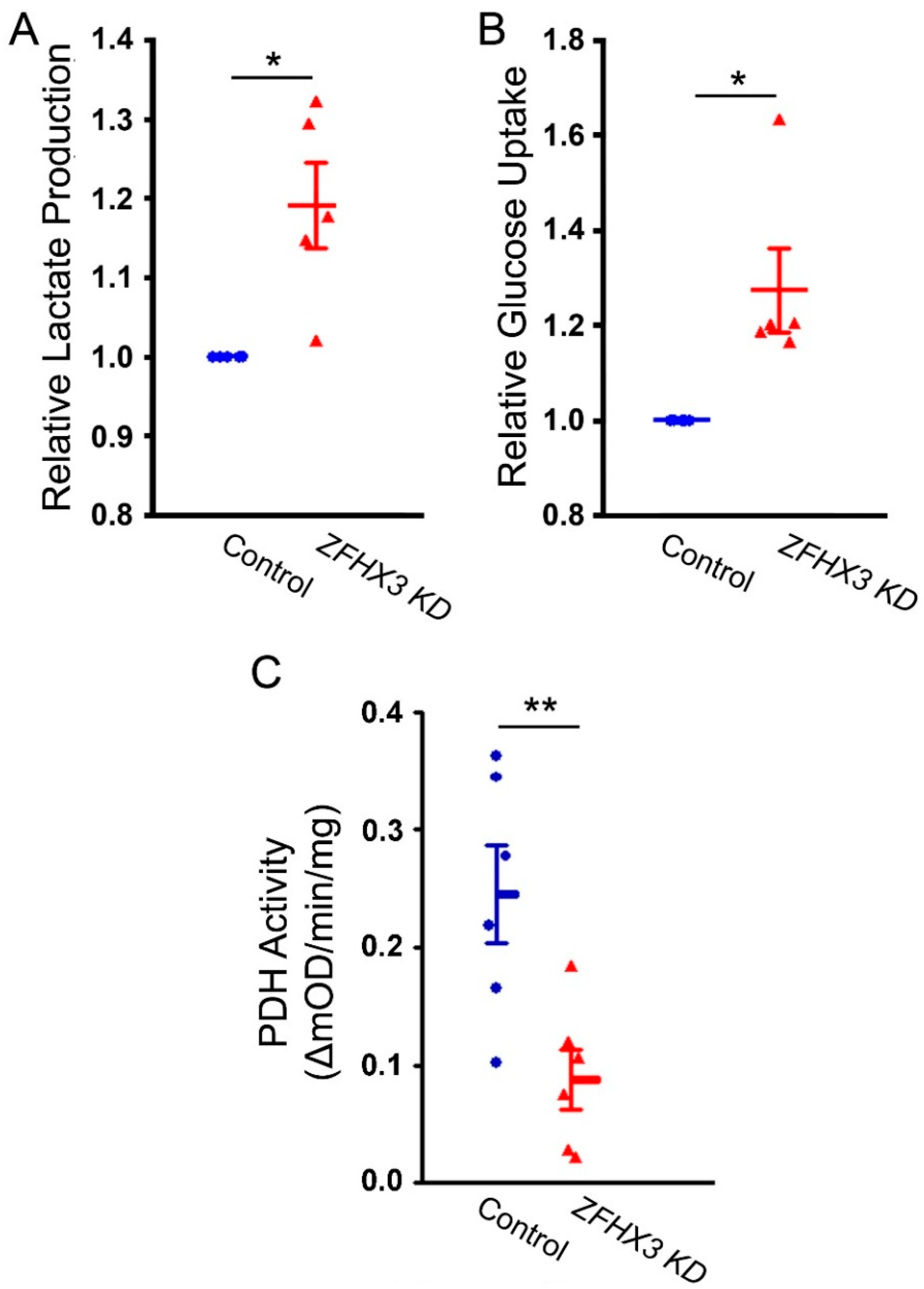
To study the impact of increased FAs utilization on cellular metabolism, we examined lactate production, glucose uptake, and PDH activity. ZFHX3 KD cells showed significantly higher lactate levels, along with increased glucose uptake and reduced PDH activity compared with control cells (Figure 2). These findings suggest that enhanced FAs utilization may suppress PDH activity, leading to an accumulation of lactate in ZFHX3 KD cells.

2.3. Effects of ZFHX3 Gene on FAs and Glucose Metabolism Regulatory Proteins

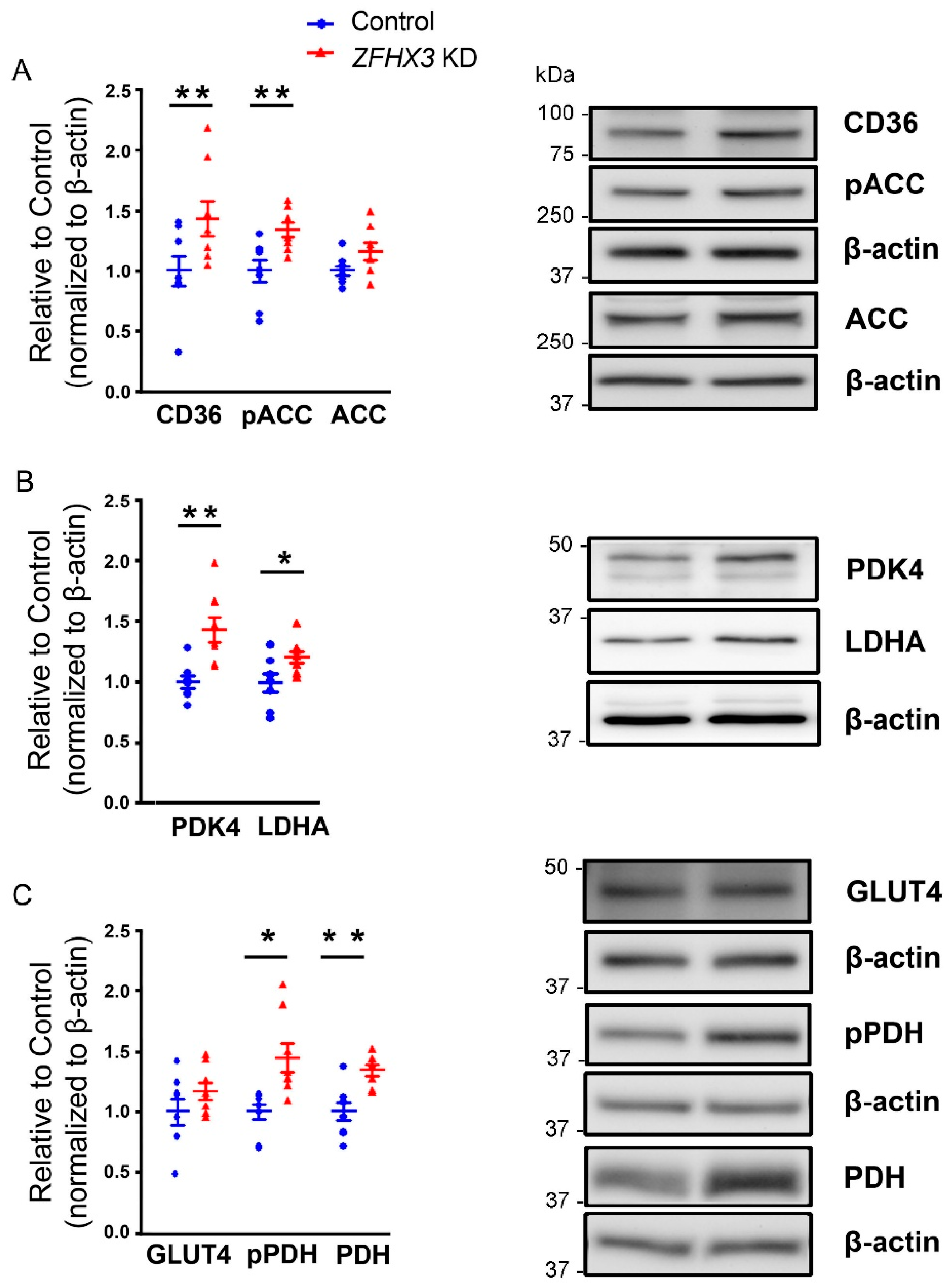
We studied the regulatory proteins involved in FAs and glucose metabolism and found that ZFHX3 KD cells exhibited higher expressions of CD36, phosphorylated ACC (pACC), phosphorylated PDH (pPDH), and total PDH proteins compared with control cells (Figure 3). However, both control and ZFHX3 KD cells showed similar expressions of ACC and GLUT4.

2.4. Trimetazidine on Lactate Production

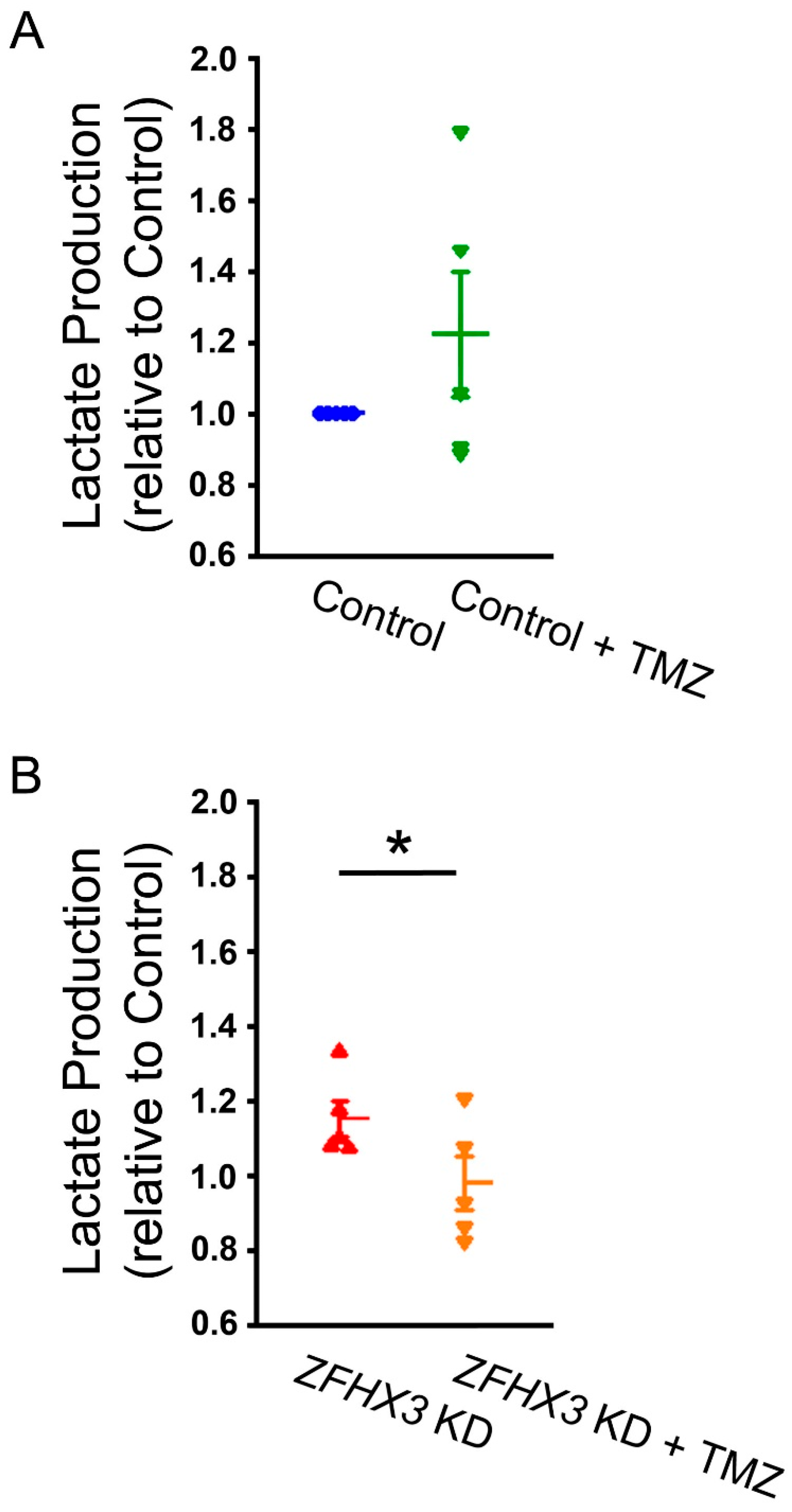
To explore the impact of increased FAs utilization on cardiac metabolism in ZFHX3 KD cells, we studied the effect of trimetazidine (TMZ), a β-oxidation inhibitor, on lactate production in HL-1 cells. As shown in Figure 4, TMZ (10 µM)-treated ZFHX3 KD cells exhibited lower lactate production compared with untreated ZFHX3 KD cells. However, lactate levels were similar between control cells and TMZ-treated control cells.

2.5. Trimetazidine on Mitochondrial Respiration and Calcium Homeostasis

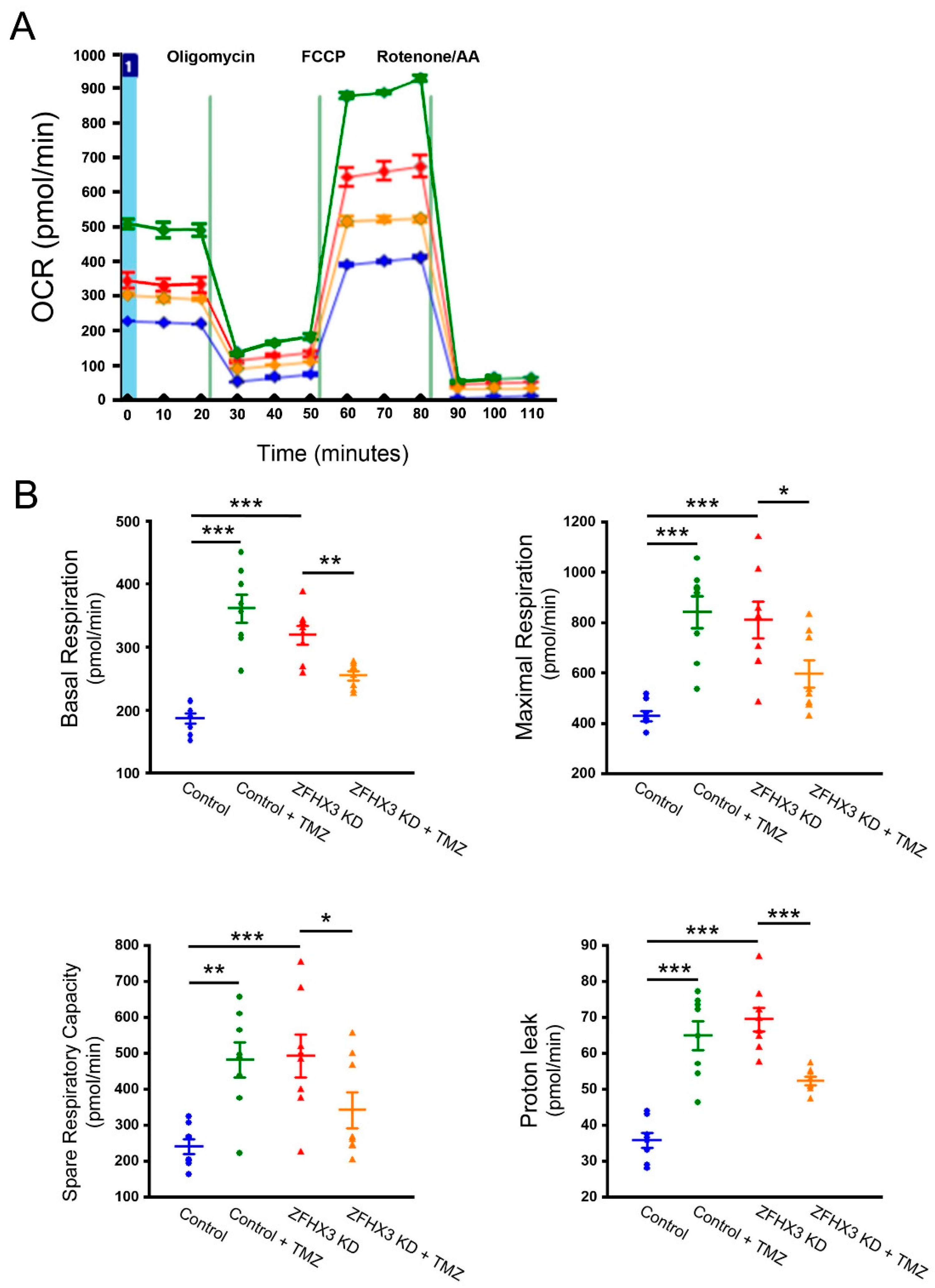
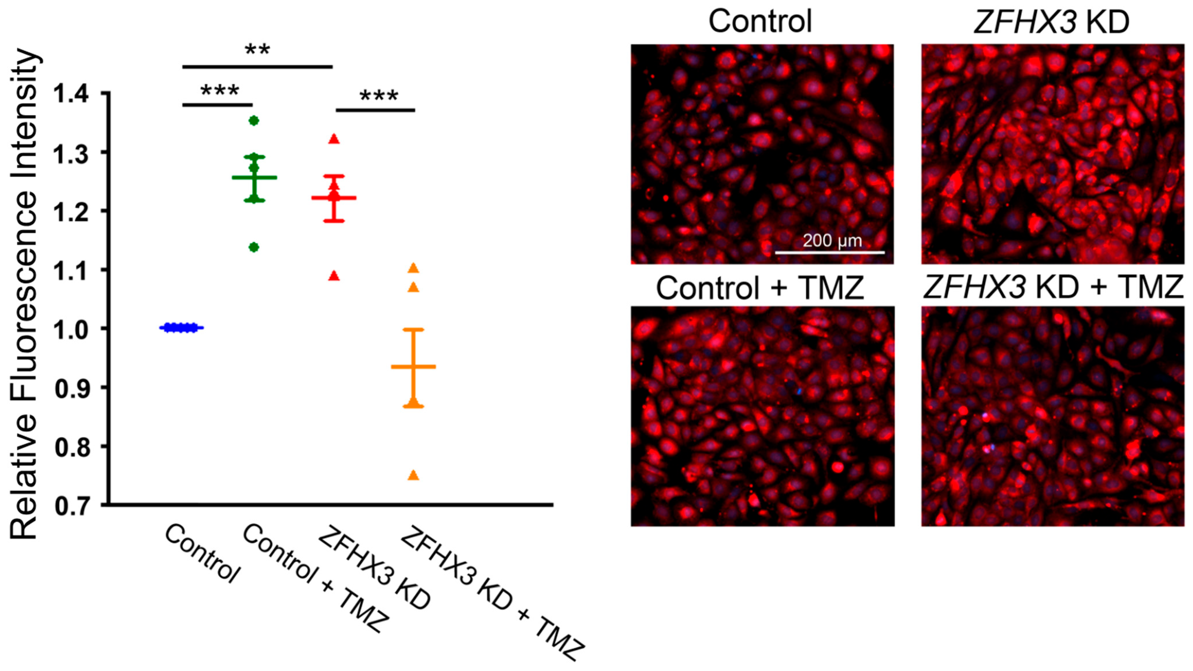
We next studied the effect of TMZ on ZFHX3 KD-induced mitochondrial respiratory distress calcium homeostasis. TMZ (10 µM) reduced basal respiration by 20% and proton leak by 26% in ZFHX3 KD cells (Figure 5). Conversely, TMZ increased basal respiration and proton leak in control cells. ZFHX3 KD cells exhibited higher mitochondrial calcium content, measured using X-Rhod-1 fluorescence, compared with control cells. This increase was reduced by TMZ, whereas TMZ treatment led to increased mitochondrial calcium content in control cells (Figure 6). Furthermore, TMZ attenuated ZFHX3 KD-induced increase in pPDH and oxidized CaMKII (ox-CaMKII) protein expression (Figure 7).

3. Discussion

Significant associations between AF and the ZFHX3 gene have been recognized for over a decade, yet the molecular mechanisms by which this gene contributes to AF remain incompletely understood. Previous studies have highlighted substantial changes in cardiac energy metabolism, including mitochondrial dysfunction and altered protein levels responsible for both FAs and glucose oxidation pathways in left atrial appendage tissues from AF patients [4]. More recently, pathways such as oxidative phosphorylation and FAs metabolism were found to be downregulated in ZFHX3-deficient murine atria [15]. In the present study, we confirmed that ZFHX3 KD cells had greater phosphorylated STAT3 expression (Supplementary Figure S1), which is consistent with previous findings [12,13] and is known to be a key regulator of FAs oxidation. Moreover, we demonstrated that ZFHX3 KD in cardiomyocytes leads to dysregulation of both FAs and glucose metabolism. ZFHX3 KD cells exhibited increased glucose uptake and lactate production, along with elevated LDHA protein expressions compared with control cells, indicating a shift toward glycolysis—a rapid, oxygen-independent but less efficient ATP-producing pathway [16,17,18]. Additionally, ZFHX3 KD cells showed decreased PDH activity, with increased levels of both total and phosphorylated PDH and PDK4 proteins. PDK phosphorylates and inactivates PDH, a rate-limiting enzyme in glucose oxidation, thereby promoting glycolysis [2]. AF has been associated with increased lactate levels, downregulation of PDH, and enhanced PDK4 protein expressions in rabbits, consistent with increased glycolysis but reduced glucose oxidation in their hearts [19,20]. These findings suggest that ZFHX3 KD results in uncoupling between glycolysis and pyruvate oxidation, confirming the presence of aerobic glycolysis (Warburg effect) in AF [21,22]. This metabolic shift leads to lactate accumulation and related metabolic abnormalities, which may contribute to dysregulated electrophysiological characteristics in atrial myocytes. Our study also found that ZFHX3 KD cells exhibited higher protein expressions of CD36 and phosphorylated ACC (pACC), while total ACC remained unchanged. ACC is inactivated by phosphorylation and is a major downstream target of AMPK, which we previously found to be upregulated in ZFHX3 KD cells [14]. AMPK is a primary energy sensor activated by metabolic stress in the heart [23]. In addition, STAT3 is implicated in the transcriptional regulation of CD36 [24], supporting the observed increase in CD36 expression in ZFHX3 KD cells. Together, these findings support a shift toward FA uptake and utilization in ZFHX3-deficient atrial myocytes. Mechanistically, ZFHX3 has been reported to regulate STAT3 signaling, and downregulation of ZFHX3 (ATBF1) can activate STAT3 via PIAS3 [13]. Activated STAT3 directly binds to the CD36 promoter, enhancing its transcription and thereby facilitating fatty acid uptake [24]. This pathway provides a plausible explanation for the observed CD36 upregulation in ZFHX3 KD cells. In contrast, we did not find evidence that STAT3 directly regulates PDK4 or LDHA. Their upregulation may involve alternative transcriptional pathways or indirect effects. For example, AMPK activation enhances LDHA and PDK4 expression through metabolic stress signaling, while PPARα is known to regulate CD36 and PDK4 transcription via promoter binding. Although additional regulators or control compounds were not examined in this study, future work will investigate the roles of AMPK, PPARα, and related pathways in mediating these effects.
Elevated free FAs and their increased cellular uptake contribute significantly to atrial arrhythmogenesis. Previous studies have demonstrated that disrupted FAs metabolism—including FAs availability, uptake, transport, and oxidation—induces atrial metabolic inflexibility and promotes AF [2]. Upregulation of CD36, a key FAs transporter, and subsequent FAs overloading have been strongly correlated with AF [4] and its risk factors, such as obesity [25,26], diabetes [27], and aging [28]. Importantly, increased FAs uptake is not always matched by proportional FAs oxidation [29]. In our study, ZFHX3 KD cells exhibited significantly higher acute response, maximal respiration, and spare capacity following Etomoxir injection compared with control cells, whereas both groups displayed similar responses to UK5099. Taken together, decreased PDH activity and heightened sensitivity to FAs oxidation inhibition suggest that ZFHX3 KD cells rely more heavily on FA metabolism in the mitochondria. Excessive FA availability and β-oxidation increase mitochondrial oxygen demand and ROS production through enhanced electron transport chain activity and superoxide leakage [30,31]. Excessive ROS oxidizes CaMKII, a central regulator of Ca2+ handling, thereby impairing calcium homeostasis and promoting mitochondrial dysfunction, inflammation, and arrhythmogenesis [30,32,33]. Our previous study demonstrated that ZFHX3 KD cells exhibited mitochondrial Ca2+ overload and elevated superoxide production [14]. We also reported that ZFHX3 KD enhances IP3 receptor (IP3R) expression and signaling [14], promoting spontaneous SR Ca2+ release and excessive mitochondrial uptake. In addition, ZFHX3 KD increased SERCA2a expression and activity, as well as RyR2 protein levels [12], collectively contributing to augment Ca2+ uptake and leak in atrial myocytes. These mechanisms, in addition to metabolic stress (lactate acidosis, ROS), establish multiple upstream pathways driving mitochondrial Ca2+ overload in ZFHX3-deficient cells. Other calcium-handling proteins may also play roles in ZFHX3 deficiency-mediated calcium dysregulation and warrant further investigation.
TMZ is a clinically used anti-angina agent that inhibits FAs oxidation, thereby reducing oxidative stress and oxygen demand [34,35]. Additionally, TMZ has been suggested as a potential therapeutic agent in heart failure through metabolic regulations [36,37]. In this study, TMZ significantly reduced mitochondrial respiration and proton leak in ZFHX3 KD cells. The greater effect on proton leak compared with basal respiration suggests that inhibiting FAs utilization helps preserve mitochondrial function under stress. TMZ also reduced lactate production in ZFHX3 KD cells, supporting the concept that increased FAs utilization drives lactate accumulation and exacerbates mitochondrial Ca2+ overload. Interestingly, TMZ exhibited differential effects in control versus ZFHX3 KD cells. In control cells, TMZ increased mitochondrial respiration and calcium content, likely due to enhanced ATP synthesis under normal conditions, whereas in ZFHX3 KD cells, TMZ exerted protective effects by reducing mitochondrial proton leak, lactate accumulation, and mitochondrial Ca2+ overload. This suggests that TMZ’s impact is context-dependent, mitigating metabolic distress in ZFHX3 KD cells while potentially enhancing mitochondrial function in healthy cells. Mechanistically, TMZ reduced PDH phosphorylation in ZFHX3 KD cells, improving glucose utilization, and lowered ox-CaMKII levels, suggesting that FAs inhibition mitigates metabolic stress-induced calcium dysregulation. Notably, at higher concentrations (10–100 μM), TMZ has been shown to enhance mitochondrial calcium uptake, supporting ATP synthesis [38]. In summary, ZFHX3 KD induces cardiac metabolic stress characterized by decreased PDH activity and increased β-oxidation, leading to mitochondrial dysregulation (calcium overload, proton leak), lactate accumulation, and enhanced ox-CaMKII, all of which may contribute to electrical remodeling and arrhythmogenesis. However, there are several limitations of our study that must be acknowledged. Our study was conducted in HL-1 atrial myocytes, a well-established in vitro model. While this system enabled mechanistic dissection of ZFHX3 KD effects under controlled conditions, the results may not fully reflect the complexity of primary cardiomyocytes or in vivo AF models. Future studies using animal models and patients-derived cells are needed to validate these findings. In addition, we examined the global effects of ZFHX3 knockdown but did not investigate specific ZFHX3 variants, which may have distinct impacts on cardiac metabolism and AF pathology. Finally, our findings are specific to atrial myocytes and may not directly apply to ventricular cardiomyocytes, which can display different metabolic and calcium-handling properties. ZFHX3 KD induces cardiac metabolic stress characterized by decreased PDH activity and increased FA oxidation, leading to mitochondrial dysfunction (proton leak, calcium overload), lactate accumulation, and enhanced ox-CaMKII. These alterations contribute to electrical remodeling and arrhythmogenesis. TMZ mitigates these effects, highlighting its therapeutic potential as a metabolic modulator in AF associated with ZFHX3 deficiency.

4. Materials and Methods

4.1. HL-1 Cell Culture and Lentiviral shRNA-Mediated ZFHX3 Knockdown

HL-1 cardiomyocytes (kindly provided by Dr. Claycomb, Louisiana State University Medical Center, New Orleans, LA, USA) were cultured in Claycomb medium (JRH Biosciences, Lenexa, KS, USA, Cat. No. 51800C) supplemented with 10% fetal bovine serum (FBS, JRH Biosciences, Cat. No. 97068-085), in flasks pre-coated with fibronectin (Sigma (St. Louis, MO, USA), Cat. No. F1141) and gelatine (Sigma, Cat. No. G9391) [14]. Cells were incubated at 37 °C in a 5% a CO2 incubator (Thermor (Saint-Jean-de-la-Ruelle, France), Model 3111). ZFHX3 knockdown was achieved using lentivirus containing shRNA targeting ZFHX3 (clone TRCN0000321285, National RNAi core Facility, Academia Sinica, Taipei, Taiwan). Cells were selected with puromycin (Sigma, Cat. No. P8833) at 2 µg/mL for 1 week and subsequently maintained at 1 µg/mL.

4.2. Intracellular Lactate Concentrations

To measure intracellular L-lactate concentrations, a colorimetric Lactate assay kit (Cat. No. MAK064, Sigma) was used according to the manufacturer’s instructions. HL-1 cells were plated into a 6-well plate and allowed to attach and normalize functionally overnight. The cells were maintained in buffered culture medium containing 10% FBS and were allowed to reach approximately 90% confluence. Cells were then treated with or without TMZ (10 µM, Sigma) for 3 h. Cells were trypsinized and the resulting cell pellet was processed according to the kit protocol. Briefly, the cell pellet was homogenized in 4 volumes of lactate assay buffer, then centrifuged at 13,000× g for 10 min to remove insoluble material. The supernatant was deproteinized using a 10 kDa MWCO spin filter to remove endogenous lactate dehydrogenase. The soluble fraction was then assayed directly.

4.3. PDH Activity Assay

The catalytic activity of PDH was measured using the PDH Enzyme Activity Microplate Assay Kit (ab109902, Abcam (Cambridge, UK)) according to the manufacturer’s instructions. Cells were plated in a 6-well plate and allowed to attach and normalize functionally overnight. After washing with PBS and trypsinizing, the cell pellet was processed as per the kit instructions. Briefly, the fresh cell pellet was resuspended in 9 volumes of PBS, and proteins were extracted by adding the detergent solution. Freshly prepared samples with a concentration of 100 µg/200 µL were loaded into microplate wells coated with an anti-PDH monoclonal antibody and incubated for 3 h at room temperature. PDH activity was determined by measuring the rate of NAD+ reduction to NADH, coupled with the reduction in a reporter dye, and monitoring the increase in absorbance of the reaction product at 450 nm. All tests were conducted in triplicate, and results were expressed as changes in absorbance per minute per milligram of protein.

4.4. Seahorse Substrate Oxidation Stress Test and Mitochondrial Oxygen Consumption Rate (OCR)

For mitochondrial OCR measurements, HL-1 cells were first cultured under standard conditions in general culture plates. On the day of the Seahorse assay, cells were detached and seeded at a density of 2 × 10⁵ cells/well in unbuffered Dulbecco’s Modified Eagle Medium (DMEM, pH 7.4) into an XFe24 Seahorse assay plate (Seahorse Bioscience, Billerica, MA, USA). The plates were incubated at 37 °C in a non-CO2 incubator for 45–60 min to allow temperature and pH equilibration. The Seahorse sensor cartridge was hydrated overnight at 37 °C in a non-CO2 incubator with sterile deionized water.
To evaluate mitochondrial substrate oxidation, we utilized the XF24 LCFA Oxidation Stress Test (Cat# 103672-100, Agilent Technologies, Santa Clara, CA, USA) and the XF24 Glucose/Pyruvate Oxidation Stress Test (Cat# 103673-100, Agilent Technologies). The assay media compositions were as follows.
LCFA Oxidation Stress Test (Figure 1A): 0.5 mM D-glucose, 1 mM L-glutamine, 0.5 mM L-carnitine, and 1% FBS. Glucose/Pyruvate Oxidation Stress Test (Figure 1B): 1 mM sodium pyruvate, 2 mM L-glutamine, and 25 mM D-glucose. Mito Stress Test (Figure 5): identical to the Glucose/Pyruvate Oxidation Test medium; in addition, cells were treated with or without trimetazidine (TMZ, 10 µM; Sigma) for 3 h prior to the assay.
OCR was measured sequentially following injections of the following compounds: Etomoxir (40 µM) to inhibit fatty acid oxidation via CPT-1a, or UK5099 (8 µM) to inhibit mitochondrial pyruvate carrier, followed by oligomycin (1.5 µM) to inhibit ATP synthase, FCCP (1 µM) to uncouple oxidative phosphorylation and induce maximal respiration, and finally, rotenone (0.5 µM) plus antimycin A (0.5 µM) to inhibit complexes I and III, thereby measuring non-mitochondrial respiration.
Absolute OCR values (in pmol/min) were recorded at each injection step. The following parameters were calculated using standard Seahorse equations. Basal respiration: Initial OCR prior to any drug injection (absolute value). Maximal respiration: FCCP-stimulated OCR minus non-mitochondrial OCR (absolute value). Spare respiratory capacity: Maximal OCR minus basal OCR (calculated delta). Acute response: OCR immediately after substrate inhibitor injection minus baseline OCR (calculated delta). Proton leak: OCR after oligomycin minus non-mitochondrial OCR (calculated delta).

4.5. Glucose Uptake

Glucose uptake of cardiomyocytes was measured using the Glucose Uptake-GloTM (Promega, Madison, WI, USA) bioluminescence assay kit following the manufacturer’s instructions. After washing the cells twice with 1× PBS, 500 µL of 1 mM 2-DG (2-deoxyglucose) diluted in PBS was added for a 10 min uptake period. Following uptake, stopping and neutralization buffers were applied and 2-deoxyglucose-6-phosphate detection reagent were added. Luminescence intensity was recorded at 1 h using a Tecan Infinite M200 plate reader.

4.6. Mitochondrial Ca2+ Measurements

Mitochondrial Ca2+ levels were assessed in intact live cells using fluorescence microscopy (EVOS FL, Invitrogen (Waltham, MA, USA)). Cells were treated with or without TMZ (10 µM, Sigma) for 3 h. After treatments, cells were stained with 2.5 μM X-Rhod-1/AM for 10 min at 37 °C, followed by a 10 min incubation with 1 mM CoCl2 in Tyrode’s solution to quench cytosolic X-Rhod-1 fluorescence.

4.7. Western Blotting

Control and ZFHX3 KD HL-1 cells with or without TMZ (10 µM, Sigma) for 3 h were lysed in M-PER Mammalian Protein Extraction Reagent (Thermo Fisher Scientific, Cat. No. 78501) with protease inhibitor cocktails (Sigma, St. Louis, MO, USA). Equal amounts of proteins from each group were separated in gradient sodium dodecylsulphate polyacrylamide gel electrophoresis under reducing conditions and electrophoretically transferred onto an equilibrated polyvinylidene difluoride membrane (Amersham Biosciences, Amersham, Buckinghamshire, UK). Blots were probed with following primary antibodies: ACC (1:1000, Abcam, Cat. No. ab6564), GLUT4 (Abcam, ab6564), CD36 (Santa Cruz Biotechnology, Dallas, TX, USA), PDH (Cell Signaling, Danvers, MA, USA), pPDH (Serine293, Sigma), pACC (#07303, Sigma), PDK4, LDHA, SLC16A1 (Antibodies.com), and oxidized CaMKII (Genetex, Irvine, CA, USA). An enhanced chemiluminescence detection system (Santa Cruz Biotechnology, Santa Cruz, CA, USA) was used to detect bound antibodies, which were then analyzed using AlphaEase FC software version 4.0. (Alpha Innotech, San Leandro, CA, USA). Targeted bands were normalized to β-actin (Antibodies) to confirm equal protein loading.

4.8. Statistical Analysis

All quantitative data are expressed as the mean ± standard error of the mean (SEM). Differences between groups were assessed using an unpaired test while comparisons among groups were made using two-way analysis of variance with repeated or non-repeated measures, followed by Turkey’s post hoc test where appropriate. Statistical significance was set at p < 0.05.

Supplementary Materials

The following supporting information can be downloaded at https://www.mdpi.com/article/10.3390/ijms26178576/s1.

Author Contributions

S.-H.L., S.H. and Y.-J.C. conceived and designed the study. B.L. conducted the experiments, analyzed the data, and created figures. B.L. and Y.-H.K. composed the first draft of the manuscript. S.-H.L., S.H., Y.-H.K. and Y.-J.C. reviewed and edited the manuscript. All authors have read and agreed to the published version of the manuscript.

Funding

This work was supported by grants from the National Science and Technology Council, Taiwan (NSTC 112-2811-B-038-049 and 113-2314-B-038-063-MY3), Taipei Medical University–Shuang Ho Hospital (114TMU-SHH-21), and Foundation for the Development of Internal Medicine in Okinawa, Japan (4-02-003, 4-02-006, 5-02-007).

Acknowledgments

We express our gratitude to the CRISPR Gene Targeting Core Laboratory at Taipei Medical University, Taiwan, for their technical support in generating ZFHX3 knockdown in HL-1 cells.

Conflicts of Interest

The authors declare no conflicts of interest.

References

  1. Ruddox, V.; Sandven, I.; Munkhaugen, J.; Skattebu, J.; Edvardsen, T.; Otterstad, J.E. Atrial fibrillation and the risk for myocardial infarction, all-cause mortality and heart failure: A systematic review and meta-analysis. Eur. J. Prev. Cardiol. 2017, 24, 1555–1566. [Google Scholar] [CrossRef]
  2. Qin, X.; Zhang, Y.; Zheng, Q. Metabolic Inflexibility as a Pathogenic Basis for Atrial Fibrillation. Int. J. Mol. Sci. 2022, 23, 8291. [Google Scholar] [CrossRef] [PubMed]
  3. Smith, R.L.; Soeters, M.R.; Wust, R.C.I.; Houtkooper, R.H. Metabolic Flexibility as an Adaptation to Energy Resources and Requirements in Health and Disease. Endocr. Rev. 2018, 39, 489–517. [Google Scholar] [CrossRef] [PubMed]
  4. Rennison, J.H.; Li, L.; Lin, C.R.; Lovano, B.S.; Castel, L.; Wass, S.Y.; Cantlay, C.C.; McHale, M.; Gillinov, A.M.; Mehra, R.; et al. Atrial fibrillation rhythm is associated with marked changes in metabolic and myofibrillar protein expression in left atrial appendage. Pflugers Arch. 2021, 473, 461–475. [Google Scholar] [CrossRef] [PubMed]
  5. Tu, T.; Zhou, S.; Liu, Z.; Li, X.; Liu, Q. Quantitative proteomics of changes in energy metabolism-related proteins in atrial tissue from valvular disease patients with permanent atrial fibrillation. Circ. J. 2014, 78, 993–1001. [Google Scholar] [CrossRef]
  6. Zhou, J.; Sun, L.; Chen, L.; Liu, S.; Zhong, L.; Cui, M. Comprehensive metabolomic and proteomic analyses reveal candidate biomarkers and related metabolic networks in atrial fibrillation. Metabolomics 2019, 15, 96. [Google Scholar] [CrossRef]
  7. Mayr, M.; Yusuf, S.; Weir, G.; Chung, Y.L.; Mayr, U.; Yin, X.; Ladroue, C.; Madhu, B.; Roberts, N.; De Souza, A.; et al. Combined metabolomic and proteomic analysis of human atrial fibrillation. J. Am. Coll. Cardiol. 2008, 51, 585–594. [Google Scholar] [CrossRef]
  8. Liu, X.; Wang, F.; Knight, A.C.; Zhao, J.; Xiao, J. Common variants for atrial fibrillation: Results from genome-wide association studies. Hum. Genet. 2012, 131, 33–39. [Google Scholar] [CrossRef]
  9. Gudbjartsson, D.F.; Holm, H.; Gretarsdottir, S.; Thorleifsson, G.; Walters, G.B.; Thorgeirsson, G.; Gulcher, J.; Mathiesen, E.B.; Njolstad, I.; Nyrnes, A.; et al. A sequence variant in ZFHX3 on 16q22 associates with atrial fibrillation and ischemic stroke. Nat. Genet. 2009, 41, 876–878. [Google Scholar] [CrossRef]
  10. Benjamin, E.J.; Rice, K.M.; Arking, D.E.; Pfeufer, A.; van Noord, C.; Smith, A.V.; Schnabel, R.B.; Bis, J.C.; Boerwinkle, E.; Sinner, M.F.; et al. Variants in ZFHX3 are associated with atrial fibrillation in individuals of European ancestry. Nat. Genet. 2009, 41, 879–881. [Google Scholar] [CrossRef]
  11. Li, C.; Wang, F.; Yang, Y.; Fu, F.; Xu, C.; Shi, L.; Li, S.; Xia, Y.; Wu, G.; Cheng, X.; et al. Significant association of SNP rs2106261 in the ZFHX3 gene with atrial fibrillation in a Chinese Han GeneID population. Hum. Genet. 2011, 129, 239–246. [Google Scholar] [CrossRef]
  12. Kao, Y.H.; Hsu, J.C.; Chen, Y.C.; Lin, Y.K.; Lkhagva, B.; Chen, S.A.; Chen, Y.J. ZFHX3 knockdown increases arrhythmogenesis and dysregulates calcium homeostasis in HL-1 atrial myocytes. Int. J. Cardiol. 2016, 210, 85–92. [Google Scholar] [CrossRef]
  13. Jiang, Q.; Ni, B.; Shi, J.; Han, Z.; Qi, R.; Xu, W.; Wang, D.; Wang, D.W.; Chen, M. Down-regulation of ATBF1 activates STAT3 signaling via PIAS3 in pacing-induced HL-1 atrial myocytes. Biochem. Biophys. Res. Commun. 2014, 449, 278–283. [Google Scholar] [CrossRef] [PubMed]
  14. Lkhagva, B.; Lin, Y.K.; Chen, Y.C.; Cheng, W.L.; Higa, S.; Kao, Y.H.; Chen, Y.J. ZFHX3 knockdown dysregulates mitochondrial adaptations to tachypacing in atrial myocytes through enhanced oxidative stress and calcium overload. Acta Physiol. 2021, 231, e13604. [Google Scholar] [CrossRef]
  15. Jameson, H.S.; Hanley, A.; Hill, M.C.; Xiao, L.; Ye, J.; Bapat, A.; Ronzier, E.; Hall, A.W.; Hucker, W.J.; Clauss, S.; et al. Loss of the Atrial Fibrillation-Related Gene, Zfhx3, Results in Atrial Dilation and Arrhythmias. Circ. Res. 2023, 133, 313–329. [Google Scholar] [CrossRef] [PubMed]
  16. Barth, A.S.; Merk, S.; Arnoldi, E.; Zwermann, L.; Kloos, P.; Gebauer, M.; Steinmeyer, K.; Bleich, M.; Kaab, S.; Hinterseer, M.; et al. Reprogramming of the human atrial transcriptome in permanent atrial fibrillation: Expression of a ventricular-like genomic signature. Circ. Res. 2005, 96, 1022–1029. [Google Scholar] [CrossRef]
  17. Ausma, J.; Litjens, N.; Lenders, M.H.; Duimel, H.; Mast, F.; Wouters, L.; Ramaekers, F.; Allessie, M.; Borgers, M. Time course of atrial fibrillation-induced cellular structural remodeling in atria of the goat. J. Mol. Cell Cardiol. 2001, 33, 2083–2094. [Google Scholar] [CrossRef]
  18. Ingwall, J.S.; Weiss, R.G. Is the failing heart energy starved? On using chemical energy to support cardiac function. Circ. Res. 2004, 95, 135–145. [Google Scholar] [CrossRef]
  19. Liu, Y.; Geng, J.; Liu, Y.; Li, Y.; Shen, J.; Xiao, X.; Sheng, L.; Yang, B.; Cheng, C.; Li, W. beta3-adrenoceptor mediates metabolic protein remodeling in a rabbit model of tachypacing-induced atrial fibrillation. Cell Physiol. Biochem. 2013, 32, 1631–1642. [Google Scholar] [CrossRef]
  20. Liu, G.Z.; Hou, T.T.; Yuan, Y.; Hang, P.Z.; Zhao, J.J.; Sun, L.; Zhao, G.Q.; Zhao, J.; Dong, J.M.; Wang, X.B.; et al. Fenofibrate inhibits atrial metabolic remodelling in atrial fibrillation through PPAR-alpha/sirtuin 1/PGC-1alpha pathway. Br. J. Pharmacol. 2016, 173, 1095–1109. [Google Scholar] [CrossRef]
  21. Hu, H.J.; Zhang, C.; Tang, Z.H.; Qu, S.L.; Jiang, Z.S. Regulating the Warburg effect on metabolic stress and myocardial fibrosis remodeling and atrial intracardiac waveform activity induced by atrial fibrillation. Biochem. Biophys. Res. Commun. 2019, 516, 653–660. [Google Scholar] [CrossRef]
  22. Liu, Y.; Bai, F.; Liu, N.; Ouyang, F.; Liu, Q. The Warburg effect: A new insight into atrial fibrillation. Clin. Chim. Acta 2019, 499, 4–12. [Google Scholar] [CrossRef] [PubMed]
  23. Lkhagva, B.; Lee, T.W.; Lin, Y.K.; Chen, Y.C.; Chung, C.C.; Higa, S.; Chen, Y.J. Disturbed Cardiac Metabolism Triggers Atrial Arrhythmogenesis in Diabetes Mellitus: Energy Substrate Alternate as a Potential Therapeutic Intervention. Cells 2022, 11, 2915. [Google Scholar] [CrossRef] [PubMed]
  24. Rozovski, U.; Harris, D.M.; Li, P.; Liu, Z.; Jain, P.; Ferrajoli, A.; Burger, J.; Thompson, P.; Jain, N.; Wierda, W.; et al. STAT3-activated CD36 facilitates fatty acid uptake in chronic lymphocytic leukemia cells. Oncotarget 2018, 9, 21268–21280. [Google Scholar] [CrossRef] [PubMed]
  25. Coort, S.L.; Hasselbaink, D.M.; Koonen, D.P.; Willems, J.; Coumans, W.A.; Chabowski, A.; van der Vusse, G.J.; Bonen, A.; Glatz, J.F.; Luiken, J.J. Enhanced sarcolemmal FAT/CD36 content and triacylglycerol storage in cardiac myocytes from obese zucker rats. Diabetes 2004, 53, 1655–1663. [Google Scholar] [CrossRef]
  26. Steinbusch, L.K.; Schwenk, R.W.; Ouwens, D.M.; Diamant, M.; Glatz, J.F.; Luiken, J.J. Subcellular trafficking of the substrate transporters GLUT4 and CD36 in cardiomyocytes. Cell Mol. Life Sci. 2011, 68, 2525–2538. [Google Scholar] [CrossRef]
  27. Bonen, A.; Tandon, N.N.; Glatz, J.F.; Luiken, J.J.; Heigenhauser, G.J. The fatty acid transporter FAT/CD36 is upregulated in subcutaneous and visceral adipose tissues in human obesity and type 2 diabetes. Int. J. Obes. 2006, 30, 877–883. [Google Scholar] [CrossRef]
  28. Einstein, F.H.; Huffman, D.M.; Fishman, S.; Jerschow, E.; Heo, H.J.; Atzmon, G.; Schechter, C.; Barzilai, N.; Muzumdar, R.H. Aging per se increases the susceptibility to free fatty acid-induced insulin resistance. J. Gerontol. A Biol. Sci. Med. Sci. 2010, 65, 800–808. [Google Scholar] [CrossRef]
  29. Lenski, M.; Schleider, G.; Kohlhaas, M.; Adrian, L.; Adam, O.; Tian, Q.; Kaestner, L.; Lipp, P.; Lehrke, M.; Maack, C.; et al. Arrhythmia causes lipid accumulation and reduced glucose uptake. Basic Res. Cardiol. 2015, 110, 40. [Google Scholar] [CrossRef]
  30. Lambertucci, R.H.; Hirabara, S.M.; Silveira Ldos, R.; Levada-Pires, A.C.; Curi, R.; Pithon-Curi, T.C. Palmitate increases superoxide production through mitochondrial electron transport chain and NADPH oxidase activity in skeletal muscle cells. J. Cell Physiol. 2008, 216, 796–804. [Google Scholar] [CrossRef]
  31. Egnatchik, R.A.; Leamy, A.K.; Noguchi, Y.; Shiota, M.; Young, J.D. Palmitate-induced activation of mitochondrial metabolism promotes oxidative stress and apoptosis in H4IIEC3 rat hepatocytes. Metabolism 2014, 63, 283–295. [Google Scholar] [CrossRef]
  32. Pabel, S.; Knierim, M.; Stehle, T.; Alebrand, F.; Paulus, M.; Sieme, M.; Herwig, M.; Barsch, F.; Kortl, T.; Poppl, A.; et al. Effects of Atrial Fibrillation on the Human Ventricle. Circ. Res. 2022, 130, 994–1010. [Google Scholar] [CrossRef]
  33. Zhang, Y.; Fu, Y.; Jiang, T.; Liu, B.; Sun, H.; Zhang, Y.; Fan, B.; Li, X.; Qin, X.; Zheng, Q. Enhancing Fatty Acids Oxidation via L-Carnitine Attenuates Obesity-Related Atrial Fibrillation and Structural Remodeling by Activating AMPK Signaling and Alleviating Cardiac Lipotoxicity. Front. Pharmacol. 2021, 12, 771940. [Google Scholar] [CrossRef] [PubMed]
  34. McCullough, P.A. Chronic angina: New medical options for treatment. Rev. Cardiovasc. Med. 2005, 6, 152–161. [Google Scholar] [PubMed]
  35. Bobescu, E.; Marceanu, L.G.; Dima, L.; Balan, A.; Strempel, C.G.; Covaciu, A. Trimetazidine Therapy in Coronary Artery Disease: The Impact on Oxidative Stress, Inflammation, Endothelial Dysfunction, and Long-Term Prognosis. Am. J. Ther. 2021, 28, e540–e547. [Google Scholar] [CrossRef] [PubMed]
  36. Shu, H.; Hang, W.; Peng, Y.; Nie, J.; Wu, L.; Zhang, W.; Wang, D.W.; Zhou, N. Trimetazidine Attenuates Heart Failure by Improving Myocardial Metabolism via AMPK. Front. Pharmacol. 2021, 12, 707399. [Google Scholar] [CrossRef]
  37. Kantor, P.F.; Lucien, A.; Kozak, R.; Lopaschuk, G.D. The antianginal drug trimetazidine shifts cardiac energy metabolism from fatty acid oxidation to glucose oxidation by inhibiting mitochondrial long-chain 3-ketoacyl coenzyme A thiolase. Circ. Res. 2000, 86, 580–588. [Google Scholar] [CrossRef]
  38. Guarnieri, C.; Finelli, C.; Zini, M.; Muscari, C. Effects of trimetazidine on the calcium transport and oxidative phosphorylation of isolated rat heart mitochondria. Basic Res. Cardiol. 1997, 92, 90–95. [Google Scholar] [CrossRef]
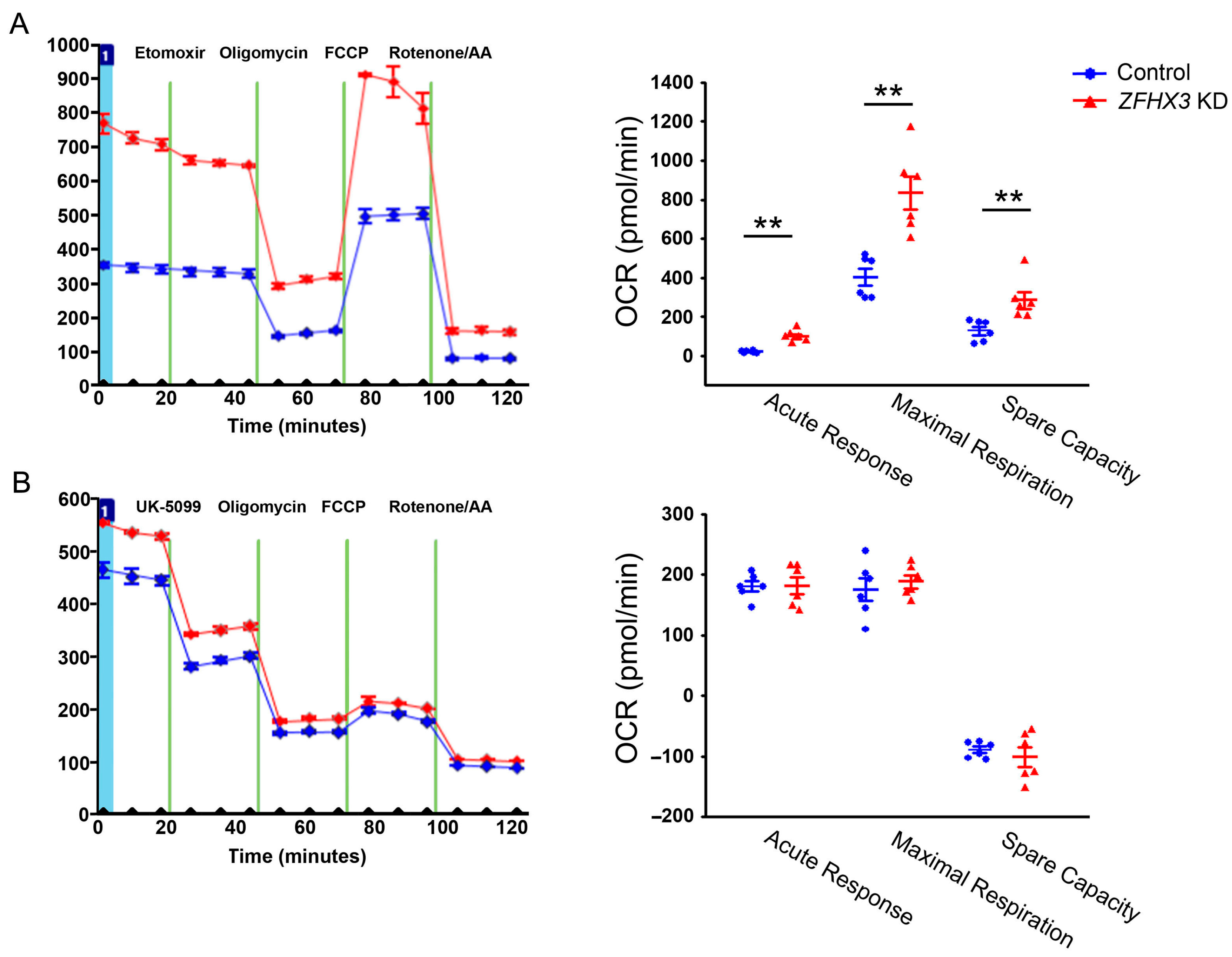
Figure 1. Effects of ZFHX3 knockdown on fatty acids (FAs) and glucose substrate utilization. Representative traces and quantified data of oxygen consumption rates (OCRs) illustrate the impacts of ZFHX3 KD on mitochondrial function in the presence of (A) Etomoxir (CPT-1a inhibitor, 40 µM) and (B) UK5099 (mitochondrial pyruvate carrier inhibitor, 8 µM). Blue circles indicate control cells, and red triangles indicate ZFHX3 KD cells. ZFHX3 KD cells exhibited significantly higher acute response, maximal respiration, and spare capacity compared with control cells following Etomoxir injection (n = 6). Control and ZFHX3 KD cells showed similar acute response, maximal respiration, and spare capacity in the presence of UK5099 (n = 6). ** p < 0.01.
Ijms 26 08576 g001
Figure 2. Effects of ZFHX3 knockdown on lactate production, glucose uptake, and pyruvate dehydrogenase (PDH) activity. Average data of lactate concentrations (A) and glucose uptake (B) in control and ZFHX3 KD HL-1 cells (n = 5). ZFHX3 KD cells showed higher lactate concentrations and glucose uptake than control cells. (C) Average PDH enzyme activity in control and ZFHX3 KD HL-1 cells, showing significantly lower PDH activity compared with control cells (n = 6). Blue circles represent control cells, and red triangles represent ZFHX3 KD cells. * p < 0.05, ** p < 0.01.
Ijms 26 08576 g002
Figure 3. Effects of ZFHX3 gene on fatty acids (FAs) and glucose metabolism regulatory proteins. (AC) Representative Western blot and quantified data revealed a significant increase in CD36, phosphorylated ACC (pACC), pyruvate dehydrogenase kinase 4 (PDK4), lactate dehydrogenase A (LDHA), phosphorylated PDH (pPDH), and total PDH levels in ZFHX3 KD HL-1 cells compared with control cells (n = 8). Blue circles represent control cells, and red triangles represent ZFHX3 KD cells. * p < 0.05, ** p < 0.01.
Ijms 26 08576 g003
Figure 4. Effects of trimetazidine (TMZ) on lactate production. (A) TMZ (10 µM) did not change lactate content in control cells (n = 5). (B) TMZ (10 µM) significantly reduced lactate production in ZFHX3 KD cells (n = 5). Blue circles represent control cells, green inverted triangles represent control + TMZ cells, red triangles represent ZFHX3 KD cells, and orange inverted triangles represent ZFHX3 KD + TMZ cells. * p < 0.05.
Ijms 26 08576 g004
Figure 5. Effects of TMZ on oxygen consumption rates (OCRs) and bioenergetics parameters. (A) Representative traces and (B) quantified data illustrate the effects of TMZ (10 µM) on basal respiration, maximal respiration, spare respiratory capacity, and proton leak in control and ZFHX3 KD cells, both with and without TMZ treatment (n = 8). Blue circles represent control cells, green circles represent control + TMZ cells, red triangles represent ZFHX3 KD cells, and orange triangles represent ZFHX3 KD + TMZ cells. * p < 0.05, ** p < 0.01, *** p < 0.005.
Ijms 26 08576 g005
Figure 6. Effects of TMZ on mitochondrial Ca2+ content. Representative images and quantified data show the effects of TMZ (10 µM) on mitochondrial Ca2+ content in control (n = 5) and ZFHX3 KD HL-1 cells (n = 5), with and without TMZ treatment. Blue circles represent control cells, green circles represent control + TMZ cells, red triangles represent ZFHX3 KD cells, and orange triangles represent ZFHX3 KD + TMZ cells. ** p < 0.01, *** p < 0.005 versus respective control groups.
Ijms 26 08576 g006
Figure 7. Effects of TMZ on pPDH and ox-CaMKII protein expressions. (A) phosphorylated PDH (pPDH) and (B) oxidized CaMKII (ox-CaMKII) protein levels were significantly decreased in ZFHX3 KD HL-1 cells treated with TMZ (10 µM) compared with cells without TMZ treatment (n = 6). * p < 0.05, ** p < 0.01.
Ijms 26 08576 g007
Disclaimer/Publisher’s Note: The statements, opinions and data contained in all publications are solely those of the individual author(s) and contributor(s) and not of MDPI and/or the editor(s). MDPI and/or the editor(s) disclaim responsibility for any injury to people or property resulting from any ideas, methods, instructions or products referred to in the content.